City, University of London Institutional Repository
Citation
: Lisi, M., Cavanagh, P. & Zorzi, M. (2015). Spatial constancy of attention across
eye movements is mediated by the presence of visual objects. Attention, Perception, & Psychophysics, 77(4), pp. 1159-1169. doi: 10.3758/s13414-015-0861-1This is the accepted version of the paper.
This version of the publication may differ from the final published
version.
Permanent repository link:
http://openaccess.city.ac.uk/19306/Link to published version
: http://dx.doi.org/10.3758/s13414-015-0861-1
Copyright and reuse:
City Research Online aims to make research
outputs of City, University of London available to a wider audience.
Copyright and Moral Rights remain with the author(s) and/or copyright
holders. URLs from City Research Online may be freely distributed and
linked to.
Spatial constancy of attention across eye movements is mediated
by the presence of visual objects
Matteo Lisi1
Patrick Cavanagh1
Marco Zorzi2,3
1 Laboratoire Psychologie de la Perception (CNRS UMR 8248), Université Paris V Descartes, 75006 Paris, France 2 Department of General Psychology and Center for Cognitive Neuroscience, University of Padova, 35131 Padua, Italy 3 IRCCS San Camillo Neurorehabilitation Hospital, 30126 Venice-Lido, Italy
Correspondence concerning this article should be addressed to Matteo Lisi, Laboratoire Psychologie de la Perception, Université Paris Descartes, 45 rue des Saints-Pères, 75006 Paris, France. E-mail:
Acknowledgments: This study was supported by grant no. 210922 from the European Research Council and by a grant from the University of Padova (Strategic Grant “NEURAT”) to MZ.
Abstract
Recent studies have shown that attentional facilitation lingers at the retinotopic coordinates of a previously attended position after an eye movement. These results are intriguing, since the retinotopic location becomes behaviorally irrelevant once the eyes have moved. Critically, in these studies participants were asked to maintain attention on a blank location of the screen. In the present study we examined whether the continuing presence of a visual object at the cued location could affect the allocation of attention across eye movements. We used a trans-saccadic cueing paradigm in which the relevant positions could be defined or not by visual objects (simple square outlines). We find an attentional benefit at the spatiotopic location of the cue only when the object (the placeholder) has been continuously present at that location. We conclude that the presence of an object at the attended location is a critical factor for the maintenance of spatial constancy of attention across eye movements, a finding that helps reconcile previous conflicting results.
In everyday life, we often find that we must keep track of several objects of interest, pedestrians and other cars as we approach an intersection, the ball and other players in sports, our friends among strangers in a swimming pool. While this tracking function itself is remarkable (Pylyshyn & Storm, 1988), it is even more remarkable that the tracking seems unimpeded by eye movements (Howe, Drew, Pinto, & Horowitz, 2011). We make about 3 eye movements each second and each one shifts the retinal input and the retinotopic (eye-centered) coordinates of our targets of interest. Clearly, if we had to rediscover each target following every eye movement, sports like soccer or basketball would be much slower paced and activities like driving much more dangerous. How do we keep our attention locked onto each target as our eyes move? One mechanism that has been proposed to explain this ability consists in the updating (remapping) of target locations to compensate for each eye movement. This process has been documented physiologically in saccade and attention areas (Duhamel, Colby, & Goldberg, 1992; Fecteau & Munoz, 2006; Sommer & Wurtz, 2006; Wurtz, Joiner, & Berman, 2011) and has been demonstrated behaviorally with probes that reveal the location of attentional benefits before and after eye movements (Hunt & Cavanagh, 2011; Jonikaitis, Szinte, Rolfs, & Cavanagh, 2013; Rolfs, Jonikaitis, Deubel, & Cavanagh, 2011). A number of review papers have suggested that the remapping process offers a sparse form of visual constancy by predicting where targets will be in retinotopic coordinates following each eye movement (Berman & Colby, 2009; Cavanagh, Hunt, Afraz, & Rolfs, 2010; Hall & Colby, 2011; Mathôt & Theeuwes, 2011; Wurtz, 2008). However, other studies report that attention is updated slowly after the eye movement and that initially, attention remains in the retinotopic location despite the fact that this location is behaviorally irrelevant following the saccade (Golomb, Chun, & Mazer, 2008; Golomb, Marino, Chun, & Mazer, 2011; Golomb, Pulido, Albrecht, Chun, & Mazer, 2010). The goal of this paper is to resolve these two different outcomes by showing the critical role played by the presence of an object at the cued location.
locations across eye movements, as has been found by Howe and colleagues in a multiple object tracking paradigm (Howe et al., 2011).
In contrast, as mentioned above, other studies report that attention is updated slowly and only after the eye movement. Golomb and colleagues developed a gaze-contingent paradigm in which participants performed an eye movement while keeping track of the position of a spatial cue (Golomb et al., 2008, 2011; Golomb, Pulido, et al., 2010). This paradigm was used to investigate the post-saccadic allocation of attention by presenting a probe at different time intervals after the saccade completion. The results showed a persisting attentional benefit at the retinotopic (eye-centered) coordinates of the cued location (even though task irrelevant) for 100 – 200 ms after an eye movement, along with growing facilitation at the spatiotopic location, reaching its maximum around 400 ms after saccade completion (see Casarotti, Lisi, Umiltà, & Zorzi, 2012, for simulations with a computational model that implements a spatial updating process). Neural signatures of this persisting retinotopic trace have been studied with EEG and fMRI for several different areas in human visual cortex (Golomb, Nguyen-Phuc, Mazer, McCarthy, & Chun, 2010; Talsma, White, Mathôt, Munoz, & Theeuwes, 2013).
Across these two groups of articles we find, on the one hand, attention can be shown to remap in advance of a saccades (Rolfs et al, 2011); on the other hand, attention lingers at the retinotopic location and is found only later at the spatiotopic location (Golomb and colleagues). Nevertheless, there may be one factor that explains the conflict in these results. Specifically, in the studies that showed attention lingering in retinal coordinates, and only in these studies, participants were asked to maintain attention on a blank location of the screen and not on a visual object (Golomb et al., 2008, 2011). In contrast, in studies that showed a spatiotopic allocation of attention, the objects were present at the relevant locations before, during and after the saccade. Golomb and colleagues (Golomb, Pulido, et al., 2010) have examined a case where a visual reference (a faint grid covering the whole display) was present throughout the trial and may have helped anchor a spatiotopic allocation. However, their grid encompassed both the retinotopic or spatiotopic locations and possibly as a consequence, yielded mixed results. They did find a spatiotopic attentional benefit present at the earliest delay tested after the saccade (75 ms), but this was accompanied by a retinotopic facilitation that remained constant up to the later delay (400 ms after the saccade).
a visual object at the attended location could support the maintenance of voluntary attention in spatiotopic coordinates across eye movements, and thereby reconcile the conflicting findings on this debated topic.
Experiment 1
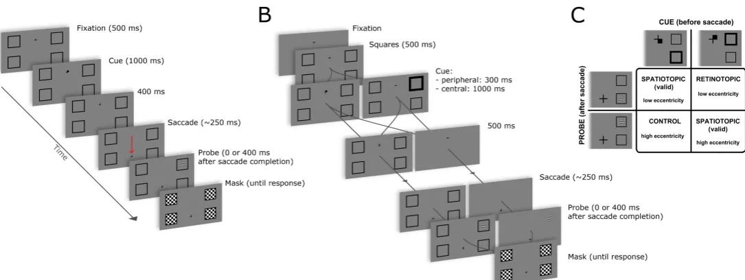
[image:5.595.36.579.363.567.2]In the first experiment, we designed a simple trans-saccadic spatial cueing paradigm to investigate how saccades affect covert orienting to a visual object that remains present throughout. Participants oriented attention to one of four placeholders (see fig. 1 panel A), and then made a guided saccade. The probe stimulus could be presented immediately after the saccade or 400 ms later. Our hypothesis is that the presence of a visual object is required to trigger the automatic remapping that keeps track of spatiotopic coordinates across saccades. In this case, with objects always present, we expected to find facilitation at the spatiotopic location regardless of the probe delay (0 or 400ms).
Method
Participants. Ten volunteers participated in Experiment 1 (4 females, 6 males, including one author, ML; mean age was 28.7). All had normal or corrected to normal vision, and gave their informed consent.
Stimuli, design, and procedure. Participants were seated in a silent and dimly lit room, with the head positioned on a chin rest at 60 cm in front of the computer screen. The experiment was run on a PC, using E-Prime 2.0 software (Psychology Software Tools, Pittsburgh, PA). Eye movements were recorded with a Tobii T120 screen-based eyetracker (Tobii Technology, Sweden), which was used also to present stimuli through its embedded 17-inch TFT monitor.
The experiment used a trans-saccadic cueing paradigm: first, one of four locations was cued, then the fixation cross was displaced and participants made a saccade to refixate its new location (see fig. 1 panel A). After the saccade, a probe was presented in one of the four locations and they made a speeded discrimination of its orientation (horizontal or vertical) on a standard keyboard using the left and right index finger (assignment of left-right key with horizontal-vertical response was counter-balanced across participants). More in detail, each trial started with a black fixation cross appearing on a gray background, horizontally centered but displaced 4° above or below the center of the screen. As soon as the participant fixated the cross, the trial started and four black square outlines (squares were 2.5° wide) appeared, arranged at the 4 corners of a rectangle of 16° width and 8° height centered on the screen. After a delay (500 ms) the cue was presented. The cue consisted in a small black square appearing for 1000 ms in one of the four sectors of the fixation cross, indicating the square in the corresponding quadrant of the screen (see fig. 1 panel C for an example). Then 400 ms after the cue disappeared, the cross was displaced up or down (depending on its initial position) of 8°, and this jump indicated to the participants to make a vertical saccade to the new fixation position. Participants were instructed to maintain fixation at the new position until response, and to maintain attention focused on the cued square. The cue was spatially congruent with the probe on half of the trials (validly cued spatiotopic trials); the other half of trials was composed of an equal proportion of retinotopic trials (probe appearing at the retinotopic cued position) and control trials (see fig. 1, panel C). We preferred to use a non-predictive cue to avoid adding any involuntary effects to the voluntary orienting (Peterson & Gibson, 2011; Risko & Stolz, 2010). In control trials the probe appeared always on the same side of the cue (left or right), to avoid additional costs of crossing the vertical meridian (Rizzolatti et al., 1987).
adaptively adjusted online. The goal of the procedure was to keep the accuracy in the spatiotopic condition approximately within the range 65% - 85%: if after a spatiotopic trial the global spatiotopic accuracy exceeded 85% or was below 65% probe duration was respectively increased or decreased by one monitor refresh cycle (~16 ms).
Eye movements were monitored with a sampling frequency of 120 Hz; trials in which subjects did not make the correct saccade were aborted and repeated within the same block. The saccade was considered completed as soon as a gaze position sample was detected 2.5° or closer to the second fixation point (the saccade target). Then the probe was immediately presented in the 0 ms delay condition, or after 400 ms in the other condition. At the end of each trial, the whole sequence of sampled eye positions was automatically inspected: if in the initial fixation phase, or during the post-saccadic delay (only in 400 ms delay condition) the gaze position deviated more than 2° from the fixation point for at least three consecutive samples (about 25ms), the trial was repeated later in the same block. Note that the transport delay (between the eye event and the availability of the gaze position sample) in the Tobii T120 eyetracker can be as much as 30-35 ms (Technology Tobii AB, 2010); by adding the duration of one monitor refresh period (about 16.6 ms) and the eyetracker sampling period (about 8.3 ms) the effective delay between saccade completion and target presentation can increase up to 55-60 ms: nonetheless we will refer to the condition with shorter delay of probe presentation as “0 ms” for simplicity, as it is the condition without any extra delay added between the detection of gaze in the final position and target presentation. Previous studies (Golomb et al., 2008, 2011) found strongest attentional benefits at the retinotopic location 75 ms after saccade completion, thus despite the unwanted unavoidable delays, our “0 ms” condition represents an appropriate comparison to test the attentional updating against the longer delay (400 ms). In the second experiment, we use a different experimental setup and have a more precise control of time delays.
Results
Trials in which the response time was shorter than 100 ms or longer than 2000 ms were excluded from subsequent analysis (0.2% of total trials). The delay between the fixation cross changing position and the gaze position crossing a circular boundary at 2° from the initial fixation position was taken as a measure of the latency of the eye movement: trials with latency shorter than 100 ms or longer than 500 ms were excluded (5% of total trials), the mean latency in the remaining trial was 206 ms (with a standard deviation across participants of 21 ms).
Probe duration was adjusted online, according to accuracy in the spatiotopic condition; the mean probe duration resulted in 79 ms (between subject standard deviation, 29 ms, min 34 ms and max 125 ms). To rule out any potential mismatch in probe duration across conditions, we carried out a repeated-measures analysis of variance (ANOVA) on probe duration with probe position (spatiotopic, retinotopic, control) as within-subjects factor. No significant differences emerged [F(2, 18) = 0.5, p = 0.62] suggesting that our probe adjustment effectively matched accuracy across participants without significantly affecting the average duration between the experimental conditions.
Response accuracy was analyzed with a repeated-measures ANOVA with probe position (spatiotopic, retinotopic, control) and Delay (0, 400 ms) as within-subject factors. This analysis revealed a main effect of probe position [F(2, 18) = 16.65, p = 0.001], whereas the main effect of delay [F(1, 9) = 0.36, p = 0.56] and the two-way interaction [F(2, 18) = 0.56, p = 0.58] were not significant. Follow-up comparisons (all t-tests paired and two-tailed) revealed significant differences between spatiotopic and retinotopic trials [t(9) = 2.27, p = 0.049], as well as between retinotopic and control trials [t(9) = 3.62, p = 0.005]. We performed additional planned comparisons between retinotopic and spatiotopic trials with similar probe eccentricity, separate for each delay: this revealed a significant spatiotopic advantage at the later delay [t(9) = 3.73, p = 0.005], and no difference at the earlier delay [t(9) = 1.77, p = 0.11] (see fig. 2 panel A). See Supplemental material for additional control analysis on response accuracy that takes into account trial by trial variations in probe duration.
Discussion
trials was higher than in retinotopic trials, however when comparing retinotopic and spatiotopic trials matched for eccentricity, at the two delays, we find that the advantage of spatiotopic over retinotopic trials was significant only at the later delay, a result that is consistent with a growing spatiotopic facilitation after the saccade and/or a decaying retinotopic trace (e.g., Golomb et al., 2008).
One difference with respect to the task used by Golomb and colleagues (2008) is that in our design the saccade was always predictable. However it is unlikely that the predictability of the saccade contributed to faster remapping, as other studies have found that attention is predictively remapped in advance of a saccade using unpredictable saccades (Jonikaitis et al., 2013; Rolfs et al., 2011).
Experiment 2
If the continuous presence of a visual object at the attended location (i.e., the placeholder) is required to maintain attention at the spatiotopic location after a saccade, removing the object after the cue and before the eye movement (as it was the case in the paradigm of Golomb et al., 2010) should impair the ability to sustain attention at the spatiotopic location, even in our simpler paradigm with only four possible probe locations. We tested this hypothesis in the second experiment, by modifying the paradigm used in experiment 1 so that the placeholders could either disappear after cue presentation or remain visible throughout the trial. These two conditions were randomly interleaved within each block. We also used two types of cues, a central, symbolic cue identical to the one used in experiment 1, and a peripheral cue, similar to the one used in previous studies (e.g., Golomb et al., 2008) to control for possible confounding effects related to the type of attentional cue.
Method
Participants. Twelve volunteers participated in experiment 2 (5 females and 7 males, including one author, ML; mean age was 29.2). All had normal or corrected to normal vision, and gave their informed consent. We added two participants with respect to experiment 1 in order to test higher order interactions (the interactions with placeholder presence and cue type) with possibly smaller effect size.
(Brainard, 1997; Pelli, 1997). Eye movements were recorded with an Eyelink 1000 Desktop Mount (SR Research, Osgoode, Ontario, Canada) with a sampling rate of 1kHz and a minimal transport delay, less than 1.8 ms (SR Research, n.d.).
The procedure was based upon the one adopted in experiment 1 (see fig. 1 panel B). In half of the trials we presented a central cue, identical to the one used in experiment 1; in the other half of the trials we presented a peripheral cue consisting in the outline of the cued placeholder increasing its thickness up to 3 times the original value (the internal area of the square remained constant during the increase, see figure 1 panel C for an example); the two types of cue were randomly interleaved within blocks. Peripheral cues are faster in orienting attention, and so we used a shorter duration (300 ms), similar to the duration of peripheral cues used in previous studies (e.g., Golomb et al., 2008).
After cue presentation, the four squares delimiting the relevant positions could either disappear or remain on the screen. Participants were instructed to ignore the disappearance of the placeholders whenever that occurred, and to maintain attention focused on the cued spatial location whether or not the placeholders were still present. After 500 ms the cross was displaced up or down, depending on its initial position, of 10°, and this jump signaled the participants to make a saccade to the new fixation position. Golomb and colleagues found an enhanced pattern of early retinotopic and late spatiotopic facilitation with larger eccentricities (at 11° vs 5° eccentricity, Golomb et al., 2008, supplemental material). We therefore increased the size of saccade and display (and thus also probe eccentricity) to maximize the possibility of stronger retinotopic effects at larger eccentricities: the 4 positions were now at the corners of a rectangle of 20° width and 10° height. The probe stimulus was again a Gabor patch (contrast 100%, spatial frequency 2 cycles/degree) presented at different delays after saccade completion (0 and 400 ms). The duration of probe presentation was adjusted online by a standard staircase procedure with criterion performance of 75% correct responses in spatiotopic trials and step of one monitor refresh cycle (~8 ms). One staircase was used for both spatiotopic trials with probe at higher and lower eccentricity. Probe duration was allowed to vary between 16 and 250 ms. We used a different procedure with respect to the previous experiment because of the higher monitor vertical refresh rate (120Hz), which allowed for a finer modulation of probe duration.
Each participant made 512 trials, 256 trials for the spatiotopic condition, and 128 for each of the other conditions, in 2 experimental sessions on different days; each session was divided into 4 blocks. Trials with different cueing conditions (spatiotopic, retinotopic and control), type of cue (central or peripheral) and presence/absence of the placeholders were randomly interleaved within blocks. As in the first experiment, before each session, participants completed 40 pre-test trials, consisting of only spatiotopic trials, in which the duration of the probe presentation was adapted according to a weighted up-down staircase procedure (Kaernbach, 1991) with criterion performance of 75% correct responses.
Results
We detected the saccades with an algorithm based on two-dimensional eye velocity (Engbert & Mergenthaler, 2006) and defined a response saccade as the first saccade that left a circular fixation region and landed inside a target-centered circular region (radii of 2°). We rejected trials with blinks or saccades larger than 1° before the response saccade, or after the saccade and before probe presentation in trials with the longer delay (400 ms). We excluded also trials in which the saccadic latency was shorter than 100 ms or longer than 500 ms; the mean latency in the remaining trials was 181 ms (with a standard deviation across participants of 36 ms). The precise time of probe onset was marked in the eye movement recordings, and was compared to the saccade landing time (detected offline): in particular in the 0 ms delay condition we excluded trials in which the onset of the probe was delayed 20 ms or more with respect to the saccade landing time (supplementary fig. 1, panel A). In all, 85% of trials were included in subsequent analysis.
Figure 2 Response accuracy. (A) Mean response accuracy in Experiment 1 in retinotopic and spatiotopic trials with similar eccentricities, represented by the difference from the control condition. (B)Mean accuracy in Experiment 2 as a function of probe position and placeholder presence/absence. The presence of placeholder significantly increased accuracy only in spatiotopic trials. Panel C and D
represent mean accuracy in retinotopic and spatiotopic trials with similar eccentricities as difference from control condition (Experiment 2). Error bars indicate SEM.
We conducted a repeated-measures ANOVA with mean accuracy as dependent variable and probe position (spatiotopic, retinotopic, control), delay (0, 400 ms), cue type (central, peripheral) and placeholders (present, absent) as within-subject factors. This analysis revealed significant main effects of probe position [F(2, 22) = 4.40, p = 0.02], delay [F(1, 11) = 110.60, p < 0.0001] and placeholders [F(1, 11) = 14.62, p = 0.001]. We found a significant interaction between delay and placeholders [F(1, 11) = 4.87, p = 0.049] and between probe position and placeholders [F(2, 22) = 6.88, p = 0.003] (fig. 2 panel B). We found also a significant three-way interaction between probe position, presence of placeholders and delay [F(2, 22) = 8.24, p =
A
B
C
D
0 5 10 15 20 25Probe delay (ms)
Accuracy difference (%)
0 400 0 5 10 15 20 25 retinotopic spatiotopic Experiment 1 70 75 80 Placeholders Accuracy (%) absent present 70 75 80 control retinotopic spatiotopic Experiment 2 Placeholders absent
Probe delay (ms)
Accuracy difference (%) -10
0
5
10
Placeholders present
Probe delay (ms)
Accuracy difference (%)
[image:12.595.136.442.99.470.2]0.002] (fig. 2 panel C and D). The cue type did not yield a significant main effect [F(1, 11) = 2.77, p = 0.12] and did not interact significantly with any other factor [condition*delay*boxes*cue type: F(2, 22) = 0.07, p = 0.92; condition*boxes*cue type: F(2, 22) = 1.54, p = 0.23; condition*cue type: F(2, 22) = 1.95, p = 0.16; delay*cue type: F(1, 11) = 1.78, p = 0.21; boxes*cue type: F(1, 11) = 1.95, p = 0.29].
Follow-up comparisons (all t-tests paired and two-tailed) revealed that, with the placeholder present, response accuracy was higher in spatiotopic trials than in both control [t(11) = 3.07, p = 0.01] and retinotopic trials [t(11) = 4.15, p = 0.002], while in contrast to experiment 1, there were no differences between retinotopic and control trials [t(11) = 0.45, p = 0.66]. On the other hand, in the placeholder-absent condition, there were no significant differences between spatiotopic and control trials [t(11) = 0.55, p = 0.59], spatiotopic and retinotopic trials [t(11) = 0.41, p = 0.69], or retinotopic and control trials [t(11) = 0.59, p = 0.56]. This pattern of results suggests that the presence of placeholders increased accuracy specifically in spatiotopic trials (fig. 2 panel B). This was further confirmed by a significant comparison between spatiotopic trials in the condition with versus without placeholders [t(11) = 5.71, p < 0.0001]; the same comparison did not reach significance for retinotopic [t(11) = 1.27, p = 0.23] or control [t(11) = 1.35, p = 0.20] trials.
We also found a significant three-way interaction between probe position, presence of placeholders and delay, and as a follow up we performed comparisons (paired and two-tailed) between the condition with placeholders present vs. the condition with placeholders absent for each probe position (spatiotopic retinotopic, control) and delay (0, 400). Spatiotopic trials resulted in a greater accuracy in the placeholder present condition at both delays [delay 0: t(11) = 3.97, p = 0.002; delay 400: t(11) = 6.57, p < 0.0001]; no difference was found in control trials [delay 0: t(11) = 0.65, p = 0.52; delay 400: t(11) = 1.30, p = 0.21]; retinotopic trials had a greater accuracy in the placeholder present condition but only at the later delay [delay 0: t(11) = 1.67, p = 0.12; delay 400: t(11) = 3.71, p = 0.003]. The three-way interaction thus arises from the different effect of placeholder absence on retinotopic trials at the two delays. Comparison with the control conditions revealed a similar pattern: in the condition without placeholders, accuracy in retinotopic trials was significantly lower than control trials at the later delay [t(11) = 2.44, p = 0.03], but no significant difference emerged at the earlier delay [t(11) = 1.23, p = 0.24].
2.58, p = 0.02] (fig. 2 panel D), but only at the later delay in the placeholder absent condition [t(11) = 2.22, p = 0.048] (fig. 2 panel C). Accuracy at the retinotopic and spatiotopic location did not differ at earlier delay in the placeholder absent condition [t(11) = 0.61, p = 0.55].
Discussion
The results of this second experiment are in agreement with our hypothesis. Analysis of response accuracy revealed that the continuous presence of placeholders enhanced performance at the spatiotopic location. The facilitation was specific to the spatiotopic location and thus not the result of a general improvement due to reduced spatial uncertainty (the presence of the placeholders had no effect on performance at the control location). Contrary to previous studies (e.g., Golomb et al., 2008), we did not find any spatiotopic facilitation in the condition with no placeholders, suggesting that participants failed to maintain attention at that location. Additionally, the three-way interaction revealed that the presence or absence of placeholders had a different effect at the retinotopic location at the two delays: when the placeholders were removed before the saccade, accuracy in retinotopic trials dropped with respect to the condition with placeholders present, but only at the longer delay. At the shorter delay with placeholders absent, accuracy was greater at cued retinotopic location (fig. 2 panel C), a pattern that resembles the retinotopic trace results from Golomb and colleagues (e.g., Golomb et al, 2008), but the difference with spatiotopic trials did not reach statistical significance in our data.
In contrast to the results of the first experiment, there was no retinotopic benefit (accuracy was similar in retinotopic and control trials, both lower than spatiotopic trials) in the condition with placeholders present. It is known that attention spreads in a gradient fashion around a cue (Downing & Pinker, 1985); additionally the gradient seems to be asymmetrical, with larger costs for probes more peripheral than the cue (Shulman, Sheehy, & Wilson, 1986). Therefore one possibility is that the difference between retinotopic and control trials in the first experiment might have been caused by the different position of the probe relative to the cue (probes are more peripheral than the cue in control trials, and vice versa in retinotopic trials). In the second experiment this effect might have been reduced because of the larger (10° vs. 8°) distance between probe locations.
ms” condition might have been delayed up to 55-60 ms due to timing lags in the eye movement monitoring and screen refresh (a delay that is still comparable with the persistence of retinotopic benefits found in previous studies, e.g. Golomb et al., 2008). In contrast, the different experimental setup used in the second experiment allowed for a finer temporal control of eye and trial events, and we were able to present stimuli almost immediately after the saccade (delay less than 20 ms confirmed by offline analyses, see supplementary fig. 1), thus making the early delay condition more challenging for participants. In any case, these less accurate responses for the short delay probe do not affect our conclusions concerning spatiotopic and retinotopic allocation of attention.
General discussion
Our findings reveal that the presence of a visual object at the attended location is a critical factor for the maintenance of the spatial constancy of attention – the ability to sustain attention in spatiotopic coordinates across eye movements. When visual attention is directed to an object, the placeholder, it is remapped to the correct spatial location with each eye movement. In contrast, when attention is directed to an empty location, participants fail to maintain it at that location across eye movements, indicating a lack of appropriate remapping.
IOR, but it was limited to the short delay after the saccade, and it turned into spatiotopic IOR at the longer delay.
An alternative view of our experiments is that the paradigm actually investigates memory rather than attention, as it involves the active maintenance of a location of interest across a saccade. There is an interesting overlap between attention and memory (Awh & Jonides, 2001), and several previous studies have used a spatial memory task to manipulate attention across saccades (Golomb et al., 2008; Golomb & Kanwisher, 2012; Golomb, Pulido, et al., 2010). However, since we did not ask participants to identify where the cue was, but only to identify a subsequent probe that may or may not appear at the same location of the cue, we have used a standard definition in the field and presented the cued performance as a measure of attention rather than memory. In particular, the cued location was continuously attended, rather than stored to be retrieved later. Unlike items stored in memory, continuously attended items do not, for example, show the decay functions of memory. Attended items show no decay at all until the focus of attention is distracted.
Overall, the pattern of results in the present study is consistent with the idea that remapping of attention across eye movements comprises two complementary but distinct processes: a rapid updating of the focus of attention to the new location, and a slower process of suppressing the attentional focus at the previous retinotopic location (Golomb, L’Heureux, & Kanwisher, 2014; Golomb, Pulido, et al., 2010). In our paradigm, the absence of placeholders seems to have prevented the updating process, but might have not blocked the suppressing process, resulting in a modulation over time of response accuracy at the retinotopic location. The idea that the two processes (spatiotopic updating and retinotopic suppression) might be relatively independent is consistent with another study by Golomb and colleagues (Golomb, Nguyen-Phuc, et al., 2010) in which they used EEG and fMRI to investigate the neural correlates of the retinotopic attentional trace: in that study participants responded only to probes presented at the central location, while ignoring the other locations. Both blood oxygen level-dependent signals and event-related potentials showed the strongest response enhancement for probes presented at the spatiotopic location, even at the shorter delay after the saccade. However, even if the spatiotopic location was facilitated at the shorter delay, they also found a robust enhancement for irrelevant probes presented at the retinotopic location, suggesting that the new spatiotopic location might be facilitated independently of when the attentional focus at the previous retinotopic location was extinguished.
location, irrespective of whether it was delimited or not by a placeholder. Nevertheless participants failed to sustain attention at the spatiotopic location when a placeholder did not mark it. This suggests that activity in areas responsible for spatial updating across saccades is modulated by object-based properties in the image. It remains a challenge for future studies to determine how this modulation takes place: one hypothesis could be that the visual representation in these maps depends mostly on grouping cells in earlier visual cortices that operate some pre-attentive figure-ground segmentation (Qiu, Sugihara, & von der Heydt, 2007). This early grouping cells could bind basic visual features into larger compounds and provide the structure for top-down attentional selection (Mihalas, Dong, von der Heydt, & Niebur, 2011). An interesting question for future research is whether this attentional modulation becomes more effective in the periphery of the visual field.
One difference between our results and previous findings (Golomb et al., 2008, 2011; Golomb, Pulido, et al., 2010) is that we did not find evidence for a growing spatiotopic benefit in the condition with placeholder absent. However, in the original paradigm of Golomb et al., attention was manipulated through a spatial memory task, and this might have enforced participants to voluntarily recover the original location of the cue after the saccade (perhaps on the basis of other spatial landmarks, like the monitor’s frame) leading to the late increase of the spatiotopic benefit. In our task, the memory of the cue location was not tested, and participants might have let attention spread over all the cued hemifield (Hughes & Zimba, 1987) instead of recovering the specific cued location.
An alternative explanation of our findings could be that a visual object simply acted as a landmark and facilitated the localization of the cued location after the eye movement (Deubel, Schneider, & Bridgeman, 2002; Deubel, 2004). However, without an anticipatory attention shift (Jonikaitis et al., 2013; Rolfs et al., 2011) localization of the cued position, even if facilitated by landmarks, would need to be followed by an attention shift to that position. The typical duration of an attention shift has been estimated to be around 150-200 ms (Khayat, Spekreijse, & Roelfsema, 2006; Montagnini & Castet, 2007), a duration much longer than the average probe duration in our second experiment (44 ms). It is thus unlikely that an attention shift occurred to the cued location after the saccade, as this would not have yielded the early spatiotopic benefit found here and in other studies (Golomb, Pulido, et al., 2010; Jonikaitis et al., 2013).
References
Awh, E., & Jonides, J. (2001). Overlapping mechanisms of attention and spatial working memory.
Trends in Cognitive Sciences, 5(3), 119–126.
Berman, R., & Colby, C. (2009). Attention and active vision. Vision Research, 49(10), 1233–48. doi:10.1016/j.visres.2008.06.017
Brainard, D. H. (1997). The Psychophysics Toolbox. Spatial Vision, 10(4), 433–436. doi:10.1163/156856897X00357
Casarotti, M., Lisi, M., Umiltà, C., & Zorzi, M. (2012). Paying attention through eye movements: a computational investigation of the premotor theory of spatial attention. Journal of Cognitive Neuroscience, 24(7), 1519–31. doi:10.1162/jocn_a_00231
Cavanagh, P., Hunt, A. R., Afraz, A., & Rolfs, M. (2010). Visual stability based on remapping of attention pointers. Trends in Cognitive Sciences, 14(4), 147–53.
doi:10.1016/j.tics.2010.01.007
Deubel, H. (2004). Localization of targets across saccades: Role of landmark objects. Visual Cognition,
11(2-3), 173–202. doi:10.1080/13506280344000284
Deubel, H., Schneider, W. X., & Bridgeman, B. (2002). Transsaccadic memory of position and form.
Progress in Brain Research, 140, 165–80. doi:10.1016/S0079-6123(02)40049-0
Downing, C. J., & Pinker, S. (1985). The spatial structure of visual attention. In M. I. Posner & O. S. M. Marin (Eds.), Attention and Performance XI: Mechanisms of Attention (pp. 171–187). Hillsdale, NJ: Erlbaum.
Duhamel, J. R., Colby, C. L., & Goldberg, M. E. (1992). The updating of the representation of visual space in parietal cortex by intended eye movements. Science (New York, N.Y.), 255(5040), 90–2.
Egly, R., Driver, J., & Rafal, R. D. (1994). Shifting visual attention between objects and locations: evidence from normal and parietal lesion subjects. Journal of Experimental Psychology. General, 123(2), 161–77.
Engbert, R., & Mergenthaler, K. (2006). Microsaccades are triggered by low retinal image slip.
Proceedings of the National Academy of Sciences of the United States of America, 103(18), 7192–7. doi:10.1073/pnas.0509557103
Fecteau, J. H., & Munoz, D. P. (2006). Salience, relevance, and firing: a priority map for target selection. Trends in Cognitive Sciences, 10(8), 382–90. doi:10.1016/j.tics.2006.06.011 Feldman, J. (2003). What is a visual object? Trends in Cognitive Sciences, 7(6), 252–256.
doi:10.1016/S1364-6613(03)00111-6
Golomb, J. D., Chun, M. M., & Mazer, J. a. (2008). The native coordinate system of spatial attention is retinotopic. The Journal of Neuroscience: The Official Journal of the Society for
Neuroscience, 28(42), 10654–62. doi:10.1523/JNEUROSCI.2525-08.2008
Golomb, J. D., & Kanwisher, N. (2012). Retinotopic memory is more precise than spatiotopic memory.
Proceedings of the National Academy of Sciences of the United States of America, 109(5), 1796–801. doi:10.1073/pnas.1113168109
doi:10.1177/0956797614522068
Golomb, J. D., Marino, A. C., Chun, M. M., & Mazer, J. a. (2011). Attention doesn’t slide: spatiotopic updating after eye movements instantiates a new, discrete attentional locus. Attention, Perception & Psychophysics, 73(1), 7–14. doi:10.3758/s13414-010-0016-3
Golomb, J. D., Nguyen-Phuc, A. Y., Mazer, J. a, McCarthy, G., & Chun, M. M. (2010). Attentional Facilitation throughout Human Visual Cortex Lingers in Retinotopic Coordinates after Eye Movements. The Journal of Neuroscience: The Official Journal of the Society for Neuroscience, 30(31), 10493–506. doi:10.1523/JNEUROSCI.1546-10.2010
Golomb, J. D., Pulido, V. Z., Albrecht, A. R., Chun, M. M., & Mazer, J. A. (2010). Robustness of the retinotopic attentional trace after eye movements. Journal of Vision, 10(3), 19.1–12. Hall, N. J., & Colby, C. L. (2011). Remapping for visual stability. Philosophical Transactions of the
Royal Society of London. Series B, Biological Sciences, 366(1564), 528–39. doi:10.1098/rstb.2010.0248
Hilchey, M. D., Klein, R. M., Satel, J., & Wang, Z. (2012). Oculomotor inhibition of return: how soon is it “recoded” into spatiotopic coordinates? Attention, Perception & Psychophysics, 74(6), 1145–53. doi:10.3758/s13414-012-0312-1
Hollingworth, A., Maxcey-Richard, A. M., & Vecera, S. P. (2012). The spatial distribution of attention within and across objects. Journal of Experimental Psychology. Human Perception and Performance, 38(1), 135–51. doi:10.1037/a0024463
Howe, P. D. L., Drew, T., Pinto, Y., & Horowitz, T. S. (2011). Remapping attention in multiple object tracking. Vision Research, 51(5), 489–95. doi:10.1016/j.visres.2011.01.001
Hughes, H. C., & Zimba, L. D. (1987). Natural boundaries for the spatial spread of directed visual attention. Neuropsychologia, 25(1), 5–18. doi:10.1016/0028-3932(87)90039-X
Hunt, A. R., & Cavanagh, P. (2011). Remapped visual masking. Journal of Vision, 11(1), 13. Jonikaitis, D., Szinte, M., Rolfs, M., & Cavanagh, P. (2013). Allocation of attention across saccades.
Journal of Neurophysiology, 109(5), 1425–34. doi:10.1152/jn.00656.2012
Kaernbach, C. (1991). Simple adaptive testing with the weighted up-down method. Perception & Psychophysics, 49(3), 227–229. doi:10.3758/BF03214307
Khayat, P. S., Spekreijse, H., & Roelfsema, P. R. (2006). Attention lights up new object representations before the old ones fade away. The Journal of Neuroscience : The Official Journal of the Society for Neuroscience, 26(1), 138–42. doi:10.1523/JNEUROSCI.2784-05.2006 Klein, R. (2000). Inhibition of return. Trends in Cognitive Sciences, 4(4), 138–147.
Krüger, H. M., & Hunt, A. R. (2013). Inhibition of return across eye and object movements: the role of prediction. Journal of Experimental Psychology. Human Perception and Performance,
39(3), 735–44. doi:10.1037/a0030092
Kusunoki, M., & Goldberg, M. E. (2003). The time course of perisaccadic receptive field shifts in the lateral intraparietal area of the monkey. Journal of Neurophysiology, 89(3), 1519–27. doi:10.1152/jn.00519.2002
Lupianez, J., Klein, R. M., & Bartolomeo, P. (2006). Inhibition of return: Twenty years after. Cognitive Neuropsychology, 23(7), 1003–14. doi:10.1080/02643290600588095
attention. Perception & Psychophysics, 67(7), 1140–9.
Mathôt, S., & Theeuwes, J. (2010). Gradual remapping results in early retinotopic and late spatiotopic inhibition of return. Psychological Science, 21(12), 1793–8. doi:10.1177/0956797610388813 Mathôt, S., & Theeuwes, J. (2011). Visual attention and stability. Philosophical Transactions of the
Royal Society of London. Series B, Biological Sciences, 366(1564), 516–27. doi:10.1098/rstb.2010.0187
Mihalas, S., Dong, Y., von der Heydt, R., & Niebur, E. (2011). Mechanisms of perceptual organization provide auto-zoom and auto-localization for attention to objects. Proceedings of the National Academy of Sciences of the United States of America, 108(18), 7583–8.
doi:10.1073/pnas.1014655108
Montagnini, A., & Castet, E. (2007). Spatiotemporal dynamics of visual attention during saccade preparation: Independence and coupling between attention and movement planning. Journal of Vision, 7(14), 8.1–16.
Pelli, D. G. (1997). The VideoToolbox software for visual psychophysics: transforming numbers into movies. Spatial Vision, 10(4), 437–442. doi:10.1163/156856897X00366
Pertzov, Y., Zohary, E., & Avidan, G. (2010). Rapid formation of spatiotopic representations as revealed by inhibition of return. The Journal of Neuroscience : The Official Journal of the Society for Neuroscience, 30(26), 8882–7. doi:10.1523/JNEUROSCI.3986-09.2010
Peterson, S. A., & Gibson, T. N. (2011). Implicit attentional orienting in a target detection task with central cues. Consciousness and Cognition, 20(4), 1532–47.
doi:10.1016/j.concog.2011.07.004
Pylyshyn, Z. W., & Storm, R. W. (1988). Tracking multiple independent targets: evidence for a parallel tracking mechanism. Spatial Vision, 3(3), 179–97.
Qiu, F. T., Sugihara, T., & von der Heydt, R. (2007). Figure-ground mechanisms provide structure for selective attention. Nature Neuroscience, 10(11), 1492–9. doi:10.1038/nn1989
Rafal, R. D., Calabresi, P. A, Brennan, C. W., & Sciolto, T. K. (1989). Saccade preparation inhibits reorienting to recently attended locations. Journal of Experimental Psychology. Human Perception and Performance, 15(4), 673–85.
Risko, E. F., & Stolz, J. a. (2010). The proportion valid effect in covert orienting: strategic control or implicit learning? Consciousness and Cognition, 19(1), 432–42.
doi:10.1016/j.concog.2009.07.013
Rizzolatti, G., Riggio, L., Dascola, I., & Umiltá, C. (1987). Reorienting attention across the horizontal and vertical meridians: evidence in favor of a premotor theory of attention. Neuropsychologia,
25(1A), 31–40.
Rolfs, M., Jonikaitis, D., Deubel, H., & Cavanagh, P. (2011). Predictive remapping of attention across eye movements. Nature Neuroscience, 14(2), 252–6. doi:10.1038/nn.2711
Satel, J., Wang, Z., Hilchey, M. D., & Klein, R. M. (2012). Examining the dissociation of retinotopic and spatiotopic inhibition of return with event-related potentials. Neuroscience Letters, 524(1), 40–4. doi:10.1016/j.neulet.2012.07.003
Scholl, B. J. (2001). Objects and attention: the state of the art. Cognition, 80(1-2), 1–46. Scholl, B. J., Pylyshyn, Z. W., & Feldman, J. (2001). What is a visual object? Evidence from target
http://www.ncbi.nlm.nih.gov/pubmed/11245843
Shulman, G. L., Sheehy, J. B., & Wilson, J. (1986). Gradients of spatial attention. Acta Psychologica,
61(2), 167–81.
Sommer, M. A., & Wurtz, R. H. (2006). Influence of the thalamus on spatial visual processing in frontal cortex. Nature, 444(7117), 374–7. doi:10.1038/nature05279
Spelke, E. S. (1990). Principles of Object Perception. Cognitive Science, 14(1), 29–56. doi:10.1207/s15516709cog1401_3
SR Research. (2013). Eyelink technical specifications. Retrieved from http://www.sr-research.com/eyelink1000.html
Talsma, D., White, B. J., Mathôt, S., Munoz, D. P., & Theeuwes, J. (2013). A retinotopic attentional trace after saccadic eye movements: evidence from event-related potentials. Journal of Cognitive Neuroscience, 25(9), 1563–77. doi:10.1162/jocn_a_00390
Technology Tobii AB. (2010). Timing Guide for Tobii Eye Trackers and Eye Tracking Software (White paper). Retrieved from: http://www.tobii.com/en/eye-tracking-research/global/library/white-papers/. Wurtz, R. H. (2008). Neuronal mechanisms of visual stability. Vision Research,
48(20), 2070–89. doi:10.1016/j.visres.2008.03.021
Wurtz, R. H., Joiner, W. M., & Berman, R. a. (2011). Neuronal mechanisms for visual stability: progress and problems. Philosophical Transactions of the Royal Society of London. Series B,
Biological Sciences, 366(1564), 492–503. doi:10.1098/rstb.2010.0186
Supplemental material
GLMM analysis of response accuracy.
We performed additional analyses of response accuracy in experiment 1 and 2 to provide a better control for the effect of trial-by-trial variations in the probe duration. To that end, we used a generalized linear mixed-effects model (Pinheiro & Bates, 2000), fitted with the open-source software R (R Development Core Team, 2012) and the lme4 library (Bates, Maechler, Bolker, & Walker, 2014). Mixed-effects models are an extension of linear models: they are called mixedbecause they include not only
fixed effects (experimental manipulations) but also random effects, associated with individual observational units drawn at random from a population. Mixed-effects models are typically used to analyze grouped data where, like in repeated measures designs, the response variable and covariates are grouped according to one or more classification factors (e.g., individual participants). Common random effects are associated to observations sharing the same level of a grouping factor, allowing mixed-effects models to flexibly represent the covariance structure induced by grouping of the data (Pinheiro & Bates, 2000).
Since our response variable was binary (response accuracy), we used a generalized effects model, with the logistic as the link function. In a mixed-effects model the response vector is taken conditionally on the random mixed-effects (Pinheiro & Bates, 2000), and is modeled as the sum of a fixed effect term X and a random effect term Z. Formally the model can be expressed as (in matrix form):
logit 𝑃(𝑌= 1|𝑏) = 𝑿𝛽+𝒁𝑏
where Y is the response vector, Xβrepresents the fixed effects term (the product of a design matrix X and a vector of fixed effects coefficients β), Zb is the random effects term (the product of a design matrix Z for the random effects and a vector of random effects coefficients b, such that 𝑏~𝑁(0,𝜎!!)). Finally logit 𝑝 = log !
!!! is the link function in the logit form.
One advantage of these models is that they can include predictors that change on a trial-by-trial basis, as is the case for probe duration in our experiments. In the following analysis we included the subject as random effect factor, and the probe duration as a continuous covariate using trial-by-trial values. The probe duration was also included in the random effect part, in interaction with subject, allowing individual variations to the shape of the relation between the response variable (accuracy) and probe duration.
all possible models that differed from the current one by dropping a single term (maintaining marginality) and compared these reduced models to the original one with a likelihood-ratio test (Pinheiro & Bates, 2000). If one or more term could be dropped without a significant increase in unexplained deviance (as indicated by the likelihood-ratio tests), we removed them and fitted a new model without these terms. Then, in the next step, we repeated the process but starting from this updated model. We stopped when no more terms could be dropped from the model, without a
significant increase in residual unexplained deviance or a violation of marginality (on the importance of marginality in regression analysis we refer the reader to Nelder, 1977; Venables, 1998).
The results of this process for each of the two experiments are presented in table 1 and 2 below. For each step in the simplification process we listed the
regression terms that could be dropped, along with the results of the correspondent likelihood ratio tests. Note that in the last row of each table, corresponding to the final step, the reported likelihood ratio tests are significant. This indicates that excluding these terms would cause a significant increase in the residual variance of the model and a significant worsening of the model fit.
The simplification excluded delay and its interaction with other probe location as predictor from the best fitting model in Experiment 1 (see Table. 1), consistently with the ANOVA reported in the results section of experiment 1.
In Experiment 2, the simplification excluded cue type (central/peripheral) and all its interaction with other factors (see Table. 2), consistently with the result of the ANOVA reported in the results section of experiment 2. The process ended when the effect of probe duration and the three-way interaction between probe position,
placeholder presence and delay resulted significant; all the other main effects and interactions could not be excluded without incurring in a violation of marginality and/or a significant increase in residual deviance. Again, the finding of a significant three-way interaction is consistent with the ANOVA in the results section of experiment 2. The significant effect of probe duration was expected, as the probe duration was updated online with a staircase procedure, but importantly with the current analysis we were able to exclude its effect when testing the significance of the other
Overall, the analysis presented here indicates that the significant effects
reported in the main text were not driven by trial-by-trial fluctuations in probe duration (the models examined here take into account trial by trial probe durations as a
covariate), or by eventual deviations from normality of the ANOVA residuals due to the proportional nature of the response variable (the models use a logistic link function to account for the binomial response variable).
Table 1
Backward stepwise simplification of GLMM model (exp. 1)
Step Regression term df χ2 p
1 probe position * delay 2 3.02 0.22
2 delay 1 0.02 0.89
3 (end) probe duration 1 14.21 0.0002
probe position 2 44.77 <0.0001
Table 2
Backward stepwise simplification of GLMM model (exp. 2)
Step Regression term df χ2 p
1 probe position * delay * placeholders presence * cue type 2 0.57 0.75
2 probe position * delay * cue type 2 3.26 0.19
probe position * placeholders presence * cue type 2 3.84 0.15
delay * placeholders presence * cue type 1 0.156 0.69
3 delay * cue type 1 0.55 0.35
placeholder presence * cue type 1 0.86 0.35
probe position * cue type 2 3.58 0.17
4 cue type 1 0.47 0.49
5 (end) probe duration 1 21.73 <0.0001
Supplemental references
Bates, D., Maechler, M., Bolker, B., & Walker, S. (2014). lme4: Linear mixed-effects models using Eigen and S4. R package version 1.1-7. Retrieved from http://cran.r-project.org/package=lme4
Nelder, J. A. (1977). A Reformulation of Linear Models. Journal of the Royal Statistical Society. Series A (General), 140(1), 48. doi:10.2307/2344517
Pinheiro, J. C., & Bates, D. M. (2000). Mixed-Effects Models in S and S-PLUS. New York: Springer-Verlag. doi:10.1007/b98882
R Development Core Team (2012). R: A language and environment for statistical computing. Vienna, Austria: R Foundation for Statistical Computing. Retrieved from http://www.r-project.org/
Supplemental tables: response accuracy
Table 3
Experiment 1: mean accuracy (SD) as a function of probe position and delay
Probe position
delay
0 400
spatiotopic (higher eccentricity) 0.78 (0.09) 0.81 (0.07)
spatiotopic (lower eccentricity) 0.83 (0.07) 0.84 (0.06)
retinotopic 0.77 (0.14) 0.76 (0.07)
control 0.70 (0.12) 0.68 (0.12)
[image:28.595.62.536.407.662.2]n=10
Table 4
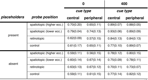
Experiment 2: mean accuracy (SD) as a function of probe position, delay, placeholders and cue type
placeholders probe position
delay
0
400
400
cue type cue type
central peripheral central peripheral
present
spatiotopic (higher ecc.) 0.70(0.20) 0.65(0.11) 0.89(0.07) 0.88(0.05)
spatiotopic (lower ecc.) 0.79(0.04) 0.74(0.13) 0.93(0.06) 0.89(0.09)
retinotopic 0.62(0.09)
0.76 (0.07)
0.57(0.15) 0.84(0.13) 0.84(0.13)
control 0.61(0.17) 0.65(0.11) 0.77(0.10) 0.89(0.07)
absent
spatiotopic (higher ecc.) 0.59(0.11) 0.56(0.15) 0.78(0.12) 0.80(0.15)
spatiotopic (lower ecc.) 0.60(0.14) 0.67(0.14) 0.75(0.09) 0.78(0.11)
retinotopic 0.63(0.13) 0.67(0.12) 0.70(0.11) 0.73(0.07)
control 0.59(0.11) 0.61(0.15) 0.77(0.14) 0.82(0.12)