Demonstration of Coherent Terahertz Transition Radiation from Relativistic
Laser-Solid Interactions
Guo-Qian Liao 1, Yu-Tong Li 1,5, Yi-Hang Zhang 1,Hao Liu 1, Xu-Lei Ge 2, Su Yang 2, Wen-Qing
Wei 2, Xiao-Hui Yuan 2,5*, Yan-Qing Deng 3, Bao-Jun Zhu 1, Zhe Zhang 1, Wei-Min Wang 1,5,
Zheng-Ming Sheng 4,2,5, Li-Ming Chen 1, Xin Lu 1, Jing-Long Ma 1, Xuan Wang 1,and Jie Zhang 2,5
1
Beijing National Laboratory for Condensed Matter Physics, Institute of Physics, Chinese Academy of Sciences, Beijing 100190, China
2
Key Laboratory for Laser Plasmas (MoE) and Department of Physics and Astronomy, Shanghai Jiao Tong University, Shanghai 200240, China
3
College of Science, National University of Defense Technology, Changsha 410073, China 4
SUPA, Department of Physics, University of Strathclyde, Glasgow G4 0NG, UK 5
Collaborative Innovation Center of IFSA (CICIFSA), Shanghai Jiao Tong University, Shanghai 200240, China
Coherent transition radiation in the terahertz (THz) region with energies of sub-mJ/pulse has
been demonstrated by relativistic laser-driven electron beams crossing the solid-vacuum boundary.
Targets including mass-limited foils and layered metal-plastic targets are used to verify the radiation
mechanism and characterize the radiation properties. Observations of THz emissions as a function
of target parameters agree well with the formation-zone and diffraction model of transition radiation.
Particle-in-cell simulations also well reproduce the observed characteristics of THz emissions. The
present THz transition radiation enables not only a potential tabletop brilliant THz source, but also a
novel noninvasive diagnostic for fast electron generation and transport in laser-plasma interactions.
PACS numbers: 52.38.Kd, 52.59.Ye, 41.60.Dk
High power terahertz (THz) sources can serve as a unique tool [1] in the exploration of
condensed-matter dynamics [2,3], biomedical imaging [4], and wireless communications [5]. Over
the last decades relativistic electron beams from conventional accelerators have been applied to
generate strong THz radiation through various mechanisms such as transition radiation [6],
Cherenkov radiation [7], synchrotron radiation [8], diffraction radiation [9], etc. The energy of THz
pulses through transition radiation in linear accelerators has reached ~600 μJ/pulse [10].
Relativistic electron beams can also be generated in the interactions of intense laser pulses with
low-density gas or high-density solid targets. For gas targets, electrons can be accelerated to a GeV
energy level with wakefields [11,12]. With such an electron beam, Leemans et al. have observed a
~0.3 μJ THz pulse through transition radiation [13]. Strong THz radiation from laser-solid
interactions has also been demonstrated [14,15,16,17]. Compared with the cases of gas targets,
electron beams from solid targets have much higher charge, up to nC-μC. Recently THz radiation
with energies of >700 μJ has been reported from the rear surface of a foil target [18,19]. Since the
THz yield is found to be correlated with the square of the proton number, it is attributed to a
transient dipole-like charge distribution structure formed by target normal sheath acceleration
(TNSA), referred to hereafter as TNSA radiation [18]. On the other hand, in principle, THz
radiation can be produced efficiently via the transition radiation process at the rear surface of a
laser-irradiated thin solid foil [20], since both the short electron bunch duration and high bunch
charge are ideal for this mechanism. Previous studies on the transition radiation from solid targets
mainly concern in the optical region [21,22]. While the radiation in the THz regime has so far not
yet been verified experimentally.
In this Letter, we will report the experimental demonstration of THz emissions via coherent
transition radiation (CTR) when the laser-produced electron beams pass through the rear
solid-vacuum interface. Various target structures and parameters are adopted to characterize the
THz radiation properties, which can be well explained by the theoretical model of CTR.
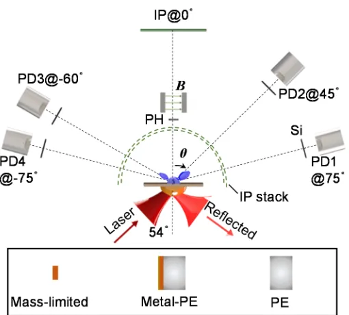
Shanghai Jiao Tong University. Figure 1 shows the layout of the experimental setup. A 2 J, 30 fs,
800 nm p-polarized laser pulse was focused onto solid targets at an incidence angle of 54° with a
peak irradiance of ~1.5×10¹⁹ W/cm2. The laser contrast ratio in the ns range was ~10-5. The THz
emissions were collected at 75°, 45°, -60° and -75° with respect to the target normal, and then
refocused into cross-calibrated pyroelectric detectors. High-resistance silicon wafers were placed in
front of THz detectors to block the visible light. The spectrum and polarization of THz radiation at
-75° were measured by a set of pre-calibrated low-pass or band-pass filters and a THz polarizer,
[image:3.595.171.426.267.497.2]respectively.
FIG. 1. Schematic of the top view of the experimental setup. The THz collection lens and windows are not shown. The lower inset sketches the three types of targets used. PD: pyroelectric detector; PH: pinhole; PE: polyethylene.
The TNSA ions were detected by an ion energy spectrometer aligned in the target rear normal
(0°). Image plate (IP) was used as the ion detector. In some shots, the angular distribution of the
forward electrons was measured by a double-layered IP stack surrounding the target. The IP stack
was wrapped with Al filters to shield from the visible light, ions and low-energy x-ray photons.
Three types of targets were used in the experiment, polyethylene (PE), mass-limited and
metal-PE targets. Mass-limited targets were 5 μm thick copper (Cu) foils with different sizes. The
different thickness in the range 0−500 μm.
Figure 2(a) shows the typical frequency spectrum of THz radiation at -75° from the metal foils
without the PE layer. The THz radiation covers a broad range up to 30 THz, which is dominated by
the low frequency component (<3 THz). Figure 2(b) shows the dependence of the measured THz
intensity on the polarizer angle and the fitting to cosine-squared function. The radiation is
elliptically polarized, mainly p-polarized but also with s-polarized components. The spectral and
polarization characteristics indicate that the observed emission is not thermal.
Blue circles in Fig. 2(c) show the typical angular distribution of the measured THz radiation
from the metal foils. The THz radiation at 0° was measured by replacing the ion energy
spectrometer with a set of THz detection system. It shows an asymmetric “double-wing-like”
distribution. The THz intensity at ±75° is much higher than that at 0°, and the radiation at 75° is
stronger than that at -75°.
FIG. 2. (a) Experimentally measured (blue circle dashed) and simulated (black solid) frequency spectra of the THz emission at -75° from the metal foils without the PE layer. (b) Measured THz intensity (blue circle) as a function of the THz polarizer angle and the corresponding fitting curve (black dashed) with the cosine-squared function. (c) THz radiation angular distributions obtained from the experiment (blue circle), numerical simulation (black dashed) and theory model (red solid), which are normalized by the THz intensity at 75°. (d) Fast electron angular
0.0 0.5 1.0
-90 -60
-30 0 30
60 90 0.0 0.5 T H z in te n s it y ( a .u
.) (b) Exp. Fit
0.1 1 10 100
0.01 0.1
1 Exp.(a)
Sim. Frequency (THz) T H z i n te n s ity ( a .u .) 0.1 1 0.0 0.5 1.0 1.5 -90 -60
-30 0 30
60 90 0.0 0.5 T H z i n te n s it y ( a .u
.) Exp. Theor. Sim. (c) 0 40 80 -90 -60
-30 0 30
60 90 0 40 e c o u n t ( a .u .)
[image:4.595.132.465.411.619.2]distributions obtained from experimental measurements (blue solid) and numerical simulations (black dashed). The red arrows in (c) and (d) indicate the laser incidence.
To understand the THz radiation and the generation of fast electrons, we have performed
two-dimensional particle-in-cell (PIC) simulations using the code KLAPS [23]. In the simulations,
a laser pulse with similar parameters to the experiment is incident onto a plasma slab with an
exponential density increasing from 0.02nc to 10nc and a density scalelength of L=3λ0 or 6λ0, where
nc is the critical density for the laser wavelength of λ0. Since the simulated results with L=3λ0 and
6λ0 are similar, here we only show the results of L=3λ0. A strong quasi-half-cycle electromagnetic
emission is observed at the rear of the plasma slab. The simulated spectrum and angular distribution
are shown in Fig. 2(a) and Fig. 2(c), respectively. Both of them are in agreement with the
experimental results.
Figure 2(d) shows angular distributions for >500 keV fast electrons escaping from the target
rear, which are measured in the experiment by the IP stack and obtained from numerical simulations,
respectively. For the measured one, one peak is near 54°, in the direction of laser incidence. The
other is near -54°, in the opposite direction of laser reflection. Fitting with a cosine-squared function,
we can obtain the divergence angles (FWHM) ~35° and 50° for the electron beams at 54° and -54°,
respectively. The PIC simulated distribution also has two peaks, which is similar to the
experimental measurement. The simulations indicate that the peaks are mainly due to the
ponderomotive acceleration of the incident and reflected laser pulses, respectively. The energy
spectra of the electrons in simulations show a quasi-thermal distribution with a temperature of ~1
MeV, in agreement with the scaling law of ponderomotive acceleration [24].
Several physical mechanisms based on laser-excited transient currents [14,15,16] or plasma
waves [17] have been proposed to explain the THz radiation from the front solid surface. However,
they do not apply here. Since the forward fast electron beam can pass through the solid rear surface,
the transition radiation could be responsible for the observed THz radiation from the rear surface
[20]. According to the model of transition radiation [13], the wavelength range λ of coherent
electron beams, ρ the target size, and γ the relativistic Lorentz factor, respectively. In our case, electrons are mainly accelerated by the laser ponderomotive force, and the corresponding electron
beam length σez~c∙τL~10 μm, where c is the light velocity in vacuum and τL the laser pulse duration,
respectively. Therefore the radiation should be mainly within 30 THz, which concurs with the
experimental and simulated results.
The transition radiation will be elliptically polarized when the electrons are emitted out of the
detection plane obliquely [25]. In principle, the polarization properties of transition radiation carry
the information of the electron direction and divergence. By use of the measured degree of
polarization ~0.78 for the THz radiation at -75°, we infer the divergence angle of the electron beam
at -54° to be ~48°, which is consistent with the measured result. More details of the estimation are
given in the Supplemental Material [26]. Using the measured electron angular distribution, we have
calculated the THz radiation angular distribution, which is shown as the red line in Fig. 2(c). The
calculated is also an asymmetric double-wing-like distribution, in agreement with the observation.
The model of transition radiation predicts that the radiation intensity is closely dependent on
target parameters such as the size and dielectric property of targets. To verify those, we have
investigated the THz radiation as a function of various target parameters systematically. Figure 3
shows the THz radiation from the 5 μm thick mass-limited Cu targets with different sizes. The THz
intensity is increased dramatically when the target size is increased from 200×200 μm to 1000×1000
μm. Thereafter it becomes saturated for larger target sizes.
0 1000 2000
0 20 40 60 80
0.0 0.5 1.0 1.5
T
H
z
i
n
te
n
s
it
y
(
a
.u
.
)
Target size (m)
Exp.
[(m)]
[3,300] [2,300] [1.4,300] [2,200] [2,500]
D
if
fr
a
c
ti
o
n
f
a
c
to
r
FIG. 3. Measured THz radiation intensity at -75° (blue circle) and calculated diffraction modification factor D for mass-limited targets with different sizes. The solid and dashed curves are obtained with different parameters (γ, λ), where γ is the relativistic Lorentz factor of electrons and λ the radiation wavelength in μm.
The observed THz radiation as a function of target sizes can be explained by the diffraction
effect in the model of transition radiation. The transverse extent of self-fields of relativistic
electrons is ~γλ. For the finite transverse target size smaller than γλ, the long-wavelength transition
radiation will be significantly suppressed due to the modification of diffraction radiation. The
radiation intensity from targets with finite sizes, Ifinite, can be approximately expressed as that from
an infinite interface I∞ times adiffraction modification factor D. For a radiator with a finite target
size of ρ [13],
2
0 1 2 2 0
2 2 2
2 2 sin 2 2 2 sin 2
[1 ( ) ( ) ( ) ( )]
( 1) sin
1 1 1
D J K J K
, (1)
where Ji and Ki are respectively the ith-order regular and modified Bessel functions, ϑ the angle
between the observation direction and electron beam direction. Given the broadband radiation and
electron energy spectra in our case, we have calculated the modification factor D as a function of
target sizes under different parameters, as shown by the curves in Fig. 3. With the increase of target
sizes, the THz radiation is predicted to increase substantially till a turning point, and subsequently
decrease to a constant saturation value. The corresponding target size at the turning point is mainly
determined by the radiation wavelength. As shown in Fig. 3, the calculated trend with λ=300 μm
agrees well with the experimental results.
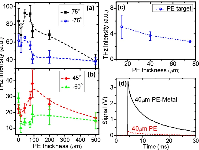
To further clarify the radiation mechanism, we have performed experiments with different
types of targets. Figure 4(a-b) shows the THz radiation from the metal-PE targets as a function of
the thickness of the PE layer. The data at 0 μm correspond to the pure metal foil without any PE.
The THz intensity drops when there is a 15 μm PE layer introduced behind the metal foil. However,
when the PE thickness is increased from 15 μm to 500 μm, the THz emissions increase first and
then decrease, indicating that there exists an optimal PE thickness for THz generation. The optimal
thickness for radiation at 75°, 45°, -60° and -75° is57-17+18,115-40+85, 140-60+50, 43-28+27 μm, respectively.
Cherenkov radiation could occur inside the PE layer. As a kind of bulk radiation, the
Cherenkov radiation scales almost linearly with the medium thickness. Figure 4(c) shows the THz
radiation from single-layer PE targets with different thickness. With the increase of PE thickness,
the THz radiation decreases, rather than increases. This indicates that Cherenkov radiation is not the
dominant contributor to the THz radiation.
FIG. 4. (a-b) Measured THz intensity at 75°(black square), 45°(red diamond), -60°(green triangle) and -75°(blue circle) from the metal-PE targets as a function of the thickness of the PE layer. (c) Measured THz intensity at -75° from the single-layer PE targets with different thickness. (d) Comparison of the typical signals recorded by the THz detector from the single-layer PE (red dashed) and the PE-metal (black solid) targets with a 40 μm PE layer.
The transition radiation depends critically on the properties of the target-vacuum boundary. To
test the effect of the interface, we have ever reversed the metal-PE target and used the laser pulse to
irradiate the PE layer, rather than the metal layer. Figure 4(d) shows the typical signals recorded by
the THz detector at -75° for the reversed PE-metal and single PE targets, where both the PE layers
are 40 μm thick. The THz radiation from the metal boundary is about 10 times higher than that from
the single PE boundary. The relative dielectric constant of the PE at the THz regime is ~2.3, while
that of the metal is much greater than one [27]. The stronger emission from the metal boundary is a
0 100 200 300 400 500
10 20 30
40 45
-60
PE thickness (m)
40 60 80 100 (b) 75 -75 T H z i n te n s it y ( a .u . ) (a)
20 40 60 80
0 4 8 (c) T H z in te n s it y ( a .u . )
PE thickness (m) PE target
0 10 20 30
0 1 2 3
40m PE
40m PE-Metal
[image:8.595.136.465.230.478.2]direct manifestation of transition radiation.
The features shown in Fig. 4(a-b) can be interpreted qualitatively by the formation-zone effect
of transition radiation. Transition radiation is not emitted instantaneously at the interface, but occurs
over a formation length [28]. For the forward radiation in the medium with a dielectric constant of
εr observed at an angle ϑ with respect to the electron motion direction, the formation length Lf is
given by
2 1 cos
f
r
L
, (2)
where β is the electron velocity normalized by the light velocity in vacuum.
For the metal-PE targets, there are actually two transition radiation sources, which are located
at the metal-PE interface inside the target and at the PE-vacuum interface at the rear target surface,
respectively. When the thickness of the PE layer is less than Lf, the forward radiation from the
metal-PE interface and the backward radiation from the PE-vacuum interface will interfere with
each other, thus suppressing the total forward radiation. Hence, the detected radiation drops when
the 15 μm PE layer is introduced behind the single metal foil. With increasing the thickness of the
PE layer, the interference will disappears gradually. Consequently, the THz radiation will become
stronger with the PE thickness thereafter. With the further increase of PE thickness, the detected
radiation from the metal-PE interface will be reduced due to the absorption of THz radiation by the
PE layer. On the other hand, as the electron beam propagates in the thick PE layer, the electron
beam will be broadened and accordingly the beam amplitude will be decreased due to the energy
dispersion. Electrons with lower energies even cannot reach to the target rear surface. Those factors
will lead to a sharp drop of the radiation from the PE-vacuum interface. Therefore, there will be an
optimal PE thickness. This agrees qualitatively with the observation.
The observed optimal PE thickness can be used to characterize the formation length Lf.
According to Eq. (2), Lf is dependent on the observation angles, i.e., the smaller the angle ϑ, the
larger the length Lf. One can find Lf at 45° and -60° is larger than that at ±75°. This explains why
The THz radiation measured does not show obvious correlation with the sheath field formation
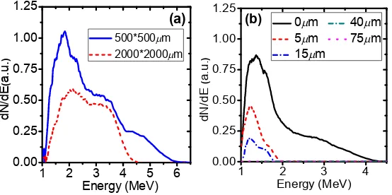
and subsequent ion acceleration via TNSA. Figure 5(a) shows the ion energy spectra from the
mass-limited Cu targets with different target sizes. When the target size is increased from 500×500
μm to 2000×2000 μm, both the number and the maximum energy of ions are decreased substantially.
A small target will confine the transverse refluxing of fast electrons, resulting in a higher sheath
field for a longer time and enhanced ion acceleration [29]. But this is completely different with the
THz radiation dependence on the target sizes shown in Fig. 3, where the THz radiation tends to
become saturated as the target size is increased.
FIG. 5. (a) Measured ion energy spectra from 500×500 μm (blue solid) and 2000×2000 μm (red dashed) mass-limited Cu targets. (b) Ion energy spectra from the metal-PE targets with different PE thickness. The maximum energy of ions from the 40 μm and 75 μm thick PE layer becomes less than the lower detection limit (1 MeV), which is caused by the shielding Al filters in front of IP.
Figure 5(b) shows the ion energy spectra from the metal-PE targets with different thickness of
the PE layer. With the increase of PE thickness, the ion number and energy drop rapidly and even
disappear for the thickness >40 μm. While the THz radiation is still emitted till 500 μm (see Fig. 4).
As fast electrons of specific divergence angles transport through thick targets, the areal density of
electrons arriving at the rear target surface is reduced. The established sheath field becomes weak,
and even too weak to accelerate ions [30].
In the experiment reported by Gopal et al. [18], a correlation between the THz yield and proton
1 2 3 4 5 6 0.00
0.25 0.50 0.75 1.00 1.25
d
N
/d
E
(
a
.u
.
)
Energy (MeV) 500*500m
2000*2000m
(a)
1 2 3 4
0.00 0.25 0.50 0.75 1.00 1.25
d
N
/d
E
(
a
.u
.
)
Energy (MeV)
0m 40m 5m 75m 15m
[image:10.595.172.449.301.439.2]numbers is observed by measuring their dependence on the laser energy. Such a correlation implies
that the THz radiation and ion acceleration may be driven by a common source. Our results show
that the source is the forward fast electrons. When the fast electrons pass through the rear
surface-vacuum boundary of a foil, they will not only radiate THz radiation via transition radiation,
but also set up a sheath field accelerating ions. Nevertheless, it should be noted that the laser
contrast in the experiment by Gopal et al. [18] is much higher than that in our case. Despite that the
THz radiation measured in our present experiment is mainly attributed to transition radiation,
different laser-plasma conditions could result in the change of the dominated THz generation
mechanisms [18,19].
There are several evidences indicating that the measured THz radiation is coherent. Firstly,
after taking into account both the detector responsivity and the transmittance of the optical
components in the detection path, the THz energy measured at 75° is ~280 μJ/sr in our experiment.
It is well known that the CTR is proportional toNe2, while the incoherent transition radiation (ITR)
scales with Ne, where Ne is the electron number. If the radiation was generated by ITR, Ne had to be
as huge as 1.75×10²⁰ to match the measured THz energy. This number is unreasonable according to
the well-known conversion efficiency from the laser energy to the fast electrons. While the CTR
model gives a reasonable Ne ~1.32×10¹⁰. Secondly, the ITR is independent on the target size, and its
spectrum is flat [13]. While in the experiment, the THz radiation is found to be strongly affected by
target sizes (see Fig. 3), and the measured spectrum is frequency-dependent [see Fig. 2(a)]. Those
features can be well explained by the model of CTR. Thirdly, the formation-zone effect is found
with the metal-PE targets, which only occurs for coherent radiation. Those evidences suggest that
the measured THz radiation is CTR.
Given the experimental observation and the theoretical model of CTR, we estimate the total
THz energy from the rear side of metal foils to be ~400 μJ/pulse under our experimental conditions,
and the corresponding energy conversion efficiency from the laser to THz radiation is ~2×10⁻⁴. The
measured THz energy has approached the energy level of the state-of-the-art THz sources based on
In conclusion, we have demonstrated intense THz transition radiation of the laser-accelerated
relativistic electron beams crossing the solid rear surface. The THz emissions from the mass-limited
and metal-PE targets are observed to strongly depend on target parameters. It can be well explained
by the model of CTR considering the effects of diffraction radiation and formation zones. The
laser-plasma-based THz transition radiation presented could be a promising compact strong-field
THz source. Moreover, it may provide an alternative diagnostic to infer the spatio-temporal
distribution of fast electron beams generated in laser-solid interactions.
We wish to acknowledge R. Yang, X. G. Qiu at the National Laboratory for Superconductivity
in Beijing for the calibration of filters. Z.M.S. acknowledges the support by a Leverhulme Trust
research grant. This work is supported by the National Basic Research Program of China
(2013CBA01500 and 2014CB339801), the National Nature Science Foundation of China
(11520101003, 11135012, 11375262, and 11421064).
1
M. Tonouchi, Nat. Photonics 1, 97 (2007).
2
R. Ulbricht, E. Hendry, J. Shan, T. F. Heinz, and M. Bonn, Rev. Mod. Phys. 83, 543 (2011).
3
T. Kampfrath, K. Tanaka, and K. A. Nelson, Nat. Photonics 7, 680 (2013).
4
A. J. Fitzgerald, E. Berry, N. N. Zinovev, G. C. Walker, M. A. Smith, and J. M. Chamberlain,
Phys. Med. Biol. 47, R67 (2002).
5
S. Koenig et al., Nat. Photonics 7, 977 (2013).
6
U. Happek, A. J. Sievers, and E. B. Blum, Phys. Rev. Lett. 67, 2962 (1991).
7
A. M. Cook, R. Tikhoplav, S. Y. Tochitsky, G. Travish, O. B. Williams, and J. B. Rosenzweig,
Phys. Rev. Lett. 103, 095003 (2009).
8
G. L. Carr, M. C. Martin, W. R. McKinney, K. Jordan, G. R. Neil, and G. P. Williams, Nature 420,
153 (2002).
9
Y. Shibata et al., Phys. Rev. E 52, 6787 (1995).
10
Z. Wu, A. S. Fisher, J. Goodfellow, M. Fuchs, D. Daranciang, M. Hogan, H. Loos, and A.
Lindenberg, Rev. Sci. Instrum. 84, 022701 (2013).
11
S. M. Hooker, Nat. Photonics 7, 775 (2013).
12
13
W. P. Leemans et al., Phys. Rev. Lett. 91, 074802 (2003); C. B. Schroeder, E. Esarey, J. van
Tilborg, and W. P. Leemans, Phys. Rev. E 69, 016501(2004).
14
H. Hamster, A. Sullivan, S. Gordon, W. White, and R. W. Falcone, Phys. Rev. Lett. 71, 2725
(1993).
15
A. Sagisaka et al., Appl. Phys. B 90, 373 (2008).
16
Y.-T. Li et al., Appl. Phys. Lett. 100, 254101 (2012); C. Li et al., Opt. Express 24, 4010 (2016);
G.-Q. Liao et al., Phys. Plasmas 23, 013104 (2016).
17
G.-Q. Liao et al., Phys. Rev. Lett. 114, 255001 (2015).
18
A. Gopal et al., Phys. Rev. Lett. 111, 074802 (2013); A. Gopal et al., New J. Phys. 14, 083012
(2012); A. Gopal et al., Opt. Lett. 38, 4705 (2013).
19
Z. Jin, private communication.
20
W. J. Ding, Z. M. Sheng, and W. S. Koh, Appl. Phys. Lett. 103, 204107 (2013).
21
J. Zheng et al., Phys. Plasmas 9, 3610 (2002); Phys. Plasmas 10, 2994 (2003).
22
J. J. Santos et al., Phys. Rev. Lett. 89, 025001 (2002); S. D. Baton et al., Phys. Rev. Lett. 91,
105001 (2003); J. Zheng et al., Phys. Rev. Lett. 92, 165001 (2004).
23
W.-M. Wang, P. Gibbon, Z.-M. Sheng, and Y.-T. Li, Phys. Rev. E 91, 013101 (2015); M. Chen, Z.
M. Sheng, J. Zheng, Y. Y. Ma, and J. Zhang, Chin. J. Comp. Phys. 25, 43 (2008) (in Chinese).
24
M. G. Haines, M. S. Wei, F. N. Beg, and R. B. Stephens, Phys. Rev. Lett. 102, 045008 (2009).
25
M. L. Ter-Mikaelian, High-Energy Electromagnetic Processes in Condensed Media,
Wiley-Intersciencer, New York, 1972. 26
See Supplemental Material for more details on the estimation of electron beam divergence from
the degree of THz radiation polarization.
27
Y.-S. Lee, Principles of Terahertz Science and Technology, Springer, New York, 2008.
28
L. C. L. Yuan, C. L. Wang, and H. Uto, Phys. Rev. Lett. 25, 1513 (1970).
29
S. Buffechoux et al., Phys. Rev. Lett. 105, 015005 (2010).
30