Int. J. Electrochem. Sci., 13 (2018) 3789 – 3798, doi: 10.20964/2018.04.20
International Journal of
ELECTROCHEMICAL
SCIENCE
www.electrochemsci.org
A Selective Medium for Pyocyanin-dependent Fast
Electrochemical Detection of Pseudomonas aeruginosa in
Environmental Microbial Samples
Shuihong Li1,2,*, Qianqian Mou2, Nuozhou Feng2, Polly H.M.Leung2
1
Hunan Province Cooperative Innovation Center for Molecular Target New Drug Study, University of South China, Hengyang 421001, China
2
Department of Health Technology and Informatics, the Hong Kong Polytechnic University, Hong Kong 999077, China
*
E-mail: zhoubaoxi520@163.com
Received: 23 October 2017 / Accepted: 24 January 2018 / Published: 6 March 2018
In this work, a selective medium was developed for pyocyanina-dependent qualitative detection of Pseudomonas aeruginosa in raw microbial samples using electrochemical analysis. To obtain an optimum nutritive formulation, the effect of different media (BYEB, LB, SDB, MHB, TSB, and NB), positive ions (K+, Na+, Ca2+, Fe3+, and Mg2+) and selected additives (1-naphthylamine, acetamide, and benzylpenicillin sodium) on the amount of synthesized pyocyanin were investigated and compared. NB was found to be the most favourable medium for the electrochemical detection of pyocyanin, and Mg2+ was shown to be the most effective supporting additive ion for the detection. 1-Naphthylamine was shown to be the most effective supporting antibiotic reagent when supplied in the medium to enhancing the presence of pyocyanina compared to the other two antibiotics. The results indicated that the improved selective medium (ISM) can effectively inhibit/limit the growth of various microorganisms other than P. aeruginosa and obviously enhance the production of pyocyanin in environmental samples that contained P. aeruginosa. This study provided a method for simple and fast electrochemical detection of P. aeruginosa in complex microbial samples.
Keywords: Pseudomonas aeruginosa; pyocyanin; electrochemical detection; selective medium; differential pulse voltammetry
1. INTRODUCTION
resistance to different antibiotics [4]. P. aeruginosa has been found in all environments including plants, animals and humans due to its ability to utilize various organic materials as the carbon resources including diesel and jet fuel [5-6]. The bacteria produces the blue-green pigment called pyocyanin, a redox-active phenazine, which is widely known to be able to kill mammalian and even bacterial cells through the generation of reactive oxygen intermediates [7]. Pyocyanin produced by P. aeruginosa is a redox active molecule [8], and theoretically, its concentration can be measured by electrochemical analysis. Two widely recognized methods have been used for pyocyanin detection: high-performance liquid chromatography (HPLC) [9] and UV spectrophotometry [10]. However, currently, the analysis of complex biological sample analysis often involves a costly and time-consuming pre-purification step. Compared to other analytical methods, electrochemical techniques show several advantages, including rapid detection, low cost, and high sensitivity [11]. Previous studies have reported the electrochemical detection of pyocyanin [12-13] in P. aeruginosa [14]. It is known that the production of pyocyanin in P. aeruginosa is regulated by the LasI/R quorum-sensing (QS) system [15]. In clinical and environmental samples, the bacterial flora were often under nutrient-deprivation conditions that led to the concentration of the P. aeruginosa QS signal molecules hardly reaching the threshold level [16-17], which suppressed the phenazines producing the genes [18] and subsequently causing low pyocyanin production.
It is necessary and useful to develop a selective medium that supports rapid proliferation of P. aeruginosa and increases pyocyanin production. This could be helpful for fast electrochemical detection of P. aeruginosa via pyocyanin in original bacterial samples.
2. EXPERIMENTAL METHODS 2.1. Materials
Pyocyanin (5-methyl-1(5H)-phenazinone, from Pseudomonas aeruginosa, ≥ 98% (HPLC), CAS No.85-66-5) (Sigma-Aldrich, China mainland) and other chemicals used in this study were of analytical grade. Mueller-Hinton broth (MHB) (Oxoid, UK), tryptic soy broth (TSB) (Oxoid UK), Luria-Bertani (LB) broth (Oxoid, UK), Sabouraud dextrose broth (SDB) (Sigma-Aldrich, Hong Kong), buffered yeast extract broth (BYEB) (Oxoid, UK) and nutrient broth (NB) (Oxoid, UK) wereutilized in this study. Silver nanoparticles (mmd=15 nm, 50 μg/mL) and gold nanoparticles (mmd=15 nm, 50 μg/mL) were purchased from Nanjing MKNANO Tech. Co. Ltd., and single-layer graphene (XFNANO®, Nanjing) was obtained through a generous gift from Prof. Tao Xiaoming, Polytechnic University.
was performed through differential pulse voltammetric (DPV) measurements carried out using a CHI1230C (CH Ins. China) electrochemical workstation at the ambient temperature.
2.2. Bacterial strains used in this study
Staphylococcus aureus ATCC29213 (S. aureus), Escherichia coli ATCC25922 (E. coli), Candida albicans ATCC10231 (C. albicans) and Pseudomonas aeruginosa PAO1 ATCC 15692 (P. aeruginosa) were obtained from the American Type Culture Collection (ATCC, USA). Acinetobacter baumanmii (A. baumanmii), Klebsiella pneumoniae (K. pneumoniae) and Sphingomonas paucimobilis (S. paucimobilis) were isolated from dust particles in a nursing home (Kwun Tong, Kowloon, Hong Kong).
2.3. Selection of electrode
The pyocyanin stock solution (1 mg/mL) was diluted with 0.1 M PBS to the final concentration of 30 μg/mL. Then, the DPV curves were recorded on the CHI1230C workstation using the three-electrode system [working three-electrode: glassy carbon three-electrode (GCE, φ 5 mm) and/or gold disc electrode (Au, φ 5 mm), counter electrode: platinum wire (Pt, φ 0.5 mm); and reference electrode: Ag/AgCl electrode (3 M KCl) and/or saturated calomel electrode (SCE, saturated KCl)].
2.4. Construction of standard curve
To generate the standard curve, the pyocyanin stock solution was diluted with fresh NB medium to a series of known concentrations ranging from 0 to 50 μg/mL. Subsequently, the DPV curves were recorded using an Ag/AgCl-GCE-Pt electrochemical cell.
2.5. Selection of culture medium
Ten microliters of the overnight log-phase cultures of P. aeruginosa were inoculated into 10 mL of each different liquid medium including MHB, TSB, LB, SDB, BYEB, NB and PBS. Subsequently, each liquid medium was incubated for 48 h at 37 °C with shaking under 200 rpm (dynamic culture). After the incubation, the cultures were routinely tested with the DPV curves of pyocyanin.
2.6. Test effects of different ions and supplements on pyocyanin production
were each prepared in 10 mL of MHB. After 48 h in the dynamic culture as described above, the samples were tested for pyocyanin by the DPV curves.
2.7. Composition of the improved selective medium
The improved selective medium (ISM) consisted of 1-naphthylamine, acetamide, penicillin G sodium salt, calcium ion and magnesium ion mixed into the NB medium (Oxoid, UK). The recipe (per litre) was as follows: D(+)-glucose, 1 g; peptone, 15 g; sodium chloride, 6 g; yeast extract, 3 g; 1-naphthylamine, 1 mg; acetamide, 2 g; CaCl2, 2 g; MgCl2, 2 g; and penicillin G sodium salt, 10 mg;
pH=7.2±0.2 (25 °C).
2.8. Effect of ISM on the growth of bacteria.
Single colonies of S. aureus, E. coli, A. baumanmii, C. albicans, K. pneumoniae, P. aeruginosa and S. paucimobilis grown on Mueller-Hinton agar (MHA) plates were respectively inoculated into 10 mL of MHB and incubated overnight under 37 °C with shaking at 200 rpm. Subsequently, the overnight bacterial broth culture was serially diluted to a final density of 1~2×10⁵ colony-forming units (CFU) /mL with fresh ISM. After a further 24 h dynamic culture, the viable number for each bacterial broth culture was evaluated by the plate counting method.
2.9. Environmental sample acquisition and assessment
Fifty-five environmental sampling swabs were randomly collected from different common areas in nursing homes in Hong Kong. All samples were labelled and inoculated into 10 mL of ISM and then incubated for 48 h at 37 °C with shaking under 200 rpm. The cultures were directly conducted through electrochemical detections according to the description above. Meanwhile, the above cultures were further inoculated onto agar media using repeated plate streaking, and different bacteria were recognized by colony morphology identification. The bacterial strains in different colonies were identified using a matrix-assisted laser desorption/ionization time-of-flight mass spectrometry (MALDI-TOF) system (Bruker, Germany) according to manufacturer’s instruction.
2.10. Optional method: using nano-modified electrodes
the aforementioned nano-modified electrodes.
3. RESULTS AND DISCUSSION
[image:5.596.104.502.163.724.2]3.1. Determination of reference curves of pyocyanin
Figure 1. DPV curves of pyocyanin using different three-electrode cells.
[image:5.596.113.484.173.438.2]
GCE electrodes showed an enhanced peak current (Ip) signal compared to the Au electrodes as shown in Fig. 1. As a result, the use of GCE electrode as the working electrode could be more favourable for pyocyanin detection than the use of the Au electrode. The value of Ip remained stable whenever Ag/AgCl or SCE was utilized as the reference electrode, and only the peak potential (Ep) varied in a certain range (from -0.288 to -0.344 V). The Ip values of the two kinds of reference electrodes are insignificant (F-test, p=0.517>0.1), implying that the reference electrode appeared to be less effective for the quantitative determination of pyocyanin. However, the Ag/AgCl electrode showed fewer disturbance peaks than the SCE electrode. The optimum three-electrode cell was obtained using Ag/AgCl, GCE and Pt electrodes. CV curves for different concentrations in the 5-50 μg/ mL range were examined, and the values of Ip were identified to be directly proportional to the concentration of pyocyanin (Fig. 2). The best-fit linear equation for pyocyanin detection was Y=4×10⁻⁷X+4×10⁻⁷ with the R2 value of 0.9956.
3.2. Effects of culture medium on pyocyanin production
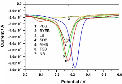
[image:6.596.119.513.449.721.2]Previous research had revealed that the nutrient source (especially the carbon source) and salt concentrations had a significant effect on pyocyanin production [19]. To acquire an optimum nutrition formula for the sufficient yield of pyocyanin in electrochemical analysis, different culture mediums were investigated. From the DPV curves (shown in Fig. 3), we can visually evaluate the strength of the Ip signal. It can be seen that the NB medium was the most favourable medium for the electrochemical detection of pyocyanin among all six investigated media.
3.3. Effects of positive ions on pyocyanin production
Figure 4. Effects of different positive ions and the selective supplements on the production of pyocyanin.
For pyocyanin production optimization, the effects of different positive ions (K+, Na+, Ca2+, Fe3+, Mg2+) and selective supplements (1-naphthylamine, acetamide, benzylpenicillin sodium) on the biosynthesis amount of pyocyanin were evaluated by the quantitative detection of pyocyanin. The contents of pyocyanin in the 48-hour P. aeruginosa culture in the presence of different positive ions were calculated according to the standard curve. As shown in Fig. 4, the enhanced pyocyanin production in P. aeruginosa culture supplied with different positive ions showed different outcomes in the order of Na+<K+<Ca2+<Fe3+<Mg2+. At the same time, 1-naphthylamine presented an obvious pyocyanin high-yield effect, and acetamide and penicillin showed no effect.
3.4. Selective effect of the ISM on bacterial growth
[image:7.596.121.475.99.335.2]
belong to the groups of Bacillus, Pseudomonas, Enterobacteriaceae, and Staphylococcus [23].
P. aeruginosa E. coli S. aureus A. baumannii C. albicans K. pneumoniae S. paucimobilis
0 1 2 3 4 5 6 7 8 9 10 Log 10 C FU /mL
Figure 5. Viable bacterial counts of different strains grown in 48-hour ISM culture.
3.5. Direct detection of the environmental P. aeruginosa samples
Bacterial contaminations have been well-documented in the living areas of nursing homes (NHs), especially in the frequent-touch areas. P. aeruginosa was identified as an important pathogen that frequently caused nosocomial and community-acquired infections especially in frail elderly population. Fifty-five sample swabs collected from nursing homes were investigated, and the results are shown in Table 1. Six samples were confirmed to have a positive test result (including P. aeruginosa) by the electrochemical response of pyocyanin after 48 h of dynamic culture in ISM. This was in agreement with the MALDI-TOF results. This indicated that ISM could be used as the medium for the easiest way to qualitatively detect pyocyanin in P. aeruginosa by an electrochemical technique.
Table 1. Distribution of P. aeruginosa in the living areas of a nursing home. The “positive samples” indicated samples that contained P. aeruginosa and pyocyanin.
Sample sites Toilet handle Dining hall table Residents room bed Publi c phon e Public key Drains of canteen Drains of toilet
Sampling quantity 5 10 10 5 5 10 10
Number of
[image:8.596.144.451.101.280.2] [image:8.596.74.523.568.718.2]
3.6. Nano silver enhanced electrochemical response of pyocyanin
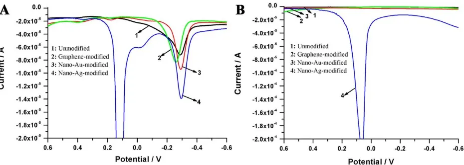
Figure 6. DPV curves of pyocyanin (A) and blank control group (B) using different nano materials modified GCEs as the working electrode.
Additionally, we discovered a new pathway to enhance the electrochemical response of pyocyanin through the electrode surface modification. As shown in Fig. 6, the values of Ip were obviously increased (F-test, p=0.0096<0.01) after nano-silver modification, and this suggested that the electrochemical analysis efficiency and the sensitivity were improved when the modified electrode was used for pyocyanin detection.
4. CONCLUSION
A selective medium was developed successfully in this study and was demonstrated to be suitable for rapid and direct detection of the content of pyocyanin in P. aeruginosa in the complex microbial samples through electrochemical analysis. This method provided a more rapid and efficient technique for the simultaneous detection of the presence of pyocyanin and P. aeruginosa in original
samples containing complicated microbial populations. ACKNOWLEDGEMENT
The financial support by the following are gratefully acknowledged: the National Natural Science Foundation of China (No.81401512), the Scientific Research Fund of Hunan Provincial Education Department (No. 14B157), the China Postdoctoral Science Foundation (No. 2016M602419), and the Hong Kong Scholars Program (No. XJ2015022).
We thank Prof. Tao Xiaoming (PolyU, HK) who kindly provided the single-layer graphene (XFNANO®, Nanjing).
References
[image:9.596.67.532.96.262.2]
2. M.L. Boulette, P.J. Baynham, P.A. Jorth, I. Kukavica-Ibrulj, A. Longoria, K. Barrera, R.C. Levesque and M. Whiteley, J. Bacteriol., 191 (2009) 6329.
3. Z. Xu, X. Fang, T.K. Wood and Z.J. Huang, PLoS ONE, 8 (2013) e57050.
4. K. Okamoto, N. Gotoh and T. Nishino, Antimicrob. Agents Chemother., 45 (2001) 1964. 5. C.L. Bojanowski, W.J. Crookes-Goodson and J.B. Robinson, Biofouling, 32 (2016) 1163. 6. L.M. Brown, T.S. Gunasekera and O.N. Ruiz, Genome Announc., 2 (2014) e01113.
7. B. Cezairliyan, N. Vinayavekhin, D. Grenfell-Lee, G.J. Yuen, A. Saghatelian and F.M. Ausubel, PLoS Pathog., 9 (2013) e1003101.
8. T. Seviour, L.E. Doyle, S.J.L. Lauw, J. Hinks, S.A. Rice, V.J. Nesatyy, R.D. Webster, S. Kjelleberg and E. Marsili, Chem. Commun., 51 (2015) 3789.
9. R.O. Fernandez and R.A. Pizarro, J. Chromatogr. A, 771 (1997) 99.
10. M.Z. El-Fouly, A.M. Sharaf, A.A.M. Shahin, H.A. El-Bialy and A.M.A. Omara, J. Radiat. Res. Appl. Sci., 8 (2015) 36.
11. S. Campuzano, P. Yáñez-Sedeño and J.M. Pingarrón, Sensors, 17 (2017) 866.
12. F.A. Atraktchi, S.B. Andersen, H.K. Johansen, S. Molin and W.E. Svendsen, Sensors, 16 (2016) 408.
13. T.A. Webster and E.D. Goluch, Lab Chip, 12 (2012) 5195.
14. T.A.Webster, H.J. Sismaet, J.L. Conte, I.J. Chan and E.D. Goluch, Biosens. Bioelectron., 60 (2014) 265.
15. J. Lee and L. Zhang, Protein Cell, 6 (2015) 26.
16. W.L. Ng and B.L. Bassler, Annu. Rev. Genet., 43 (2009) 197. 17. S. Atkinson and P. Williams, J. R. Soc. Interface, 6 (2009) 959.
18. D.V. Mavrodi, R.F. Bonsall, S.M. Delaney, M.J. Soule, G. Phillips and L.S. Thomashow, J. Bacteriol., 183 (2001) 6454.
19. B.R. Lundgren, W. Thornton, M.H. Dornan, L.R. Villegas-Peñaranda, C.N. Boddy, and C.T. Nomura, J. Bacteriol., 195 (2013) 2087.
20. K. Poole, J. Mol. Microbiol. Biotechnol., 3 (2001) 255.
21. P.S. Vizuete, B. Orgaz, S. Aymerich, D.L. Coq and R. Briandet, Front Microbiol., 6 (2015) 705. 22. K. Okamoto, N. Gotoh and T. Nishino, Antimicrob. Agents Chemother., 45 (2001) 1964.
23. G. Szita, V. Tabajdi, A. Fábián, G. Biró, O. Reichart and P.S. Körmöczy, Int. J. Food Microbiol., 43 (1998) 123.