THE STRUCTURAL PROTEINS
OF
INFl,UENZA VIRUS
by
IAN PETER GRIFFITH
A thesis submitted (1968) for the degree of
DOCTOR OF PHILOSOPHY
of the
AUSTRALIAN NATIONAL UNIVERSITY
Addendum
i
STATEMENT.
The work presented in this addendum is based on my own ideas and was performed entirely by myself with no
supervision at the Commonwealth Serum Laboratories,
Parkville, Victoria, except as indicated in the Acknowledge-ments.
-\:\'j~~
ACKNOWLEDGEMENTS.
I am greatly indebted to the Commonwealth Serum Laboratories for generous financial support and for
allowing me the use of their facilities to carry out the work presented in this addendum; in particular, I should like to thank Dr. Frank Warburton for encouraging this worko
I am also grateful to Professors D.O. White and S.J. Leach, and Drs. Elizabeth Haslam and I.M. Cheyne for helpful discussions; to Messrs. N.G. Wrigley, M. Taylor and R. Hamilton for the electron micrographs; to
Mr. J. Ride for the computer analysis; to Dr. B. Milligan for the gifts of dyes; and to Drs. D.C. Shaw and WoR.
Sawyer for gifts of proteins.
PREFACE.
This addendum is presented in the form of papers prepared for publication; hence, the figure numbering for each paper applies to that paper only. Additional references have been listed at the end of the addendum. The abbreviations used were generally those recommended by the Biochemical Journal
(Biochem. J. (1967) 102, 1) or have been listed pre-viously (previous submission, p.ix)~
TABLE OF CONTENTS.
S TATElvfENT
ACKNOWLEDGEMENTS
PREFACE
TABLE OF CONTENTS
GENERAL INTRODUCTION TO THE AD
D
ENDUM
PAPER I.
The fine structure of influenza virus.PAPER
2. Imm~diate visualisation of protein indodecyl sulphate-polyacrylamide gels by prestaining with a Remazol dyeo PAPER 3.. The preparation of highly infectious
influenza virus.
PAPER 4. Th~ effect of cystine cross-links on the mobility of prot~ins in dodecyl sulphate-polyacrylamide gels.
PAPER
5.
The polypeptides of the influenza virion.PAPER
6.
A simple procedure for the recovery ofprotein from preparative gel slabs.
GENERAL DISCUSSION OF
RESULTS.
REFERENCES
iv
Page i ii iii iv 1 10
34
47
61
81
INTRODUCTION
Earlier work (previous submission) described attempts to prepare a specific antiserum to 'the' ttsoluble antigenlt (chapter 1) and to purify titt from infected tissue sources (chapter
2).
Chapter 3 was concerned with purification of virus, chapter 4
described attempts to disrupt the virion, and to separate and iden-tify the split products. Chapter
5
was concerned with analysis of some isolated protein fractionso1
Conclusions from the earlier work were based on a number of assumptions; among these were:
1) that only a single internal antigen was present in the virion, the RNP, and that specific antiserum could be prepared against this by injecting crude concentrates of infected tissues into rabbits (Chapters 1 and 2); also that the polypeptide R was derived from the RNP (Chapters
4
and5).
2) that erythrocyte adsorption-elution was the best method of
initial concentration and purification of the BEL strain (Chapter
3).
3) that the pure virus was completely separated into its constituent polypeptides by oxidation with performic acid followed by polyacryla-mide gel electrophoresis in the presence of 8 M urea and SDS.4) that the
s.%
polyacrylamide gel electrophoresis technique, both analytical and preparative, satisfactorily separated the viral poly-peptides into three major species.2 One only (paper 1) is in conjunction with another worker (N.G. Wrigley). The work reported is largely new work, undertaken for a variety of
reasons.
Paper 1 is concerned with the fine structure of the virion and its structural proteins as revealed by the electron microscope. This paper embodies work reported earlier (Chapter 4) and is expanded to include new observations. The two most pertinent to the previous work are the observation of an additional internal component in the virion, and that the head of the neuraminidase may have a molecular weight of the order of 64,000 consisting of
4
subunits of molecular weight 16,000.Paper 2 describes a new technique for staining proteins with reactive dyes of the Remazol type prior to application to poly-acylamide gels. This work was undertaken for the following reasons: 1) A rapid method of staining proteins is of value for quickly assessing the purity of different virus fractions during virus or protein purification. It was considered that, in the absence of
immune serum of proven specificity for the RNP, analytical polyacry-lamide gels were better guides to the progress of internal antigen purification.
2) A more sensitive staining method would more readily reveal minor protein components of the virus preparation.
would assist denaturation and solubilisation of the viral polypep-tides at neutral or alkaline pH.
4) A more satisfactory alternative to bromophenol blue was sought as a reference marker for molecular weight determinations by the polyacrylamide gel method of Shapiro et ~. (1967), as used by Weber and Osborn (1969) and E.A. Haslam (personal communication).
Bromo-hlu~
phenol(is an unsatisfactory marker because it migrates as a broad band, it migrates more slowly than several polypeptides (e.go those of molecular weight 17,000 or less) and it elutes from the gel
during staining-destaining procedures. The latter problem was over-come by Weber and Osborn (1969) who measured the distance migrated by the bromophenol blue and the length of the gel before and after staining, and from these values estimated the position the
reference dye would have had in the stained gels; this procedure is time consuming and could compound errors in measurement. Glucagon as a reference marker has several advantages over bromophenol blue; it has a higher mobility and smaller spread, and does not elute from the gel during staining and destaining procedures. However, glucagon stains poorly with amido black and Coomassie blue, and cannot be used as a reference marker in gels being stained for gly-coprotein with the periodic acid-Schiff staining procedure.
Glucagon prestained with Remazol blue or red has none of these dis-advantages and was thus used as a reference marker in later work.
5) The intensity of staining of proteins with reactive dyes is
~.
t
I'
I
•
II
~.
~
1963).
Prestaining with Remazol blue (also a reactive dye) would thus permit a more accurate estimation of relative proportions of viral polypeptides (by scanning gels with a densitometer and inte-grating the area under the peaks) than would be possible with sub-stantive dyes (e.g. amido black or Coomassie blue) 0 As no densito-meter was available, this has yet to be done.6) Protein stained with Remazol blue may be used to monitor poly-acrylamide gel systems and evaluate the method of recovering protein from the gels. Difficulty was encountered earlier (Chapter 5) in estimating recoveries of protein from gel slabs since the presence of bound SDS in recovered protein was not excluded. By measuring dyed protein spectroscopically, this difficulty may be overcome. Moreover, the efficacy of desalting procedures may be checked by
correlating the optical density of the protein before and after with its dry weight. The preparative polyacrylamide gel technique, and recovery and desalting procedures, have been modified (Paper
6)
and Remazol-dyed proteins have been used in their evolution and evalu-ation.Paper 3 is concerned with the preparation of pure and highly infectious virus of the BEL strain. Although two cycles of adsorption-elution on human erythrocytes are adequate for preparing, in quantity, purified virus of a number of strains, for work on the protein chemistry of the virus, this procedure has a number of
drawbacks (enumerated by Frommhagen and Knight,
1959),
especially..
4f
I
~!
I':
i
II
~.
for preparing virus of high infectivity. Cycles of
adsorption-elution of BEL with human erythrocytes are not possible (fowl
cells were not available) since extensive haemolysis results even
on the third cycleo
A
variety of initial concentration proceduresas alternatives to red cell adsorption-elution were tried, and
batch adsorption-elution on aluminium phosphate proved the most
satisfactory. No satisfactory alternative to centrifugation was
found for concentrating eluates. The buoyant-density and rate-zonal
techniques have been modified by including glycerol, which is known
to preserve viral infectivity. SDS has been used to destroy
preferen-tially non-infectious virions of the BEL strain, leaving a highly
infectious preparation of virus. The use of the Remazol prestaining
technique for monitoring viral purity is illustrated in Papers 2
and
3.
When a circulating pump became available, the
polyacryla-mide gel technique used in the previous work (Appendix 2) was
l.\
abandoned in favOff of the buffer system of r,'Iaizel (1966; but without
urea) for a number of reasons:
1 ) The presence of glycine and urea in the buffers used in the pre-vious work unnecessarily complicated experiments with the isolated
polypeptides. The high buffering capacity of the tris-glycine buffer
was no longer important when SDS-phosphate buffer in the electrode
compartments could be recirculated.
2) The presence of a buffer discontinuity increased the risk of low
molecular weight proteins "stacking" at, and moving with, the buffer
--..
5
front.
3) By using gel systems used by other workers, results could be more directly compared.
4) The use of the SDS-phosphate buffer system permitted many
estimates to be made on. the molecular weight of the viral
polypep-tides.
In work described in Paper
5
it became apparent that thelargest major polypeptide of BEL migrated more slowly when
denatu-red without simultaneous denatu-reduction. To interpret this finding, it
was necessary to first establish whether the rate of migration of
polypeptides in SDS-polyacrylamide gels is affected by unfolding of
the polypeptide following cleavage of disulphide bonds. The effect
cystine bridges have on the mobility of polypeptides in ~ ~ol¥nen
tiilp-fl ; 1'1' ,,% polyacrylamide gels is described in Paper 4. This work
had to be performed with proteins of known cystine content. The
use of polypeptides linked with glutaraldehyde is described, and
three conclusions were reached which were relevant to
polyacryla-mide gel analysis of influenza polypeptides. Firstly, failure to
disrupt cystine bonds results in a significant increase in mobility
in most cases; this finding might explain variations in the
mole-cular weights ascribed to influenza virus polypeptides by different
workers, since some have used for standard curves cystine
cross-linked proteins of high molecular weight. Secondly, the mobility
of the polypeptides relative to glucagon was remarkably constant
from one experiment to another for most proteins, indicating that
Iii"
the technique was reproducible and reliable, although it was shown
that low molecular weight proteins did not always migrate at a rate
proportional to the log10 of their molecular weight.
Thirdly, the high temperature, high pH denaturation procedure
employed appeared to give no polypeptide chain cleavage with the
proteins used.
This paper also demonstrated that glutaraldehyde
cross-linked proteins migrate as relatively compact polypeptide bands.
A project was initiated to investigate (using glutaraldehyde) which
polypeptides were in intimate contact in the intact virion, since
only those polypeptides which are virtually touching may both
simul-taneously react with the same glutaraldehyde molecule, it was hoped
that this work would provide evidence for some of the associations
suggested in Paper 1, especially that between the RNP and the
proposed 'core' antigen. However, completion of this project
await-ed the introduction of a denaturation procawait-edure which could be shown
to break down the virion proteins into the smallest units linked by
covalent bonds other than cystine.
In Paper 5, the conversion of the virion into its smallest
subunits was investigated. For this work, egg-grown virus was used
for the following reasons:
1) Larger amounts of virus may be grown for a given expenditure in
time and materials. Projected work on the protein chemistry of the
virion requires large amounts of virus, which can only be
satisfac-torily produced in eggs.
f
I
2) Egg-grown virus generally contains a greater proportion of infectious virus than that grown in tissue culture. It is not practicable to label egg-grown virus with radioactive amino-acids or aminosugars, since this can only economically be done in de -embryonated eggs, which produce much incomplete virus.
3) It was considered that, due to differences in Ithost-antigen" content, disruption techniques applicable to egg-grown virus might give different results with tissue culture-grown virus. This has been found to be the case by others (E.A. Haslam, personal commun-ication). In preliminary experiments, rapid qualitative analysis is more desirable than accurate quantitative analysis, and for this reason (and those indicated above) disruption of the virion was in-vestigated using egg-grown virus stained with dyes for protein and carbohydrate.
The results presented in Paper 5 indicated that certain of the larger virion polypeptides were only partially converted into subunits, the extent to which this occurred depended on the denatu-ration procedure used. It was concluded that the only effective dena-turation procedure unlikely to cause peptide bond cleavage is oxida-tion with performic acid followed by reducoxida-tion in nonionic detergent .~ solutions saturated with guanidine hydrochloride and urea,
follo-wed by treatment with SDS and urea under reducing conditions. Virus treated in this way had a number of minor components of variable molecular weight, and two major polypeptides of quite low molecular weight, all of which appear to be derived from high molecular weight
glycoproteins.
The addendum is concluded with a discussion of all the
work presented in this thesis. Those portions of the previous
submission which may still be valid, but on which no additional
work has been done, are indicated. Also indicated are, those
por-tions which are no longer valid, those which may still be valid,
and those which are definitely valid in the light of more recent
work reported here, and by other workers elsewhereo
•
w
=
M
.~
~
~
~
d
~
=
~~
d
.~
•
~
~
0
~
~
~
M
~
~
<
~
~
=
M~
w
~
d
.~
~
~
~
I Ifl'RODUCTI ON •
Particles of non-filamentous strains of influenza virus
are often pleomorphic, their largest dilnension being
800
-
1800~ with an envelope70
-
100R thick from which arise spikes90 -
100Rlong,
15
-
20R wide and70
-
80~ apart; the envelope is alipo-protein and encloses a coil of RNP (Hoyle,
1952;
Horne et alo,1960;
Hoyle et al.,
1961).
Two surface antigens of influenza virus havebeen separated and identified, namely the HA (Laver,
1964;
Laverand Webster,
1968;
Laver and Valentine,1969)
and the neuraminidase(Laver,
1963;
Kilbourne et al.,- -
1968;
Laver and Valentine,1969).
The subunits investigated by these workers have been derived from
influenza virus strains containing HA or neuraminidase resistant
to the detergent sodium dodecyl sUlphate
(SDS).
It has long beenthought that the HA corresponded to the viral 'spikes" described by
Horne et al.,
(1960);
Laver and Valentine(1969)
confirmed this, andalso showed that the neuraminidase resembled a tmushroom' in shape,
and postulated that this structure was present on the surface of
the virion.
At present only a single internal antigen is generally
recognised: the RNP. The quaternary structure of this RNP is
variable, but is generally believed to be a protein-RNA complex,
50
-
70R in diameter, coiled into a helix 600R in diameter and ofvariable length (Hoyle et al.,
1961;
Waterson et al.,1963;
Apostolovand Flewett,
1965).
This RNP has also been purified from disruptedvirus, but it then appears to be in fragments
500
-
1300R
in lengthand
75 -
150R
in width containing subunits30
-
45~
in diameter(Hoyle et al., 1961; Ruttkay-Nedecky et al., 1968; Pons et al.,
1 969) •
This paper is largely concerned with the fine structure of
the BEL and X7F1 strains of influenza A , as revealed by partial
disruption of the virion resulting from prolonged storage, or
treat-ment with detergents. The origin of pleomorphic virus and the
possible existence of a second internal antigen are discussed, and
evidence is presented for the existence of the ENP helix in a
variety of forms. Evidence is also presented suggesting that the
haemagglutinin spike has a forked substructure, that the Ao and A2
neuraminidase have similar structure, and that some BEL virions are
resistant to ionic detergents.
IJfATERIALS AND METHODS.
Detergents.
Sodium dodecyl sulphate (SDS), (obtained from Natheson,
Coleman and Bell) was used as a 1~0 (w/v) solution. Sodium deoxycholate (DOC) was prepared by neutralisation of solid
recrystallised deoxycholtc acid with NaOH. Deoxycholic acid was prepared by acidifying a solution of DOC (reagent grade, British Drug Houses) and recrystallising the recovered precipitate from acetic
12
acid-wa ter. Tween 80, obtained from Atlas Powder Company, \'iilmington, Delaware, was deionised as a 10% (v/v) solution by passage through a
column of mixed bed resin.
Virus.
Virus was obtained from stocks held at _700 at the
Australian National Universityo BEL virus seed had an 1D50 of 108•66 uni ts/ml on thaw'ing and 1D50/HA value of 104 • 71. The Ao - A2
recombinent X7F
1 was that described by Kilbourne (Kilbourne et al., 1967). About 0.05 ml of a 1:1000 dilution of virus 1'1[aS inoculated
old
into the allantoic cavity of 10 - 11 daYLfertile chicken eggs. Eggs were incubated at
36
0 for about 40 hours before chilling at _100 forone hour.
Purification of virus.
Infected allantoic fluid was filtered through a double
layer of fine-mesh nylon cloth, chilled, and adsorbed with
recovered by centrifugation
(gOO
g, 5 minutes) were washed oncewith ice-cold saline and suspended in 1/10 the allantoic fluid
volume of Hanks BSS at 370 for 1 - 2 hours. Virus recovered by
elution from these cells was clarified by centrifugation (2,000 g,
15 minutes) and concentrated by centrifugation (one hour, 19,000
revs/min in the Spinco 21 L rotor) onto a 0.40 ml sucrose cushion
(6a;~ in 0015 r·1-NaCl). On completion of centrifugation, the loosely
packed pellet and sucrose cushion were resuspended in a little of
the supernate and subjected at 00 to ultrasonication (2 - 3 minutes,
maximum output of a rJIullard ultrasonic drill) follovled by dialysis
overnight against 50 - 100 volumes of 0.15 M-NaCl containing 2;b (w/v)
sucrose. The virus was resonicated, clarified by low speed
centri-fugation (4,000 g, 15 minutes) and layered on a 26 ml linear sucrose
gradient (5 - 2()}b T,v/v sucrose in 0.15 r.-I-NaCl buffered with 0.01 N
-tris-HC1, pH 7.2). Tubes were centrifuged for 40 minutes at 12,000 revs/
min in the Spinco S'tJ 25.1 rotor. The virus band was recovered from
these gradients through a hole pierced at the bottom of the tube to one
side of the pellet, and concentrated by centrifugation onto a sucrose
cushion as described above. 'rhis virus, resuspended with sonication,
was visibly contaminated wi th red cell debris. The latter ",as
separated from the virus by centrifugation into a 25 ml preformed
linear density gradient consisting of a mixture of sucrose and
potassium tartrate (Griffith, 1968). A stock solution containing
261b (w/v) sucrose and 17/0 (w/v) potassium tartrate and 0.17~ (VT/V)
sodium azide was used as the heavy component. The light component
was prepared by mixing three volumes of this with one volume of
distilled water. Gradients were centrifuged for 90 minutes at
23,000 revs/min in a Spinco SW 25.1 rotor. The lower band containing
the virus was recovered and stored for four weeks at 4°.
Pr~paration of virus for electron microsco~~.
14
Virus was dialysed at 40 against 100 volumes of 0.15 M-NaCl,
buffered at pH 7.2 with 0.01 M-tris-HC1, to remove sucrose and
tart-rate. BEL virus was treated with two equivalents (weight for weight)
of sodium deoxycholate or sodium dodecyl sulphate at room temperature
for 30 minutes. X7F1 was sonicated (5 minutes, maximum output of a
MSE ultrasonic drill) in the presence of two equivalents (weight for
weight) of Tween 80 at 00 ; the haemagglutinin titre was reduced by
approximately 90.%, but the neuraminidase activity was unaltered.
Virus treated with DOC was similarly dialysed, but the
detergent formed a weak gel and was only slowly removed; thus
dialysis was continued with one change for a further 24 hourso
Dialysed virus was diluted approximately tenfold with 0.15
M-ammonium acetate at room temperature. One drop of this material
was placed on a carbon-coated electron microscope grid, followed by
a drop of
4%
(w/v) aqueous sodiuUl silicotungstate. Excess fluid wasdrained off with the aid of filter-paper, and the grids air-dried.
BEL virus preparations were examined with a Philips EM200 electron
microscope, operated at 60kV and X7F
Results and Discussion.
The origins of pleomorphic virus.
The BEL prepared as described contained much pleomorphic
virus, but was seen to be largely free of non-virion material (]'ig. 1).
The reason for the presence of pleomorphic virus is not clear. One
explanation may lie with the BEL seed used. This gave one ID50 per
60 - 100 virus particles on different occasions before purification;
9~b of this infectivity was lost during purification (Griffith, in +
preparation) . The production of pleomorphic virus appears to occur
most readily in cells in an abnormal physiological state, e.g. those
treated with vitamin A (Blough, 1964a), or those present in
de-embryonated eggs (Hanig and Steim, 1960). As one may regard an
infected cell as becomi ng increasingly abnormal as infection proceeds,
the production of pleomorphic virus late in infection may be a
"normalll occurrence (von 1'llagnus, 1957), especially in strains which
grovi to a high titre. In the extreme, with certain strains, this may
take the form of filaments, as found by Ada et ale (1958); they
showed that the Ryan F strain produced spherical virus during the first
21 hours after infection, and subsequently both short and long filaments.
Short filaments were occasionally found in this preparation of BJ:!JL
(arroY/a~ Fig. 1). Diploid and triploid forms of parainfluenza
particles exist (Hosaka et al., 1966) and it has been proposed that
these arise through random incorporation of genomes into virions during
budding of virus from the cell surface (Dahlberg and Simon, 1 S'69).
+Chapter 3
Fig. 1. The BEL strain of influenza virus; pleomorphic virions, two short filaments may
Some particles show 'tongues' which have few
('b' arrows). Scale line = 2,000
1.
In addition to many
[image:22.808.9.793.15.977.2]In addition to being formed during maturation, pleomorphic
virus may have arisen in other ways during purification and
subsequent storage. The virus used here had been resuspended with
sonication, and stored for a month at
4
0 in a high concentration ofpotassium tartrate. This may have caused the formation of
pleomor-phic virus, since such virus is reported to be formed on "moderate
sonication" (Simpson, 1964) or on storage of virus at
4
0 or in thepresence of hypertonic saline (Blough, 1964b); no mechanism for this
transformation was proposed by these workers. Some pleomorphism is
undoubtedly due to dehydration of particles, perhaps by osmosis
during storage or by evaporation during staining and drying on the
electron microscope grid; this might especially be the case with
viral filaments (Hoyle, 1954) or smaller particles incompletely
filled with RNP. However, the ability of influenza virus to fuse
with cytoplasmic membranes (Hoyle et al., 1962; Hoyle, 1962; Morgan
and Rose, 1968) suggested to us that in the absence of available
cell membrane (as is the case with purified virus) virions may fuse
with each other. This would in part explain the heteroploid virus
commonly observed in myxo- and paramyxovirus preparations. Sonication,
'ageing' or exposure to high salt concentrations may help this process
by removing absorbed material (such as nucleic aCids, or inhibitory
mucoids) from the surface of the viriono The process may also be
helped by high-speed centrifugation of virus onto tube surfaces, a
procedure which is accompanied by significant loss of infectivity
with influenza virus (Griffith, unpublished observations), as vTell as
with a number of other viruses (Anderson and Cline, 1967) 0
Spherical particles were frequently found in pairs (Fig. 2 - 5).
These pairs may be particles in various stages of fusion, as is well
shown in Figo 6. If fusion does occur, one may predict that with
increasing time of storage, the proportion of monoploid virus will
decrease, while that of heteroploid virus will increase. This would
be difficult to demonstrate experimentally by absolute particle
counts at well spaced intervals, since particles have a tendency to
disintegrate spontaneously during storage. Particles were often seen
to contain what appeared to be 'tongues' of membrane (Figs. 1b, 2b,
4b) sometimes containing only a few spikes (Fig. 12d) or having a
double membrane appearance (Fig. 13c). This could be the point of
last contact between the virion and the cell membrane during virus
release from infected cells. It may be the point of contact of the
fusing virions, and the point of irreversible first contact during
the viral invasion of uninfected cells.
The lipoprotein shell of the virion.
The virus preparation contained many particles penetrated
by negative stain. At high magnification, an area free of stain and
of width varying from 40 - 130J{ was seen below the surface spikes of
many of these particles (arrow II a", Fig. 7). In one particle, it vlas
evident that the haemagglutinin spikes (150 J{ in length) were embedded
in this layer to a depth of 60 - 80~ (Fig. 7b). This particle also
showed elements of a second layer beneath the outer membrane.
Occasionally, this stain-free area was resolved as a double layer
01
4JCD
I.-
~
CD
Fig.
7.
Several virions of the BEL strain were penetrated by neg-ative stain and showed an area free of stain below the 8urf"cespikes ('a' arrows). The spikes were occasionally seen to be
embedd-ed in this layer ('b' arrows). The arrow head indicated wnut may be a neuraminidase molecule. Scale line - 500
X.
Fig . 8. Two particles showing the double layered structur~ of the outer shell of the virion (inner component arrowed). The ItNP
coil is clearly separated from this component in the largest
[image:26.803.26.784.23.986.2]which was clearly not part of the IU~P coil (arrowed, Fig. 8). The
outer of these two layers is probably the lipid in which the spikes
are embedded (Laver and Valentine,
1969)
while the layer between thisand the P~P coil is possibly a second internal antigen, and may
correspond to the Ifinner leaflet" described by Compans and Dimmock
(1969),
or the "granular layerll of Apostolov et al.,(1969),
or the"inner apposition" of Bachi et al.,
(1969).
To account for multipleprecipitin lines obtained on immunodiffusion of "solubletl
antigen
preparations against specific antisera, Hana and Hoyle
(1966)
suggested that more than one antigenic form of the HNP existed; the
presence of a second internal antigen would, in part, also explain
their findings.
A polypeptide (with a molecular weight of about 23,000)
which has not yet been associated with any of the known viral antigens
18
may be isolated by electrophoretic separation of the virion polypeptides
in SDS-acrylamide gels; many neutral peptides are found in peptide
+
maps of tryptic digests of this polypeptide (Griffith, in preparation).
This might be expected of a protein 1-1hose function is to stabilise a
membrane by hydrophobic interaction vJi th the membrane lipid. If a
second internal antigen distinct from the RNP does exist and is
lipophiliC in character, one might expect it to react with polar lipid
or detergents without being denatured or disorganised. The remarkable
resistance of some influenza virions to disruption by ionic detergents
(~ebster and Laver,
1966)
may thus be explained in part by thestabilising effect this protein might have on the lipoprotein shell
+ .
Chapter 2 ~ Chapter 5
of the virion. 'We may further speculate that this protein assists
assembly of the virion by simultaneously recognising and interacting
with the hydrophobic foot of the haemagglutinin spike, as well as the
membrane lipid and the HNP coil. Alternatively, this protein may
playa role during infection of cells by assisting the dissolution
of the cytoplasmic membrane 'which Horgan and Rose (1968) believe
accompanies viral invasion of the cell.
The structure of the ribonucleoprotein.
The RNP coil shown in Fig. 8 appears to consist of a helix
of 19 or 20 turns. Other particles with a smaller number of turns
to the helix were also found (Figs. 9, 10, 11); in one case, the
diameter of the helix decreased at the lower end (arrowed, Fig. 11).
The diameter of the coil varied from 350 - 600~, while each turn was
50 - 65R thick and separated by a gap 25 - 40~ wide. ~~hese particles
show parallel bar structures vThich "Tere interpreted by Hoyle et al.,
( 1961) as a parallel arrangement ofHNP fragments. It is nOvl more
generally believed that the RNP is present as a helix in the form of
an extension spring ( Waterson et al., 1 962; Apostolov and ]'levIett,
1965). Apostolov and Flewett reported particles containing 6, 12 or
18 turns to the helix, and postulated that a basic llNP unit containing
19
6 turns was normally present, and that the other particles were "diploid"
or "triploid" states. In our virus preparation, this was not the rule.
Occasionally, particles apparently containing disorganised
®
a
b
c
d
e
~
Q)
~ ~I
/ 'i
1000
A
•
•
®
9f
•
•
.
.
,
.
•
•
•
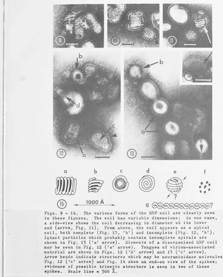
Figs. 9 - 1~. The various forms of the RNP coil are clearly seen
in these figures. The coil has variable dimensions; in one case, a side-view shows the coil decreasing in diameter at its lower
end (arrow, Fig. 11). From above, the coil appears as a spiral coil, both complete (Fig. 13, 'b') and incomplete (Fig. 12, 'be). Intact particles which probably contain incomplete spirals are
shown in Fig. 13 ('a' arrow). Elements of a disorganised RNP coil may be seen in Fig. 12 ('a' arrow). Tongues of virion-associated material are shown in Figs. 12 ('d' arrow) and 13 ('c' arrow).
Arrow heads indicate structures which may be neuraminidase molecules. Fig. 12 (tc t arrow) and Fig. 1~ show an end-on view of the spikes; evidence of possible trimeric structure is seen in two of these spikes. Scale line ~ 500
i.
[image:29.804.26.779.23.957.2]~
III
I
:
Fig. 12(a)0
p~ spirals resembling watch springs have been described
by others (Horne et al., 1960; de The and O'Connor, 1966;
Ruttkay-Nedecky et al., 1968) . Some similar structures were also clearly
seen where particles had partially disintegrated (li'igs. 12b, 13b).
These coils ''Jere 350 - 450i? in diameter; that shown in li'ig. 13 had a strand width of about 50i?. One of these spiral coils had a hollow centre (Fig. 12); presumably it represents an end-on view of a
structure of the type shown in 1!'ig. 11 . The outer layer of the coil
shovln in I!'ig. 12 appeared to differ in staining properties from the
rest of the RNP. Perhaps this was because the outer layer is a part of the proposed second internal antigen, or perhaps because it repre-sents a single thickness of RNP, the centre of the coil consisting of several superimposed strands. Intact particles with dark centres \llere also present (Ii'ig. 13a) and these presumably contain RNP coils of the type shown in Fig. 12. In Fig. 13 a particle is visible ln which some internal component appeared to be protruding through the
outer shell of the virion (arrow I C ' ) . This may be residual
cytoplasmic membrane as proposed earlier, or since each had the same
width (about 50R) as the coiled RNP strand (arrow 'bY), it may be two strands of RNP.
It is evident from our results, and those of others, that
the RNP may be arranged 'wi thin the virion in a nwnber of ways, and
these are indicated diagramatically in Fig.
1S
(a-d). No structures were seen which might correspond to a side-view of the complete-..
20
spiral coil, and we can only speculate on its appearanceo The
lTIak~tsy loop model shown in Figo 16e is one possibility. 'fhis is
similar to the model proposed by Horne and Wildy (1961) or the
"ball-of-string" model of Blough (1964a). HOvTever, to account for the observations of Compans and Dimmock (1969), we suggest that a strand of ruqp passes through the middle as shown. A thin
cross-section of this model, at the points indicated by arrows, would have
21
the appearance indicated in Fig. 1tf. Such an arrangement of dark spots (including the central one), each 50Ji: in diameter, is common in thin
sections of influenza particles (Compans and Dimmock, 1969).
Influenza virus RNP can only be extraced in several short fragments of varying length (Duesberg, 1969; Pons et al., 1969;
Kingsbury and v'~ebster, 1969). Furthermore, long-lived, high molecular weight ID~A (Duesberg and Robinson, 1967; Pons and Hirst, 1968) or RNP (Duesberg, 1969) precursors of these fragments cannot be found 'within infected cells. The appearance of the RNP in disintegrating
particles as a coil without any obvious breaks is difficult to
recon-cile 1;-li th these findingso It has been suggested that the HNP fragments are linked end-to-end by weak secondary forces readily broken
mechanically, (Pons et al., 1969). This could explain the failure to
observe intact coils (other than in particles penetrated by stain)
RNA (and hence RNP) is not cleaved into fragments by hydrodynamic
forces during isolation, as is the case under certain circumstances
with Turnip yellow mosaic virus (Matthews and Ralph, 1966) and mottle
Cowpea chlorotieVvirus (Bancroft et al., 1968)0 The RNA in influenza
virus RNP may be cleaved by ribonuclease, and it is possible that
some of the influenza virus RNA is fragmented by contaminating
nucleases, as is found with the RNA of Cucumber mosaic virus
(Francki, 1968) and Alfalfa mosaic virus (Boll and Veldstra, 1969).
We favour an alternative explanation for the failure to
find RNP in lengths of several 1000 .~. In the preparation of RNP
described by Pons et al. (1969) may be seen a structure 150 .~ wide
and nearly 2,000 ~ long, containing a helical mid-section and
terminal loops. If the RNP coil shown in J?ig. 1Se had free ends, then on its release from the virion it might be expected to uncoil
giving a single strand several 1000 ~ long and 50
R
wideo If it waspresent as a loop of RNP this could not occur, but rearrangement of
the coil, perhaps with some tightening, might give the structure
shown in Fig. 15g, which depicts diagrammatically the structures
shown by Pons et al.(1969). The 2,000
R
structure of these workerswould thus be a single RNP strand (considerably longer than 4,000
R)
in the form of a loop which, perhaps because of some internal stress,
had twisted into a rope-like structure, which would be at least twice
the width of the strands seen in the R~P coils of collapsing particles.
This continuous loop model is consistent with the suggestion
(Mac-kenzie, 196~ that the RNA of influenza virus behaves in the infected
..
22
cell as a circular functional unit.
We propose that the influenza virus RNP is incorporated
into the spherical particle as a continuous loop of variable length,
but probably 8,000-10,000
i
long(FrischNiggemeyer,1961),coiled intoa Takatsy loop-like structureo
It is not known what fills the interior of these RNP coils.
The cavity might be filled with fluid, or a matrix of RNP and lipid
(Kates et al., 1962) or a haemagglutinin gel (Hoyle, 1968). Alternat-ively passAlternat-ively incorporated cell cytoplasm might be present (Hoyle,
1954), perhaps containing nucleases; the presence of cell material
is suggested by the work of Ada and Perry (1958) w'ho showed that
host-cell RNA is incorporated into viral filaments. It is also
possi-ble that the RNP coil is embedded in the proposed second internal
antigen. Two viral polypeptides are found initially in the nucleus
of infected cells (Taylor et al., 1969). The larger of these
(mole-cular weight 50,000) is derived from the protein of the RNP (White
et ~l, 1970) and the smaller (molecular weight 20,000) contains as
much as 40;1
0 of the radioactive amino-acids incorporated into
egg-grovln virus (E.A. Haslam, personal communication). As suggested
earlier this smaller unidentj,fied polypeptide may be part of a second
internal antigen, and it is possible that the virion is filled with
a core (which is assembled in the nucleus of infected cells)
consis-ting of RNP embedded in a matrix of this internal antigen. '1'he
susceptibility of purified influenza RNP to ribonuclease (Hoyle, 1952;
•
I
Schlifer and Zillig, 1954-; Duesberg, 1969; Pons at al., 1969) suggests
that the RNA of the RNP is exposed. The presence of many basic
24
peptides (as w~ll as neutral peptides) in tryptic digests of this
polypeptide (Griffith, in preparation) is consistent with a protective
interaction between the basic residues of this protein and the acidic
phosphate groups of the exposed RNA. 11e suggest that this postulated
second internal antigen be called the 'core' or 'packing' antigen.
The HA and, RNp-fl.:f POC:-disrupted vj.rus,.
The naked coils shown in Figs. 12 and 13 were surrounded bl
rO'lps of short rods, ""hich resemble the viral HA isolated by Laver
and Valentine (1969). In addj.tion, many triangular particles \vi th a
side of about 45 ~ were visible scattered over the back~round. Since
the triangles could also be seen in intact particles (Figs. 12c, 1+ ),
it was thought likely that they represente~ the HA spikes vie"'led
end-on. Triancular structures have been reported previously (Rowe and
\'Jilliams, cited by Cruickshank, 1964), and vwre thought to be
neura-ninidase. On the basis of the ~'lork of Laver and Valelltine (1969), and
tha t reported here, it is nore likely that these vJere in fact HA
monomers mounted vertically on the grid. The reason for their
verti-cal stance is presumably a result of the hydrophobic foot of the
molecule (postulated by Laver and Valentine, 1969) interacting "lith
the hydrophobic carbon on the grid. It was thought that in the
presence of an ionic detergent this interaction might be prevented.
Fig. 16. The BEL strain, treated with DOC, is seen to b~ reduced
to fragments of varying size. Rod-like HA subunits (ta' ~rr0ws)
and mushroom-like' subuni ts ('d' arrows) have been released from
the virions. Fragments of what is probably RNP may al~u he seen
('c' arrows). The shorter HA monomers sometimes seem to b~ forked
at one end ('a' arrows). The end-on view of the RA spikes again
suggests a trimeric subunit structure ('b' arrows). The
neuraminid-ase molecules, alone or grouped with other suhunits, Rre fr~quently
seen ('d' arrows). Their dimensions vary dependin~ on how obliquely
the molecules are being viewed (see Fig. 24). Scale l i n e : 1,000
1
~
150l+
~
45l
a
t
-
30l®
b
c
d
Fig.
17.
Schematic interpretation of the llA sub-structure.(a) end-on view of the HA spike. (b) side-view of the HA spike.
[image:35.801.5.785.16.963.2]the presence of traces of this detergent. The nWQber of individual monomers found standing vertically was much reduced ("fi'ig. 16) although groups (or \I rosettes") were still present. I~any of the rods were
wider at one end and sometimes seemed forked (]Iigo 16a). The trian-gular end-on view of these rods suggested that this forking may be three-pronged. Such a structure "tvas seen in end-on views of the HA spikes in virions both before (Fig.12c,
1
4
)
and after DOC-treatment~ig. 16b). The detached spike was about 30
R
wide at the base, butbroadened to 40-50 ~ tow'ards -L ts forked end. The length of the HA monomers appeared to vary from 80
R
to 150~; presumably the shorterro~s were not lying flat on the grid and thus appeared foreshortened. Our interpretation of the fine structure of the HA monomer is shown in Flig. 170 Irhis figure shows how foreshortening may affect monomer
length, and how the forking (if it exists) could be obscured, depen-ding on the angle from which the rod is viewed.
A molecular ·weight of 150,000 has been proposed for the HA monomer (Laver and Valentine, 1969). Separation of virion poly-peptides (after reaction with performic acid) by electrophoresis in SDS-polyacrylamide gels reveals none with a molecular weight of this
which
order; in fact the largest major polypeptide presen~~as a molecular weight of about 60,000, is a glycoprotein and has been equated with the viral HA (Griffith, 1968, 1969).
In
making these estimates the presence of carbohydrate was not taken into account, and so the values are probably rather inaccurate, but they do indicate that the HAmonomer very probably has a subunit structure.
IVlaterial resembling the RNP fragments of Hoyle et a+.
(1961) may also be seen in Fig. 16 (arrow c). In these there is
little evidence of the secondary coiling demonstrated in RUP
frag-ments by others (Ruttgay-Neckedy et al., 1968; Pons et al., 1969).
This RNP is considerably wider than that present in the larger
coils described earlier, being 110-130 ~ in width. The two fragments
shown were about 600
R
long; they may represent shorter and moretightly coiled structures than that depicted diagrammatically in
Fig. 15g.
The neuraminidase of influenza virus.
In addition to fragments of RNP and HA, the DOC-treated
BEL preparations also contained tmushroomt-shaped structures
resembling the neuraminidase of Asian (A2) influenza isolated from
the recombinant X7F
1 and described by Laver and Valentine (1969),
although the tstem' and 'footV of many were barely visible. These
structures are present alone, or grouped with other subunits (Figo
16d). The dimensions of the 'head' varied in both length (70-90 ~)
and width (35-65 ~), while the stem-pIus-foot appeared of variable
length (60-120~) presumably because of foreshortening in those
subunits viewed obliquely.
On occasions side-on views of the neuraminidase suggested
that the head vias composed of tvlO spheres each 30-35
R
in diameter,while oblique views suggested a more complex sub-structure (II'ig.16d)
which in one case (top centre, arrow d, Fig. 16) appeared to be
tetrameric.
[image:38.806.19.794.26.988.2]Since the HA and neuraminidase of BIEL and X-7:F'I differ
chemically in their resistance to SDS, it was wondered whether they
differed morphologically also. Even though the HA of X-7FI had been
largely destroyed by sonication with Tween 80, structures were seen
resembling the HA spikes of BEL, both in end-on and side-on
projec-tion (Figs. 18a, 19a), and they appeared to have similar dimensions
to those of the BEL strain. Subunits resembling the neuraminidase
isolated by Laver and Valentine (1969) from the X-TI'I recombinant
were also readily recognized in preparations of this virus. Some
clearly had a head consisting of two spheres, while others, viewed
obliquely, showed heads of more complex substructureo (Fig. 18b.).
Also present were structures which might represent the foot and
s tern and head of the neuraminidase detached from each other. ("E'igs.
18c, 20a). In Fig. 18 the stem had a length of about 110
R
and thefoot a diameter of about 20
R.
What may have been the detached headin one case appeared to have elements of a tetrameric substructure
(Fig. 18c). The loss of its supporting sten may have resulted in the
subunits moving closer to each other, since the subunits of two
similar tetramers present on the surface of an intact virion (Fig.
18d) were more widely spaced. The dimensions of these two tetramers
were 70-75
i
along the side, and about 85R
across the diagonal; thesubunits had a diameter of 30-35
R.
Fig. 24 shows a schematicinter-pretation of the fine structure of the neuraminidase molecule of the
influenza virus as revealed in Figs. 16, 18 - 20. The effect
fore-shortening m'ight have on the appearance and dimensions of the head and
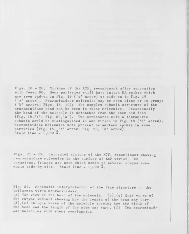
Figs. 18 - 20. Virions of the X7F recombinant after sonlcation with Tween 80. Some particles still have intact HA spikes which are seen end-on in Fig. 18 ('a' arrow) or side-on in rig. 19
r ea' arrow). Neuraminidase molecules may be seen alone or in groups ('b' arrows, Figs. 18, 19); the complex subunit structure of the neuraminidase head can be seen in these molecules. Occaslonally
the head of the molecule is detatched from the stem and foot (Fig. 18,'c'; Fig. 20,'a'). Two structures with a tetrameric
subunit could be distinguished in one virion in Fig. 18 ('d' arrow). Neuraminidase molecules were present as surface spikes in some
particles (Fig. 18, Ie' arrow; Fig. 20, 'be arrow). Scale line = 1,000
X.
Figs. 21 - 2). Untreated virions of the X7F
l recombinant showing neuraminidase molecules in the surface of the virion. On
occasions, fringes are seen which could be several enzyme sub-units side-by-side. Scale line = 1,000
X.
Fig. 24. Schematic interpretation of the fine structure - the influenza virus neuraminidase.
[image:40.801.11.775.24.968.2]I
oct
0
-0
...
~
Q)
oc(
0
It)
...
~
oc(
~
0
~
It)
~+
.-U
rot
oc(
~~~
.-8
0
....0
-ooc:l
0
N
Rtffit~
c
stem are clearly illustrated in Fig. 24.
'rhe length of the neuraminidase molecule is similar to that
of the HA monomer (about 150
g).
This suggests that the foot and partof the stem of the neuraminidase molecule are embedded in lipid to
the same depth as the HA spike, i.e. 60-80
R
(Fig.7).
Thus at leasta portion of the stem is likely to be lipophilic, and an attraction
between the stems of two neuraminidase molecules would account for
the presence of what appear to be pairs of these subunits with stems
overlapping (Pig. 16d), as shown diagrammatically in Fig. 24f.
The head of the neuraminidase and
HA
molecule may often b~ distinguished by their different staining properties. Thus spikes ofintact particles were often seen which were probably side-on views
of the neuraminidase (Fig. 18e, 20b, 21, 22, 23). \1here many
neurami-nidase subunits were present side-by-side, part~cles had a 'fringed'
appearance, especially where the surface of the virion had little
curvature. (]'igs. 22, 23); it is clear that in these particles the
'crown' of the neuraminidase head projects to the same extent as
that of the HA spike. \vhat could be neuramini.dase spikes at the
sur-face of BEL virions are indicated by arrow-heads in Figs. 2, 3,
4, 5,
10, 12.
The molecular weight of the neuraminidase head has been
calculated as 130,000 (Laver and Valentine, 1969) on the assumption
it is a cylinder 85
R
long and 50R
in diameter. The failure by these workers to resolve the fine structure of the head may be related totheir method of isolation of the molecule in the presence of SDS,
which is accompanied by considerable losses of enzymic activity
(Griffith, unpubliehed observations), and perhaps the detergent
caused SOTIe disorganisation of the head of the molecule. The
mole-cular weight of a sphere of protein (partial specific volume
0.73 cm3/g.) 35
~
in diameter is close to 16,000. A tetramer ofthese subunits would give the head a molecular weight of 64,000.
This is in good agreement with the value of 60,000 assigned to the
major polypeptide believed to be derived from the neuraminidase
(E.A. Haslam, personal cOIDITlunication). The sedimentation coefficient
of purified influenza A2 neuraminidase obtained by Rafelson et al.,
(1966) was 4.25. This indicates that the molecular weight of the
neu-raminidase is probably a little less than that of bovine serum albumin
(67,000), which has a sedimentation coefficient of
4.3
(Tanford, 1961).It is not clear whether Rafelson et a1.(1966) had isolated just the
head of the neuraminidase; our results and those of g.A. Haslam
sug-gest that this was likely. The presence of the stem and foot in the
molecule may explain the higher S values obta.ined by other ~vorkers 0
(Noll et ale 1962; Laver 1963; Laver and Valentine, 1969). \{hether
the head is a single molecule or consists of subunits is not yet known.
The ease of separation of the head and stem of the molecu.le
demonstra-ted in F:ig. 18 suggests that the head and stem are probably not joined
by covalent bonds, although the possibility that traces of
contamina-ting proteases jn the viru.s preparation had separated the head from
the stem cannot be excluded.
+ Chapter 4
structures resembling the neuraminidase of Drzeniek et a,l.
(1968) were not seen. The structures reported by these workers may
in fact be aggregates of RNP subunits, since they closely resemble
rings present in the RNP preparations of Hoyle et q~., (1961),
Ruttkay-Nedecl:::y .§.1
..fl1.,
(1968) and Pons et al., (1969) both in appearance anddimensions.
The_.§.:ffe_ct
Q.tJ2.QC
and SDS ___ Q1Lyj.rioll_illi§.grit..Y..
Although DOC almost abolishes infectivity of a number of
'n:1uer~za strains (Smith, 1939; Burnet and IJush, 1940; Sunaga.§l al.,
1959; Webster and Laver, 1966), partially disrupted particles, such
as those shown in Figo 16 were common in DOC-treated BEL preparations,
indicating that this detergent is incapable of completely disrupting
this strain. This probably accounts for the failure of this detergent
to produce haemagglutinating subunits of uniform size from the BEL
strain (~Jebster and Laver, 1966). DOC also fails to produce
haemagglu-tinating subunits of uniform size with a number of other strains
(Nizutani and Hizutani, 1966; ~iebster and Laver, 1966) and does not
permi t the complete separation of neuraminidase and HA of the LEi';
strain by electrophoresis (Laver, 1963).
In contrast to DOC, 0DS is reported to reduce B~L HA to
subunits (Laver, 1964; Laver and Webster, 1966; ~'Iebster and Laver,
1966), al thour,h nearly 40ib of the infect; vi ty of B~-::L survived SDS
treatment (Webster and Laver, 1966)0 Electron micrographs of BEL
treated with SDS revealed few recognizable virus particles, although
many agr:regates of (".\IDorphous material were present vlhich confirmed
the assrunption (Laver,
1964)
that on the whole BEL is disrupted bySDS (Fig. ?5)o Those few particles which remained intact were small
and generally spherical, being
700 -
goo .R
in diameter. An exampleat higher magnification is shown in Fig.
26.
The variable lipid composition of the
MEL
strain ofinfluen-za virus grown in the same host cell (Kates et al.,
1962)
suggeststhat for the assembly of the virion, the precise nature of the lipid
is not important. However, stability of the virion is probably
dependent u~on its lipid, since infectious virions of the
MEL
straingrown in different host cells differ in their resistance to SDS
(Burnet and Lush,
1940),
while infectious virions of several strainsfrown in different tissues of the same host differ in susceptibility
to phospholipase
C
(Simpson and Hauser,1966).
Filamentous virusresembles cytoplasmic membranes in several respects (Burnet and Lind,
1957)
and our findings strongly suggest that pleomorphic virus toodiffers in some way from small spherical virus, which appears to be
more resistant to SDS. The difference may lie with the lipid in the
virion, since pleomorphic virus has a lipid conposition which differs
from that of normal virus (Blough et al.
1967).
Although this mayhold for the majority of the pleomorphic virus, any formed by fusion
of two small SDS-resistant spherical virions might also be expected
to survive treatment with SDS; an example of such a particle is
shown in Fig.
2
7
.
Fig. 25. Amorphous material and small intact spherical particles after treatment of BEL virus with SDS. Scale line ~ 2,000
X
.
[image:46.801.24.782.13.979.2]At present it is not possible to draw conclusions on the
composition or genetic behaviour of infectious influenza virus
particles by studying the structure or behaviour of virus populations,
since the 'latter contain a majority of non-infectious particles. For
this reason, we do not know which of the many forms of the RNP is
present in infectious virions. ,The ease with which ~extension spring
RNP coils are found in influenza virus preparations may be a
reflec-tion on the instability of particles containing these structures. The
complete spiral of our Takatsy loop structure (w'hich was only rarely
seen) may be the form present in the more stable spherical particles
and may therefore be closer to the form of the HNP present in the
infectious virion, if only the spherical particles are infectious.
However, it has been demonstrated that heteroploid parainfluenza
virus may be infectious (Dahlberg and Simon,
1969)
and this iscertainly the Case with influenza virus filaments (Ada et al.,
1958)
and probably also the case with pleomorphic influenza viruso But the
smallest particle capable of harbouring a full complement of RNP makes
the best use of available virion coat proteins, and would therefore
be the preferred form of the infectious virion. Indeed, filamentous
forms are generally replaced by spherical mutants during repeated
passage of influenza virus ;n the allantoic cavity of chiclr embryos.
We believe that the spherical particles shown in Fig. 25 represent
the infectious parti.cles of BEL which are known to survive SDS
treatment (v[ebster and Laver,
1966)
and the possibility of using SDSto prepare virus with a high proportion of infectious particles is
currently being jnvestigated in Melbourne.
PAPER 2.
Immediate visualisation of proteins in dodecyl
Immediate visualisation of proteins in dodecyl-sulphate polyacrylamide
gels by prestaining with a Remazol dyeo
Polyacrylamide gel electrophoresis has gained wide
acceptance as an analytical guide to purity at different stages in the purification of proteins. Proteins have been located in the gels by staining with amido black (Davis,
1964)
or by the moresensitive Coomassie blue (Maizel,
1966)
followed by removal of excess dye, or by prestaining proteins with fluorescent dyes (Kierszenbaum etalo,1969).
The time required to stain and destain polyacrylamide gels has been reduced by the introduction of electrophoreticdestaining procedures (Peterson,
1968)
or by using 0.05% Coomassie G\c.ehvblue in
to'fo
trichlorilo"acid (Chrambach etal.,1967),
but generally, several hours are required for these procedures. Recently, a rapid method for staining proteins in polyacrylamide gels has beendescribed by Hartman and Udenfriend
(1969),
but this method was found to be of low sensitivity for staining gels containing SDS, asthe latter produces a background fluorescence.
In this paper, a procedure is described for reacting biological material with a dye (Remazol B,rilliant_ Blue R) during denaturation prior to analysis in dodecyl-sulphate polyacrylamide gelso The sensitivity of Remazol Brilliant Hlue prestaining was better than that obtained wi th Coomassie Brilliant B,lue aloneo The method is illustrated by the rapid evaluation of purity of influenza virus fractionated in density gradientso
MATERIALS.
1~ Remazol Brilliant Blue Rand Remazol Red B were gifts from
Australian Hoechst Ltd.
2. Coomassie B.rilliant B,lue R250 was a gift from Imperial Chemical Industries (Australia) Ltd.
3
.
Influenza virus; the filamento.us recombinant X1F1 of Kilbourne
(Kilbourne e·tal., 1967), and influenza B/Vic/65 were used.
4. Buff'e~s used were: sodium carhonate-bic.arbonate (pH10, 1M), sodium carbonate-sodium dihydrogenphosphate buffer (pH 9.0, 1M),
and sodium phosphate buffer (pR 8.0, 1M)~
METHODS
.
Virus propagation and purification.
Influenza virus was inoculated into the allantoic cavity
of 10-day o.ld chicken embryos and the allantoic fluid harvested after
46 hours. B/Vic/65 and X7F1 virus were purified by two cycle's of adsorption onto and elution from human erythrocytes, followed by
differential, buoyant-density and rate-zonal centrifugation as will
4-be descri4-bed elsewhere (Griffith, in preparation). High and low density virus was separated by 10t hour centrifugation into a
preformed sucrose-tartrate buoyant density gradiento These fractions were further separated into slowly and rapidly sedimenting components
using a preformed 5 - 20% sucrose gradient.
+Chapter 3