Int. J. Electrochem. Sci., 12 (2017) 6863 – 6873, doi: 10.20964/2017.07.67
International Journal of
ELECTROCHEMICAL
SCIENCE
www.electrochemsci.org
Development of Triglyceride Biosensor Based on the
Polydopamine-Gold Nanocomposite
Min Zhu1, Zhang Yang2, Yongxia Cheng3, Yuanbo Sun4, Jian Xing5, Jieru Wei1, Jiping Sun1, He Liu1 and Xiandong Song5,*
1
Department of Radiology, HongQi Hospital Affiliated MuDanJiang Medical University, MuDanJiang, HeiLongJiang Province, P.R. China
2
Department of Anatomy, MuDanJiang Medical University, MuDanJiang, HeiLongJiangProvince, P.R. China
3
Department of Pathology, MuDanJiang Medical University, MuDanJiang, HeiLongJiangProvince, P.R. China
4
Department of Nephrology, HongQi Hospital Affiliated MuDanJiang Medical University, MuDanJiang,HeiLongJiang Province, P.R. China
5
Department of Radiology, HongQi Hospital Affiliated MuDanJiang Medical University,MuDanJiang,HeiLongJiang Province, P.R. China
*
E-mail: [email protected]
Received: 26 March 2017 / Accepted: 5 May 2017 / Published: 12 June 2017
One significant factor in statin-induced type 2 diabetes (T2DM) predication is the concentration of triglyceride (TG). This study presents a polydopamine-gold nanocomposite (PDA/AuNPs)-based TG biosensor that features significant sensitivity. Au nanoparticles were found to exert remarkable enhancement of electrochemical responses. A double linear detection range with a low detection limit (DL) was exhibited by the prepared TG biosensor, which performed desirably in anti-interference. Hence, the designed PDA/AuNPs have potential as remarkable alternatives to TG detectors.
Keywords: Triglyceride; Type 2 diabetes; Gold; Polydopamin; Electrochemical sensor; Enzyme
1. INTRODUCTION
abnormalities [6] could be predicted by increases in blood pressure, glucose, and TG concentrations. It could be reasonably supposed that persons with insulin resistance are comparatively more likely to suffer from statin-induced diabetes because T2DM can be predicted by insulin resistance [7, 8]. If so, it has clinical usefulness for the identification of a subset of patients (with insulin resistance) to receive statin treatment. The absence of a standardized insulin technique would lessen the use of plasma insulin measurements and/or plasma insulin concentrations as an insulin resistance formula for this purpose and increase the clinical impracticality of specific measurements of insulin resistance. The remarkably significant relationship between hyperinsulinemia, insulin resistance & hypertriglyceridemia has been perennially acknowledgement [9, 10]. Moreover, compared to the concentration of plasma insulin, TG concentration has undergone standardized clinical laboratory detection. Hence, the role of a surrogate estimate was possibly taken by the concentration of plasma TG during the identification of insulin-resistant persons, where this possibility seemed worth evaluation. In comparison to obviously healthy East Asian persons suffering slightly elevated low-density lipoprotein cholesterol (LDL-C) concentrations, those suffering from both increased TG concentrations and LDL-C were insulin-resistant [11]. This possibility was further confirmed by the research discussed herein. More recently, findings with respect to a greater population of obviously healthy persons suffering high LDL-C concentrations were issued in a Letter to the Editor. Herein, a subgroup with glucose intolerance and insulin resistance were identified through subdividing the subjects (mainly European ancestry) on the basis of an accompanying increase in TG concentration [12]. The similar success of this technique during application in high T2DM risk sufferers before statin therapy activation was not necessarily indicated by the capacity of an increased TG concentration in its identification of obviously healthy persons with insulin resistance and elevated LDL-C concentration. Because no less than 1 of the American Association of Clinical Endocrinology (AACE) Consensus Criteria with respect to increased T2DM risk was displayed for statin-treated individuals, they were regarded as those at higher T2DM risk prior to statin treatment activation. Furthermore, they functioned as subjects in the current analysis to solve the aforementioned problem [13-17].
As a result of the increasing numbers of abnormal TG level-induced clinical disorders issued, the field of clinical diagnostics/analysis are experiencing the growing significance of TG detection in food, urine and blood. Because of complex specimen pre-processing and costly tools, high performance liquid chromatographic (HPLC), chromatographic, bioluminescent, fluorimetric and chemical technique-based standard TG detection (in serum) strategies are not suitable for real-time detection. Biosensors have thus been regarded as remarkable alternatives for the detection of TGs because of their desirable selectivity, convenience of application and comparative inexpensiveness. [18, 19]. Therefore, the fabrication of TG biosensors with favorable selectivity, sensitivity, rapidness, and disposability has been extensively studied by many researchers for years.
coatings exhibit the following useful traits: (i) even under moderate conditions or air at ambient temperatures, the polymerization reaction would occur; (ii) a thin PDA layer would adhere on some surfaces via self-polymerized biological amines [23]; (iii) molecules containing preliminary thiol & amine species would covalently bond to the PDA surface as a result of its appealing secondary reaction functional entities [24]; and (iv) the reduction of some noble metal salts into metallic nanoparticles (NPs) would occur via surface multifunctional species at the surface of the PDA film [25, 26]. Because of the desirable biological, electrochemical, electrical, and optical traits of metal NP coatings on diverse polymeric surfaces, they have been applied to nanomedicine, photocatalysis, biodetection, electrical and other fields. In particular, substrates of nearly any type could be connected via the rather active material, Au NPs. The dihydroxy and amine functional species of PDA substrates on the surface have been shown to undergo convenient linking with Au NPs via covalent bonding. During the course of Au deposition, the PDA coating could take on the role of a reducing agent.
To achieve desirable TG determination sensitivity, unremittent Au NP film was attained because of the active support of the PDA coating’s adhesive layer. Unremittent Au NP growth was accomplished under favorable circumstances obtained by the existence of functional species on the surface of PDA. The working electrode could be enhanced in specific surface area, surface negativity and conductivity through unremittent Au film growth. The proposed PDA/Au NPs platform can be effectively used for enzyme immobilization and consequently for TG determination. Thus, a very high sensitivity electrochemical sensor was achieved.
2. EXPERIMENTS
2.1. Chemicals
Glutaraldehyde, hydrogen tetrachloroaurate (III), sodium-4-polystyrenesulfonate, 3,4-ethylenedioxythiophene, dopamine hydrochloride (DA), potassium ferrocyanide, 4-aminophenazone, adenosine 5′-triphosphate disodium salt hydrate, triolein, glycerol-3-phosphate oxidase, glycerol kinase and lipase were purchased from Sigma-Aldrich. The dissolution of Na2H2PO4·2H2O and
Na2HPO4·12H2O in certain quantities of deionize (DI) water was performed to prepare the phosphate
buffer solution (PBS). The entire set of reagents were of analytical grade and thus were employed without additional purification.
2.2. Characterization
A CHI−660D electrochemical workstation with triple-electrode configuration was employed for the entire set of electrochemical measurements, where Hg/Hg2Cl2/3 M KCl standard calomel
electrode (SCE) and Pt wire served as the reference and counter electrodes, respectively, and the entire range of electrochemical reactions were conducted under nitrogen (N2) conditions. An
composition of PDA and Au, whereas a Lambda 750 UV-Vis/NIR spectrophotometer was employed for the measurement of film thickness.
2.3. Preparation of PDA/Au NPs nanocomposite
A positive CV scan ranging from –400 to 300 mV (versus SCE) with a scan rate of 10 mV/s in DA (3 mM, pH 7.5, 10 mM PBS) was conducted to achieve the deposition of oxidized DA molecules onto an FTO substrate, thus obtaining the PDA product under N2 conditions. Before being applied to
future characterization, the aforementioned PDA substrates were stored at 4°C. Being modified in several aspects, the earlier issued CV technique was employed to accomplish Au NP deposition. Double negative CV scans ranging from 800 to –500 mV (versus SCE) with a scan rate of 5 mV/s in HAuCl4 solution (2 mM) were employed to conduct the reaction.
2.4. Preparation of PDA/Au NPs-enzyme electrode
The dissolution of 4-aminoantipyrine, adenosine triphosphate (ATP), GPO (500 U), GK (80 U) and lipase (110 U) (3.0 mg) in PBS buffer (1 mL, 0.02 M) with a pH of 7.0 contributed to the successful preparation of the enzyme mixture solution (10 μL), which was then dropped on the surface of the electrode, followed by 120 min absorption at a temperature of 25 °C. To ensure maximal activity in the PBS buffer solution, an enzyme of a designed unit ratio was employed. The as-prepared enzyme/Au/PEDOT-PSS-designed electrode was subsequently stored at 25 ± 2 °C prior to electrochemical measurements.
2.5. Electrochemical measurement
To achieve optimization with respect to the electrochemical response of the PDA/Au NPs-enzyme, this work preliminarily employed CV in ferri/ferrocyanide solution (10 mM), which was obtained via the mixture of phosphate buffer solution (0.1 M, pH 7.0) and specified quantities of KCl (0.1 M) and K4Fe(CN)6. The measurement of CV response was conducted at a scan rate of 0.1 V/s, a
3. RESULTS AND DISCUSSION
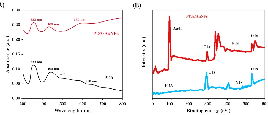
As indicated in Figure 1, XPS & UV-Vis verified the existence of PDA/AuNPs and PDA. The deposition of Au NPs on the surface of the PDA was suggested by the 550 nm peak for Au, together with the 650, 495, 400 & 333 nm absorption peaks (in the vicinity) for the melanin−like structure of PDA, as shown in the UV-Vis. results (Figure 1A) [28]. Moreover, Figure 1B shows the presence of 89.3 eV (Au 4f5/2) and 85.3 eV (Au 4f7/2) Au peaks together with 532.0 eV (O 1s), 400 eV (N 1s) and
286.0 eV (C 1s) PDA peaks in the XPS measurements [29, 30]. The coatings of PDA/AuNPs and PDA have been successfully deposited on the surface of FTO, as indicated by the XPS and UV-Vis results.
Figure 1. (A) UV-Vis. absorption and (B) XPS spectra of PDA and PDA/AuNPs electrode.
[image:5.596.68.519.251.445.2]
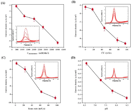
Figure 2. Optimization of PDA growth parameters by varying (A) monomer concentration, (B) CV segments, (C) scan rate and (D) pH value.
The deposition of Au NPs on PDA coatings was performed under various circumstances, with their electrocatalytic activity compared to that in the presence of DA (0.01 mM). As indicated in Figure 3, the PDA/AuNPs electrode exhibited maximal current density in the presence of monomer DA (3 mM) with a pH of 7.5 and scan rate of 10 mV/s. The conductivity, surface negativity and specific surface area of the electrode was comparatively decreased in the case of small-sized Au NPs, whereas the charge transfer process was disturbed in large-sized Au NPs containing thick film [32]. The following tests were conducted on the basis of the aforementioned optimal conditions.
[image:6.596.67.529.79.469.2]
the adjusted potential rising in a range of 0.1-0.4 V, there was an instant rise in steady-state reduction current, which subsequently experienced an insignificant decline upon additional potential increase. Thus, 0.4 V (approximately) was determined to be the optimal reduction current, and the following measurements all adopted this potential.
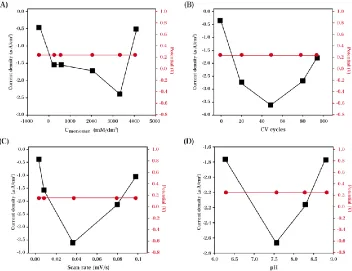
Figure 3. Electrocatalytic activity of PDA/AuNPs electrode with varying (A) monomer concentration, (B) CV cycles, (C) scan rate and (D) pH in the presence of 0.01 mM DA.
[image:7.596.114.466.146.417.2] [image:7.596.182.427.495.685.2]
By adjusting the pH range between 6.0 and 8.0, this work addressed the influence that the buffer pH exerts on the current response to triolein. The amperometric response of the co-immobilized PDA/Au NPs-enzyme is characterized in Figure 5 in a buffer with varying pH values in the presence of triolein (2 mM). As the pH value rises in the range of 6.0-7.0, an instant increase in the steady-state reduction current is observed, whereas upon additional increase in buffer pH value up to 8.0, there is a remarkable decline in the amperometric response. This result is comparable to that obtained in other reports [34-37], which demonstrated that the optimum pH for PDA/Au NPs mixed enzymes for electrochemical detection is in the range of 6.5–7.5. Hence, the all of the following measurements adopted 7.0 as the optimum pH value for TG determination.
[image:8.596.180.427.244.430.2]Figure 5. Effect of buffer pH on the amperometric response of the co-immobilized PDA/Au NPs-enzyme in triolein solution (2 mM).
[image:8.596.170.424.530.720.2]
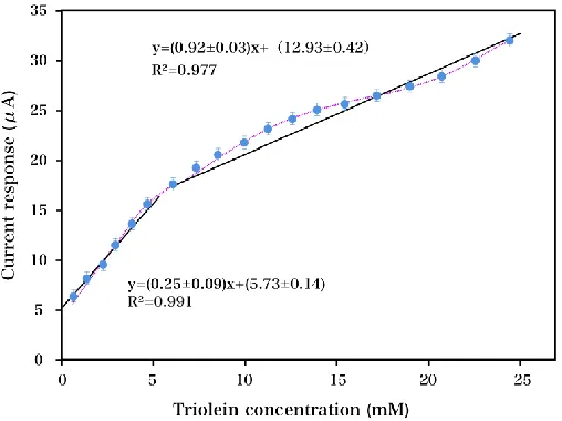
On the basis of the aforementioned measurements, the optimal buffer pH value and determination potential were applied during the conduction of amperometric TG determination with a triolein concentration range of 0 to 25 mM, followed by the estimation of the DL, response time and calibration profile from the amperometric responses. Figure 6 exhibits the characterization of PDA/Au NPs-enzyme via calibration profiles. High and low concentration domains were obviously observed in double ranges of linear concentration dependence with respect to electrode. At a low concentration domain of 0 to 6 mM with 0.075 mM DL (3S/N), the most extended linear range is displayed with respect to the electrode, whereas a nearly identical linear range of 6 to 25 mM was obtained for PDA/Au NPs-enzyme at high concentration domains. Herein, it is regarded as the most desirable alternative with respect to TG (high-concentration) detection. Additionally, we have compared this electrode with some other modified electrodes for the simultaneous determination of TG, as summarized in Table 1.
Table 1. Comparison of the major characteristics of electrochemical sensors used in the determination of TG.
Detection method DL (mM) LR (mM) Reference
Capillary gas chromatography 3-70 2.4 [38]
Spectrophotometric procedure 5-20 3.3 [39]
Colorimetric Hantzsch condensation ― 5 [40]
Amperometric enzymic sensor 2-50 1.6 [36]
Colorimetric determination 0.5-4 0.21 [41]
PDA/Au NPs-enzyme 1-25 0.075 This work
With respect to to-be-employed biosensors in practical cases, the most important factor for consideration is specificity. Glucose, acetaminophen, uric acid, ascorbic acid, and cholesterol (all 2 mM) as well as other significant interference groups were employed with physiological concentrations for interference testing to achieve the successful evaluation of sensor specificity. Compared to 2 mM triolein response, an approximately 30% interference signal was obtained from cholesterol, whereas that from glucose, acetaminophen, uric acid and ascorbic acid were negligible. To ensure the dominance of enzyme mixture (GPO, GK and LP)-induced specific oxidative reactions, 0.4 V was obtained for the working electrode potential; thus, there was a desirably low interference from the aforementioned groups. Nevertheless, partial reaction with an enzyme mixture would unavoidably occur because of their significant similarity in structure; thus, cholesterol exhibited some interference. An enzyme system with higher selectivity could be adopted to address the interference to cholesterol while it remains under investigation.
[image:9.596.58.543.330.435.2]
sensors) and repeatability (from a single sensor), respectively, of current responses (Table 2). Both of these parameters are considered desirable for potential clinical diagnosis applications. The overall performance of the sensor is considerably better than the standard method, which requires a time-consuming assay procedure. The superior performances of the TG biosensor may be attributed to effective enzyme immobilization by glutaraldehyde on PDA/Au NPs-enzyme and the excellent electrochemical properties of gold nanoparticles and PDA/Au NPs composite structures, which provide a large effective surface area, high catalytic activity and high electrochemical conductivity with excellent stability.
Table 2. Reproducibility and stability of the proposed TG sensor
Sensor Detection number RSD
PDA/Au NPs-enzyme 15 (individual) 7.35
PDA/Au NPs-enzyme 10 (same one) 3.66
4. CONCLUSION
This contribution initially employed a CV technique to achieve the successful synthesis of adhesive PDA coatings on the surface of FTO under optimal circumstances. This work was followed by electrochemical TG determination via PDA, which actively supported the growth of unremittent Au NPs. A remarkable enhancement in electrochemical responses was obtained through the Au nanoparticles. TG amperometric determination in blood could be favorably accomplished via PDA/Au NPs-enzyme featuring desirable optical behaviors, where the dynamic range was extended to 0-25 mM.
ACKNOWLEDGEMENTS
Authors acknowledge National Natural Science Founds for Young Scholar of China (81500629, The research of effect and mechanism of miR-1 on the lipidmetabolism in diabetic cardiomyopathy by targeting LXR)
References
1. N. Sattar, D. Preiss, H. Murray, P. Welsh, B. Buckley, A. Craen, S. Seshasai, J. McMurray, D. Freeman and J. Jukema, The Lancet, 375 (2010) 735.
2. D. Preiss, S. Seshasai, P. Welsh, S.A. Murphy, J. Ho, D. Waters, D. DeMicco, P. Barter, C. Cannon and M. Sabatine, Jama, 305 (2011) 2556.
3. P. Ridker, A. Pradhan, J. MacFadyen, P. Libby and R. Glynn, The Lancet, 380 (2012) 565. 4. D.C. Goff Jr, D. Lloyd-Jones, G. Bennett, S. Coady, R. D'Agostino Sr, R. Gibbons, P. Greenland,
D. Lackland, D. Levy and C. O'Donnell, Journal of the American College of Cardiology, 63 (2014) 2935.
5. D. Waters, J. Ho, D. DeMicco, A. Breazna, B. Arsenault, C. Wun, J. Kastelein, H. Colhoun and P. Barter, Journal of the American College of Cardiology, 57 (2011) 1535.
[image:10.596.50.538.247.293.2]
7. S. Lillioja, D. Mott, M. Spraul, R. Ferraro, J. Foley, E. Ravussin, W. Knowler, P. Bennett and C. Bogardus, N. Engl. J. Med., 329 (1993) 1988.
8. S. Lillioja, B.L. Nyomba, M. Saad, R. Ferraro, C. Castillo, P. Bennett and C. Bogardus, The Journal of Clinical Endocrinology & Metabolism, 73 (1991) 866.
9. G. Reaven, R. Lerner, M. Stern and J. Farquhar, Journal of Clinical Investigation, 46 (1967) 1756. 10.J. Olefsky, J. Farquhar and G. Reaven, The American journal of medicine, 57 (1974) 551.
11.W. Sheu, S. Shieh, M. Fuh, D. Shen, C. Jeng, Y. Chen and G. Reaven, Arteriosclerosis, Thrombosis, and Vascular Biology, 13 (1993) 367.
12.F. Abbasi and G. Reaven, Journal of Internal Medicine, 277 (2015) 498.
13.A. Garber, Y. Handelsman, D. Einhorn, D. Bergman, Z. Bloomgarden, V. Fonseca, W. Garvey, J. Gavin Iii, G. Grunberger and E. Horton, Endocrine Practice, (2008)
14.V. Gupta, S. Jain and U. Khurana, Electroanalysis, 9 (1997) 478.
15.V. Gupta, A. Jain, G. Maheshwari, H. Lang and Z. Ishtaiwi, Sensors and Actuators B: Chemical, 117 (2006) 99.
16.V. Gupta, A. Singh, S. Mehtab and B. Gupta, Anal. Chim. Acta., 566 (2006) 5.
17.V. Gupta, A. Jain and P. Kumar, Sensors and Actuators B: Chemical, 120 (2006) 259. 18.J. Narang, Minakshi, M. Bhambi and C. Pundir, Analytical Letters, 43 (2009) 1.
19.J. Narang, M. Bhambi and C. Pundir, International Journal of Biological Macromolecules, 47 (2010) 691.
20.H. Lee, S. Dellatore, W. Miller and P. Messersmith, Science, 318 (2007) 426.
21.J. Dalsin, B. Hu, B. Lee and P. Messersmith, Journal of the American Chemical Society, 125 (2003) 4253.
22.S. Kang, J. Rho, I. Choi, P. Messersmith and H. Lee, Journal of the American Chemical Society, 131 (2009) 13224.
23.B. Li, W. Liu, Z. Jiang, X. Dong, B. Wang and Y. Zhong, Langmuir, 25 (2009) 7368. 24.H. Lee, J. Rho and P. Messersmith, Adv. Mater., 21 (2009) 431.
25.M. Sureshkumar, D. Siswanto and C. Lee, Journal of Materials Chemistry, 20 (2010) 6948. 26.Y. Lee and T. Park, Langmuir, 27 (2011) 2965.
27.P. Nilsson-Ehle and M. Schotz, Journal of Lipid Research, 17 (1976) 536. 28.J. McGinness, P. Corry and P. Proctor, Science, 183 (1974) 853.
29.L. Zhu, Y. Lu, Y. Wang, L. Zhang and W. Wang, Appl. Surf. Sci., 258 (2012) 5387.
30.J. Hu, W. Li, J. Chen, X. Zhang and X. Zhao, Surface and Coatings Technology, 202 (2008) 2922. 31.S. Palanisamy, Electrochimica Acta, 138 (2014) 302.
32.V. Ball, D. Del Frari, V. Toniazzo and D. Ruch, Journal of Colloid and Interface Science, 386 (2012) 366.
33.H. Liu, S. Xu, Z. He, A. Deng and J.-J. Zhu, Anal. Chem., 85 (2013) 3385. 34.J. Narang, Minakshi, M. Bhambi and C. Pundir, Analytical Letters, 43 (2009) 1. 35.S. Webb, Annals of the New York Academy of Sciences, 247 (1975) 327.
36.C. Pundir, Sensors and Actuators B: Chemical, 133 (2008) 251. 37.C. Pundir, Indian. J. Biochem. Biophys., 45 (2008) 111.
38.C. Plank and E. Lorbeer, Journal of Chromatography A, 697 (1995) 461. 39.S. Gottfried and B. Rosenberg, Clinical Chemistry, 19 (1973) 1077. 40.L. Foster and R. Dunn, Clinical Chemistry, 19 (1973) 338.
41.M. McGowan, J. Artiss, D. Strandbergh and B. Zak, Clinical Chemistry, 29 (1983) 538.