Int. J. Electrochem. Sci., 9 (2014) 1537 - 1546
International Journal of
ELECTROCHEMICAL
SCIENCE
www.electrochemsci.org
Electrochemical Determination of Cisplatin in Serum at
Graphene Oxide/Multi-walled Carbon Nanotubes Modified
Glassy Carbon Electrode
Liqing Ye1,2, Mingwu Xiang1,2, Yiwen Lu1,2,Yuntao Gao1,2,*, Pengfei Pang1,2 1
Key Laboratory of Ethnic Medicine Resource Chemistry, State Ethnic Affairs Commission & Ministry of Education, Yunnan University of Nationalities, Kunming, 650500, P. R. China 2
School of Chemistry and Biotechnology, Yunnan University of Nationalities, Kunming, 650500, P. R. China
*
E-mail: 2314972096@qq.com
Received: 25 October 2013 / Accepted: 17 November 2013 / Published: 5 January 2014
Graphene oxide dispersed multi-walled carbon nanotubes composite modified glassy carbon electrode (GO-MWNTs/GCE) was prepared and the GO-MWNTs/GCE was employed for the electrochemical determination of anticancer drug cisplatin. A pair of well-defined redox peak of cisplatin was observed at the GO-MWNTs/GCE in 0.05 M KCl solution (pH 7.4) and electrode process is adsorption-controlled. The result indicates that the GO-MWNTs composite materials can effectively improve the sensibility and performance of the electrode for cisplatin. Based on the GO-MWNTs/GCE, a differential pulse voltammetry (DPV) method for the determination of cisplatin was proposed. The analytical performance of the method was further evaluated in the presence of human 5% serum. A good linear relationship was obtained within the concentration range from 1.30 μM to 26.0 μM in the presence of 5% serum, with the linear regression equation Ipa (μA)= 345947c+3.2421 (r=0.9990). The
detection limit for cisplatin in the presence of 5% human serum can reach to 0.113 μM.
Keywords: Cisplatin; Differential pulse voltammetry; Graphene oxide; Multi-walled carbon nanotubes; Serum.
1. INTRODUCTION
Cisplatin, cisplatinum or cis-diamminedichloroplatinum(II) (CDDP) is a platinum-based chemotherapy drug which was widely used to treat various types of cancers, including sarcomas, some carcinomas (e.g. small cell lung cancer, and ovarian cancer), lymphomas and germ cell tumors.[1]
(ICP-AES), inductively coupled plasma mass spectrometry (ICP-MS),[2-4] high performance liquid chromatography (HPLC) with UV detection and capillary electrophoresis technique etc.[5-8] Several modified electrodes have also been reported for the electrochemical detection of cisplatin, including metallothionein (MT) modified hanging mercury drop electrode and DNA-modified glassy carbon electrode.[9-10]
Since its discovery in 2004,[11] graphene has captured the attention of researchers due to its excellent electrical properties, the ultrahigh ratio of surface area to volume and the extreme sensitivity of its surface atoms to any surface adsorption reactions. Graphene-based electrodes have shown superior performance in terms of electrocatalytic activity and macroscopic scale conductivity than carbon nanotubes (CNTs)-based ones.[12-14] Graphene oxide (GO), which is a derivate of graphene, has attracted increasing interest because of its unique properties and ease of fabrication.[15, 16] It can be readily exfoliated into individual GO sheets to yield stable suspensions in water because of the hydrophilic oxygen groups attached to the strongly oxygenated graphene sheets, thus render it water dispersible, easily stick to glass carbon electrode and fall off less easily.[17] It has recently been demonstrated that GO can be used as surfactant to disperse carbon nanotube (CNTs) in water due to its high solubility and adhesion of CNTs onto the flat GO sheets through strong π-π stacking interaction.[18-23] A novel GO multiwall carbon nanotube (MWNTs) self-assembled composite could be formed by mixing GO and MWNTs in water.[24] This GO-MWNTs composite was highly stable without any precipitation for more than 6 months.[20, 21] The graphene oxide dispersed carbon nanotube composite has been used in various fields such as dye sensitised solar cells, [25] supercapacitors,[18] proton exchange membrane fuel cell,[26] biofuel cells,[27] sensors and biosensors.[28-29] Hence, it is believed that the GO-MWNTs opens new ways for researchers in designing hybrid materials for various potential applications and may find wide potential applications in the fabrication of biosensors, biomedical devices, and bioelectronics.
In our previous work, we have demonstrated that the use of multi-walled carbon nanotubes modified electrode can improve the sensitivity for electrochemical analysis of cisplatin and picoplatin.[30, 31] In this work, we describe a novel graphene oxide dispersed carbon nanotube composite modified glassy carbon electrode (GO-MWNTs) for the electrochemical analysis of cisplatin. Furthermore, we tested the influence of complex biological matrix (human blood serum) on the cisplatin determination.
2. EXPERIMENTAL
2.1. Instruments, materials and reagents
ZAHNER Zennium IM6 Electrochemical Workstation (ZAHNER-elektrik GmbH & Co. KG, Kronach, Germany). A three-electrode system, including a GO-MWNTs/GCE working electrode, a saturated calomel reference electrode (SCE) and a platinum wire counter electrode.
Other reagents used were of analytical-reagent grade. Twice-distilled water was used throughout all experiments.
2.2. Experimental Methods
2.2.1. Preparation of graphene oxide
Graphene oxide (GO) was firstly synthesized from graphite according to the Hummers and Offeman method.[32] Then the GO was reduced and followed a typical procedure: the resulting GO dispersion (100 mL) was mixed with 70 μL of hydrazine solution (50 wt.% in water) and 0.7 mL of ammonia solution (28 wt.% in water). The mixture was stirred for 1 h at the temperature of 95°C. Finally, black hydrophobic reduced graphene oxide (GR) sheets were obtained by filtration and dried in vacuum.[33]
2.2.2. Preparation of GO-MWNTs modified graphite electrode
A GO-MWNTs suspension was obtained by dispersing the treated GO (15 mg) and MWNTs (5 mg) into 15.0 mL distilled water with the aid of intensive sonication (120 W, 40 KHz, 3 h).
A glassy carbon electrode (5 mm in diameter) was polished to a mirror-like surface, with metallographi sand paper and 0.05 μm Al2O3 suspension, respectively. After rinsed thoroughly with doubly distilled water between each polishing step, the electrode was subjected successively with 50% nitric acid, ethanol and doubly distilled water in ultrasonic bath, and dried in air. The electrode was then electrochemically cleaned in 0.5 M H2SO4 by cycling potentials between -1.4 and +2.0 V at 0.1 V s-1 until steady cyclic voltammogram was obtained. Graphene oxide/multi-walled carbon nanotubes modified electrode (GO-MWNTs/GCE) was prepared by casting 15 μL of the GO-MWNTs suspensio on the GCE and dried under an infrared lamp.
2.2.3. Electrochemical analysis
Cyclic voltammetry (CV) and differential pulse voltammeter (DPV) were performed in the three-electrode cell in pH 7.4 KCl solution between the potential range of -0.8 and +0.8 V at a scan rate of 0.06 V s-1. The DPV conditions were: pulse width 100 ms, pulse amplitude 40 mV and pulse interval 150 ms.
3. RESULTS AND DISCUSSION
3.1. Influence of supporting electrolyte and pH
supporting electrolyte. Well-defined CV response with high redox peak of cispaltin was obtained in 0.05 M KCl solution.
1 2 3 4 5 6
3 4 5 6 7 8 9
pH
Ipa
/μ
A
Figure 1. The influence of pH on peak current, in KCl solution at the scan rate of 0.6 mV s-1.
The influence of pH was then investigated in 0.05 M KCl solution (shown in Fig. 1), the oxidation peak current of cisplatin increases with the pH increasing from 3.5 to 7.4. while the oxidation peak current decreases with as pH increasing above 7.4. The maximum value of oxidation peak current with the lowest background current was obtained at pH 7.4. Therefore 0.05 M KCl solution (pH 7.4) was chosen as the optimal supporting electrolyte for subsequent experiments.
3.2. Influence of the composition of the electrode materials
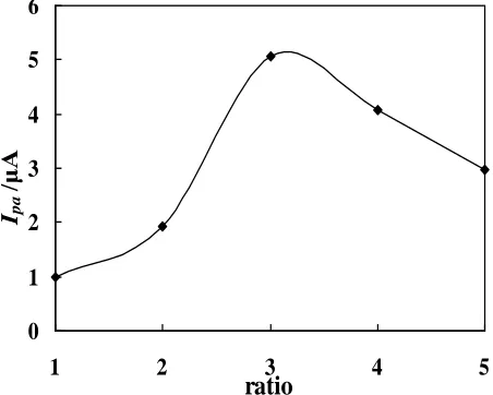
0 1 2 3 4 5 6
1 2 3 4 5
ratio
Ipa
/μ
[image:4.596.182.411.131.314.2]A
[image:4.596.184.410.516.697.2]
The influence of the composition of the composite electrode materials on the electrochemical response of cispaltin was investigated by changing the mass ratio of GO to MWNTs. Fig. 2 displays that the oxidation peak DPV response of cisplatin increases with GO-MWNTs mass ratio varied from 1:1 to 3:1, a maximum of DPV response was obtained at GO-MWNTs mass ratio of 3:1. While it decreases as GO-MWNTs mass ratio varying from 3:1 to 5:1. The GO-MWNTs mass ratio of 3:1 was therefore chosen for subsequent experiments.
3.3. Influence of the scan rate
-0.8 -0.6 -0.4 -0.2 0.0 0.2 0.4 0.6 0.8
-15 -10 -5 0 5 10 15 20 25
0.04 V s-1
I
/
μ
A
E/V(vs. SCE)
0.10 V s-1
Figure 3. The cyclic voltammetry curves of cisplatin at different scan rates in pH 7.4 KCl solution
0.04 0.05 0.06 0.07 0.08 0.09 0.10 -10
-5 0 5 10 15 B
v (V s-1)
I
/
μ
A
[image:5.596.193.413.230.407.2]Ipa Ipc
Figure 4. The linear relationship between the peak currents (Ipa, Ipc) and the scan rate(v).
The influence of scan rate (υ) on the voltammetric peak has been investigated. Fig. 3 illustrates the effect of scan rate on the CV response of cisplatin at GO-MWNTs /GCE in pH 7.4 KCl solution. It is found that both the oxidation peak current (Ipa) and reduction peak current (Ipc) are linear to the scan
[image:5.596.183.409.456.633.2]
(µA) = 0.0978υ + 5.6823 (r=0.9964), respectively, indicating that the electrochemical process of cispaltin at GO-MWNTs/GCE is adsorption-controlled.[34] The maximum achievable peak signal-to-noise ratio for cispaltin was obtained at the scan rate of 0.06 V s-1. The scan rate of 0.06 V s-1 was therefore selected in this work.
3.4. The cyclic voltammetry behaviors of cisplatin at different electrodes
-0.8 -0.6 -0.4 -0.2 0.0 0.2 0.4 0.6 0.8
-15 -10 -5 0 5 10 15 20
E/V(vs.SCE)
d b
I
/
μ
A
a
[image:6.596.185.407.198.374.2]c
Figure 5. Cyclic voltammograms of cisplatin at different electrodes in pH 7.4 KCl solution at the scan rate of 0.6 mV s-1. (a) GO-MWNTs/GCE (b) MWNTs/GCE (c) GO/GCE and (d) GCE
Fig. 5 shows the CV curves of GCE (d), GO/GCE (c), MWNTs/GCE (b) and GO-MWNTs/GCE (a) in 0.05 M KCl solution (pH 7.4) at the scan rate of 0.06 V s-1. A pair of redox peak was obtained for MWNTs/GCE and GO-MWNTs/GCE electrodes, while the oxidation and reduction peak currents of cisplatin at GO-MWNTs/GCE is much higher than that at MWNTs/GCE, which means that GO-MWNTs composite electrode materials can effectively improve the sensibility and performance of the electrode for the determination of cisplatin.
The oxidation peak potential (Epa) and reduction peak potential (Epc) of cispaltin at
GO-MWNTs/GCE were 0.092 V and -0.239 V (vs. SCE), respectively, △E=0.331 V (vs. SCE). The ratio of oxidation peak current (Ipa) and reduction peak (Ipc) was 2.07, implying that the electrode process of
cispaltin at GO-MWNTs/GCE is quasi-reversible.
3.5. Sensor Stability and Repeatability
0.13%.These experiments demonstrate that the GO-MWNTs/GCE has good stability, repeatability and reproducibility for the determination of cisplatin.
3.6. Analytical performance of the GO-MWNTs/GCE
The analytical performance of GO-MWNTs/GCE for the DPV determination of cisplatin was evaluated in the presence and absence of human serum. As table 1 shown, Good linear relationship was obtained between the oxidation peak current and the concentration of cisplatin within the concentration range from 65.0 μM to 358.0 μM for the absence of serum, and from 1.30 μM to 26.0 μM for the presence of serum, respectively. According to the method recommended by IUPAC, detection limit (CL)=3Sb/Sx, where, 3 is confidence factor, Sb is background noise standard deviation, Sx is the measurement sensitivity (The slope of the standard curve), as the result, the detection limit for cisplatin in the absence and presence of serum can reach to 0.192 μM and 0.113 μM, respectively, indicating that the sensitivity of GO-MWNTs/GCE is improved significantly as compared with that of MWNTs modified electrode [30]. Morever, the above results suggest that the GO-MWNTs/GCE has quite similar analytical performance for the absence and presence of human serum, indicating that the GO-MWNTs/GCE is much suitable for the determination of cisplatin in biomaterials.
Table 1. The Analytical performance of the GO-MWNTs/GCE for the determination of cisplatin in the presence or absence of 5% serum.
Table 2. Comparison of the detection limit for cisplatin with other previously reported methods
[a] Atomic Absorption Spectroscopy; [b] High Performance Liquid Chromatography; [c] Liquid Chromatography-tandem Mass Spectrometry; [d] Inductively Coupled Plasma-Atomic Emission Spectrometry; [e] Multi-walled Modified Glassy Carbon Electrode; [f] Graphene Oxide/ Multi-walled Modified Glassy Carbon Electrode.
Concentration range (μM)
Linear regression equation
Correlation coefficient
Detection limit (μM)
In the presence of 5% serum
1.30 ~26.0 Ipa (μA)=
345947c+3.2421
0.9990 0.113 In the absence of
serum
65.0 ~ 358.0 Ipa (μA)=
7294.2c+1.4056
0.9991 0.192
Methods LOD (μM) References
AAS [a] 0.10 [35]
HPLC [b] 3.33 [36]
HPLC 47.0 [37]
LC-MS [c] 0.033 [38]
ICP-AES [d] 0.12 [39]
Electrochemical method using a MWNTs/GCE [e] 39.0 [30]
The detection limit of the proposed method has been compared with that of the other previously reported methods for the determination of cisplation shown in table 2. It is evident that the proposed electrochemical method shows highly sensitive with the lower detection limit, indicating that GO MWNTs/GCE can be used as a sensor for the sensitive electrochemical detection of cisplatin in various samples.
3.7. Determination of cisplatin in spiked human serum
The standard addition recovery test and precision were also performed through analysis of 5% serum samples spiked with deferent concentration of cisplatin. The result was shown in table 3. The percentage recoveries based on the average of four separate determinations were from 93.18 to 104.89% with the RSD of 1.33 to 3.14%. In accordance with the equation (1):
V R c
6503 . 0
5
(1)
in the literature,[39] where, 5 is dilution ratio, R is measured platinum concentration in 5% serum samples, 0.6503 is the content of in platinum cisplatin (μg mL-1
), V is divide the volume of serum (mL), an amount of 1.5854 μg mL-1
of Pt in the 5% serum sample was obtained. Table 3. Measurement results of cisplatin in 5% serum (n=5).
No. Added (μM) Found (μM)
Recovery (%)
RSD (%)
1 13.00 13.23
2 19.50 20.01 104.89% 3.14
3 22.75 23.03 97.28% 2.47
4 26.00 25.35 93.18% 1.33
4. CONCLUSIONS
ACKNOWLEDGEMENTS
This work was supported by the National Natural Science Fundation of China (B070303, 51062018 and 21205104), the Natural Science Fundation of Yunnan (2010FXW004), Program for Innovative Research Team (in Science and Technology) in University of Yunnan Province (2010UY08, 2011UYN09) and Program for Yunnan Provincial Innovation Team (2011HC008).
References
1. R. B. Weiss and M. C. Christian, Drugs, 46 (1993) 360.
2. M. Breda, M. Maffini, A. Mangia, C. Mucchino and M. Musci, J. Pharm. Biomed. Anal., 48 (2008) 435.
3. Z. F. Fan, Z. C. Jiang, F. Yang and B. Hu, Anal. Chim. Acta, 510 (2004) 45.
4. M. Krachler, A. Alimonti, F. Petrucci, K. J. Irgolic, F. Forastiere and S. Caroli, Anal. Chim. Acta, 363 (1998) 1.
5. S. N. Lanjwani, R. K. Zhu, M. Y. Khuhawar and Z. F. Ding, J. Pharm. Biomed. Anal., 40 (2006), 833.
6. L. S. Foteeva and A. R. Timerbaev, J. Anal. Chem,. 64 (2009) 1206. 7. A. R. Timerbaev, Talanta, 52 (2000) 573.
8. A. R. Timerbaev, Electrophoresis, 23 (2002), 3884.
9. J. Petrlova, D. Potesil, J. Zehnalek, B. Sures, V. Adam, L. Trnkov and R. Kizek, Electrochim. Acta, 51 (2006), 5169.
10.A. M. Brett, A. Muuro, M. A. L. Scalea , S. H. Serrano, Electroanalysis, 8 (1996) 992.
11.K. S. Novoselov, A. K. Geim, S. V. Morozov, D. Jiang, Y. Zhang, S. V. Dubonos, I. V. Grigorieva and A. A. Firsov, Science, 306 (2004) 666.
12.Y. L. Zhang, Y. P. Liu, J. M. He, P. F. Pang, Y. T. Gao and Q. F. Hu, Electroanalysis, 25 (2013) 1230.
13.Y. Y. Shao, J. Wang, H. Wu, J. Liu, I. A. Aksay and Y. H. Lin, Electroanalysis, 22 (2010), 1027. 14.D. Zheng, S. K. Vashist, M. M. Dykas, S. Saha, K, Al-Rubeaan, E. Lam, John H. T. Luong and F.
S. Sheu, Materials, 6 (2013), 1011-1027.
15.J. Kim, L. J. Cote, F. Kim and J. Huang, J. Am. Chem. Soc., 132 (2010) 8180.
16.H. L.Wang, Q. L.Hao, X. J. Yang, L. D.Lu and X. Wang, ACS Appl. Mater. Interfaces, 2 (2010) 821.
17.S. Sun, M. Q. Zhang, Y. J. Li and X. W. He, Sensors, 13 (2013) 5493.
18.X. C. Dong, G. C. Xing, M. B. Chan-Park, W. H. Shi, N. Xiao, J. Wang, Q. Y. Yan, T. C. Sum, W. Huang and P. Chen, Carbon, 49 (2011) 5071.
19.V. Mani, A. T. Ezhil Vilian and S. M. Chen, Int. J. Electrochem. Sci., 7 (2012) 12774. 20.C. Zhang, L. L. Ren, X. Y. Wang and T. X. Liu, J. Phys. Chem. C., 114 (2010) 11435.
21.L. Qiu, X. W. Yang, X. L. Gou, W. R. Yang, Z. F. Ma and L. Chem. Eur. J., 16 (2010) 10653. 22.L. L. Tian, M. J. Meziani, F. S. Lu, C. Y. Kong, L. Cao, T. J. Thorne and Y. P. Sun, ACS Appl.
Mater. Interfaces, 2 (2010) 3217.
23.D. H. Kim, Y. S. Yun and H. J. Jin, Curr. Appl. Phys., 12 (2012) 637.
24.Q. Zhang, S. H. Yang, J. Zhang, L. Zhang, P. L. Kang, J. H. Li, J. W. Xu, H. Zhou and X. M. Song, Nanotechnology, 22 (2011) 4940101.
25.C. K. Hsieh, M. C. Tsai, M. Y. Yen, C. Y. Su, K. F. Chen, C. C. Ma, F. R. Chen and C. H. Tsai, Chem. Phys., 14 (2012) 4058.
26.R. I. Jafri, T. Arockiados, N. Rajalakshmi and S. Ramaprabhua, J. Electrochem. Soc., 157 (2010) B874.
27.B. Devadas, V. Mani and S. M. Chen, Int. J. Electrochem. Sci., 7 (2012) 8064.
29.V. Mani, B. Devadas and S. M. Chen, Biosens. Bioelectron., 41 (2013) 309.
30.M. L. Fang, Q. L. Li, Q. Liu, Y. X. Bei and Y. T. Gao, Chin. J. Pharm. Anal., 31 (2011) 1165. 31.L. L.Yan, Y. T. Gao, Z. F. Wang and Y. X. Bei, Chin. J. Pharm. Anal., 32 (2012) 1431. 32.W. Hummers, R. Offeman, J. Am. Chem. Soc., 80 (1958) 1339.
33.S. Yu, X. Y. Cao, M. Yu, Microchem. J., 103 (2012) 125.
34.S. J. Dong, G. L. Che, Y. B. Xie, Chemistry Modified Electrode, Revised edition, Wiley (2003). 35.S. B. Kangarloo, S. B. Gangopadhyay, S. Glück, J. E.A. Wolff, Turkish J. Cancer, 34 (2004) 71. 36.K. H. Kaushik, V. K. Sripuram, S. Bedada, N. Y. Reddy, G. I. Priyadarshini, K. R. Devarakonda,
Clin. Res. Regul. Affairs, 27 (2010) 1.
37.Y. Ramos, C. Hernández, L. A. Fernandez, M. Bataller, E. Veliz, R. Small, Quím. Nova, 34 (2011) 1450
38.D. V. Yaroshenko, A. V. Grigoriev, A. A. Sidorova, L. A. Kartsova, J. Anal. Chem., 68 (2013) 156. 39.Y. M. Yang, Y. P. He, J. S. Chen, J. Li, Chin. J. Health Lboratory Technol., 10 (2000) 259.