0022-538XI891031059-10$02.00/0
CopyrightC 1989, AmericanSocietyforMicrobiology
Characterization
of DNA
Sequence-Common
and
Sequence-Specific
Proteins Binding
to
cis-Acting
Sites
for
Cleavage of the Terminal a
Sequence of
the Herpes
Simplex Virus
1
Genome
JOANY CHOUAND BERNARDROIZMAN*
The
Marjorie
B. Kovler ViralOncologyLaboratories, the Universityof Chicago, 910 East58th Street,Chicago,
Illinois
60637Received13 September 1988/Accepted 12 November 1988
The terminal 500-base-pair a sequence of the herpes simplex virus 1 genome contains signals for cleavage (Pacl and Pac2) of unit-length DNA molecules from concatemers inuniquestretches ofsequencesdesignated Ub and
Uc,
respectively, and a cis site forcleavage designated DR1. We report that nuclear extracts from infected cellscontain factors which form two DNA-virus-specific protein complexes with components of the a sequence. Purffication of the factors forming the V2 complex yielded a protein with an apparent molecular weight of 82,000 binding to DNA in a non-sequence-specific manner. Addition ofMg2+
to the purified protein-DNA probe mixture resulted inexonucleolytic degradation of the DNA. The protein was identified as thevirus-specific DNase with monoclonal antibodyspecific for the viral enzyme. The purification of the proteins forming the V4 complex yielded two proteins with molecular weights of >250,000 and 140,000 corresponding to infected cellprotein 1 and to an as yet unidentified protein, respectively. These proteins formed two DNA sequence-common bands with a number of DNA probes and onesequence-specific band with probes containing both Pac2 andDR1 but not withprobes containing either site alone or Pacl and DR1. Since the DNA probe containing Pac2 and DR1 inserted into viral genome or into amplicons induced specific cleavage of the DR1 sequence whereas the nonreactive probes failed to induce thecleavage, the formation of this sequence-specific DNA-protein complex is significant and may reflect a DNA-protein interaction essential for cleavage. The possible role of the proteins identified in this study for the cleavage-packaging of viral DNA into capsids is presented.The terminal sequence, designated sequence a, of the
herpes simplex virus 1 (HSV-1) genome (Fig. 1) encodes
several cis-acting sites involved in (i) the circularization of
viral DNA after infection, (ii) the inversion of the two
covalently linkedcomponentsofHSV-1DNA, long(L) and
short(S), relativetoeachother,(iii)thecleavage of
genom-ic-lengthDNAfromcirclesorconcatemersandpackaging of
theexcised molecules into capsids, and(iv)theexpression of
anmRNAextending from theasequenceintothereiterated
sequenceofthe L component(2-7, 21-23, 25, 26, 28,29). In
the expectation that specific viral or host proteins or both
interact with the DNA at the specific cis sites, we
con-structedseveral viralDNAprobes
encompassing
mostofthe a sequence and tested these forreactivity
with proteinscontained innuclear extractsofmock-infected and infected
cells.Wereport that twoDNA-protein complexes formed by
the probes with nuclear extracts of infected cells each
contain a
virus-specified
protein. Theproperties
of theseproteinsare described.
Relevant to this report are the structure and nucleotide
sequence arrangementofthe a sequence. InHSV-1 strain F
[HSV-1(F)],
theasequencesituatedatthejunction
between the L and S components andrepeated inan invertedorien-tationatthetermini ofthe genomeconsistsofa
20-base-pair
(bp) terminal repeat
designated
DR1, a64-bp unique
se-quence designated Ub, 22 repeats of a 12-bp sequence
designated
direct repeat 2 (DR2), three repeats ofa37-bp
sequence
designated
directrepeat 4(DR4)andcontaining
11 of the 12 nucleotides of DR2, aunique
58-bp
sequence*Corresponding author.
designated
Uc,
and a second copy of DR1 (23). Tandemrepeats ofthe a sequence share the intervening DR1. The number ofcopies ofDR2 and DR4 vary, andpartial copiesof these sequences may also be repeated. This variability accountsformostoftheobserved a sequencepolymorphism (4-6, 21, 23, 28).
The observations that theterminal DR1 ofthe L
compo-nent consists of18 bp plus a single nucleotide extended 3'
andthatthe terminalDR1oftheScomponentconsists of1
bp plus a single nucleotide extended 3' producing, when
ligated,anintactDR1 led tothe conclusionthat the
genomic-lengthDNAisexcised fromconcatemersbycleavage within
DR1 sequences(23). Recentstudieshavedemonstratedthat
the signals for the cleavage and
packaging
of the DNAdesignatedPac2 andPacl(Fig. 1)arelocatedin the
Uc
andUbsequences, respectively, of thea sequence(6,28).
(These resultswerepresentedattheInternational
Herpes-virusWorkshop, Irvine, Calif., August 1988.) MATERIALS ANDMETHODS
Cells, viruses, and protein extracts. HeLa cells grown to
confluency in 850-cm2 roller bottles were either mock
in-fected or inin-fected with 5 PFU ofHSV-1(F)orherpes
simplex
virus (HSV-2) strain G
[HSV-2(G)]
per cell. Cells wereharvested 20 to 24 h afterinfection ormock
infection,
and nuclear extracts werepreparedin 0.42 MKCI asdescribed previously (8).Preparation ofDNAprobesandcompetitorDNAand DNA band shift assays for DNA-binding proteins. DNA
probes
used in thetext wereprepared
by
digesting
theplasmids
with 1059on November 10, 2019 by guest
http://jvi.asm.org/
1060 CHOU AND ROIZMAN
F probe
z-a,omb
L ">S@
Cpac-I A probe pac-2
DRI
U(DR2)22
(DR4)
DRIX Dral
ApaI
__ .IEprobe pac-2 1
H
probe
Hprobe rr DR4DRIDRI
G probe
[1
BamHI
AvaUI
ApaI
BamHI
Jprobe C probe
D probe
B probe Kprobe
l
L probe
A
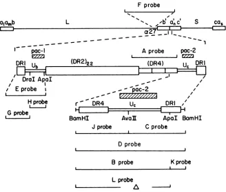
FIG. 1. Schematic diagram ofthe sequence arrangementsin the HSV-1genomeand in theasequenceand thepositionin thegenomeof the DNAprobes used in this study. Top line:Sequencearrangementof the HSV-1genome.a,anda.refertotheterminalasequencesofthe LandScomponents.Subscriptn, One or moreasequences;subscriptm,noneto morethanone asequences.The insert indicates theposition ofthe F probe consisting of the 48 bp ofthe regulatory domain ofthe HSV-1(F) a27gene frompRB606 (13). Second line: Sequence arrangementof theasequence.Thewild-typeasequencecontainsa20-bp directly repeatedelement(DR1)flanking theasequence;a64-bp uniquesequence(Ub); 22copies of the12-bpdirectlyrepeatedelement(DR2);3copies of the 12-bp directly repeated element (DR4); anda
58-bpuniquesequence(Uc). The AprobeencompassingthreecopiesofDR4and onecopyofDR2wasderived fromtheplasmidA-1 and generated by BssHII andApalrestrictiondigestoftheA-1 clone(6).The DandEprobesderived frompRB3389andpRB3387,respectively, weregenerated by cleaving the parentplasmid withBamHI forprobe DandBamHI toApaIfor probe E.The pRBplasmids have been previously described (2). TheLprobewasgenerated byBamHIandApal cleavageofaplasmidwhich containsaBssHII deletion ofpRB3389 as shown; the C, J, B, and K probes were generated from pRB3389 by taking theBamHI-to-AvaII subfragment, the AvaII-to-BamHI fragment, the BamHI-to-ApaIfragment, and theApaI-to-BamHIfragment, respectively, asindicated. TheGand Hprobesweregenerated from pRB3387 bytaking the BamHI-to-DraI fragmentandtheDral-to-Apal fragment, respectively.
appropriate restriction enzymes. The DNAs were then
de-phosphorylated with calf intestinal alkaline phosphatase
(Boehringer Mannheim Biochemicals, Indianapolis, Ind.) andloadedona5% nondenaturingpolyacrylamide gel.The
fragmentsof interestwerethenpurified frompolyacrylamide
gels and 5' end labeled with [32P]ATP (>7,000 Ci/mmol; Dupont, NEN Research Products, Boston, Mass.) and T4
polynucleotide kinase (United States Biochemical Corp.,
Cleveland, Ohio) to an activity of approximately 30,000
cpm/ngof DNAfragment(17).
The standard competitor DNA usedin these assays was
synthetic poly(dI-dC)- poly(dI-dC) (Pharmacia, Molecular Biology Division, Piscataway, N.J.). Competitor polymers
were used at 2 to 3 ,ug per reaction of standard nuclear
extract and 0.5 ,ug per reaction ofpurified proteins in gel binding retardationassays.
Binding reaction mixtures (12pl)contained approximately 0.1ngoflabeledDNA, 2to3,ug of competitor DNA, 9
RI
of binding reaction buffer (20 mM Trishydrochloride[pH 7.4], 50 mM KCl, 0.05% Nonidet P-40, 5% glycerol, 50 ,ug of bovine serum albuminper ml, 5 mM 3-mercaptoethanol, 1 mMEDTA), and 2to 3 ,ug ofextractprotein. After incuba-tionatroomtemperaturefor 30min,reaction mixtureswereloadedon a5% polyacrylamide gel in 50 mM Tris-borate-1 mMEDTAbuffer(pH 8.3)prerunat230 V for 2 h. Gelswere
run at 230 V until bromophenol blue dye reached the gel
bottom; then they were dried and autoradiographed (27).
Whenmonoclonal
antibody
wasusedin thebinding reaction,
1
RI
ofspecificdilutedmonoclonalantibodywasadded tothereactionmixture aftertheincubation periodin the standard
reactions and the reaction was allowed to incubate for
additional30minbefore
loading
ontogels.Chromatographic purificationofproteins. The
purification
scheme used in these studies is shown in
Fig.
2. For P11phosphocellulose chromatography, 50 to 70 mg of nuclear
extract stored at 12.9 mg/ml in buffer D (20 mM Tris
hydrochloride
[pH 7.9],
0.42MKCl,
1.5mMMgCI2,
0.2mMEDTA, 0.5 mMdithiothreitol,0.5mM
phenylmethylsulfonyl
fluoride,25%glycerol)wasdiluted with buffer C (20mMTris
hydrochloride
[pH
7.9],
0.2 mMEDTA,0.5 mMdithiothre-itol, 25% glycerol,0.5 mMphenylmethylsulfonylfluoride)to
afinalKCIconcentration of0.125 M. Thedilutedextractwas
loadedontoapreequilibrated P11 phosphocellulose column
(Whatman, Inc., Clifton, N.J.) prepared according to the
instructions ofthe manufacturer. The column was washed
with 2 column volumesofbuffer Ccontaining0.15 M
KCl.
Astepgradientinbuffer C
containing
KClranging
from0.15to0.6 Min stepsof0.05Mwasthen
applied,
and the fractionswere collected. The fractions were then analyzed for the
J.VIROL.
on November 10, 2019 by guest
http://jvi.asm.org/
[image:2.612.152.480.71.349.2]HSV(1) Nuclear
Extract
Pi1 buffer C
0.15M KC1
Flowthrough 0.25-0.30M KC1
buffer c Hepa ri n buffer C
0.15M KC1 Agarose 0.15M KC1
0.30-0.35M KC1 0.35-0.40M KC1
DEAE
buffer C
jabuffer
C0.10M KC1
Sehcl
0.10M KC10.15-0.20M KC1
buffer C
0. 1014 KC1
Superc
12 Fractions
13-14 V2
0.15-0.20M KC1
:)SO buffer C
0.10M KC1 Fractions
10-11
[image:3.612.84.269.65.369.2]V4
FIG. 2. Flow diagram of theprocedures for purification of the factors forming the V2 and V4 DNA-protein complexes. HSV-1 infections in HeLacells, preparation of the nuclearextracts, and chromatographic purificationof theproteinsaredescribed in
Mate-rials and Methods.Column sizes used in these purifications
proce-duresare6 mlof P11 phosphocellulose, 3 ml ofheparin-agarose, 1
mlofDEAE-Sephacel,24 mlof Bio-Gel A-0.5M(0.8 by47cm), and 25 mlof fastprotein liquid chromatography Superose 12.
abilitytoformcomplexeswithprobeDNA ingelretardation
assays. Theactive fractionswere pooled.
Forheparin-agarosecolumnchromatography, active frac-tions eluted from the P11 column were pooled and diluted
with buffer CtoaKCI concentration of 0. 15 M and loadedon a preequilibrated heparin-agarose column (Sigma Chemical Co., St. Louis, Mo.). After the column was washed with 2 column volumes of buffer C-0.15 M KCI, a step gradient consistingof buffer C with a salt concentration from 0.2 to 0.5 MKCIwasdeveloped andfractionswere collected and
assayed by the binding gel retardation assay. Active frac-tions were pooledandprocessed.
For DEAE-Sephacel column chromatography, protein fractions pooled from the preceding column were diluted
with buffer Cto a KClconcentration of 0.1 M and loaded
ontoapreequilibratedDEAE-Sephacelcolumn(Pharmacia). Elution procedures werethesameasabove.
For Bio-Gel A-0.5M(25 ml; Bio-Rad Laboratories, Rich-mond, Calif.) orfast-protein liquid chromatography
Super-ose12(24 ml; Pharmacia)chromatography, proteinfractions
wereconcentratedtoless than 200 pJ,1% of the column bed volume, withaCentricon 30 concentration (Amicon Corp., Danvers, Mass.) prior to loading. After loading, proteins
wereeluted with buffer Ccontaining0.1MKClandfractions
were collected on a fraction collector and assayed for binding activity.
Gel electrophoresis of proteins. Electrophoretic separa-tions of denatured proteins were done in 9.25% polyacryl-amide gels cross-linked with N,N'-diallyltartardiamide con-taining sodiumdodecyl sulfate as described by Morse et al. (24). The gels were stained with silver for visualization (20) and mounted forphotography.
RESULTS
Experimental design. Preliminary experiments established that nuclear extracts of infected cells contained factors capable of binding to DNA fragments derived from the a sequence. Intheexperiments we reporthere, labeled DNA fragments were usedasprobes to monitor the purification of these factors, to determine the specificity of binding of the viral proteins recognizing DNA sequences contained in the terminal a sequence, and last, to identify the proteins involved in forming the complexes. As shown in Fig. 1, probe A contained the reiterated sequence DR4 and one copyofthe DR2 sequence. ProbesB, D,C,and Jcontained all or portions ofthe DR4,
Uc,
and DR1 domains of the a sequence. ProbesG, H, and E similarly covered the Ub andDR1 domains of the a sequence. Last, probe F containing
unique sequences of the L component was used as the
representative of non-a sequence domains of the HSV-1
genome. Probes B and C were used for detecting and
monitoring the purification of viral proteins capable of
binding to the a sequence. These probes overlap; whereas
the C probe contains most if not all of the Pac2 sequence
required forcleavage recognition and the cleavage site, the B
probe contains only the cleavage recognition sequence. All
theprobes wereusedfordefiningthe specificity of binding.
Duringpreliminary experiments involvingDNA bandshift
assays, weidentified in the nuclearextracts of infectedand
mock-infected cells several factors capable of binding the
labeled DNA probes used in these studies. Of particular
interest were two factors designated V2 and V4 detected
only in extracts of infected cells. To differentiate between
these factors and to study their properties, we used the
concentrationand partial purificationscheme shown inFig.
2.
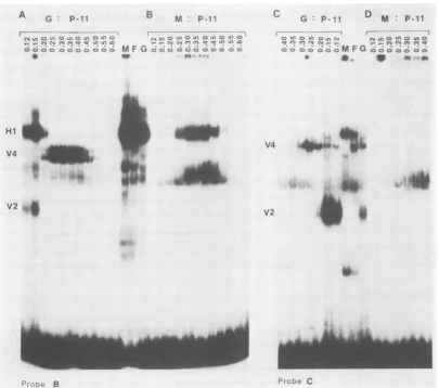
Demonstration of factors from infected cells capable of binding to the DNAprobe sequences. Figure 3 shows DNA
band shiftassaysutilizing probesB (panelsAand B) andC
(panels C and D) and both crudeandP11-fractionated (Fig.
2) nuclear extracts ofinfected andmock-infectedcells. The resultswere as follows.
(i) Both labeled DNAprobes formed multiple bands
con-taining DNA-protein complexes when reactedwith nuclear
extracts ofmock-infected and HSV-2(G)-infected cells. In
the experiments shown, the competitor DNA was
poly(dI-dC) poly(dI-dC), but substantially identical results were
obtained with other synthetic polymers
[poly(dI-dC);
datanot shown).
(ii)Severalof theDNA-protein complexes(e.g., the band
designated asHi in Fig. 3A and B) were detected in DNA
band shift assays withextractsof nuclei from mock-infected andfrom HSV-1- or HSV-2-infected cells. In contrast, the
DNA-protein complexes designated V2 and V4 were
de-tectedonlyin assays donewithextractsofinfected cells. In
general,thehost
protein-DNA complexes
tendedtoobscuretheinfectedcell-specific protein-DNA complexesformed
by
crude extracts ofinfected cells
(for example,
comparethe lanes marked M[mock]
and G[HSV-2]
shown betweenpanels with the lanes showing the reactions of fractionated
proteins in extracts of mock-infected
[panels
B andD]
andon November 10, 2019 by guest
http://jvi.asm.org/
1062 CHOU AND ROIZMAN
A
G:
P-fl
B M P-11 C G: P-l D M: P-1I
....
Lo
U) 'rufC2K0 U)OO()*n (_ :,a)0 r3 a)Unr) 'rC.<3il'r
C:>ICcDc3a oC.- 5OC M F G C OO O Oc a a °
-Hl1
V4
_Irb...
V2
14;1~~~~~~~~~~~~~~~~~~~~~~
CDU) C)U') CnU:S C U') Lo CD
OsCDDOC:3 0 F G O COO CD°
V4 X 4
*
i~~a-*.
i
Probc C
FIG. 3. Autoradiographic images of labeled DNA-protein complexeselectrophoretically separatedin nondenaturing gels. DNA probe fragments(Fig. 1)generated bydigestionof theplasmidswiththeappropriate enzymesweredephosphorylatedwithcalf intestinal alkaline phosphatase, purified frompolyacrylamide gels, and 5'end labeled with[.y-32P]ATP (>7,000Ci/mmol)byT4polynucleotide kinasetoan
activity of approximately 30,000cpm/ngofDNAfragment (17). (A and B) Reactionofprobe B DNAwith chromatographicfractions of nuclearextractprepared fromHSV-2(G)-infected cells (A) and mock-infected cells(B).(Cand D)Reaction ofprobeCDNAwith thesame
chromatographic fractions as in panels A and B. Lanes M, F, and G, Reactions of nuclear extracts of mock-, HSV-1(F)-, and HSV-2(G)-infected cells. Numbersatthe top of thegel indicatetheKCI concentration inbuffer C usedtoelute theproteinfractionsfrom column. V2andV4designatetheviralprotein-DNAcomplexes. Hi is discussed in thetext.
infected [panels A and C] cells). Moreover, the infected cell-specific protein-DNA complexesformed byextracts of HSV-2(G)-infected cellswere strongerandmore prominent
than those formed byextracts ofHSV-1(F)-infected cells. (iii) The properties and the specificity ofbinding of the factors responsible for the V2 and V4 DNA-protein
com-plexes aredealtwith in the text below. Of the hostfactors, the Hi factordefinedby P11 column chromatography (Fig. 3)wasshowninotherexperimentsdescribed later in thetext tobindstronglytoprobes B, J,and D butnottothe Cprobe. The designation of Hi assigned to the comigrating desig-nated bands inFig. 3A andB isprovisional; either the host DNA-binding proteinsin the mock-infectedcell extract are
different fromthose detected in the infected cellextractsor
theirpropertieshave beenalteredduringinfection inasmuch
astheir elutionprofiles were reproduciblydifferent.
Concentration andpartial purificationof V2and V4 factors from nuclear extractsof infected cells. The protocol for the purification of the proteins involved in the formation of V2 and V4DNA-protein complexes is shown in Fig. 2 and is
described inMaterials and Methods and in thelegendtoFig.
2. ThepurificationwasmonitoredbyDNAbandshiftassays
(Fig. 4) andbysilverstaining of proteins
electrophoretically
separated in denaturing 9.3%
polyacrylamide
gels (Fig. 5Aand B). In each step of the
purification procedure,
the fractionscontainingtheactivitydetectedby
DNA bandshift assayswerepooled
and usedasstarting
materialin thenext step.Asnotedearlierin the text,activity ofthefactorsin the V2
and V4 DNA-protein complexes were
barely
detectable incrude nuclear extracts of HSV-1-infected cells but were more abundant in those of HSV-2(G)-infected cells. The factors forming the V2DNA-protein complex eluted in the
flowthrough fraction from P11 columnchromatography and
at aslightlylower salt concentration than V4from
heparin-agarose chromatography (Fig. 4). The Superose 12column fraction containingmost ofthe V2 activitymaterialyielded
on electrophoresis in a denaturing sodium dodecyl
sulfate-polyacrylamide gela
single major
silver-stainedpolypeptide
band with anapparentmolecular
weight
of85,000for HSV-J. VIROL.on November 10, 2019 by guest
http://jvi.asm.org/
[image:4.612.112.517.72.430.2]HSV-1 a SEQUENCE-SPECIFIC PROTEINS 1063
B. Heparin Agarose
I~~n a i~nn QCin
N
C%N
MFGC eaooa o oo
t
p
_ eu*C. DEAE Sephacel
MFQ
an
a CoMFG0 o o o o
. _-. -iw-- 1
0. Superose12
M F G P 3 4 5 B7
s1O12U14151S17*1S2l2;
8W be
V2
a
V2AIAa--.__LAs
E.P11 F. HeparinAgarose
0.25 0O925
P11 un o C? ° P11 o
MFG' FG W-gIci CD r-0 CD° M1: GNMf G o
4
G. OEAE Sephael
o an a cMs Go a o2o ocoa M F Go o c
o o o es
V4 4
Wv4
iqp~
~~~~~~W
3
IV4 swOr weV4
a. L
FIG. 4. Autoradiographic images of labeledDNA-protein complexes electrophoretically separated innondenaturing gels. (AtoD)The
autoradiograms show the binding activityof theV2proteinfractionatedinsequentialorder by (A) P11 phosphocellulose,(B)heparin-agarose, (C) DEAE-Sephacel, and (D) Superose 12 columns.Thechromatographicprocedureswere asdescribed in the legendtoFig. 2.LanesM, F,
and Gcontainingreactionmixturesofnuclearextractsofmock-,HSV-1(F)-,andHSV-2(G)-infectedcells,respectively,withprobe CDNA
indicate the position of V2 protein-DNA complexes. Fractions collected from each column were identified by the KCI content (P11, heparin-agarose,andDEAE-Sephacel)orserially (Superose 12). (EtoH)Sameas panels A and DexceptthatprobeBDNAwasusedto
probeV4DNA-protein complexes.LanesM, F,and G, Reaction mixtures of nuclearextractsof mock-,HSV-2(F)-,andHSV-2(G)-infected cellswithprobeDNAs. ThelettersV2and V4markthe position ofthecorresponding bands;asnoted in thetext,thesebandsarefrequently
notseeninassaysof crude nuclearextracts.
1(F) (Fig. 5, right panel) or 82,000 for HSV-2(G) (data not shown) and avery faint band with an apparent molecular weight of60,000.Thefaintbandwasnotalwayspresent;it is
seen in lane 4 but not in lane 3 of Fig. 5 and may be a
degradation productof thehigher-molecular-weight protein. The material eluting from the Superose 12 column and forming the V4 DNA-protein complex formed two,
appar-ently equimolar, major silver-stained bands with apparent
molecular weights of >250,000 and 140,000 (Fig. 5, left panel),referredtoasproteinsAandB,respectively. Similar bands were obtained from extracts of HSV-2(G)-infected cells,andanalyses done with monoclonal antibodies reactive with infectedcell protein 1(ICP1) suggest that proteinAis ICP1 (data not shown). It should be stressed, as will be shownbelow,that atallstages ofpurificationof theproteins formingthe V4complex,thefractionscontainingtheactivity
A.P11
c MFG
V2*9 V2
H. Superose 12
MFG8 101112 .AMAIL A&Ak
mm
VOL.63, 1989
V4
on November 10, 2019 by guest
http://jvi.asm.org/
1064 CHOU AND ROIZMAN
HSV-1(F):V4 HSV-1(F):V2
KPG M F'4 3 2 1 F 1 2 3 4 ICP
4
4
11
S
1717
* * _. 19
2425
[image:6.612.64.304.73.356.2].- II^
FIG. 5. Photographofasilver-stained denaturing (sodium
dode-cyl sulfate)9.3%polyacrylamide gelcontainingelectrophoretically separated proteins from various stages ofpurification ofproteins contained in V2 and V4DNA-protein complexes.LanesM, F,and Gwere loaded withapproximately20 jig eachof the crude nuclear
extracts ofmock-infected, HSV-1(F)-infected, or
HSV-2(G)-infec-ted cells, respectively. Lanes 1, 2, 3, and 4 in both left andright panelswereloaded withpooled activefractioncontainingeither V4 (left panel)orV2(right panel) activityandobtainedduringstepwise
purification of the proteins formingthe V2 and V4complexesfrom HSV-1(F)-infected cell extracts by chromatography on P11
phos-phocellulose (lanes 1), heparin-agarose (lanes 2), DEAE-Sephacel (lanes 3), and Superose 12 (lanes4), respectively. Thepurification protocol is shown in Fig. 2. Numbers to the right of the figure indicate the ICP numberdesignations of Honess and Roizman (12)
asamendedby Morseet al.(24).
yieldedtwobands in DNAretardation gels with probe B and threebands with probe C.
Biologic properties and identificationofprotein formingthe V2 DNA-protein complex. During studies of the biologic properties of the proteins forming theV2complex,wenoted
thataddition of the purified V2 (Fig. 5,right panel, lane 4)to
the labeled DNA probes in the presence of 10 mM Mg2+ resulted in the loss of the terminal 32p label from the probe DNAs (Fig. 6, right panel). One hypothesis that could account forthe apparentloss of thelabeled DNA probes is that addition of
Mg2'
activated a DNase. To test the possibilitythat theprotein containedin theV2DNA-protein complexistheviralDNase, weperformed band shiftassaysin the presence and absence of monoclonal antibody Ql
obtained from K. L. Powell of the Wellcome Research Laboratories and directed against the HSV-1 and HSV-2
enzymes. Earlier we showed that antibody specific to the DNA-binding proteins frequently binds the protein in the DNA-protein complexandfurther retards the mobility of the DNA(14). The resultsshown in Fig. 6,left panel, and other studies not shownindicated thefollowing.
(i) Only a singleDNA-protein complex was observed in all the assays done with partially purified fractions of the V2 factor (Fig. 5, right panel, lane 4). In each instance, the addition of the Ql monoclonalantibodyretarded the
migra-tion of the V2 complex, as would be expected from the
specific interaction of the monoclonal antibody with the protein factorcontained in the complex (14). A faint DNA-protein complex was also noted in the assays done with crude nuclear extracts ofHSV-2(G)-infected cells and the E probe. Themigration ofthis complex was alsoretarded by the antibody.
(ii) The formation of the V2 DNA-protein complex in assays with the probe F DNA, a 48-bp fragment derived from the promoter-regulatory domain of the a27 gene (13),
indicated that V2 factoris notspecificfor DNA sequences
containedin the asequence. The reliability of the band shift
assays is underscored by the observation that the F probe DNAdid react with theaH1proteinand thevirus-specifica trans inducing factor (aTIF) to produce the DNA complex
showninFig. 6. Theidentityof thecomplex was verified by
thedemonstrationthatantibody to the aTIF protein retarded
themigrationofthecomplex innondenaturing gels (datanot
shown), as was previously demonstrated (19).
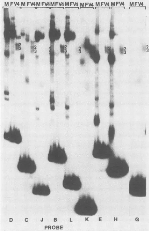
Bindingspecificity of V4. Thepurified fraction which forms the V4 DNA-protein complex consists of two equimolar amounts of viral proteinscorrespondingin apparent
molec-ularweighttoICP1 and to a yet unidentifiedprotein(Fig. 5,
left panel). To define the specificity ofthe binding ofthese proteins to DNA, we tested alltheprobesgenerated inthis study for theirreactivityandabilitytoform theV4 protein-DNAcomplex with thepurifiedV4fraction shownin Fig.5, leftpanel, lane 4, and with crude nuclear extracts of
mock-infected cellsand cells infectedwithHSV-1(F). Theresults
indicate that probes D and C reacted with the purified
proteins formingthe V4complex toyieldthreebands. Bands
2and 3formedwith allprobes,except the bandsformedwith the J probe werebarely visible andthose formedwiththe L
andGprobeswerefainterthan thoseobtained with the other
probes. Band 1 was reproducibly present only in lanes
showingtheprotein complexesformed by probes D andC.
DISCUSSION
We identified several DNA-protein complexes that were
formed by nuclear extracts of both infected and mock-infected cells and two DNA-protein complexes designated V2and V4 that werespecific for nuclear extractsofinfected cells. Thefocus ofthestudies reported here was the
identi-fication of viral proteins which reacted with the a sequence
of HSV-1. It is convenient to consider the identity and
significance ofthe proteins formingthe various complexes
separately.
V4protein-DNA complex. The most purified preparation which formed the probe C DNA-V4 protein complex con-tained two viral proteins in approximately equimolar amounts. Protein A was identified as ICP1 by itsapparent molecularweight and its reactivity with a monoclonal anit-body (9). Protein B comigrated with a protein of a molecular weight predicted forICP7, but itsidentity is notknown. Of the three bands formed with probe C, the two designated bands 2 and 3(Fig. 7) were alsoformed with other probes, whereas thecomplex designated as band 1 formed only with
probes D and C, i.e., the probes that contain at least a
portionof the
Uc
sequence and DR1. We mayconcludefromthese observations that either the specificity of binding
requires the interaction of both proteins or one of the two
J. VIROL.
on November 10, 2019 by guest
http://jvi.asm.org/
Qi Oi Oi Oi
+ , t
t F OGV2V2M F V2WN
h IT I I
V4 .
V2
%
w.
0101 O Qi
+ + + I
AF GGV2V2M F GGVT2
_1
iaTIF 1 ~2 3
Mg
3 -.... MW4 5
_IM
E
240
bp
S
125bp
2~ 86
bp
q_p
56 bp
4
39bp
[image:7.612.98.521.76.370.2]Probe:E D A F
Probe
Cwith
V2
Protein
FIG. 6. Autoradiographic images of labeled DNA electrophoretically separated in nondenaturing gels. Right panel: Images of purified V2 protein-labeled probe C DNA complexes formed in the presence or absence ofMg2+in thebinding reaction. Lane 1, Labeled probe without protein added; lane 2, probe DNAand 0.2 ,ul ofV2fraction containing noMg2". Lane 3, Same as lane 2 except that the binding buffer contained10mMMg2+.Lanes 4and 5,Sameaslanes2and 3, respectively, except that theconcentration of V2 protein was 0.5p.1perbinding reaction. The V2 preparation istheone shown in Fig. 5, right panel, lane 4. The probe C DNA and the V2fraction in a total of 20 ,u1were reacted at37°C for 1 h. The reaction was terminated by the addition of sodium dodecyl sulfate and proteinase K to final concentrations of 0.4% and100,ug/ml, respectively. The DNA was extracted with phenol, ethanol precipitated, dried, and subjected to electrophoresis on a 5% nondenaturinggel asdescribed intheResults. Left panel: Reactivity of V2 protein-DNA complexes with monoclonal antibodyQlspecific for HSV DNase. Lanes M, F, and G, Reactions of nuclear extracts of mock-infected, HSV-1(F)-infected, and HSV-2(G)-infected cells described above with various probe DNAs; lanes designated V2, reaction of labeled DNA probes identified on the bottom of the lanes with purified V2 protein shown in Fig. 5, right panel, lane 4. In lanes designated +Q1, the reaction mixture contained in addition to the stated ingredients1 p.lof monoclonal antibodytoHSV-1 DNase. The dot to the right of the band identifies the V2 protein-DNA complexes. Note thatprobeFcontains the cis sitefor the induction of a genes by the virion aTIF (19). With HSV-1(F)- or HSV-2(G)-infected cell extracts and the competitor DNAs, probeFDNAformscomplexes with a host protein designated as aH1(15) and with both aH1 and aTIF. The Ql antibody hasnoeffect on the formation ofaH1oraTIFbands (comparelane G, probe F, with lane G+ Ql, probe F). V2 forms a complex withprobe F, and this complex is retarded in its electrophoretic mobility by antibodyQl (rightmost two lanes). MW, Molecular weight.
proteins was copurified adventitiously and binds to DNA
nonspecifically.
ProteinsA (ICP1) andB have notbeen extensively
char-acterized. ICP1 isastructural protein(VP1); it is located in
the tegument and is not readily removed from virions
de-enveloped by treatment with nonionic detergents (16).
Fewer than 150 molecules of ICP1 per virion have been
estimated to bepresentbased on its then estimated
molec-ular weight of 260,000 to 270,000 (9). Since its translated
molecular weight is currently predicted tobe 336,000 (18),
the actual numberof molecules per virion might be
signifi-cantly smaller.
Independently derived data link ICP1 with encapsidated
DNA and more specifically with the release of viral DNA
from capsids (1). Thus, in cells infected with
HSV-1(HFEM)tsB7 at the nonpermissive temperature, the
cap-sidsaretransportedtothenuclear pore, but viral DNA isnot
released into the nucleus. The DNA is released upon shift
downtothepermissivetemperature. The mutation in
HSV-1(HFEM)tsB7 wasmapped by marker rescuetechniques to
thedomain oftheICP1gene. Itis conceivablethatICP1may
be involved in the shuttling ofthe DNA in and out ofthe
capsids, but atleast the packagingfunction remains to be
proved.
The genespecifying proteinB has notbeen identified. In
earlierstudies, proteinBwouldhavebeen obscuredbyother
virion proteinswith similarelectrophoretic mobilities(9).
V2 protein-DNA complex. The most purified V2 protein
preparation containedasinglepolypeptide.The
electropho-reticmobility ofthe protein, its enzymatic activity, and its
reactivitywith monoclonal antibodyare consistent with its
beingthevirally encodedDNase. Thebindingof the DNase
toDNA, although notunexpected, has notbeenpreviously
reported. Although the virally encoded DNase has strong
exonucleolytic
activity,
it has beenreported
to exhibitnon-sequence-specific endonucleolytic activity in the
pres-enceof
Mg2+
(10,11). Thebindingof the DNasetotheviralDNAfragments doesnotappeartobe sequence
specific.
It0 acHi
on November 10, 2019 by guest
http://jvi.asm.org/
1066 CHOU AND ROIZMAN
TABLE 1. Correlation ofsusceptibility to cleavage with the formation ofsequence-specific complexes
Sequence present
Probe asequencecomponent present Cleavage inampliconorvirus
cissite
A DR2 + (DR4)3 - - - NDa
B Uc + portionofDR1 - + - ND
C Portion ofUc + DR1 (portionofprobe D) - + + ND
D Uc+ DR1 - + + +(6)
E Ub+ DR1 + - + -(6)
F None(regulatory region of the ca27 gene) - - - ND
G Portion ofUb+ DR1(portion ofprobe E) - - + _b
H Ubsubfragment (portion ofprobe E) + - - ND
J PortionofU,, (portion of probe D) - - - ND
L Uc, sameasprobe D,but minus the BssHIIfragment - - -C
a ND, Not determined.
bThis is the smallestfragment containinganintact DR1 sequence. The DR1 sequencewastestedin the virus and in theampliconsystemwithnegativeresults
(6).
cTheeffect of the deletion of the BssHIIfragment fromUcwastested inthe contextofalargerfragment containing DR1,theUcminusthe BssHIIfragment,
and thereiterated DR4 and DR2 sequences andUbsequence.TheDNAfragmentwas notcleavedin theampliconsystem,indicatingthat the deleted sequence wasessential for cleavage (6).
should be noted, however, thatDNA binding andcleavage
by the DNasecould be sequence specific ifits activitywas
directedbya protein whichcoupled tothe DNaseandwas
bound to the viral DNA in a sequence-specific fashion. A
MFV4MFV4MFVY4MFV4MFV4 MFV4 MFV4MFV4 MFV4
w..t~~~~~~~~~~~~~
2fti,4 | i s
a
\ t~i'
w
0
.:#I
p
4-role for the viral DNase may also be deduced from the available data on thecleavage ofunit-lengthDNAmolecules fromconcatemers orcircles.
Specifically, (i) the Lcomponent terminus and the
junc-tion between the L and S components of the HSV-1 DNA
eachcontainone to several a sequences. The Scomponent
terminus containsonly one a sequence(23). Studies onthe
structureof the asequences attheterminiandthe
junction
of theLand Scomponentsledtotheconclusion that the site
of the cleavage of concatemers orcircles into linear
unit-length DNAmoleculesiswithintheDR1 sequence shared
by
two adjacent a sequences and results in single-base 3'
extensions in the ends of the linear molecules (23). The
endonuclease
responsible
for thecleavage
of the DNA hasnot been identified. Although the cleavage and
packaging
appear to be related (7), it is not inconceivable that viral DNase in connection with other site-directed proteins is
responsiblefor thiscleavage. The HSV DNase can act asan
endonuclease.
(ii) Akey question regarding
cleavage-packaging
of viralDNA was the fate ofnewly synthesized molecules with a
singleasequence at the componentjunctions. Insertion ofa
singleasequence into virus resulted in oneof two events. In
someinstances,the asequence wasreduplicatedintandem,
yieldinguponcleavagetwoidentifiable endseach
containing
an asequence. When the a sequencewasnotamplified,the
cleavageeventoccurredin thedistal
(post-U,)
DR1and theterminus containing solely the residualportion of DR1 but
not the remaining components ofthe a sequence was no
longer detectable andwaspresumedtobedegraded (28). If
the same sequence of events was to occur in
cleavage-D C J B
PROBE
FIG. 7. Autoradiographic images of the complexes formed by labeled DNA probes with crude extracts and purified fractions containing the proteins forming the V4complex. The complexes formedby each of theprobes shownonthebottom of thefigure with nuclearextracts of mock-infectedcells (M)or HSV-1(F)-infected
cells(F)orwithpurifiedV4
(V4)
areshown in the threecorrespond-inglanes ofeach
triplet.
The V4 bandsunique
toprobes
Dand Careidentifiedasband1.Bands 2 and 3 appeartoform withmostprobes tested inthis study. The purified V4is that shown in Fig. 5, left
H G panel, lane4.Thecomplexeswereelectrophoretically separatedon
EHG nondenaturing gels. The bands formed withextractsof infectedand mock-infected cells and migrating slightly slower than band 1 correspondtothosedesignatedHi in Fig.3.
J. VIROL.
on November 10, 2019 by guest
http://jvi.asm.org/
[image:8.612.68.562.87.224.2] [image:8.612.60.303.344.721.2]packaging
of viral genomes from concatemers, it wouldbeexpected
that theDNAmoleculelacking
anasequenceatitsterminuswould be
degraded exonucleolytically
untilacom-ponent
junction containing
acompetentasequence wouldbereached. The HSV DNasecan act as anexonuclease. Possible role of
proteins forming
the V4 complexincleav-age-packaging
of HSV-1 DNA.Thesignificant
aspectsofthe interaction of theproteins
in thepurified preparation
with the DNAprobes
rest onthecorrelation betweenthe forma-tion of thesequence-specific
band 1inFig.
7and the in vivoability
of theprobe
sequencesto act astargetsforcleavage-packaging
inviral DNAorinamplicons (Table 1).
Previous studieshaveshown that the relevantcis-acting
sites for bona fidecleavage
areinthesequencesdesignated
PaclinUb
and Pac2 inUc
and,
in intact a sequences, theintervening
DR1 sequence whichcontains the actual siteofcleavage
ofviralDNAfrom circles orconcatemersinto
unit-length
moleculesandfor
packaging
intocapsids
(6, 7, 28).
Theresultsreported
here indicatethatthe
complex exemplified by
band 1inFig.
7 isformed
only
withprobes
D andCwhichcontainthePac2 andDR1
sequences but notwithprobe
Econtaining
Pacl and theDR1sequence. The upperband(band 1)
didnotform with DNAfragments containing
only
DR1 or Pac2 (e.g.,probes
B andG).
The concordant dataare thatinsertion ofprobe
D intoamplicons
or into viral DNA results in thecleavage
of the DR1 of theprobe
sequence, whereas theinsertion of
probe
E or of theappropriate
DNAfragments
withadeletion identicaltothat in
probe
Ldoes notresultincleavage (6).
Theseobservationspredict
that the V4proteins
formpart ofthe
complex recognizing
theDR1-Uc
sequencerequired
for thecleavage-packaging
of the viralDNAsfromconcatemers. Theidentification of the viralDNaseasoneof the
proteins binding
to ourDNAfragments,
albeitnonspe-cifically,
may befortuitous,
butatthistimewe cannotreject
the
possibility
thatcleavage
of the DR1 sequenceby
anendonuclease is
specifically
directedby
the V4protein
complexes and,
as notedabove,
that viral DNase has theappropriate
attributestofulfill thisrole. ACKNOWLEDGMENTSWe thank K. L.Powell,Wellcome ResearchLaboratories,for the monoclonal antibody Ql to the viral DNase and J. Wesley, the
University
ofChicago,
for use of the Superose 12 column forpurification
ofproteins
in this work.These studies were aided by Public Health Service grants
CA08494,
CA19264, and CA47451 from the NationalCancerInsti-tute and A1124009 from the National Institute for Allergy and Infectious Diseases andbygrant MV2X from the American Cancer
Society.
LITERATURE CITED
1. Batterson, W., D. Furlong, and B. Roizman. 1983. Molecular
genetics
ofherpessimplex
virus. VIII. Further characterizationofatemperature-sensitive mutant defective in release of viral
DNA and in other stages of the viral reproductive cycle. J. Virol. 45:397-407.
2. Chou, J,and B. Roizman. 1985.Isomerization ofherpes simplex
virus1 genome: identificationof thecis-actingand recombina-tional sites within the domain of the a sequence. Cell 41: 803-811.
3. Chou, J.,and B.Roizman.1985. The terminalasequence ofthe
herpes simplex
virus genome containsthepromoter ofagene located intherepeat sequences ofthe Lcomponent. J. Virol. 57:629-637.4. Davison, A.J. 1984. Structure of the genome termini of
vari-cella-zostervirus.J. Gen. Virol. 65:1969-1977.
5. Davison,A.J.,andN. M. Wilkie.1981. Nucleotide sequencesof
thejoint between the L and S segments ofherpessimplex virus type 1 and 2. J. Gen. Virol. 55:315-331.
6. Deiss, L. P.,J. Chou, and N. Frenkel. 1986. Functional domains within the a sequence involved in the cleavage-packaging of herpes simplex virus DNA. J. Virol. 59:605-618.
7. Deiss, L. P., and N. Frenkel. 1986. The herpes simplex ampli-con:cleavage of concatemeric DNA is linkedtopackaging and involves the amplification of the terminally reiterated a se-quence.J. Virol. 57:933-941.
8. Dignam, J. D., R. M. Lebovitz, andR.Roeder. 1983. Accurate transcription initiation by RNA polymerase II in a soluble extractfrom mammalian nuclei. Nucleic Acids Res. 11:1475-1489.
9. Heine, J. W., R. W. Honess, E. Cassai, and B. Roizman. 1974. Proteins specified by herpes simplex virus. XII. The virion polypeptides of type 1 strains. J. Virol. 14:640-651.
10. Hoffman,P.J. 1981. Mechanism of degradation of duplexDNA by the DNase induced by herpes simplex virus. J. Virol. 38:1005-1014.
11. Hoffmann, P. J., and Y. C. Cheng. 1977.Thedeoxyribonuclease induced after infection of KB cells by herpes simplex virus type 1 ortype 2. J. Biol. Chem. 253:3557-3562.
12. Honess,R.W.,and B. Roizman.1984.Regulation of herpesvirus macromolecularsynthesis. I. Cascade regulation of the synthe-sis of three groups of viralproteins. J. Virol. 14:8-19. 13. Kristie,T.M.,and B. Roizman. 1984.Separation of sequences
definingbasalexpressionfrom thoseconferring a gene recogni-tion within theregulatory domains of herpes simplex virus 1 a genes. Proc. Natl. Acad. Sci. USA 81:4065-4069.
14. Kristie,T.M.,and B. Roizman.1986. a4, the major regulatory proteinofherpes simplexvirus type 1, isstably and specifically associated withpromoter-regulatory domains of a genes and of selected other viral genes. Proc. Natl. Acad. Sci. USA 83: 3218-3222.
15. Kristie,T.M.,and B.Roizman.1987. Host cellproteins bindto the cis-acting site required for virion-mediated induction of herpes simplexvirus 1 a genes. Proc. Natl. Acad. Sci. USA 84:71-75.
16. Lemaster, S., and B. Roizman. 1980. Herpes simplex virus phosphoproteins. II. Characterization of the virion protein kinase and of thepolypeptidesphosphorylated in the virion. J. Virol. 35:798-811.
17. Maniatis, T.,E. F.Fritsch,andJ. Sambrook. 1982. Molecular cloning, alaboratorymanual. ColdSpring Harbor Laboratory, ColdSpring Harbor,N.Y.
18. McGeoch, D. J., M. A. Dalrymple, A. J. Davison, A. Dolan, M. C.Frame,D.McNab,L.J. Perry, J.E.Scott,and P.Taylor. 1988. ThecompleteDNAsequence of thelong unique region in the genome of herpes simplex virus type 1. J. Gen. Virol. 69:1531-1574.
19. McKnight, J. L. C., T. M. Kristie, and B. Roizman. 1987. Binding of the virion protein mediating a gene induction in herpes simplex virus 1-infected cells to its cis site requires cellularproteins. Proc.Natl.Acad. Sci. USA 84:7061-7065. 20. Merril, C. R., D. Goldman, S. A. Sedman,and M. H. Ebert.
1981. Ultrasensitive stain for proteinsin polyacrylamide gels showsregionalvariation incerebrospinalfluidproteins.Science 211:1437-1438.
21. Mocarski,E.S.,L. P.Deiss,and N. Frenkel. 1985.The nucleo-tide sequence and structural features ofanovelUs-ajunction present in adefective herpessimplex virus genome. J. Virol. 55:140-146.
22. Mocarski,E. S.,L. E. Post, and B. Roizman.1980. Molecular engineeringoftheherpes simplexvirus genome: insertion ofa secondL-Sjunctioninto the genomecausesadditionalgenome inversions. Cell22:243-255.
23. Mocarski,E.S.,and B. Roizman.1982.Thestructureandrole of theherpes simplexvirus DNAtermini ininversion, circulariza-tion,andgenerationof virionDNA. Cell31:89-97.
24. Morse,L.S.,L.Pereira,B.Roizman,and P. A. Schaffer.1978. Anatomyofherpes simplexvirus DNA. XI. Mappingof viral genes by analysis ofpolypeptides and functions specified by HSV-1 x HSV-2 recombinants. J. Virol. 26:389-410.
on November 10, 2019 by guest
http://jvi.asm.org/
1068 CHOU AND ROIZMAN
25. Poffenberger, K. L., and B. Roizman. 1985. A noninverting
genomeofaviable herpes simplex virus 1: presence of
head-to-tail linkages in packaged genomes and requirements for
circularizationafterinfection.J. Virol. 53:587-595.
26. Roizman, B. 1979. The structureand isomerization ofherpes simplex virusgenomes.Cell16:481-494.
27. Strauss, F., and A. Varshavsky. 1984. A protein binds to a
satellite DNA repeat at three specific sites that would be broughtinto mutualproximity by DNA folding in the
nucleo-some.Cell37:889-901.
28. Varmuza, S. L., and J. R. Smiley.1985.Signals for site-specific cleavageof herpessimplex virus DNA: maturation involvestwo separatecleavageevents at sitesdistaltotherecognition site. Cell41:792-802.
29. Vlazny,D. A., A. D.Kwong, and N. Frenkel. 1982.Sitespecific cleavage/packagingofherpessimplex virusDNAandthe selec-tive maturation of nucleocapsids containing full length viral DNA. Proc.Natl. Acad. Sci. USA 79:1423-1427.
J.VIROL.