0022-538X/92/063355-08$02.00/0
Copyright©1992, AmericanSocietyfor Microbiology
Binding of
EBNA-1
to
DNA
Creates
aProtease-Resistant
Domain
That
Encompasses the
DNA
Recognition
and Dimerization Functions
WARIS A. SHAH,1RICHARD F. AMBINDER,lt GARY S. HAYWARD,1 ANDS. DIANE HAYWARD2* Virology Laboratories, Department ofPhannacology and Molecular Sciences,1and Department of Neurology,2
JohnsHopkins School of Medicine, 600 North Wolfe Street, Baltimore, Maryland21205
Received 11 December 1991/Accepted20February1992
TheEpstein-Barr virusnuclear antigen EBNA-1 is essential for replication oftheviralDNA during latency.
EBNA-1 bindsas adimertopalindromic recognitionsequenceswithin the plasmid origin of replication,ori-P.
In this study, proteinase Ksusceptibilityhas been used to further characterize the DNA-binding domain of
EBNA-1. Limited protease digestion of EBNA-1 (amino acids 408to 641) generateda smaller DNA-binding
species that hadadegreeof inherentproteaseresistance. When EBNA-1waspreincubated withaspecific DNA
probe, the protease resistance of the smaller binding species increased 100-fold, suggesting that the
conformation of EBNA-1changesonbinding. The protease-resistant species comprisedan18-kDa polypeptide
that was further cleaved at high levels ofprotease to 11- and 5.4-kDa products. A model of the proposed
protease-resistant domain structureis presented. Constructions carrying serial, internaldeletionsacross the
18-kDadomainwerecreated. Each of the deletionsperturbeddimerizationabilityand abolishedDNA binding.
These studies suggestthat the DNA-binding and dimerization motifs of EBNA-1 lie withinaconformationally
discrete domainwhose overallintegrityisnecessaryfor EBNA-1-DNA interaction.
Inlatently infectedBcells, the Epstein-Barrvirus (EBV) genome is maintained as an episome that replicates in
synchrony with cell division(39), and viralgene expression
is limited (reviewed in reference 12). One of the latency proteins, EBNA-1, plays a key role in the maintenance of
latent infection. EBNA-1 isfunctionally pleiotropic.A con-tributiontolatencygeneregulation is implied from its ability to transactivate the latency C promoter (32) and from the positioningof the region III EBNA-1-binding sites immedi-atelydownstream of the recently identified latency F pro-moter(11, 27, 29, 31). EBNA-1 also has anestablished role in DNA replication and is the sole virus-encoded protein required for replication of the episomal form of the EBV genome (40). EBNA-1 mediates its activity through direct
interaction withthe viral DNAand binds with high affinityto
palindromic recognitionsequenceswithinthelatency origin
of plasmid replication, ori-P (11, 24). The two structural domains ofori-P, the family ofrepeats and the dyad sym-metry region, contain 20 and 4 copies of the
EBNA-1-binding site, respectively (24, 26, 37).EBNA-1bindingtothe
family of repeats provides an enhancer function that is
requiredfor activereplication (25, 36). Thedyad symmetry
region is the site of initiation of replication (7), and the contribution of EBNA-1 binding at this location is poorly
understood.LikelyrolesincludefacilitatingDNA conforma-tional changes and recruitment of cellular replication
pro-teins.
TheEBNA-1-bindingsite hasbeenextensively character-ized (2). High-affinity binding requires a 16-bp palindromic
recognition sequencewith the consensus GA/GTAGCATA
TGCTAC/TC. The relative spacingbetween the halves of therecognitionsequenceiscritical,andbindingis abolished
by the addition or removal of bases at the center of the
*Correspondingauthor.
tPresent address: Department of Oncology, Johns Hopkins School ofMedicine, Baltimore,MD21205.
palindrome. Examination of all possible single-base substi-tutions within theconsensushalf-binding site revealed that
positions 3 through 8 of the recognition sequence had the most stringent requirements, with transversions at these
positions reducingoreliminating binding. Methylation inter-ference studies also indicated contacts between EBNA-1 and bases within the major groove in this segment of the
bindingsite (2, 14).
The EBNA-1protein comprises641amino acids(aa),and information is accumulating on the functional domains within EBNA-1.One-thirdof EBNA-1(aa90to325)consists ofa repetitive arrayofglycine and alanine residues which canlargelybe deleted withoutaffectingfunction in transient
replication and plasmid maintenance assays (22, 38).
EBNA-1 is found in the nuclearcompartmentof thecell,and aa 379 to 387 mediate nuclear localization (1). The active
DNA-bindingform of EBNA-1 is a dimer, and both
DNA-binding and dimerization functions map to the
carboxy-terminal one-third of EBNA-1(1).Since EBNA-1 function is
integrallylinkedtoDNAbinding,priorityhas beengivento characterization of theDNA-bindingdomain of theprotein.
We initially performed DNase I footprinting studies and demonstrated that a bacterial fusion protein containing aa 450to 641 of EBNA-1 [EBNA-l(aa450-641)] bound specifi-cally to target sequences within ori-P (24). Subsequently, mobilityretardationassayswereusedtomonitor the
DNA-binding properties of serial amino-terminal and
carboxy-terminal deletions of thisaa450to641domain. These studies revealed that retention ofwild-type levels of DNAbinding required aa 459 to 607 (1). Low levels of DNA binding (<10% of the wild-type level) were observed with some additional constructions, for example, EBNA-l(aa470-641)
andEBNA-l(aa450-584). However, itwas unclearwhether the decreased binding ability demonstrated by these poly-peptideswas dueto encroachment on specific DNA recog-nition and dimerization motifs or rather to disruption of nativepolypeptideconformation. In thisstudy,theprotease 3355
on November 9, 2019 by guest
http://jvi.asm.org/
susceptibility ofDNA-bound EBNA-1 has been usedtogain further insight into the structure of the DNA-binding
do-main. The generation of a protease-resistant polypeptide
mapping within EBNA-1 aa459to 617 and the inabilityof
this region to tolerate internal deletions provides evidence
that the DNA recognition and dimerization motifs of
EBNA-1 lie within a conformationally discrete domain
whose integrity isnecessaryfor the interaction of EBNA-1
with DNA.
MATERIALSANDMETHODS
Plasmid constructions. The plasmids used for in vitro
transcription and translation of EBNA-1 were as follows.
EBNA-1(aa408-641)wasexpressedfrompRA362,which has
been describedpreviously (1).EBNA-l(aa459-641),pWS61,
wasgenerated from pRA362 by using oligonucleotide prim-ers and thepolymerase chain reaction (PCR). Theinternal
deletion serieswerecreated intwosteps. First,
oligonucle-otide-based site-directedmutagenesiswasusedtointroduce auniqueXbaI restriction siteatEBNA-1 codons 479 and480
(pRA324), 499 and 500(pRA330), 521and 522(pRA333), 555
and 556(pRA325), and 615 and 616(pRA331). Second,PCR wasusedtoamplify pairs of productsin which the XbaIsite served as either the 5' or 3' anchor sequence. The PCR fragment pairs were then cleaved withXbaI, ligated, and cloned into the in vitrotranscriptionvectorpBD7 (5).Inthis
way, we created pRA374 (A480-521), pRA376 (A500-521),
pRA378 (A525-532), pWS52A (A537-553), pWS43 (A554-576), pWS46 (A577-615), and pWS47 (A599-615). All dele-tions were confirmed by DNAsequencing (3). Thecontrol
plasmid for the cross-linking experiments, pWS17, con-tained a truncated Zta gene terminating at the PstI site in codon 198. The protein expressed from this plasmid lacks the Zta dimerization domain (4).
Plasmids carrying 30-mer consensus EBNA-1 binding sites were pGH66 and pGH65, which have one and two
copies, respectively (11), andpWS10, which has one copy cloned into the XbaI site of the vector pBEND2 (13). Plasmid pWS13 is similar to pWS10 except that the insert
carries a2-bp deletion at the centerof thepalindrome that
abolishes EBNA-1binding (2).
In vitro transcription-translation. Plasmid DNA was
lin-earized downstream of thecodingsequenceof EBNA-1and incubated with T7 RNA polymerase to prepare capped
mRNA, using an in vitro transcription kit (Stratagene, La
Jolla, Calif.). Invitro translation was carried out byusing
rabbit reticulocyte lysate (Promega, Madison, Wis.). A standard in vitrotranslation reaction contained 1to 2 ,ugof mRNA in a50-pul reaction mixture and 50 puCi of
[35S]me-thionine (800 Ci/mmol) or
[35S]cysteine
(1 Ci/mmol) (NewEngland Nuclear, Wilmington, Del.). Labeled proteinswere stored at -70°C.
Mobility shiftassay and proteinase K digestion. A 156-bp
specific DNA probe was prepared by BglII cleavage of
pWS10, and a comparable 154-bp nonspecific probe was
generated by BglII cleavage of pWS13. Monomer (90-bp) and dimer (120-bp) binding-site probes were prepared by
EcoRI and XbaI cleavage of plasmids pGH66 and pGH65, respectively. Radiolabeled probes were prepared by
end-labeling with [32P]dATPand Klenow polymerase. Ina
typi-cal binding assay, EBNA-1 was incubated with probe (1
fmol)for30minat4°C in 25 pul of bindingbuffer[25mM
N-2-hydroxyethylpiperazine-N'-2-ethanesulfonic acid (HEPES;
pH 7.5), 1 mM dithiothreitol, 0.05% Nonidet P-40, 8 pug of
poly(dI-dC) poly(dI-dc)perml. Thereaction mixtureswere
loaded onto a5% polyacrylamide gel in TBE (0.05 M Tris, 0.05Mboricacid,1 mMEDTA)andelectrophoresedin TBE at 10V/cm for 2 hat roomtemperature.
Digestion with proteinase K (from Tritirachium album; Sigma, St. Louis, Mo.)wasperformed in binding buffer after
preincubationwithout DNA orwith nonspecificor specific DNA probes. After electrophoresis, the positions of the DNA-bound complexes were determined by
autoradiogra-phyand thecomplexeswereexcised. Theproteinwaseluted by the method of Hager and Burgess (9) and analyzed by
electrophoresis through a sodium dodecyl sulfate
(SDS)-15% polyacrylamide gel (15). Gelswere dried andexposed
forautoradiography.
Tryptic peptide fingerprinting. A modification of the method ofGibson et al. (8) was used for two-dimensional analysis oftryptic peptides. Following electrophoresis, the gel was soaked in 500 ml ofwatercontaining 10 g of mixed bedresin(RG501-X8; Bio-Rad)for 30 min. Thegelwasthen dried andexposed for autoradiography. The desired bands were excised and rehydrated for 20 min in formic acid-methanol (4:1). One-fifth volume of performic acid was
added, and thegelslicesweresoaked for 2 hat -8°C.After repeatedlyophilization to remove acid, the gel sliceswere
rehydratedfor 20 min in 0.05 MNH4HCO3.The
hydrated
gelwasfragmented by passage throughasteel meshand incu-bated at room temperature overnight with 40
pJ
ofTPCK-trypsin (1 mg/ml; Worthington). The acrylamide was
re-movedby passagethroughtwodisks ofaMillipore prefilter
(AP25 04200). Hydrolyzed proteinwas lyophilized and
re-suspended in peptide electrophoresis buffer (acetic
acid-formic acid-water, 78:25:897). The peptides were spotted
onto a corner ofa thin-layer cellulose plate, the
plate
wasdampened with peptide electrophoresis buffer,and electro-phoresiswascarriedout at800Vfor 30 min under pressure (100lb/in2).Theplateswerethendried,chromatographedin the second dimension in peptide chromatography buffer
(butanol-pyridine-acetic acid-water,90:75:15:60)for 6h,and exposed forautoradiography.
Protein cross-linking and immunoprecipitation. In vitro-translated
35S-labeled
proteinsweredilutedto90 ,ulwith 10 mM potassium phosphate buffer (pH 8.0), and 10 ,ul of freshly diluted0.1% glutaraldehydewasadded. After1 hatroomtemperature,400 ,ul ofTSET buffer(150mMNaCl, 50 mMTris-HCl,0.1 mMEDTA,2%TritonX-100, pH8.0)and rabbit anti-EBNA-1 (19) or rabbit anti-Zta (4) antiserum were added, and the mixture was incubated at 4°C for 90 min. Protein A-Sepharose was added, and after 1 h of
incubationat4°C, the precipitatewaspelleted and washed threetimes in TSET. The sample was resuspended in buffer
(62.5 mM Tris [pH 6.8], 10% glycerol, 4% SDS, 0.001% bromophenolblue) and boiled for5 min before electropho-resison anSDS-10% polyacrylamide gel.
RESULTS
DNAbinding decreases susceptibility of EBNA-1 toprotease digestion. In a previous study (1), a series of 5' and 3' deletions of the EBNA-1 open reading frame was used to map the location of the DNA-binding domain. Wild-type levels ofDNA-bindingactivity were observed in the 5' series with EBNA-1polypeptides commencing at or before aa 459 and in the 3'series with an EBNA-1 polypeptide terminating
at aa 607. These two constructions also expressed stable polypeptides, asdetermined byimmunoblot analyses. How-ever, incursion inside the aa 459 and aa 607 boundaries
significantly reduced the stability of the bacterially
on November 9, 2019 by guest
http://jvi.asm.org/
A30-mer 30-mer - DNApre-incubn
0*|0 ej \ NC z - - - - Protease lug)
I shifted
digestion
complexes
21.5-Q0
unshifted 12.5S 1 2 3 4 5 6 7 8 9 10 1112 13 1415
FIG. 1. BindingtoDNA decreases theproteasesusceptibilityof EBNA-1. Amobility retardation assay wasperformed toexamine the ability of in vitro-translated EBNA-1(aa408-641) to bind to a
radiolabeled, consensus binding-site probe after treatment with proteinase K (0.01to5 pLg). Protease digestionwasperformedeither
before addition ofDNA(lanes 1 to3), afteraddition ofamutated
sequence incapable of bindingto EBNA-1 (lanes 4to 6), orafter
additionofaconsensusbinding site (lanes7to11).Lanes 12to15 containcontrol reactions consisting of mutated nonbinding probe (lane 12), mutated probe plus EBNA-1 (lane 13), consensus DNA
probe (lane 14), andconsensusprobe plusEBNA-1(lane 15).
pressed EBNA-1 polypeptides. This observation suggested that the region between aa 459 and 607 might represent a
structural domain.To furtherexamine thisaspectof EBNA-1-DNA interaction, proteolytic digestion was used as a
means of identifying structurally stable domains of the
protein.
Invitro-translated EBNA-1 (aa408-641)wasdigested with
0.01, 0.1, or 1.0 ,ug of proteinase K and then tested for
specific DNA binding in a mobility shift assay (Fig. 1).
Digestionwith0.01 and 0.1jigof proteinaseKresultedinthe
generation of polypeptides that continued to bind to a
specificDNAprobe butweresmallerinsize,asindicated by theincreasedmobility of theshiftedcomplex relativetothe parentalEBNA-1 (Fig. 1,lanes 1and2).Incubationwith 1.0
,ug ofproteinase completely eliminated DNA binding (lane 3). To determine whether the proteinase sensitivity of
EBNA-1 was altered by interaction with DNA,
EBNA-1(aa408-641)waspreincubated with eitheranonspecificor a
specificDNA probe priorto proteinase Kdigestion. Prein-cubation with nonspecificDNA hadnoeffecton proteinase sensitivity (lanes 4 to 6). Proteinase digestion after incuba-tion with a specific 156-bp DNA probe resulted in the
generation of a dominant binding species with the same
mobilityinthegelshiftassay asthesmallestbindingspecies
generated in the absence of DNA or in the presence of
nonspecific DNA. However, after preincubation with
spe-cific probe, thisDNA-bindingspeciesdemonstratedgreatly
increased resistancetoproteinase digestion, and DNA
bind-ingremained undiminished even after exposure to 5 ,ug of
proteinaseK(lanes 7to 11).
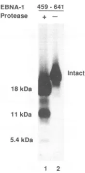
Protease resistance identifies an 18-kDa DNA-binding do-main. The generation of a protease-resistant DNA-binding
speciesindicates that theDNA-bindingfunctionof EBNA-1 lies within a physically and conformationally discrete do-main. Todetermine the size andapproximateboundariesof this domain, the protected species generated from three different EBNA-1 constructions were compared. Methio-nine-labeled EBNA-1(aa408-641), EBNA-1(aa459-641), and
EBNA-1(aa459-617) were subjected to proteinase K
diges-1 2 3 4 5 6 7 8
FIG. 2. Protease digestion generates an 18-kDa DNA-binding
domainthatmapswithin theaa459to617segmentof EBNA-1, as
determined by gel electrophoretic analysis of the protease-resistant EBNA-1 polypeptidespresentinDNA-bound complexes. [35S]me-thionine-labeled, in vitro-translated 1(aa408-641), EBNA-1(aa459-641), and EBNA-1(aa459-617)wereincubated with protein-ase K in the presence of unlabeled consensus DNA probe. The
DNA-boundcomplexeswereseparated from unbound protein and DNA by electrophoresis through a native 5% polyacrylamide gel
and isolated, and the polypeptides present were analyzed on an
SDS-15% polyacrylamide gel. Results are shown for
EBNA-1(aa408-641) minus (lane 1) and plus (lane 2) protease, EBNA-1(aa459-641) plus (lanes 3 and 6) and minus (lanes4and5)protease, andEBNA-1(aa459-617) plus (lane 7) and minus (lane 8) protease.
tion after preincubation with specific probe. The
DNA-bound species were separated from unbound protein by
using a mobility shift assay. The shifted complex was
isolatedfrom thegel, and the sizes oftheprotected
polypep-tides inthecomplexwerethendeterminedby electrophore-sis through a denaturing polyacrylamide gel (Fig. 2). The
proteinase-resistant species generated from EBNA-1(aa408-641) consisted of a dominant polypeptide of 18 kDa and a less abundant 11-kDa polypeptide (Fig. 2, lane 2). The
proteinase-resistant species generated from EBNA-1(aa459-641) (lanes 3 and 6) and EBNA-1(aa459-617) (lane 7) gave
rise to polypeptides of the same size as did the larger EBNA-1(aa408-641) form.Thus, the 18-kDa
protease-resis-tantdomain lies within the 158-aaregionboundedbyaa459 and617. The size of thisregioniscompatiblewiththe 139-aa sizeestimated foran 18-kDapolypeptide.
Binding of EBNA-1 to adjacent sites does not modify
protease resistance. Each of the three EBNA-1-binding loci in the EBV genome consists of multiple adjacent binding
sites. ori-Pcontains two subdomains: thefamilyof repeats whichincludes 20 EBNA-1-bindingsitestandemly repeated at 30-bp intervals, and the dyad symmetry locus, which consists oftwo paired EBNA-1-binding sites spaced 21 bp
apart. The physically distant Q locus also contains two
overlapping EBNA-1-binding sites with their centers 25 bp apart (2). We previously examined EBNA-1 binding in competition assays (11) and did not find any evidence for cooperative bindingwhentwoorfourconsensus30-bpsites
were present. The observation that protease resistance
changes upon DNA binding potentially provides another
waytoprobeforcooperative EBNA-1interactions. Methio-nine-labeled EBNA-1(aa408-641) was preincubated with DNA containing either monomer or dimer 30-bp binding
sites and then digested with proteinase K. In the mobility
40E4 -459641 Protease - + +
-kDa 30 -
-45R-fi4 459-617
_ +F +-_
-4
0 tIS IkDa I
11kDo
on November 9, 2019 by guest
http://jvi.asm.org/
[image:3.612.74.281.76.216.2] [image:3.612.358.516.77.240.2](A)
(A)
Protease - - 2 2 5 10 50 100 Protelnase K (ug)
Intact
* %0 dimer(2)
ISoi
dimer(1) monomer_ ^ _ ^ Digested
a a a Complexes
1 2 3 4 5 6 7
(B)
Protease
Intact -30 kDa
-21.5
18 kDa
11kDC t.
[image:4.612.332.549.80.322.2]1 2 3 4
FIG. 3. BindingofEBNA-1toadjacentconsensussites doesnot alterproteasesusceptibility.Invitro-translated, methionine-labeled EBNA-1(aa408-641)wasincubated with unlabeled DNAfragments
containing1or2consensusbindingsites. After proteasedigestion, the DNA-bound complexes were separated by electrophoresis through anative polyacrylamide gel. (A) Amonomerbinding-site
probewasusedwithincreasing amountsof protease (lanes1 to4) and in the absence ofprotease (lane 5).Adimerbinding-site probe
wasused with increasingprotease(lanes 6to9)and in theabsence of protease (lane 10). (B)Thepolypeptides present in theexcised DNA-boundcomplexeswereanalyzed bySDS-polyacrylamide gel
electrophoresis. No difference was observed between peptides
presentin themonomerand dimercomplexes. Lanes: 1,monomer
complexfrom lanes 2to4ofpanel A; 2, complexfrom the dimer probewithasingle site occupied,lanes 7to9ofpanel A; 3, complex fromthe dimerprobe with both sites occupied, lanes7to9ofpanel A; 4, mixed monomer and dimer complexes without protease digestion, lanes5 and 10 ofpanelA.
shift assay (Fig. 3A), the monomer probe gave a single shiftedcomplex (lanes1to5)and thedimerprobeproduced two complexes representing the probe DNA with one site filled (dimer 1)orwith both sitesfilled (dimer 2) (lanes 6 to
10).These shiftedcomplexeswereisolated, andthesizesof the protease-resistant polypeptides were examined on a
denaturing acrylamide gel (Fig. 3B). The protease-resistant speciesisolated afterpreincubation with themonomer
bind-ing-site probegave the expected 18-kDa andless abundant 11-kDapolypeptides (lane 1). Exactly thesamepolypeptides were released from the protease-resistant complexes that had been preincubated with the dimer binding-site probe, and thiswas true of the complexes representing one filled
site(lane 2) and both sites filled (lane 3). Therefore, at this level ofanalysis, thepresenceof protein boundto adjacent sites at30-bp intervals does notaffect the formation of the
protease-resistant domain, nordoes it alter the size of the
protected polypeptides.
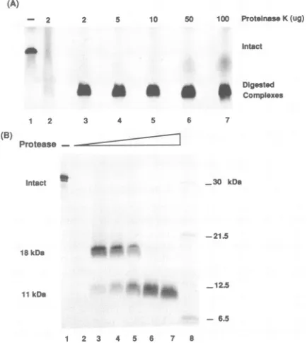
The 18-kDa domain is susceptibletospecific internal cleav-age at high levels ofprotease. In addition to the dominant
18-kDa polypeptide, analysis of the protease-resistant
com-plexrevealedalessabundant 11-kDa species. Todetermine
- 6.5
1 2 3 4 5 6 7 8
FIG. 4. The18-kDapolypeptideiscleavedtoan11-kDaproduct athighlevels of protease.(A)Electrophoretic separationon anative 5%acrylamide gelof DNA-boundcomplexesformed after incuba-tion of [35S]methionine-labeled EBNA-1(aa408-641) with an unla-beled consensus DNAprobe, followed byexposure to increasing amounts of proteinase K. Lanes: 1, EBNA-1(aa408-641) minus protease; 2, DNA probe added after protease digestion; 3 to 7, incubationwithincreasingamountsof protease.(B) SDS-polyacryl-amidegel electrophoretic analysisof thepolypeptidespresent in the DNA-boundcomplexes.Lanes 1to7 show thepolypeptidespresent inthecomplexes fromthecorrespondinglanes inpanelA.
the derivation of this smaller polypeptide, methionine-la-beled EBNA-l(aa408-641) was subjected to digestion with increasingamountsofproteinaseKrangingfrom 2 to 100,ug ofenzyme.AsshowninFig. 4A,aDNA-bindingspecieswas obtained even after digestionwith 100 ,ug of protease, and the mobilityof the bound complexremained similarto that formed afterdigestionwith 2,ugofproteinaseK. Whenthe
polypeptides present within these DNA-bound complexes
weredisplayedon adenaturingpolyacrylamide gel, a differ-ent picture emerged. Digestion with 2 ,ug of protease
pro-duced thepreviouslyobservedpatternofadominant 18-kDa
species and a less abundant 11-kDa species (Fig. 4B). However,withincreasingamountsofprotease, the relative abundance of thetwopolypeptides changed until, at100 jig
ofproteinase K, onlythe 11-kDa bandwasobserved. Thegenerationof the 11-kDapolypeptide at theexpense
of the 18-kDa polypeptide suggested a precursor-product
relationship. That the 11-kDapolypeptide doesindeed rep-resent a subfragment of the 18-kDa polypeptide was con-firmed by a comparisonof methionine-labeled tryptic pep-tides(Fig. 5).Theidenticalnatureofthemethionine-labeled tryptic peptidescombinedwith the asymmetric distribution
ofmethionine residues in EBNA-1(408-641) (Fig. 6B) indi-catedthat thelabeled 11-kDapolypeptidewasderivedfrom the carboxy terminus of the 18-kDa species. The 11-kDa
polypeptidecould begeneratedeitherbycontinued
proteo-lytic digestion sequentially removing amino acids from the amino-terminal end of the 18-kDapolypeptideorby specific
I
hi
(B)
1 2 3 4 5 6 7 8 9 10
1BkDam _
11kDa -12-5
on November 9, 2019 by guest
http://jvi.asm.org/
[image:4.612.92.273.80.318.2]18kDa
* i
I0
1 1 kDa (A)
o 61o
:j661
_m.
-eSkOD
4=
466
4 4W0 m mo no
L
P-
C---P----P-G@PGPGP-PGPus so
FIG. 5. Trypticpeptide analysis confirmsthatthe 11-kDa poly-peptide is derived from the 18-kDa species. After DNA binding, [35S]Met-labeled EBNA-1(aa408-641)wasdigested withproteinase
K, the resistant complexwasisolated, and thepolypeptidespresent
wereseparatedon adenaturing 15% polyacrylamide gel.The 18-and
11-kDa peptides were excised from the gel and incubated with
trypsin, and the tryptic digestion products were subjected to a
two-dimensionalanalysisonthin-layer cellulose plates. The identity of thetryptic digests confirms therelationship between the11-and 18-kDaspecies.
cleavage at susceptible internal sites within the 18-kDa polypeptide. Since the relative mobility of the native EBNA-1-DNA complex does not change appreciably during the conversionof the18-kDa formtothe11-kDaform, the latter
seems more likely, but these alternatives cannot be
ad-dressed by using methionine-labeled EBNA-1. The four
available methionines are located between aa 543 and 613
(Fig. 6B), andtherefore any digestion products terminating
prior to aa 543 would not be labeled and would not be
detectable.
Toobtain more information on the mechanism by which the 11-kDapolypeptidewasgenerated, EBNA-l(aa459-641) wastranslatedin vitro in thepresenceof
[35S]cysteine.
The labeled protein was subjected to the same protocol ofproteinase Kdigestion andanalysis of the
proteinase-resis-tant binding species on denaturing polyacrylamide gels. Therearefour cysteineresidues inthe 18-kDapolypeptide, andtheir distribution is similar tothatofmethionineexcept thatthemostamino-terminalcysteineislocatedataa530,as
opposed toaa542 for the equivalent methionine (Fig. 6B). Size analysis of the cysteine-labeled proteinase-resistant speciesrevealedboth the 18- and 11-kDa bandsplus a new band at 5.4 kDa (Fig. 7). The presence of the additional smallercysteine-labeled polypeptideis takentoindicatethat the11-kDa speciesisgenerated primarily byinternal prote-asecleavage ataspecific susceptible site within the 18-kDa
polypeptide.The5.4-kDafragmentwasdetectable byusing radiolabeled cysteine but not methionine. The differential
recovery of Cys-530 but not Met-542 within the 5.4-kDa
species suggested that the protease cleavage event that generatesthe 11-kDaspeciesmayoccurwithin theaa531to 541 segment ofEBNA-1 (Fig. 6B). Cleavage in this region
should generate the 11- and 5.4-kDa polypeptides with an apparent 3:1 molar ratio, since theycontain three and one
cysteine residues, respectively. However,the5.4-kDa frag-ment has a considerably lower abundance than predicted. Sincethecleavageeventwouldoccurveryclosetocysteine
(C)
INSOLUTION SOUND`TO DNA
FIG. 6. Diagrammatic representation of the location and
pro-posed structure of the protease-resistant DNA-binding domain of EBNA-1. (A) EBNA-1(aa408-641), EBNA-1(aa459-641), and EBNA-1(aa459-617) each give risetothe 18-kDa protease-resistant polypeptide, indicating that this polypeptidemapswithin theaa459 to617segmentofEBNA-1.(B)The11-kDapolypeptide is generated by furtherproteasecleavage of the18-kDa species, and analysis of methionine-labeled tryptic digestion products places the 11-kDa peptideatthecarboxy terminus of the18-kDa species. The detec-tionofa5.4-kDaprotease-resistant polypeptide with cysteine- but not methionine-labeled EBNA-1 suggests that the cleavage event generating the 11-kDa species occurs between cysteine 530 and
methionine 542 inaproline-and glycine-richsegmentof the protein.
*, methionine; 0, cysteine. (C) Model illustrating the
conforma-tional changes that may occur when EBNA-1 binds DNA. The DNA-binding/dimerization domain becomesproteaseresistant, with
acentralsegment (@) of this domain being themostsusceptibleto furthercleavageathighproteaseconcentrations.
530,it ispossiblethatsubsequentproteolytic activityresults inloss of label fromapercentageofthe5.4-kDapolypeptides
or, aswill be discussed later, thatthe 5.4-kDa fragment is
preferentiallyreleased from the DNA-bound complex. The dataobtainedon theprotease-resistant domain are summa-rized inFig.6,which also showsamodelillustratingpossible
conformational featurespredicted bytheseexperiments.
Deletions within the 18-kDa domain destroyDNA binding
and perturb EBNA-1 dimerization. The 18-kDa protease-resistant fragment is located between aa 459 and 617 of EBNA-1 and appears to represent a discrete structural domain. The DNA-binding activityof this regionand
previ-ous analyses ofbacterially expressed EBNA-1 (1) indicate that the domain must contain both DNA recognition and EBNA-1 dimerization functions. The generation of the 11-kDa super-resistant polypeptide at high levels of protease raised the possibility that these two functions might be located inphysically distinguishable subdomains. Asastep towardexaminingthisquestion,aseries of internal deletions was created across the domain in an effort to identify any nonessential sequences. The deletions encompassed be-tween 7 and 41 aa (Fig. 8). Although all of the deleted constructions yielded stable in vitro-translated products
ol u.
(B)
1e
llk1d
0
a II
A 4m
.l
on November 9, 2019 by guest
http://jvi.asm.org/
[image:5.612.322.555.81.311.2] [image:5.612.87.268.83.248.2]A EBNA-1 459- 641
Protease +
-Intact 18kDa
11kDa
5.4kDa
1 2
FIG. 7. Labeling with [35S]cysteine identifiesan additional
5.4-kDa protease-resistant polypeptide. An SDS-polyacrylamide gel electrophoretic analysis of the polypeptides present in a DNA-bound protease-resistant complex formed by using [35S]cysteine-labeled EBNA-1(aa459-641) with (lane 1) and without (lane 2) protease.
t>b qltOOA<;ff5 ,15cz1bbl 9b oe. I',,,
6i z- hn- \V shifted
0 a ~~~~~~~~~~~~~shifted
as
unshittedB
X-linking - + - + - + + kOa
69 up
dimer
(datanotshown),noneof themproved capableofbindingto DNA inamobility retardationassay(Fig. 9A).
Since both DNA recognition and EBNA-1 dimerization arerequired for DNAbinding, each of the deletions thatwe created could beintruding intoone orother of these
subdo-mains, with aresultant loss ofDNA-binding activity. DNA
recognition byEBNA-1cannotbe assessed in the absence of EBNA-1 dimerization. However, EBNA-1 dimerization oc-curs in solution in the absence ofDNA, and hence dimer-izationabilitycanbe measuredas an independentfunction.
Consequently, we performed glutaraldehyde cross-linking
experiments on the same set of EBNA-1 deletions.
Cross-linkingof theparental EBNA-1(aa408-641) polypeptidegave
a strong band at the dimer position (Fig. 9B and C). However, each of the deletions severely compromised
dimerizationability. Sinceit isunlikelythat all six deletions
impinge directly on the dimerization motif, this result sug-gests that anydisruption ofthe conformational integrity of
the18-kDa domainadversely affects the ability of the protein todimerize and tointeract with DNA.
DISCUSSION
We have used the susceptibility of EBNA-1 to protease
digestion to examine aspects of the interaction between
EBNA-1 and its DNA-binding site. Previous studies using
jEBNA1
30_-__M
C
X-llinking - + - + - +
kDa
69_ I
- - |mOnornef
%95
dimer
30
40_
|monomerFIG. 9. Deletions within the 18-kDa protease-resistant domain abolish DNAbindingandperturbdimerization.(A) Mobility retar-dation assayusing a32P-labeled DNA fragment containing a con-sensus binding site. The parental EBNA-1(aa408-641) bound the probe, but none of the deletion mutants retained DNA-binding activity. (BandC) Glutaraldehyde cross-linkingof
[35S]methionine-labeled, deletedpolypeptides. Abandat the dimerpositionin the SDS-polyacrylamide gel is readily detectable with the parental EBNA-1(aa408-641). However, only marginal dimerization is ob-served with the deletedpolypeptides.
A4g0 im
-______________________ :OM
[image:6.612.135.223.78.257.2]Am
FIG. 8. Diagrammatic representation of internal deletions
cre-ated within the 18-kDadomain. The locations of predicted alpha-helical regions (10)areindicated.
deleted amino- and carboxy-terminal variants of EBNA-1 had demonstrated that wild-type levels of DNA binding requiredanEBNA-1 domain locatedbetweenaa459 and 608
(1). EBNA-1 binds DNA as a dimer, and the dimerization motif also resides within thissegment(1). Limitedprotease
digestion of EBNA-1(aa408-641) in solution generated a
gb oil
as as "I
on November 9, 2019 by guest
http://jvi.asm.org/
[image:6.612.343.532.79.542.2] [image:6.612.61.300.609.696.2]smallerpolypeptide that bound DNA in a mobility retarda-tion assay, indicating that the DNA-binding domain of EBNA-1 has a degree of inherent resistance to protease. EBNA-1 exists in solution almost exclusively in the dimer form (1, 6), and the partial protease resistance may be created by dimerization. The leucine zipper dimerization motifcommon intranscriptional activator proteins forms a helical coiled coil (16, 20, 23). Exposure of the leucine zipper protein C/EBP to trypsin in solution generates a protease-resistant fragment that encompasses the leucine zipper dimerization domain (30). However, the basic DNA recog-nition motif of free C/EBP is not protected, whereas the EBNA-1 recognition motif apparently is protected since DNAbinding can still be demonstrated with the protease-treated EBNA-1.
When EBNA-1 is bound to DNA prior to exposure to protease, the protease resistance of the DNA-binding do-main is increased at least 100-fold. This indicates a change in the EBNA-1 protein conformation on binding. The transcrip-tional activators Jun/Fos and GCN4 show an increase in alpha helicity on DNA binding (21, 34), and this conforma-tional change may be necessary for stable protein-DNA contacts. The change in conformation of TFIID on binding has also been suggested to be necessary for stable binding (17). It is possible that conformational changes also unmask
protein domains and in this way contribute to biological
function. The change in conformation undergone by the yeasttranscriptional activator PTRF on binding to a respon-sive site is believed to unmask an activator domain on that protein (33). In the case of EBNA-1 binding, induced
changes could result in the exposure of a transcriptional
activation domain or in unmasking sites for interactions between EBNA-1 and cellular replication proteins. The
exact role played by EBNA-1 in episomal EBV DNA replication remains unclear. A working hypothesis is that EBNA-1 induces or stabilizes an accessible DNA structure
attheoriginand may thenprovideascaffolding for assembly of the active DNA replication initiation complex. If the EBNA-1 domains involved in the initial steps of complex
assemblywere available onlyon DNA-bound EBNA-1 and
not on free protein, this would provide a mechanism for
minimizing nonproductive protein interactions. An alterna-tive induced protein-protein interaction could involve EBNA-1toEBNA-1 contacts. The existence ofhigher-order
EBNA-1DNA-boundcomplexes isapossibility (18). Earlier studies had determined that wild-type levels of DNAbindingrequiredanEBNA-1domainencompassingaa
459to608. Weintroduced asetof six internal deletions into this region and found that each of the deletions abolished DNA binding. Dimerization ability was also severely im-paired by each of the deletions, and the inability to form a
stable dimer is likely to have contributed to the observed loss of DNA binding. The dimerization and DNA-binding
motifs of EBNA-1 have yet tobeidentified experimentally.
Inoue et al. (10) noted a significant amino acid sequence
homologybetween thebasic-helix-loop-helix
(HLH)
domainof
proteins
suchasc-Myc,MyoD, andE12/E47
and EBNA-1 intheregion ofaa467through 583. However,in the caseof EBNA-1, the basic-helix 1 and helix 2 domains(aa467to488 and 571 to583)areseparated byanextended loopof 83 aa,comparedwithonly10to 12aa inMyoD. Thisarrangement has some similarities to the basic-helix-span-helix
(HSH)
DNA recognition/dimerization motif of the transcriptional
activatorAP-2, inwhichtwohelixmotifsareseparated bya
span of82 aa(35). Inthe caseofAP-2, all deletions across
theHSHmotif, including onein the spanregion, abolished
DNA binding and dimerization (35). This observation is reminiscent of our results with the deleted EBNA-1
poly-peptides. Only two of our six deletions, A480-521 and
A556-576,wouldimpinge on the critical helix domains (Fig. 8) but all deletions affected both DNA binding and dimeriza-tion.
Athigh levels ofprotease, the 18-kDaprotease-resistant polypeptide was clipped into an 11-kDa and a 5.4-kDa species. On the basis of the differential labeling of the 5.4-kDa fragment with cysteine and methionine, the cleav-ageatthejunction with the 11-kDa fragment was mapped to aregion between aa 531 and 541 that is rich in proline and glycine residues (Fig. 6B). A 5.4-kDa fragment would con-tainapproximately 42 aa, which coincides with the distance betweenthe cleavage in theproline-glycine region (aa 531 to 541) and the 3' end of the predicted helix 1 (aa 493). One interpretation of the protease data is that at high levels of protease, asecond cleavage occurs near the junction of helix 1 with the intervening span DNA and that the 5.4-kDa fragment represents cleaved-out span sequences. Such a model would predict a loss of the 5.4-kDa fragment from the DNA-bound complex, and this could explain the relatively lowmolarityobserved experimentally for this fragment. The protease experiments also raise an interesting point regard-ing the physical interaction between the DNA and EBNA-1. Both helix 1 and helix 2 of an HLH or HSH protein are required for dimerization and DNA binding. However, the data suggest that once binding has occurred, helix 2 and perhaps helix 1 can maintainadegree of association with the DNA evenwhen theprimary covalent linkage between them has been lost.
Itis also interesting to note that protease sensitivity may incertain circumstances have biological relevance. In phage lambda, the lambda repressor is synthesized from the
lyso-genic phage DNA, and its function is to stimulate its own synthesis while repressing transcription of the lytic phage genes. Lambda repressor binds specifically, with high affin-ityas adimerto17-bpoperatorsites. Inactivation of lambda repressor occursthrough cleavage of the repressor polypep-tidein a reaction that is partly autocatalytic and is facilitated by RecA protein. This cleavage event destabilizes the re-pressor dimer, which reduces operator binding and conse-quently allows initiation of thelyticphagegrowth cycle(28).
Theproteaseclipping of bound EBNA-1 may simply reflect the exposure ofa relatively unstructured segment within a
more densely packed alpha-helical region of the protein.
However, the lambda repressor model has intriguing impli-cations for the switch in EBVfrom latentori-P-basedDNA
replicationtoori-Lyt replication.
On the basis of the existence of a protease-resistant
domain encompassing the DNA-binding and dimerization functions and the drasticeffectsofinternaldeletions within thisregion,weconclude that the EBNA-1 DNArecognition
and dimerization motifs are intimately linked and form a
highly structured domainthat has features in commonwith both the HLH and HSHmotifs. On the otherhand,itshould benoted that
differences
alsoexist.Forexample,
the patternofmethylationprotectionof bases in theEBNA-1consensus
binding site differs significantly from that exhibited
by
thecharacterized MyoD HLH
proteins.
ACKNOWLEDGMENTS
We thank Wade Gibson for advice and help with the tryptic
digests, Mabel Chiu fortechnical assistance, and Sarah Heaggans
and PamelaWrightforpreparationof the manuscript.
on November 9, 2019 by guest
http://jvi.asm.org/
This work was funded by Public Health Service grants RO1-CA42243 (S.D.H.), RO1-CA22130 (G.S.H.), and K11AI00648 (R.F.A.). W.A.S. was supported by a Pakistan Ministryof Science andTechnology scholarship.
REFERENCES
1. Ambinder, R. F., M. Mullen, Y. N. Chang, G. S. Hayward, and S.D.Hayward. 1991. Functional domains of Epstein-Barr virus nuclear antigen EBNA-1. J. Virol. 65:1466-1478.
2. Ambinder, R. F., W. A. Shah, D. R. Rawlins, G. S. Hayward, and S. D. Hayward. 1990. Definition of the sequence require-ments for binding of the EBNA-1 protein to its palindromic target sites inEpstein-Barrvirus DNA. J. Virol. 64:2369-2379. 3. Baer, R., A. T. Bankier, M. D. Biggin, P. L. Deininger, P. J. Farrell,T.J.Gibson, G. Hatfull, G. S. Hudson, S. C. Satchwell, C. Sequin, P. S. Tuffhell, and B. G. Barrell. 1984. Organization of the B95-8Epstein-Barrvirus genome. Nature (London) 310:207-211. 4. Chang, Y.-N., D. L.-Y. Dong, G. S. Hayward, and S. D. Hayward. 1990.The EBV Zta transactivator: a member of the bZip familywith unique DNA binding specificity and a dimer-ization domain that lacks the characteristic heptad leucine zippermotif. J. Virol. 64:3358-3369.
5. Dasmahapatra, B., E. J. Rozhon, and J. Schwartz. 1987. pBD7, a novel cell free expression vector with efficient translation initiationsignal. Nucleic Acids Res. 15:3933.
6. Frappier, L., and M. O'Donnell. 1991. Overproduction, purifi-cation, and characterization of EBNA-1, the origin binding protein of EBV. J. Biol. Chem. 266:7819-7826.
7. Gahn,T.A., and C. L. Schildkraut. 1989. The Epstein-Barr virus origin ofplasmid replication, ori-P, contains both the initiation andtermination sites of DNA replication. Cell 58:527-535. 8. Gibson,W. D., A. I. Marcy, J. C. Comolli, and J. Lee. 1990.
Identification of precursor tocytomegalovirus capsid assembly protein and evidence that processing results in loss of its carboxy-terminal end. J. Virol. 64:1241-1249.
9. Hager, D. A., and R. R. Burgess. 1980. Elution of proteins from SDS-PAGE, removal of SDS and renaturation of enzymatic activity:results with 8 subunit of E. coli RNA polymerase, DNA topoisomeraseandotherenzymes.Anal. Biochem. 109:76-86. 10. Inoue, N., S. Harada, T. Honma, T.Kitamura, and K. Yanagi.
1991. The domain of Epstein-Barr virus nuclear antigen 1 essential for binding to oriP region has a sequence fitted for the hypothetical basic-helix-loop structure. Virology 182:84-93. 11. Jones, C. H., S. D. Hayward, and D. R. Rawlins. 1989.
Interac-tionoflymphocyte-derived Epstein-Barr virus nuclear antigen EBNA-1with its DNA binding sites. J. Virol. 63:101-110. 12. Kieff, E., and D. Liebowitz. 1990. Epstein-Barr virus and its
replication, p. 1889-1920. In B. N. Fields, D. M. Knipe, et al. (ed.), Virology, 2nded. Raven Press, New York.
13. Kim, J., C. Zwieb, C. Wu, and S. Adhya. 1989. Bending of DNA by gene regulatory proteins: construction and use of a DNA bendingvector. Gene 85:15-23.
14. Kimball, A. S., G. Milman, and T. D. Tullius. 1989. High resolution footprints of the DNA-binding domain of Epstein-Barrvirus nuclearantigen-1. Mol. Cell. Biol. 9:2738-2742. 15. Laemmli, U. K. 1970. Cleavage of structural proteins during the
assembly of the head of bacteriophage T4. Nature (London) 227:680-685.
16. Landschulz,W. H., P. F.Johnson, and S. L. McKnight. 1988. Theleucine zipper: a hypothetical structure common to a new class of DNAbinding proteins. Science 240:1759-1764. 17. Lieberman, P. M., M. C.Schmidt, C. C. Kao, and A. J. Berk.
1991. Twodistinct domains in the yeast transcription factorIID andevidence for aTATAbox-induced conformational change. Mol. Cell. Biol. 11:63-74.
18. Milman, G., and E. S.Hwang. 1987.Epstein-Barr virus nuclear antigen forms a complex that binds with high concentration dependencetoasingle DNA-binding site. J. Virol. 61:465-471. 19. Milman, G., A. L.Scott, M.-S. Cho, S. C.Hartman, D. K. Ades, G. S.Hayward,P.G. Ki, J. T.August, and S. D.Hayward. 1985. Carboxy-terminal domain of the Epstein-Barr virus nuclear antigen is highly immunogenic in man. Proc. Natl. Acad. Sci.
USA 82:6300-6304.
20. O'Shea, E. K., R. Rutkowski, and P.S. Kim. 1989. Evidence that the leucinezipper is a coiled coil. Science 243:538-542. 21. Patel, L., C. Abate, and T. Curran. 1991. Altered protein
conformation on DNAbinding by Fos and Jun. Nature (Lon-don) 347:572-575.
22. Polvino-Bodnar, M., J. Kiso, and P. A. Schaffer. 1988. Muta-tional analysis ofEpstein-Barrvirus nuclear antigen1 (EBNA-1).NucleicAcids Res. 16:3415-3435.
23. Rasmussen, R., D. Benvegnu, E. K. O'Shea, P.S. Kim, and T. Alber.1991. X-rayscatteringindicates that theleucinezippers is acoiled:coil. Proc. Natl. Acad. Sci. USA88:561-564. 24. Rawlins, D. R., G. Milman,S. D. Hayward, andG. S. Hayward.
1985. Sequence specific DNA-binding of EBNA-1 to clustered sites in the plasmid maintenance region. Cell 42:859-868. 25. Reisman, D., and B. Sugden. 1986. Transactivation of an
Ep-stein-Barr viral transcriptional enhancer by the Epstein-Barr viral nuclear antigen 1.Mol. Cell. Biol.6:3838-3846.
26. Reisman, D., J. Yates, and B. Sugden. 1985. Aputative originof replication of plasmids derived from Epstein-Barr virus is composed of two cis-acting components. Mol. Cell. Biol. 5:1822-1832.
27. Sample, J., L. Brooks, C. Sample, L. Young, M. Rowe, C. Gregory, A. Rickinson, and E. Kieff. 1991. Restricted EBV protein expression in Burkitt lymphoma is due to a different Epstein-Barr nuclear antigen 1 transcriptional initiation site. Proc. Natl. Acad. Sci. USA 88:6343-6347.
28. Sauer, R. T., R. Jordan, and C. 0. Pabo. 1990.ARepressor: a model system for understanding protein-DNA interactions and protein stability. Adv. Protein Chem. 40:1-61.
29. Schaefer, B. C., M. Woisetschlaeger, J. L. Strominger, and S. H. Speck. 1991.Exclusive expression of EBV nuclear antigen 1in Burkitt lymphoma arises from a third promoter, distinct from the promoters used in latently infected lymphocytes. Proc.NatI. Acad. Sci. USA88:6550-6554.
30. Shuman, J. D., C. R. Vinson, and S. L. McKnight. 1990. Evidence of changes in protease sensitivity and subunit ex-change rate on DNA binding by C/EBP. Science 249:771-774. 31. Smith, P. R., and B. E. Griffin. 1992. Transcription of the
Epstein-Barr virus gene EBNA-1 from different promoters in nasopharyngeal carcinoma and B-lymphoblastoid cells. J. Virol. 66:706-714.
32. Sugden, B., and N.Warmn. 1989. A promoter of Epstein-Barr virus that can function during latent infection can be transacti-vated by EBNA-1, a viral protein required for viral DNA replication during latent infection. J. Virol. 63:2644-2649. 33. Tan, S., and T. J. Richmond. 1990.
DNA-binding-conforma-tional change of the yeast transcripDNA-binding-conforma-tional activator PRTF. Cell 62:367-377.
34. Weiss, M. A., T. Ellenberger, C. R. Wobbe, J. P. Lee, S. C. Harrison, and K. Struhl. 1990. Folding transition in the DNA binding domain of GCN4 on specific binding to DNA. Nature (London) 247:575-578.
35. Williams, T., and R. Tjian. 1991. Characterization of a dimer-ization motif in AP-2 and its function in heterologous DNA binding proteins. Science 251:1067-1071.
36. Wysokenski, D. A., and J. L. Yates. 1989. Multiple EBNA-1 binding sites are required to form an EBNA-1-dependent en-hancer and to activate a minimal replicative origin within oriP of Epstein-Barr virus. J. Virol. 63:2657-2666.
37. Yates, J., N. Warren, D. Reisman, and B. Sugden. 1984. A cis acting element from the Epstein-Barr viral genome that permits stable replication of recombinant plasmids in latently infected cells. Proc. Natl. Acad. Sci. USA81:3806-3810.
38. Yates, J. L., and S. M. Camiolo. 1988. Dissection of DNA replication and enhancer activation functions of Epstein-Barr virus nuclear antigen 1. Cancer Cells 6:197-205.
39. Yates, J. L., and N. Guan. 1991. Epstein-Barr virus-derived plasmids replicate only once per cell cycle and are not amplified after entry into cells. J. Virol. 65:483-488.
40. Yates, J. L., N. Warren, and B. Sugden. 1985. Stable replication of plasmids derived from Epstein-Barr virus in a variety of mammalian cells. Nature (London) 313:812-815.

![FIG. 5.wereoftrypsin,peptideK,two-dimensional18-kDa11-kDa[35S]Met-labeled the Tryptic peptide analysis confirms that the 11-kDa poly- is derived from the 18-kDa species](https://thumb-us.123doks.com/thumbv2/123dok_us/1307701.84025/5.612.87.268.83.248/wereoftrypsin-peptidek-dimensional-tryptic-peptide-analysis-confirms-derived.webp)