City, University of London Institutional Repository
Citation
:
Yang, G., Zhuang, X., Khan, H., Haldar, S., Nyktari, E., Ye, X., Slabaugh, G. G.,
Wong, T., Mohiaddin, R., Keegan, J. and Firman, D. (2017). Segmenting Atrial Fibrosis from
late Gadolinium-Enhanced Cardiac MRI by Deep-Learned Features with Stacked Sparse
Auto-Encoders. Paper presented at the Medical Image Understanding and Analysis (MIUA)
2017, 11 Jul 2017, Edinburgh, UK.
This is the accepted version of the paper.
This version of the publication may differ from the final published
version.
Permanent repository link:
http://openaccess.city.ac.uk/id/eprint/17828/
Link to published version
:
Copyright and reuse:
City Research Online aims to make research
outputs of City, University of London available to a wider audience.
Copyright and Moral Rights remain with the author(s) and/or copyright
holders. URLs from City Research Online may be freely distributed and
linked to.
City Research Online:
http://openaccess.city.ac.uk/
publications@city.ac.uk
adfa, p. 1, 2011.
© Springer-Verlag Berlin Heidelberg 2011
Segmenting Atrial Fibrosis from Late
Gadolinium-Enhanced Cardiac MRI by Deep-Learned Features with
Stacked Sparse Auto-Encoders
Guang Yang *1,2, Xiahai Zhuang *3, Habib Khan 1,2, Shouvik Haldar 1, Eva Nyktari 1, Xujiong Ye 4, Greg Slabaugh 5, Tom Wong 1,
Raad Mohiaddin 1,2, Jennifer Keegan 1,2, David Firmin 1,2
1
Cardiovascular Biomedical Research Unit, Royal Brompton Hospital, SW3 6NP, London, UK
2
National Heart and Lung Institute, Imperial College London, SW7 2AZ, London, UK
3
School of Data Science, Fudan University, Shanghai, 201203, China
4
School of Computer Science, University of Lincoln, LN6 7TS, Lincoln, UK
5
Department of Computer Science, City University London, EC1V 0HB, London, UK
* Corresponding authors: GY—g.yang@imperial.ac.uk and XZ— zxh@fudan.edu.cn
Abstract. The late gadolinium-enhanced (LGE) MRI technique is a well-validated method for fibrosis detection in the myocardium. With this technique, the altered wash-in and wash-out contrast agent kinetics in fibrotic and healthy myocardium results in scar tissue being seen with high or enhanced signal rela-tive to normal tissue which is ‘nulled’. Recently, great progress on LGE MRI has resulted in improved visualization of fibrosis in the left atrium (LA). This provides valuable information for treatment planning, image-based procedure guidance and clinical management in patients with atrial fibrillation (AF). Nev-ertheless, precise and objective atrial fibrosis segmentation (AFS) is required for accurate assessment of AF patients using LGE MRI. This is a very challeng-ing task, not only because of the limited quality and resolution of the LGE MRI images acquired in AF but also due to the thinner wall and unpredictable mor-phology of the LA. Accurate and reliable segmentation of the anatomical struc-ture of the LA myocardium is a prerequisite for accurate AFS. Most current studies rely on manual segmentation of the anatomical structures, which is very labor-intensive and subject to inter- and intra-observer variability. The subse-quent AFS is normally based on unsupervised learning methods, e.g., using thresholding, histogram analysis, clustering and graph-cut based approaches, which have variable accuracy. In this study, we present a fully-automated multi-atlas propagation based whole heart segmentation method to derive the anatom-ical structure of the LA myocardium and pulmonary veins. This is followed by a supervised deep learning method for AFS. Twenty clinical LGE MRI scans from longstanding persistent AF patients were entered into this study retrospec-tively. We have demonstrated that our fully automatic method can achieve ac-curate and reliable AFS compared to manual delineated ground truth.
1
Introduction
pop-ulation [1]. AF can cause substantial morbidity and mortality; for example, it is asso-ciated with a five-fold incidence of stroke, three-fold risk of congestive heart failure and doubles the possibility of dementia that has a major worldwide public health im-pact [2]. Currently, the pathophysiology of AF is not fully understood; however, pre-vious studies on both animal and human experimental models have shown that multi-ple disease pathways, e.g., structural, contractile, or electrical alterations, can promote abnormal electrical impulse formation and propagation [3]. Fibrosis in the left atrium (LA) is the hallmark of atrial structural remodeling, and is one of the major risk fac-tors for AF progression [4, 5]. Moreover, studies have shown that ectopic beats from the pulmonary veins (PVs) can frequently trigger the AF [6]. Based on these findings, minimally invasive radio-frequency catheter ablation (CA) using the pulmonary vein antrum isolation method has been developed as a front-line therapy for symptomatic AF patients refractory to drug treatment [7], but suffers a >30% recurrence rate [8].
In order to understand AF and facilitate better management and prognosis, tech-niques have been developed to evaluate the LA wall composition and assess the cir-cumferential PVs scarring that results from CA. Currently, the electro-anatomical mapping (EAM) system is used as a clinical reference standard technique for the as-sessment of extent and distribution of native atrial fibrosis and post-ablation scar. This is normally performed during an electrophysiological procedure and has the major drawback of invasiveness and suboptimal accuracy, which has been reported as being up to 10 mm in the localization of atrial fibrosis [9, 10]. Moreover, there are the po-tentially hazardous effects of ionizing radiation for the patients using EAM.
In contrast, the late gadolinium-enhanced (LGE) MRI, which is noninvasive and without ionizing radiation, allows the detection and quantification of native fibrosis and post-ablation scars by highlighting the slow washout kinetics of the gadolinium in these tissues [11–15]. Firstly, the extent and distribution of native fibrosis identified in the pre-ablation baseline LGE MRI scan has emerged as the strongest independent predictor of AF recurrence after the first ablation [13]. Secondly, LGE MRI can be used as a powerful tool to detect ablation-induced fibrosis formed by radiofrequency energy delivered in the atrial myocardium [11]. This has a potential role in recogniz-ing ablation line gaps, which are the main reason of ablation failure [11, 16]. In addi-tion, LGE MRI can be also useful to guide the ablation procedure [11, 17].
Essentially, there are two main steps required to analyze fibrotic tissues from LGE MRI images: (1) segmentation of the anatomical structure of the LA and PVs and (2) the atrial fibrosis segmentation (AFS).
For the segmentation of the anatomical structure of the LA and PVs, most previous studies have relied on manual delineation [7, 13, 18, 19], which potentially suffers from large inter- and intra-observer variability and is also very time-consuming. Semi-automatic and automatic methods have been proposed to solve this task, e.g., using thresholding with region growing [20], statistical shape model [21] and atlas propagation [22] based approaches. However, these methods required further opera-tor’s manual intervention [20, 21] or used un-gated first-pass MR angiography (MRA) data [22], which may cause difficulties in co-registration with the respiratory and cardiac gated LGE MRI data.
For the AFS, to the best of our knowledge, most studies applied unsupervised learning based methods, e.g., using histogram analysis [13], k-means clustering [18] and graph-cut [21] based approaches. In addition, maximum intensity projection (MIP) can provide intuitive visualization of the atrial fibrosis [11, 12, 20, 22]; howev-er, this is only a visualization technique for hyper-enhancement regions, rather than a segmentation method that can result in volumetric quantification [21]. Recently, a grand challenge was carried out to benchmark different algorithms for solving AFS [19] including 8 submissions for the competition. These benchmarked algorithms were all unsupervised learning based methods [19]. The challenge included data ac-quired from multiple institutions, and the LA endocardium and cavity for each scan were provided to all the participants beforehand. Promising results were achieved for the best performing algorithms. However, there were large variances in the perfor-mances especially for the pre-ablation cases, which may be attributed to the fact that the image quality was generally worse and the native fibrosis is more diffuse. There-fore, the challenge of atrial fibrosis segmentation and assessment remains open. Moreover, the inaccurate AFS could be one of the major reasons for poor reproduci-bility of the correlation between atrial fibrosis identified by LGE MRI (enhanced regions) and EAM (low voltage regions) [23], [24].
In this study, we present a fully automatic framework for an efficient and objective atrial fibrosis assessment using: (1) a fully-automated multi-atlas based whole heart segmentation (MA-WHS) method to solve the LA and PVs anatomy and (2) a fully automatic supervised deep learning method for the AFS. Compared with the ground truth formed by manual delineation, our fully automatic framework obtains promising segmentation results, which are comparable to other state-of-the-art methods.
2
Method
2.1 Cardiac MRI Data Acquisition
minutes after gadolinium (Gd) administration when a transient steady-state of Gd wash-in and wash-out of normal myocardium had been reached [26]. LGE MRI im-ages were scanned with a field-of-view 380×380mm2 and reconstructed to 60–68 slices at 0.75×0.75×2mm3.
In the LGE MRI images, healthy myocardium is ‘nulled’ and only fibrotic tissues are seen with high signal; therefore, it is hard to extract the anatomical structure of the LA and PVs directly from LGE MRI images. Instead of using un-gated MRA as pre-vious studies described, in our study, a respiratory and cardiac gated 3D Roadmap image, which is acquired using a balanced steady state free precession sequence (TE/TR 1ms/2.3ms), has been scanned for each patient to derive the anatomical struc-ture of the LA and PVs. Our roadmap data were acquired with a field-of-view 380×380mm2 and reconstructed to 160 slices at 0.8×0.8×1.6mm3.
Both 3D LGE MRI and Roadmap data were acquired during free-breathing using a crossed-pairs navigator positioned over the dome of the right hemi-diaphragm with a navigator acceptance window size of 5mm and continuously adaptive windowing strategy based respiratory motion control [27].
2.2 Patients
Cardiac MRI was performed in longstanding persistent AF patients between 2011– 2013 in agreement with the local regional ethics committee. A Likert-type scale was applied to score the image quality of each LGE MRI scan, e.g., 0 (non-diagnostic), 1 (poor), 2 (fair), 3 (good) and 4 (very good) depending on the level of SNR, appropri-ate inversion time, and the existence of navigator beam and ghost artifacts.
Ten pre-ablation scans with image quality ≥2 have been retrospectively entered into this study (~60% of all the scanned pre-ablation cases). To make a balanced dataset, we randomly selected 10 post-ablation cases from all the 26 post-ablation scans with image quality ≥2 (~92% of all the scanned post-ablation cases).
2.3 Anatomical Structure Delineation for the LA and PVs
In this study, we applied a MA-WHS method to segment the anatomical structure of the LA and PVs [28]. This has been done on the Roadmap images and then mapped to LGE MRI. Our segmentation consists of two major steps: (1) atlas propagation based on image registration algorithms and (2) label fusion from multi-atlas propagat-ed segmentation results as describpropagat-ed in [28].
fusion (MSP-LF),
∑
where and are the labels of the background and LA and PVs, respectively, and
the local weight is determined by the local similarity between the target image and the atlas. is the Kronecker delta function which returns 1 when and returns 0 otherwise.
In MSP-LF, the local similarity of patches using a multi-scale strategy is computed as follows,
∑ ( )
in which is the target image from scale-space that is computed from the convolution of the target image with Gaussian kernel function with scale . Here, we computed the local similarity in multi-scale images using the conditional probability of the images, that is
( )
where and and the conditional image probability is obtained from the joint and marginal image probability which can be calculated using the Par-zen window estimation [30].
For each patient case, the Roadmap dataset was then registered to the LGE MRI da-taset using the DICOM header data, and then refined by affine and nonrigid registra-tion steps. The resulting transformaregistra-tion was applied to the MA-WHS derived cardiac anatomy to segment the anatomical structure of the LA and PVs on the LGE MRI dataset.
In this study, we validated the anatomical structure segmentation against estab-lished ground truth from manual segmentations by experienced expert cardiologists specialized in cardiac MRI. We evaluated the segmentation accuracy using Dice score [31], Hausdorff distance (HD) [32] and Average Surface Distance (ASD) [31].
2.4 Training Datasets and Ground Truth Construction for AFS
The fixed-size window or regular pixel-grid is a traditional way to select patches from larger images prior to feature extraction [33], and has been widely used in deep convolutional neural networks based classification. In this study, we used a Simple Linear Iterative Clustering (SLIC) [34] based method to partition pixels into meaning-ful ‘atomic’ regions, i.e., super-pixels (SPs), based on certain similarity metrics (Fig. 1 (c)). In addition, SLIC based over-segmentation can be more consistent with the object boundaries in the image and is able to avoid producing outputs with zigzag boundaries that can be normally observed using relatively large sliding windows [33].
without the SPs grid overlaid, as this would reduce the visibility of the enhancement on LGE MRI images. The coordinates of the mouse clicks were used to select the enhanced SPs. Only one mouse click is taken into account if multiple clicks dwell in the same super-pixel.
As aforementioned, the anatomical structure of the LA and PVs was segmented us-ing a MA-WHS method. Then a morphological dilation was applied (assumus-ing the LA wall thickness is 3mm) to extract the LA wall and PVs. The blood pool regions were derived by another morphological erosion (5mm) from the endocardial LA boundary and the pixel intensities throughout the image were normalized according to the blood pool intensities [19]. We masked the selected enhanced SPs using the LA wall and PVs segmentation. Only the SPs having a defined overlap (≥20%) with the LA wall and PVs segmentation were selected as enhancement (these enhanced SPs were then labeled as 1). Other enhanced SPs were discarded as they were considered to be enhancement from other confounded tissues. As seen in (Fig. 1 (e)), the other SPs overlapped with the LA wall and PVs but not selected as enhancement were con-sidered as unenhanced (these unenhanced SPs were then labeled as 0).
[image:7.595.131.461.393.506.2]Once we extracted the enhanced SPs, they were combined to create a binary image, i.e., 1 for enhanced SPs and 0 for unenhanced. The binary image was overlaid on the original LGE MRI images and our cardiologists performed manual corrections to create the final boundaries (ground truths) for the enhanced atrial fibrosis.
Fig. 1. (a) Original LGE MRI image of a post-ablation example case; (b) Manual mouse clicks performed by cardiologists to identify enhanced fibrotic tissues; (c) SLIC over-segmentation of the original LGE MRI image (only a ROI of the LA region shown); (d) 3D rendering of the MA-WHS results with 3D MIP based visualization of the enhanced fibrotic tissues; (e) Labeled
enhanced (yellow) and unenhanced (blue) SPs that will be used to train our classification mod-el; (f) Zero-padded SP patches (yellow box: enhanced SPs and blue box: unenhanced SPs); (g) SSAE based classification; (h) Visualization of the weights derived from the first auto-encoder.
2.5 Deep Learning via Stacked Sparse Auto-Encoders for AFS
storing all the input training vectors . Each input vector was formed by: (1) zero-padding all the SPs into a matrix, which is the smallest bounding box for the largest super-pixel dimensions (Fig. 1 (f)), and (2) vectorizing the matrix into a vector. The cost function of this pre-training can be written as
∑‖ ̂ ‖
‖ ‖ ∑ ̂
where is the number of input training vectors, is the number of hidden nodes, is the coefficient for the L2 regularization term, is the weight of sparsity penalty, is
the Kullback-Leibler divergence function | ̂)
̂ ̂ ,
is sparsity parameter that specifies the desired level of sparsity, ̂ is probability of
firing activity that is ̂ ∑ . The unsupervised pre-training is performed one layer at a time by minimizing the error in reconstructing its input and learning an encoder and a decoder, which yields an optimal set of weights and biases stored in . If the number of hidden nodes is less than the number of visible input nodes , then the network is forced to learn a compressed and sparse representation of the input [35] (Fig. 1 (g) and (h)).
Second, a Softmax layer was added as the activity classification model to accomplish the SPs classification task [35]. In addition, it can be jointly trained with the SSAE during fine-tuning of the parameters with labeled instances in a supervised
fashion. The weight matrix is obtained by solving the convex optimization problem
as following.
∑ ∑
‖ ‖
where is the class label, ̃ repre-sents a set of labeled training instances, and the last term for the L2 regularization.
Finally, fine-tuning was applied to boost the classification performance, and it treats all layers of the SSAE and the Softmax layer as a single model and improves all the weights of all layers in the network by using the backpropagation technique [35].
Hyper-parameters were not optimized explicitly but were determined via trial and error. Here are the final defined hyper-parameters (values) used in this study: maxi-mum epochs of the SSAE (200), maximaxi-mum epochs of the Softmax and fine-tuning (500), hidden layers size of the SSAE (100 and 50), sparsity parameter (0.1), sparsi-ty penalsparsi-ty (5), L2 regularization term for the SSAE and the Softmax (0.0001).
For evaluation of the AFS, we used leave-one-patient-out cross-validation (LOO CV) and reported the cross-validated accuracy, sensitivity, specificity, average area under the receiver operating characteristic (ROC) curve (AUC), and the Dice score.
3
Results and Discussion
Fig. 1 (d) and (e) show the 3D MIP results overlaid on the segmented LA and PVs and LA wall and PVs boundaries overlaid on a LGE MRI slice respectively. The quantitative evaluations show that the MA-WHS based method achieved 0.90±0.12 Dice score, 9.53±6.01mm HD and 1.47±0.89mm ASD.
[image:9.595.132.470.231.398.2]For the SSAE based SPs classification, we obtained LOO CV accuracy of 0.91, sensitivity of 0.95, specificity of 0.75, AUC of 0.95, and the Dice score for the final AFS was found to be 0.82±0.05.
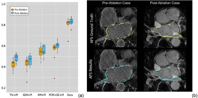
Fig. 2. (a) Boxplot for the comparison results of the Dice scores obtained by our fully automat-ic framework and other four methods (Thr, SD4, KM and FCM+GC) with manual delineated LA wall and PVs (+M); (b) Final AFS results (cyan regions) for an example pre-ablation (left)
and an example post-ablation (right) case compared to the ground truth (yellow regions).
benchmarking work and this may have resulted in a large variance in the images which could affect the final AFS segmentation. One of our future studies will be ap-plying our fully automatic framework on multi-scanners LGE datasets to validate its robustness.
Fig. 2 (b) demonstrates that qualitatively our fully automatic AFS is in accordance with the manual segmented atrial fibrosis. However, if there are enhancements from the nearby mitral valve or blood pool regions, our method may mis-classify them as enhanced atrial fibrosis that is the major contribution for the false positives.
Another possible limitation of our study is that the SSAE based classifier has many hyper-parameters, which need to be carefully tuned, e.g., maximum epochs of the SSAE, maximum epochs of the Softmax and fine-tuning, hidden layers size of the SSAE, sparsity parameter , sparsity penalty , L2 regularization term for the SSAE
and the Softmax. Currently these hyper-parameters were tuned via trial and error, which may limit the final classification accuracy.
4
Conclusion
In conclusion, we have developed and validated a fully automatic framework to segment atrial fibrosis from LGE MRI images that is based on accurate anatomical structure delineation via a MA-WHS algorithm. The evaluation has been done on 20 LGE MRI scans for longstanding persistent AF patients that contain both pre-ablation and post-ablation cases. Based on the results, we can envisage a straightforward de-ployment of our framework for clinical usage. As a future direction, we will develop a more robust parameter tuning method for the applications on multi-scanners datasets.
References
1. Feinberg, W.M., Blackshear, J.L., Laupacis, A., Kronmal, R., Hart, R.G.: Prevalence, age distribution, and gender of patients with atrial fibrillation. Analysis and implications. Arch. Intern. Med. 155, 469–73 (1995).
2. January, C.T., Wann, L.S., Alpert, J.S., Calkins, H., Cigarroa, J.E., Cleveland, J.C., Conti, J.B., Ellinor, P.T., Ezekowitz, M.D., Field, M.E., Murray, K.T., Sacco, R.L., Stevenson, W.G., Tchou, P.J., Tracy, C.M., Yancy, C.W.: 2014 AHA/ACC/HRS Guideline for the Management of Patients With Atrial Fibrillation: Executive Summary. J. Am. Coll. Cardiol. 64, 2246–2280 (2014).
3. Pontecorboli, G., Figueras I Ventura, R.M., Carlosena, A., Benito, E., Prat-Gonzales, S., Padeletti, L., Mont, L.: Use of delayed-enhancement magnetic resonance imaging for fibrosis detection in the atria: a review. Europace. (2016).
4. Allessie, M.: Electrical, contractile and structural remodeling during atrial fibrillation. Cardiovasc. Res. 54, 230–246 (2002).
fibrillation with and without underlying mitral valve disease. Heart. 90, 400– 405 (2004).
6. Haïssaguerre, M., Jaïs, P., Shah, D.C., Takahashi, A., Hocini, M., Quiniou, G., Garrigue, S., Le Mouroux, A., Le Métayer, P., Clémenty, J.: Spontaneous Initiation of Atrial Fibrillation by Ectopic Beats Originating in the Pulmonary Veins. N. Engl. J. Med. 339, 659–666 (1998).
7. Ravanelli, D., Dal Piaz, E.C., Centonze, M., Casagranda, G., Marini, M., Del Greco, M., Karim, R., Rhode, K., Valentini, A.: A novel skeleton based quantification and 3-D volumetric visualization of left atrium fibrosis using late gadolinium enhancement magnetic resonance imaging. IEEE Trans. Med. Imaging. 33, 566–576 (2014).
8. Fichtner, S., Sparn, K., Reents, T., Ammar, S., Semmler, V., Dillier, R., Buiatti, A., Kathan, S., Hessling, G., Deisenhofer, I.: Recurrence of paroxysmal atrial fibrillation after pulmonary vein isolation: is repeat pulmonary vein isolation enough? A prospective, randomized trial. Europace. 17, 1371–5 (2015).
9. Schmidt, E.J., Mallozzi, R.P., Thiagalingam, A., Holmvang, G., D’Avila, A., Guhde, R., Darrow, R., Slavin, G.S., Fung, M.M., Dando, J., Foley, L., Dumoulin, C.L., Reddy, V.Y.: Electroanatomic mapping and radiofrequency ablation of porcine left atria and atrioventricular nodes using magnetic resonance catheter tracking. Circ. Arrhythm. Electrophysiol. 2, 695–704 (2009).
10. Zhong, H., Lacomis, J.M., Schwartzman, D.: On the accuracy of CartoMerge for guiding posterior left atrial ablation in man. Heart Rhythm. 4, 595–602 (2007).
11. Peters, D.C., Wylie, J. V, Hauser, T.H., Kissinger, K. V, Botnar, R.M., Essebag, V., Josephson, M.E., Manning, W.J.: Detection of pulmonary vein and left atrial scar after catheter ablation with three-dimensional navigator-gated delayed enhancement MR imaging: initial experience. Radiology. 243, 690–695 (2007).
12. McGann, C.J., Kholmovski, E.G., Oakes, R.S., Blauer, J.J.E., Daccarett, M., Segerson, N., Airey, K.J., Akoum, N., Fish, E., Badger, T.J., DiBella, E.V.R., Parker, D., MacLeod, R.S., Marrouche, N.F.: New Magnetic Resonance Imaging-Based Method for Defining the Extent of Left Atrial Wall Injury After the Ablation of Atrial Fibrillation. J. Am. Coll. Cardiol. 52, 1263–1271 (2008).
13. Oakes, R.S., Badger, T.J., Kholmovski, E.G., Akoum, N., Burgon, N.S., Fish, E.N., Blauer, J.J.E., Rao, S.N., Dibella, E.V.R., Segerson, N.M., Daccarett, M., Windfelder, J., McGann, C.J., Parker, D., MacLeod, R.S., Marrouche, N.F.: Detection and quantification of left atrial structural remodeling with delayed-enhancement magnetic resonance imaging in patients with atrial fibrillation. Circulation. 119, 1758–1767 (2009).
enhancement MRI and left ventricular hypertrophy in patients with atrial fibrillation. Europace. 15, 1725–1732 (2013).
15. Bisbal, F., Guiu, E., Cabanas-Grandío, P., Berruezo, A., Prat-Gonzalez, S., Vidal, B., Garrido, C., Andreu, D., Fernandez-Armenta, J., Tolosana, J.M., Arbelo, E., De Caralt, T.M., Perea, R.J., Brugada, J., Mont, L.: CMR-guided approach to localize and ablate gaps in repeat AF ablation procedure. JACC Cardiovasc. Imaging. 7, 653–663 (2014).
16. Badger, T.J., Daccarett, M., Akoum, N.W., Adjei-Poku, Y. a., Burgon, N.S., Haslam, T.S., Kalvaitis, S., Kuppahally, S., Vergara, G., McMullen, L., Anderson, P. a., Kholmovski, E., MacLeod, R.S., Marrouche, N.F.: Evaluation of left atrial lesions after initial and repeat atrial fibrillation ablation; Lessons learned from delayed-enhancement MRI in repeat ablation procedures. Circ. Arrhythmia Electrophysiol. 3, 249–259 (2010).
17. Arujuna, A., Karim, R., Zarinabad, N., Gill, J., Rhode, K., Schaeffter, T., Wright, M., Rinaldi, C.A., Cooklin, M., Razavi, R., O’Neill, M.D., Gill, J.S.: A randomized prospective mechanistic cardiac magnetic resonance study correlating catheter stability, late gadolinium enhancement and 3 year clinical outcomes in robotically assisted vs. standard catheter ablation. Europace. 17, 1241–50 (2015).
18. Perry, D., Morris, A., Burgon, N., McGann, C., MacLeod, R., Cates, J.: Automatic classification of scar tissue in late gadolinium enhancement cardiac MRI for the assessment of left-atrial wall injury after radiofrequency ablation. In: van Ginneken, B. and Novak, C.L. (eds.) In SPIE Medical Imaging. p. 83151D (2012).
19. Karim, R., Housden, R.J., Balasubramaniam, M., Chen, Z., Perry, D., Uddin, A., Al-Beyatti, Y., Palkhi, E., Acheampong, P., Obom, S., Hennemuth, A., Lu, Y., Bai, W., Shi, W., Gao, Y., Peitgen, H.-O., Radau, P., Razavi, R., Tannenbaum, A., Rueckert, D., Cates, J., Schaeffter, T., Peters, D., MacLeod, R., Rhode, K.: Evaluation of current algorithms for segmentation of scar tissue from late gadolinium enhancement cardiovascular magnetic resonance of the left atrium: an open-access grand challenge. J. Cardiovasc. Magn. Reson. 15, 105–122 (2013).
20. Knowles, B.R., Caulfield, D., Cooklin, M., Rinaldi, C.A., Gill, J., Bostock, J., Razavi, R., Schaeffter, T., Rhode, K.S.: 3-D visualization of acute RF ablation lesions using MRI for the simultaneous determination of the patterns of necrosis and edema. IEEE Trans. Biomed. Eng. 57, 1467–1475 (2010). 21. Karim, R., Arujuna, A., Housden, R.J., Gill, J., Cliffe, H., Matharu, K.,
Rinaldi, C. a, O’Neill, M., Rueckert, D., Razavi, R., Schaeffter, T., Rhode, K.: A Method to Standardize Quantification of Left Atrial Scar from Delayed-Enhancement MR Images. Transl. Eng. Heal. Med. IEEE J. 2, 1–15 (2014). 22. Tao, Q., Ipek, E.G., Shahzad, R., Berendsen, F.F., Nazarian, S., van der
Geest, R.J.: Fully automatic segmentation of left atrium and pulmonary veins in late gadolinium-enhanced MRI: Towards objective atrial scar assessment. J. Magn. Reson. Imaging. 44, 346–354 (2016).
Wilson, B., Cates, J., Harrison, A., Ranjan, R., Burgon, N.S., Greene, T., Kim, D., DiBella, E.V.R., Parker, D., MacLeod, R.S., Marrouche, N.F.: Atrial fibrillation ablation outcome is predicted by left atrial remodeling on MRI. Circ. Arrhythmia Electrophysiol. 7, 23–30 (2014).
24. Harrison, J.L., Sohns, C., Linton, N.W., Karim, R., Williams, S.E., Rhode, K.S., Gill, J., Cooklin, M., Rinaldi, C. a., Wright, M., Schaeffter, T., Razavi, R.S., O’Neill, M.D.: Repeat Left Atrial Catheter Ablation: Cardiac Magnetic Resonance Prediction of Endocardial Voltage and Gaps in Ablation Lesion Sets. Circ. Arrhythmia Electrophysiol. 8, 270–278 (2015).
25. Peters, D.C., Wylie, J. V., Hauser, T.H., Nezafat, R., Han, Y., Woo, J.J., Taclas, J., Kissinger, K. V., Goddu, B., Josephson, M.E., Manning, W.J.: Recurrence of Atrial Fibrillation Correlates With the Extent of Post-Procedural Late Gadolinium Enhancement. A Pilot Study. JACC Cardiovasc. Imaging. 2, 308–316 (2009).
26. Keegan, J., Jhooti, P., Babu-Narayan, S. V., Drivas, P., Ernst, S., Firmin, D.N.: Improved respiratory efficiency of 3D late gadolinium enhancement imaging using the continuously adaptive windowing strategy (CLAWS). Magn. Reson. Med. 71, 1064–1074 (2014).
27. Keegan, J., Drivas, P., Firmin, D.N.: Navigator artifact reduction in three-dimensional late gadolinium enhancement imaging of the atria. Magn. Reson. Med. 785, 779–785 (2013).
28. Zhuang, X., Shen, J.: Multi-scale patch and multi-modality atlases for whole heart segmentation of MRI. Med. Image Anal. 31, 77–87 (2016).
29. Tobon-Gomez, C., Geers, A., Peters, J., Weese, J., Pinto, K., Karim, R., Schaeffter, T., Razavi, R., Rhode, K.: Benchmark for algorithms segmenting the left atrium from 3D CT and MRI datasets. IEEE Trans. Med. Imaging. 34, 1460–1473 (2015).
30. Thévenaz, P., Unser, M.: Optimization of mutual information for multiresolution image registration. IEEE Trans. Image Process. 9, 2083–99 (2000).
31. Zhuang, X.: Challenges and methodologies of fully automatic whole heart segmentation: a review. J. Healthc. Eng. 4, 371–408 (2013).
32. Huttenlocher, D.P., Klanderman, G.A., Rucklidge, W.J.: Comparing images using the Hausdorff distance. IEEE Trans. Pattern Anal. Mach. Intell. 15, 850–863 (1993).
33. Xu, J., Luo, X., Wang, G., Gilmore, H., Madabhushi, A.: A Deep Convolutional Neural Network for segmenting and classifying epithelial and stromal regions in histopathological images. Neurocomputing. 191, 214–223 (2016).
34. Achanta, R., Shaji, A., Smith, K., Lucchi, A.: SLIC superpixels compared to state-of-the-art superpixel methods. IEEE Trans. Pattern Anal. Mach. Intell. 34, 2274–2281 (2012).