Vol. 66, No. 12 0022-538X/92/126885-08$02.00/0
Copyright © 1992, American Society for Microbiology
A
Novel Sequence-Specific DNA-Binding Protein, LCP-1,
Interacts
with Single-Stranded DNA and Differentially
Regulates Early
Gene Expression of the Human
Neurotropic
JC
Virus
HIROOMI TADAAND KAMELKHALILI*
MolecularNeurovirology Section, Jefferson Institute of Molecular Medicine, and Department of Biochemistry and Molecular Biology, Thomas Jefferson University,
Philadelphia, Pennsylvania19107-5541
Received 24 June 1992/Accepted 24 August 1992
We haveidentifiedanovelbrain-derived single-stranded-DNA-binding protein that interacts with a region of the humanneurotropic JC virus enhancer designated thelyticcontrol element (LCE). Thisnuclear factor, LCP-1
(forlyticcontrol element-binding protein 1),specifically recognizes the LCE, asdeterminedby gel retardation
assays. Alkldation interference showed that specific nucleotides within the LCE were contacted by LCP-1. Subsequent experiments revealed that point mutations within the LCEdifferentially affected LCP-1 binding. UV cross-linking and competition
analysis
suggested that the LCP-1 DNA-protein complexes were 50 to 52 and 100 to120 kDa in size. Promoter mutations that affected LCP-1 binding reducedearly
mRNAtranscription during theearly phaseof thelyticcycle.However, upon DNA replication in the presence of JC virus T antigen, whenearly
mRNAinitiation shiftsto newlocations indicative of the latephase, the LCP-1 mutations had no effect. We suggest that theJC virusearly
transcription unit isdifferentially regulated
by LCP-1 prior to but not after DNA replication, suggesting a novel mechanism by which DNA structure regulateseukaryotic
geneexpression.JC virus(JCV)isanexcellent model system forexamining
the regulationoftissue-specific gene expression in the
cen-tralnervous system. JCV is anopportunisticpolyomavirus
responsible for the fatal demyelinating disease progressive
multifocal leukoencephalopathy(26, 27).Unlike simian virus
40(SV40) and otherpolyomaviruses, JCV hasanunusually
narrow tissue tropism. Inimmunocompromised hosts, JCV lytically infectsoligodendroglialcells, themyelin-producing
cellsin the centralnervoussystem,while incellculture,JCV
replicates efficiently only inprimary humanfetal glial cells
(26).Thenarrowtissuetropismof JCVcanbeattributed,at
least in part, tothetranscriptionally restricted expression of
the early genes to glial cells, as examined by
transient-transfection assays (8, 15, 37), cellfusion experiments (3),
andexperimentswith transgenicmice(32, 39).
It has been shown previously that the viral enhancer containsarepeatedpentanucleotidesequencewithin theOP1
region,
5'-AATGG
CTG-3', whichdownregulates transcription initiation from the viral late
promoter as well as a heterologous promoter in glial cells
(36).Others have found that this A+G-rich motifisrequired
forT-antigen(T-Ag)-mediatedreplication of JCV DNA(23)
andthe related humanpolyomavirus BKvirus
(6).
Multiplenuclear factors derived fromglial cells have been found to interact with this sequence, in particular a 56-kDa protein
(30,
36),
although the functions oftheseproteins
have notbeen determined. More recent results with a heterologous
promoter haveindicated that thepentanucleotiderepeat may function as an orientation-dependent activator/repressor
(35a), suggesting that this region may represent a novel
multifunctional elementcapableofinteractions with several nuclear proteins involved in the
regulation
of both viraltranscription and DNA
replication.
Therefore, we have*
Corresponding
author.designated the pentanucleotide repeat sequence the lytic
control element(LCE) for JCV.
In the present study,we have begunto identify,
charac-terize, and purify the nuclear proteins derived from brain
tissue which interactwiththe LCE. We have found several
complexes which appear to interact with this region. In
particular, we have identified a novel sequence-specific
complex, lytic control element-binding protein 1 (LCP-1),
which
recognizes
onlysingle-strandedDNA. Wehaveiden-tified sets of point mutations that specifically affect the
binding ofLCP-1 to its target sequences within the LCE.
Thesepoint mutations,whenplacedbackinto thefull-length
promoter, affected transcription from the early promoter
priortoDNAreplication(early-earlymRNAs[EE mRNAs])
but not after DNA replication (late-early mRNAs [LE
mRNAs]). We discuss how LCP-1 may be involved in the
regulationof theearlypromoterduringthecourseof the JCV
lytic cycleinglialcellsaswellasthepotentialrole of similar
transcriptionfactors in theregulationof cellulargrowth and
gene expression.
MATERIALSANDMETHODS
DNA-protein interactions. Oligonucleotides were
synthe-sizedcommercially by
Oligos
Etc.,Guilford, Conn.,
andgel
purified by denaturing gel
electrophoresis
and UVshadow-ing priorto use.The sequences of the
oligonucleotides
were:OP1L5'-GATCCAAAAAAAAGGGAAGGGATGGCTG-3'
3'- GTTTTTTTTCCCTTCCCTACCGACCTAG-5' OPlE
0P2L
5'-GATOCAAAAAAAACAGATCTAATGGCTG-3'
3'-
GTTTTTTTTGTCTAGATTACCGACCTAG-5'
0P2E
OP3L5'-GATCCAAAAAAAATGGAACGGAG-3'OP4L 5'-GATOCAAAAAAAGTGAAGCGAG-3' OP5L5'-GATCCAAAAAAAAGGTAAGGCAG-3'
6885
on November 9, 2019 by guest
http://jvi.asm.org/
Fresh monkey brain was obtained from Perkasave
(Quakertown, Pa.) and
kept
frozen at-70°C
until use.Nuclear extracts werepreparedasdescribed
previously (1).
The protein concentration was measured with a kit fromBio-Rad and determinedtobe 2
mg/ml.
Gel retardationexperimentswereperformed
essentially
asdescribed before
(36).
Briefly, double- andsingle-stranded
DNAs wereend labeledwith32Pandgel
purified.
Approxi-mately 30,000 cpm of probe was incubated with 4jig
ofnuclear extract on ice for 15 min
prior
toelectrophoresis
on native 9%polyacrylamide-0.5x
TBEgels.
Incompetition
experiments,extracts wereincubated with unlabeled DNAs
onice for 15 min before the
probe
was added.Formethylationandcarbethoxylation interferenceassays,
end-labeled oligonucleotides were
alkylated
withdimethyl
sulfide (DMS) or diethylpyrocarbonate
(DEPC) (Sigma)
asdescribed previously
(35).
Binding
reaction mixes werescaled upfivefold, using 300,000cpm of modified
probe.
Gelretardation was carried out as
above,
and thewetgel
wasexposed
overnight.
Free and boundoligonucleotides
wereisolated from the
gel
and cleaved for30minat95°C
in1 Mpiperidine (Sigma). Followingseveral rounds of
lyophiliza-tion,theproductswere
electrophoresed
on adenaturing
20%polyacrylamide gel.
InUVcross-linkingexperiments,
binding
reactionswerecarriedout asabovewith1 ,ugof total
protein
frompartially
purifiedfractions.Complexeswerecross-linked for 30 minat
roomtemperature with a hand-heldlong-waveUV
light.
Theresulting complexeswereeitheranalyzeddirectly bysodium
dodecyl sulfate-polyacrylamide gel electrophoresis
(SDS-PAGE) or loaded onto native gels and then
analyzed
bySDS-PAGEasdescribedin thetext.Thedried
gel
showninFig.5 wasexposedto a
phosphor
screenand thecomplexes
were detected with a PhosphorImager(Molecular
Dynam-ics), whereas the gels shown in
Fig.
6 werevisualizedby
standard autoradiography.
Plasmids and site-directed mutagenesis. All enzymes and reagents used in cloning and
sequencing
were purchasedfrom either New England Biolabs, Boehringer Mannheim
Biochemicals, orU.S. Biochemicals and usedaccordingto
the supplier's recommendations. The plasmid
pBEL2-JC
wasconstructedbyreplacing theSV40controlregionin the vectorpBEL2
(41)
withthe controlregionof the Mad Istrainof JCV. TheNcoI
fragment
fromplasmid pBJC
(1)
contain-ing theearlyand late promoterregionswasbluntedwith T4
DNA polymerase and gel purified. The SV40 sequences
were removed from the vector withHindIII, and the ends were blunted with T4 DNApolymerase and ligated tothe
JCVfragment.
The sameNcoIfragmentwascloned into theSmaIsiteof
M13
mpl9
and mutagenized as described previously (19).The sequences of the mutagenic oligonucleotides (mutated
bases are underlined) were: 3A, 5'-AAAAAAAAIGGAA
CGGATGGCTGCCAGCC-3';4A,5'-AAAAAAAAGIGAAG
CGATGGCTGCCAGCC-3'; and 5A,
5'-AAAAAAAAGGI
AAGGCATGGCTGCCAGCC-3'.
The resulting mutantswere sequenced to
identify
clones containing the expectedmutations only in the "A" 98-bp repeat (see Fig. 1). The
HindIII-PvuII fragment containingtheorigin and the 98-bp
repeats from each mutantreplaced the same fragment in the parentplasmidtoyield pBEL2-3A,-4A,and-SA.This entire insert was sequenced again to ensure that no additional
mutations were present in the promoters of the
pBEL2-derived vectors.
Cell culture, transfections, and
Si
nuclease protection. U87-MG cells were maintained in Dulbecco's modifiedEa-gle's medium supplemented with 10% fetal bovine serum
(GIBCO) and antibiotics. Cellswereplatedatadensity of2
x 106 cells per10-cm dish andgivenfreshmedium4hprior
to transfection by the calcium phosphate coprecipitation
method (10). Twenty-five micrograms of each
pBEL2-de-rivedplasmidwascotransfected with5 ,ugof either pUC19
orpBJC-T,aplasmidencoding JCV T-Ag under the control
of theherpes simplex virus ICP4promoter (38).
Total RNA was isolated 40 h after transfection for S1
nuclease protection assays. Cells were washed twice with
phosphate-buffered saline andlysed in abuffer
containing
4Mguanidinium
isothiocyanate,
10 mMEDTA, and140 mM2-mercaptoethanol. Thelysates fromtwo10-cmplateswere
pooled and pelleted through a cushion of 5.7 M cesium
chloride-10mM EDTAinaTL-100 rotor(Beckman) for 4.25
h at 70,000 rpm. Input plasmid DNA was subsequently
removedbydigestionwith RNase-free DNase I(Boehringer Mannheim).
TheS1nucleaseprotectionprobe wasderivedessentially
as described previously (38). The fragment spanning the
globin cDNA sequence from +108 through the JCV early
region to nucleotide 111 was cloned into the SmaI site in
M13mpl9.The universalprimerwas usedtodirect
synthe-sisof auniformlylabeledsingle-stranded probe622
nucleo-tides inlength. TotalRNA(30,ug)wasresuspended with the
probe (20,000 cpm) in 20 ,u of 80% formamide-40 mM
PIPES [piperazine-N,N'-bis(ethanesulfonic acid), pH
6.5]-400 mM NaCl-1 mMEDTA, denaturedfor 15 min at70°C,
and hybridizedovernight at 37°C. S1 digestionwascarried
out at37°C for 1 hby adding180 ,ul of 30 mM sodiumacetate
(pH
4.6)-250
mM NaCl-1 mMZnSO4-30
,ug ofdenaturedpUC19 DNA per ml-120 U of S1 nuclease (Boehringer
Mannheim). The reaction mixes weresubsequently
phenol-chloroform extracted, ethanol precipitated, and electro-phoresed on a denaturing 6% polyacrylamide gel.
RESULTS
Identification of a novel DNA-binding protein, LCP-1. Our attentionhas focusedonthe LCE withintheJCVenhancer,
a regionwhich may be involved in the regulation of both
viraltranscriptionand DNAreplication. The region
contain-ingtheLCEin the A98-bprepeatproximal to the origin (Fig.
1) displaysanunusual DNA structurethatishighlysensitive
to S1 nuclease and bromoacetaldehyde (2). Therefore, we
asked whether nuclear proteins could recognize different
structural forms of theLCE.Nuclear extractsfrom monkey
brain were prepared and tested for specific DNA-binding
activity with single- and double-stranded oligonucleotides
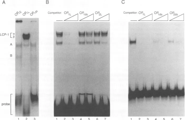
containing these sequences (Fig. 2A). Both the duplex
oligonucleotide OP1ds and the early-coding-strand
oligonu-cleotide OP1E formed two complexes, A and B, of similar
mobilities in a gelretardation assay (Fig. 2A, lanes 1 and 3).
Thelate-coding-strand oligonucleotide0P1L,whichcontains
the AGGGAAGGGA sequence, formed a minor complex
similar in mobility to complex A. Interestingly, the major DNA-proteincomplexes formed by0P1L,a doublet labeled LCP-lot andLCP-1,B (Fig. 2A, lane 2),were unique tothis
probe.
Competition experimentswere performed with single- and
double-stranded oligonucleotides containing either the
wild-type sequence
(0P1)
or a mutant variant (OP2) inorder to testboth the structuraland DNAsequence requirementsofthecomplexes (Fig. 2B).In thisassay,unlabeled competitor
DNAs were incubated withnuclear extract prior to addition of theOP1Lprobe. UnlabeledOP1L(Fig.2B, lanes 2 and 3)
on November 9, 2019 by guest
http://jvi.asm.org/
SINGLE-STRANDED-DNA-BINDING PROTEIN LCP-1 6887
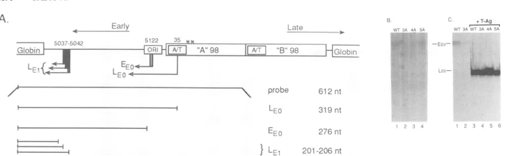
FIG. 1. JCV controlregion consists ofanorigin of DNA repli-cation (ori) and a 98-bp direct repeat. The EE and LE mRNA initiation sitesareshown abovethe control region, whereas the late mRNAinitiation sites areshown below, asdetermined previously (14, 17). The box within each 98-bp repeat denotes the A+T-rich region. Thesequencessurrounding the LCE (box)areshown for the proximalA98-bprepeatandare foundonthe late coding strand. The shaded box below thesequence denotes the region of hyper-sensitivity determined previously (2) that is found only in the A 98-bprepeat.
A B
~I
_R- , l
R-LCP-1
[F
to.
A
competed efficiently for the complex, whereas
OPjds
(Fig. 2B, lanes 4 and5)wasunableto sequesterLCP-1. Compar-ison of OP1L (Fig. 2B, lanes 2 and 3) with OP2L (Fig. 2B, lanes 6 and 7) indicated that the AGGGAAGGGAsequence wasrequired for the formation of the LCP-1 complex.The complex comigrating with bandA(shown in Fig. 2B) revealed wider sequence specificity than LCP-1. The inten-sity ofthis complex was diminished only when OP2Lwas used at ahigher concentrationas acompetitor. ComplexA butnotthe LCP-1 complexes was observed when the OP1ds probe was used (Fig. 2C, lane 1). The competition experi-ments indicated that complex A had a different sequence specificity than LCP-1. Both single- and double-stranded OP1 oligonucleotides competed effectively for this complex (Fig. 2C, lanes 2 to 6). Moreover, OP2ds (lanes6 and7)and
OP1E (not shown)
alsocompeted
for thisactivity.
Complex
B was common to OP1E and OP1ds; however, it was not reproducibly observed in our studies. Therefore, it appears that the LCP-1 complexes were formed only on single-stranded DNA containing the AGGGAAGGGA sequence andmay represent novelsequence-specific, single-stranded-DNA-binding complexes present in brain nuclear extracts.
Specific nucleotide contacts made by the LCP-1 complexes. Methylation andcarbethoxylation interference assayswere used to determinewhich purine nucleotides were in close contactwith theLCP-1 proteins. DMS methylates DNAat N-7 of guanosyl and N-3 of adenosyl residues (G > A). DEPC carbethoxylates purines at N-7 (A > G). In these experiments, OP1L was 32P-end-labeled and chemically
C
]X, c P ;_
VW
&_
-Mi
probeL j
1 2 3 2 3 4 5 6 7
FIG. 2. (A) 5'-End-labeled single-strandedandduplex oligonucleotideswereanalyzedonnativegelsfor theirabilitytoformcomplexes withaproteinpresentinmonkeybrain nuclearextract. OP1Erepresentstheearlycodingstrand(lane 1), OP1Lrepresentsthe latecoding strand(lane2),and
OPIds
representsduplex probe (lane3).Thedoublet labeledLCP-laandLCP-11 wasuniqueto0P1L,and its sequence specificity is analyzedinpanelB.ComplexAwas common toall threeoligonucleotides,and itsspecificityisanalyzedinpanelsBandC. ComplexBwas common to0PjE
andOPjds
butwas notreproduciblyobserved.The sequencesoftheoligonucleotidesareshown inMaterialsandMethods.(B) Complexes formed bytheOP1L single-strandedprobe(lane1).Theupperdoublet isLCP-1a/,;the lower doubletiscomplex
A.Thespecificity of these complexeswastestedbypreincubationof theextractwith 10or100 ngofOP1L(lanes2 and3,respectively),OP1ds (lanes4and5,respectively), and themutantOP2L(lanes6and7,respectively).Thenewband thatappearedin lanes 4 and5atthebottom of thegelis freeduplexoligonucleotides.(C) ComplexAformedbythe
OPjds
oligonucleotide (lane 1).ThespecificityofcomplexAwastestedbypreincubationof theextractwith 10or100 ng ofsingle-strandedOP1L(lanes2 and3,respectively), duplexOP1ds(lanes4 and5, respectively),and themutantduplex OP2ds(lanes6and7,respectively).Thissetofsampleswas run onthesamegelasthesamplesinpanel B,but thefigurewasseparated forsimplicity.Themigration ofcomplexAwasidenticaltothat inpanelB.
VOL.66,1992
on November 9, 2019 by guest
http://jvi.asm.org/
[image:3.612.64.298.71.267.2] [image:3.612.140.491.385.613.2]DMS
F B F
DEPC
F B F
G7G
*G
8-7
-
~G5-*G
4
21
-5 - A A A A A A A A G G G A A G G G A T G G C T G -3
1 2 3 4 5 6 7 8
FIG. 3. Free (F) and bound (B) oligonucleotides which were premodified byeitherDMS(left)orDEPC(right)wereisolated from native gels, cleaved with piperidine, and analyzed on 20% acryl-amidesequencing gels.TheJCV sequences presentwithin the OP1L oligonucleotideareshownatthebottom,where the G residuesare numbered from the 5'endoftheoligonucleotide.G residues marked withanasteriskinterferewith LCP-1bindingafter modification.
modified with either DMS or DEPC so that, on average, therewasless thanonemodification peroligonucleotide. In gel retardation assays, modified residues which interfered with LCP-1 binding were underrepresented in the bound
population of oligonucleotides. Therefore, free probe and
DNAbound in theLCP-lot and-13complexeswereisolated after nativegel electrophoresis,cleaved withpiperidine,and
comparedon sequencing gels.
The results with the DMS-modified oligonucleotide
showed that the LCP-1 complexes interacted specifically
with the LCE as well as with several adjacent residues.
Comparisonof the boundand free DNAs indicated thatG-1,
G-2, G-4, G-5,and G-6 of thepentanucleotiderepeat aswell
as the 3' residues G-7 and G-8 were in close contact with LCP-1 (Fig. 3). Similarly, the DEPC-modified
oligonucleo-tide identified the same guanosyl nucleotides as being in contact with LCP-1. It does not appear that G-3 and the 3'-most G residue interfered with LCP-1 binding when modifiedatN-7, and they maynotbe in closecontactwith
the complex. In addition, none of the adenosyl residues
appeartomake criticalcontactswith LCP-1ateither the N-3
orN-7position.
,bFJ1
[ kd:S>. ;_t''''''03:^w'D~P
~
4L~~~~~~~~~~1
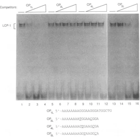
FIG. 4. Extractswereincubated with increasingconcentrations of either single-stranded homologous ormutantcompetitor DNAs
priortoaddition of the
0PlL
probe.Thefirst lane in everyset(lanes1, 5, 9,and13)containsprobealone;other lanescontained1, 10,or
100 ng,respectively,of thehomologous
0PlL
(lanes2to4)andthe mutant0O3L(lanes6to8),0P4L
(lanes10to12),and0P5L
(lanes14 to16)DNAs. Thesequences of thecompetitorDNAsareshownat the bottom. This figure is a composite of two gels which wereelectrophoresed in parallel and represents equivalent exposures.
ComplexAwas seenweaklyin this exposure,migratingfaster than
LCP-1, but is not marked. Note that longer exposure of the
autoradiogram showed that all threemutants effectively competed
forcomplexAat100 ng (not shown).
Nucleotide
requirements
of the LCP-1 complexes. Theprevious experiment
identifiedspecific
residues withinOPjL
whichwere critical for LCP-1 interactions. We were inter-ested indetermining
theimportance
of theG residues within thepentanucleotide
repeat itself forbinding.
Therefore,oligonucleotides
whichweremissing
contacts G-7 and G-8 and containedpairs
ofpoint
mutations weresynthesized.
These mutant
oligonucleotides
were used ascompetitors
ingel
retardationexperiments
to test thespecificity
of the LCP-1complex
for the core AGGGAAGGGA sequence(Fig. 4).
Formation of the LCP-1complexes
wereabolishedby
10and 100 ng of thehomologous
0P1L_
competitor
(Fig.
4,lanes 3 and
4).
The0P5L_
mutantretainedareducedability
tocompete for LCP-1. At 1 and 10 ng,
0P5L_
did notdiminishtheLCP-1
complexes,
butitcompeted
wellat100 ng(Fig.
4,lanes14to
16).
However, the mutations:P3:presentin and0P4L_
severely
affectedtheability
oftheseoligonucleotides
tobind andsequesterLCP-1
(Fig.
4,lanes6to8 and10to12,respectively).
Even at 100 ng, whichrepresented
a greaterthan 50-fold molar excess, no
competition by
these two mutantswasobserved.These data
appeared
to be consistent with the results of themethylation
andcarbethoxylation
interferenceexperi-ments.All threemutant
oligonucleotides
weremissing
con-tact
points
G-7 and G-8. In addition, 0O3L and0P4L
had transversion mutations at two contactpoints
in the coresequence
(G-1 plus
G-4 and G-2plus
G-5,respectively).
Therefore,
only
three of seven contactpoints
for LCP-1remained intact in these mutants. On theother
hand,
0P51L
G3
-'.G2
-;"'G1
on November 9, 2019 by guest
http://jvi.asm.org/
[image:4.612.64.287.61.367.2] [image:4.612.324.554.72.297.2]SINGLE-STRANDED-DNA-BINDING PROTEIN LCP-1 6889
A 7
CLm
o o
_100-120kDLE
50-52
kDE
B
Competitor: ORI- OP3L OP4L 0P5L
kD
200---t100-120[
97-
68-50-52 1.4
43
-1 2
28
[image:5.612.140.481.77.380.2]-1 2 3 4 5 6 7 8 9
FIG. 5. (A) The LCP-1 complexeswereUVcross-linkedtotheOP1Lprobe priortoloadingon anativegel. LCP-la (lane1) and LCP-1, (lane 2)werecutseparatelyfromthe wet gel,eluted, and loaded ontoSDS-10% PAGE gels. The dried gel was exposed toaphosphor screen, and thecomplexesweredetected withaPhosphorImager(MolecularDynamics). (B) Partially purified LCP-1wascovalentlycross-linkedto labeledOP1Lprobe byUVlightinthe absence(lane 1)andpresence(lanes2to9) of10(lanes 2, 4, 6, and8) and100(lanes 3,5,7, and9) ng of the unlabeledhomologousor mutantcompetitorDNAsused in Fig.4. Theresulting DNA-protein complexes were analyzed directly on
SDS-10%PAGEgels and visualized by autoradiography. Two resolvable complexes of 50to52 kDaand alargercomplex of100 to120kDa
arelabeled.Thesmearbelow the labeledcomplexes may bedegradation products.
contains transversion mutationsatG-3 andG-6,butonly one
of the mutations affected a contact point within the LCE
(G-6). We suggest that since
OP5L
contained four ofsevencontactpoints, it retained areduced abilitytobind LCP-1, whereas the threecontactpointspresentin0P3Land
0P4L
were notsufficienttobind LCP-1.
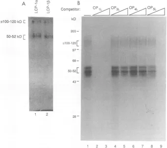
Characterization of the protein components of LCP-1. LCP-1 was partially purified from monkey brain nuclear extractsby ion-exchange chromotographyin orderto study
these proteins biochemically. The LCP-1 complexes were isolated from the other OP1 region-binding proteinswith a
purification
ofapproximately 17-fold(35a).
These partiallypurified protein fractions were used to characterize the
protein components of LCP-1 by UV cross-linking and
competition.
Initially, UV cross-linkingwasusedtodeterminewhether
therewere differences between the
protein
components ofLCP-lcx and
LCP-113.
Proteins were cross-linked to end-labeledOP1L byexposuretolong-waveUVlight
for 30 minprior to analysis by gel retardation. LCP-la and LCP-1lB
were cut separately from the wet gel, eluted into
protein
sample buffer, and analyzed by SDS-PAGE. Our results
showedtwocross-linkedspeciespresentin bothLCP-la and
LCP-11,
asmallercomplexof 50to52 kDa andalarger
oneof 100 to 120 kDa (Fig.
5A).
Although it was difficult toresolveaclearsize differenceinthis
particular
experiment,
it appeared that the 50- to 52-kDa component of
LCP-1f
migrated slightlyfaster than that of LCP-la. Moreover, a
faint minor band above the 50-to52-kDa bandwasdetected in theLCP-la complex.Theintegrityofthis bandhas yetto be determined. Later experiments in which cross-linked
proteinswere loaded directlyon protein gels resolved two
distinct complexesin this size range.
The sequencespecificityof thesecomplexeswastestedby
performingcompetition experiments withtheOP3, OP4,and
OP5
mutant oligonucleotides. These unlabeled competitors were incubated with the partially purified protein fractionpriortoaddition ofOP1L probe, cross-linked, and analyzed
immediately by SDS-PAGE. Resolvable 50- and 52-kDa
complexesaswellasthe 100-to120-kDa bandweredetected
(Fig. SB, lane 1). The smeared bands smaller than 50 kDa
appear to be
degradation
products.
Thehomologous
com-petitor abolished
binding
of all of theproteins
in thesample
at 10 and 100 ng (Fig. SB, lanes 2 and 3). The OP5I_
oligonucleotidedidnotdiminish
protein
binding
muchat10ng but competed well at 100 ng
(Fig.
SB,
lanes 8 and9).
Neither OP3L (Fig. SB, lanes 4 and
5)
nor OP4L(Fig. SB,
lanes 6 and 7) competed at either concentration of DNA. These results mirrored the gel retardation results inFig.
4 and confirmed that theseDNA-protein
complexes
werecomponents ofLCP-1.
Functional
analysis
of LCP-1promoter mutations. Inorder VOL. 66,1992FM.
lp
"W
iw,
on November 9, 2019 by guest
http://jvi.asm.org/
A.
Early ;i."+:-.AT-Ag-A5A
..)
L.
7p
;")122, KA ___ D1 3 's 8_
_~
EZI
',A'98L
FE`-
0'JLl 201-206nt
FIG. 6. (A)Structures of thepBEL2-derived plasmids, showingthe locationsof theEEandLE transcriptswithin theJCVcontrolregion. Belowareindicated the full-length S1 probeandthe sizes of thefragments protected bythevariousearlyRNAs. The two asterisks above
the A98-bprepeat showthepositions of the3A, 4A, and 5Amutations. Thesequencesof the mutations canbefound in Materialsand
Methods.nt,nucleotides. (B)Si nucleasedigestion productsprotected byRNAs isolatedfromtransfection of thepBEL2-derived plasmids into U87-MG glial cells. The EEO mRNAs resulting from transfection of thewild-type (wt) plasmid pBEL2-JC (lane 1) or the mutants
pBEL2-3A (lane 2), pBEL2-4A (lane 3),andpBEL2-5A (lane 4)areindicated.(C)S1 nuclease-protectedRNAsresultingfrom cotransfection
of pBEL2 plasmids with pBJC-T. pBEL2-JC and pBEL2-3A plasmids were transfected either alone (lanes 1 and 2, respectively) or
cotransfected withpBJC-T (lanes3 and4,respectively). OnlytheEEOproductwasdetected in the absenceofT-Ag (lanes 1 and 2),whereas
in thepresenceofT-Ag,theearlyinitiation sites shiftedasindicated(lanes3and4).Cotransfection ofpBEL2-4AandpBEL2-5AwithpBJC-T (lanes 5 and 6,respectively)gavesimilar results.LEOwasdetected afterlongerexposureand did notdifferamongthesamples (not shown).
toidentifythepossible function(s)of LCP-1intheregulation ofJCV, site-directed mutationscorresponding toOP3,OP4, and OP5 were introduced into the A 98-bp repeat of the
full-length JCV promoter. The B 98-bp repeat was not
mutagenized because it has been shown to be much less sensitivetoSi nuclease andbromoacetaldehyde (2)and thus lesslikelytobeunpairedandcapableofbindingLCP-1. The
wild-type and mutant promoters were placed into plasmid pBEL2 (13, 41), a vector which contains two divergent copies of the rabbit P-globingene.
These plasmids were transfected either alone or with a
plasmid encoding JCV T-Ag into U87-MG cells. This cell linewasderived fromahumanglioblastomawhichcontained
aprotein complexindistinguishablefromLCP-1(35a).Total RNAwasextractedat40 hposttransfection andhybridized
toauniformlylabeled Si probe complementarytotheglobin cDNA and the JCV early region (Fig. 6A). This probe mapped the early RNAs according to their initiation sites, whereas the late RNAs protected a single fragment
corre-sponding to the break point between the globin and late leadersequences.
Analysis of RNA derived from transfection of the wild-type constructpBEL2-JC alone showed oneprotected spe-cies, marked EEO, which corresponded to the major EE mRNAs (Fig. 6B, lane 1). RNA isolated from cells trans-fectedwith the mutantplasmids pBEL2-3Aand pBEL2-4A reducedthe levels of theEEO transcripts (Fig. 6B,lanes 2and 3). Transfection with the pBEL2-5A mutant gave approxi-mately wild-type levels of the EEO transcription products (Fig. 6B,lane4).Theseresultswereconsistentwith thoseof the binding studies and suggested that LCP-1 regulates EE transcriptioninitiation ofJCV.
Cotransfection of pBEL2-JC with T-Ag, which initiates DNAreplication, mimics the late phase of thelytic cycle by causingashift in theearlymRNAinitiationsites from EEOto
twonewlocations, LE1and
LEO
(17). Interestingly,theearlyRNAs isolated from the cotransfection of pBEL2-3A with T-Agshowed the samepattern and abundance oflate-early transcriptsasinthewildtype(Fig. 6C,comparelanes 3and
4), whereas in the absence ofT-Ag, the EEO product level
wassignificantlylower than inthe wildtype(Fig. 6BandC, compare lanes 1and2). Theothermutants, pBEL2-4Aand pBEL2-5A, showed the samepatternand abundance of the late-earlyRNAs(Fig. 6C,lanes5and6). Further studiesare
inprogress toinvestigate therole ofLCP-1 intranscription of theJCV late promoterbefore and after DNAreplication (4).
DISCUSSION
Wehavepresentedevidence thata
single-stranded-DNA-binding protein in brain nuclear extract specifically recog-nized the LCE. The binding studies showed that LCP-1 recognized a specific sequence within the enhancer, and pointmutations thatdifferentiallyaffectedLCP-1 bindingto that sequence were determined. These LCE point
muta-tions,whenplacedinthecontextof thefull-lengthpromoter, affected JCVearlygenetranscription initiating from the EEO butnotfrom eithertheLE0(showninFig.6C)orthe
LE,
(notshown). Thecorrelation betweenthebinding and functional
assayssuggested thatLCP-1 regulates earlygene
transcrip-tionduringtheearly phaseof the lytic cycle, priortoDNA replication. However, duringthe latephase following repli-cation, the mutations had no effect on LE transcription, suggesting a novel mechanism by which changes in DNA
structuremayaffect the interaction betweenatranscription
factorand its cognate bindingsite to regulate transcription initiation.
Themajorityofeukaryotic transcription factorsrepresent a classofproteinswhich recognize and bind specific DNA
sequencestoactivateorrepresstranscription(11, 12, 21, 22, 34). The DNA target sequences for this growing class of proteinshave beenfairly wellcharacterized, but muchless is known about potential alternative structures of these cis elements that may influence DNA-protein interactions. From the existence of distinct DNase I- and Si-sensitive sites within the genome, it has been suggested that sub-classes of sequence-specific proteins whichrecognize
non-B-DNAstructuresmayexist(20). Forexample, DNA
bend-ing has been shown to strengthen the interaction of the
F14
(s:',ob
612nt
319nt
276 nl
IE
"B 98on November 9, 2019 by guest
http://jvi.asm.org/
[image:6.612.64.560.64.217.2]Drosophila zinc finger protein suppressor ofhairy wing
[su(HW)] with the octamer motif within the gypsy element
(33).Thetranscription factor MF-3 has recently been shown
to recognize both double- and single-stranded DNAs
con-taining both the MCAT and CArG motifs present in the
promoters of some muscle-specific genes (28). The protein ssARS-T fromSaccharomyces cerevisiaehas beenidentified
asasequence-specific single-stranded-DNA-bindingprotein
which regulates the initiation of DNA replication in yeast
cells (29). Thus, it appears likely that new classes of
se-quence-specificDNA-bindingproteins might recognize
alter-native DNA structures to regulate both transcription and
replication.
The LCE has been previously shown to adopt a unique DNA structure, described as a non-B-DNA right-handed helix (2). Interestingly, the LCE in the A 98-bp repeat
proximaltotheorigin and early-early mRNA initiation site is
highly sensitive to Si nuclease and bromoacetaldehyde in negativelysupercoiledDNA. In contrast,the same sequence in the distal(B)98-bp repeatshowslittlesensitivitytothese reagents,suggestingthatneighboringsequences,suchasthe
origin, may influence the unusual structure of this region.
This differencemayhavefunctionalconsequences, sincethe
EE mRNAs initiate predominantly from the A repeat TATATA box(17). Thus, while other factors may also be involved, the structure of the LCE may be an important
determinant in the positioning ofthe EE mRNA initiation
complex.
The LCE lies between the TATATA box and the recog-nition site for a brain-specific activator protein (1, 16, 18).
Wehypothesizethat theaffinityof LCP-1 forsingle-stranded
DNAcontributesto the assembly of theEE RNA initiation
complexatthe A repeat TATATA box. We suggestthatthe
LCE innegativelysupercoiled inputDNA issingle stranded,
allowing LCP-1 to bind and subsequently interact with the
transcription machinery. Following DNA replication, the
LCP-1binding sitemaynolonger beaccessible, possiblydue torelaxation of thetemplate by topoisomerase orthe
repli-cationmachinery. Failuretobind LCP-1 may thensignal the
transcription complextoassembleatnewlocations on both
the early and late promoters. Therefore, the LCE may
regulatethe early-to-late shift bychanging the structure of
theregion upon DNA replication, signaling transcription to initiate at other locations within the viral control region. Thus, the structure of the LCE may be important in
regu-lating JCVgene expression throughoutthe virallytic cycle.
What role doesJCVT-Agplayinmediatingthe shift of the
early mRNAinitiation sites? We have already discussed a
possible indirectrole forT-Agvia stimulationofviralDNA
replication, resultinginanalteredchromatinstructurethat is
nolonger recognized byLCP-1. In the case ofSV40, it has
been suggestedthat there may be competitionbetween the
EE and LE start sites for transcription initiation, so that occupancyof theorigin byT-Agstericallyhinders initiation attheEE site(25, 34).Wedonotfavorthishypothesis,since the presence ofJCV T-Ag in the absence of DNA
replica-tion, asobservedin thehamstergliomacell line HJC
(8,
9),is not sufficient toinduce the shift from the early initiation
site in vivo and in vitro(17; unpublished data). On the other
hand, it is possible thatJCV T-Agmay play a more direct
role, such as inactivating orsequestering LCP-1 so that it can nolongerbindDNA, ashas been shown forSV40T-Ag
and AP-2(24).
Viruses representinterestingandconvenient systems with whichtostudytherelationshipbetweenDNA structureand
mRNAtranscriptionineukaryoticcells. Our results suggest
anovel mechanismoftranscription regulation in which the interaction ofatranscriptionfactor with its binding site may
bedirectly influencedbythe structure of that region.
Eluci-dation of the mechanisms by which the structure of cis elementsregulates viral mRNAtranscription will have im-portant implications for the regulation of cellular mRNA
synthesis, particularly in rapidly proliferating cells and in
differentiatingtissues.
At present, we have not identified any cellular genes which may be regulated by LCP-1, but we have found related DNA motifs inanumber ofcellularpromoters. The promoters of myelin basic protein and proteolipid protein
contain
proximal
elements that resemble the LCE sequence,differing by a single nucleotide (5, 7). The neurofilament
heavy-chaingene promotercontainsanA+G-richelement,
Pal-1, that appearsto beinvolved in regulation of neurofil-ament heavy-chain expression (21a). Interestingly, this se-quenceis presentas apalindrome which canpotentiallyalter DNA structure and, perhaps,gene expression.
ACKNOWLEDGMENTS
We thankRobertLazzarini forcommunicating his results before publication, Richard Kuhn for providing the dutung mutant Esch-erichia coli BW313, Alan Wildeman for providing the pBEL2 plasmid, Darwin Prockop for the use of the PhosphorImager, FrancesFeiglforartwork,and membersof the Molecular Neuro-virology Section of the JeffersonInstituteof MolecularMedicinefor theirhelpful discussions andcomments.
This work was supported by grants CA47996 and A128272, awardedbytheNational InstitutesofHealth,and grantRG2054A1, awardedbytheNationalMultiple Sclerosis Society,toK.Khalili.
REFERENCES
1. Ahmed, S., J. Rappaport, H. Tada, D. Kerr, and K. Khalili.
1990. A nuclear protein derived from brain cells stimulates transcription of the human neurotropic viruspromoter,JCVE,in vitro.J. Biol.Chem. 265:13899-13905.
2. Amirhaeri, S.,F.Wohirab,E.0.Major,and R. D. Wells. 1988. UnusualDNA structure in theregulatoryregion of the human papovavirus JC virus.J. Virol. 62:922-931.
3. Beggs, A.H., R. J. Frisque, and G. A. Scangos. 1988.Extinction of JC virus tumor-antigen expression in glial cell-fibroblast hybrids.Proc. Natl.Acad. Sci. USA 85:7632-7636.
4. Chang, C.-F.,et al.Unpublished data.
5. deFerra, F.,H. Engh,L. Hudson, J. Kamholz, C. Puckett, S. Molineaux, and R. A. Lazzarini. 1985. Alternativesplicing for thefour formsofmyelinbasicprotein. Cell43:721-727. 6. DelVecchio, A.M.,R. A. Steinman, and R. P. Ricciardi. 1989.
An element of the BK virus enhancer required for DNA replication. J.Virol. 63:1514-1524.
7. Diehl,H.J.,M.Schaich,R. M.Budzinski,and W.Stoffel.1986. Individual exons encode the integral membrane domains of humanmyelin proteolipid protein.Proc.Natl. Acad. Sci.USA 83:9801-9811.
8. Feigenbaum, L.,K.Khalili,E.0.Major,andG.Khoury.1987. Regulation of thehost rangeofhumanpapovavirusJCV. Proc. Natl.Acad. Sci. USA 84:3695-3698.
9. Frisque, R.J.,D. B.Rifldn,and D. L. Walker.1980. Transfor-mation ofprimary hamster brain cells with JC virus and its DNA. J.Virol. 35:265-269.
10. Graham, F. L.,andA.J.vanderEb.1973.A newtechniquefor the assay ofinfectivityof human adenovirus 5 DNA. Virology 52:456-467.
11. He, X., andM. G. Rosenfeld. 1991. Mechanisms ofcomplex transcriptional regulation: implicationsfor brain development.
Neuron7:183-196.
12. Johnson, P.F.,andS. L.McKnight.1989.Eukaryotic
transcrip-tionalregulatoryproteins.Annu. Rev. Biochem. 88:799-839. 13. Kelly, J. J., J. M. Munholland, and A. G. Wildeman. 1989.
Comeasurement of simian virus 40 early and late promoter VOL. 66, 1992
on November 9, 2019 by guest
http://jvi.asm.org/
activityinHeLaand 293 cells inthepresence ofTantigen. J. Virol. 63:383-391.
14. Kenney,S.,V.Natarajan, and N. P.Salzman. 1986. Mapping 5' terminiof JCvirus late RNA.J.Virol. 58:216-219.
15. Kenney, S., V. Natarajan, V. Strika, G. Khoury, and N. P. Salzman. 1984. JC virus enhancer-promoter active in human brain cells. Science 226:1337-1339.
16. Kerr, D., and K.Khalili. 1991.Arecombinant cDNAderived from human brain encodesaDNAbinding protein that stimu-lates transcription of the humanneurotropic virus JCV.J.Biol. Chem. 266:15876-15881.
17. Khalili, K., L. Feigenbaum, and G.Khoury.1987. Evidencefor ashiftin5'-termini ofearly viralRNAduring thelytic cycleof JCvirus.Virology 158:469-472.
18. Khalili, K., J. Rappaport, and G.Khoury. 1988. Nuclear factors in human brain cells bind specifically to the JCV regulatory region. EMBO J. 7:1205-1210.
19. Kunkel, T. A.1985. Rapid and efficientsite-specific mutagenesis without phenotypic selection. Proc. Natl. Acad. Sci. USA 82:488-492.
20. Larsen, A.,and H. Weintraub. 1983. Analtered DNA confor-mation detected byS1 nuclease occurs at specific regions in active chickglobin chromatin. Cell 29:609-622.
21. Latchman, D. S. 1990. Eukaryotic transcription factors. Bio-chem. J.270:281-289.
21a.Lazzarini,R.Personal communication.
22. Levine, M.,andJ. L.Manley. 1989.Transcriptionalrepression ofeukaryoticpromoters.Cell 59:405-408.
23. Lynch, K. J., and R. J.Frisque. 1990. Identification ofcritical elements within the JC virusDNAreplication origin. J. Virol. 64:5812-5822.
24. Mitchell, P. J., C. Wang, and R. Tjian. 1987. Positive and negative regulation oftranscription in vitro: enhancer-binding proteinAP-2is inhibitedby SV40T-Ag. Cell 50:847-861. 25. Myers, R.M.,D. C. Rio,A. K. Robbins,and R.Tjian. 1981.
SV40geneexpression is modulatedbythecooperative binding ofTantigentoDNA.Cell 25:4445-4449.
26. Padgett,B.L.,andD. L. Walker.1976. Newhuman
papovavi-ruses.Prog. Med. Virol. 22:1-35.
27. Padgett, B. L., D. L.Walker,G. M.ZuRhein,R.J. Eckroade, and B. H. Dessel. 1971. Cultivationofapapovavirus-likevirus from human brain withprogressive multifocal leukoencephalop-athy. Lancet i:1257-1260.
28. Santoro,I.M.,T. M.Yi,and K. Walsh.1991. Identification of single-stranded-DNA-binding proteins that interact with muscle geneelements. Mol. Cell. Biol. 11:1944-1953.
29. Schmidt, A. M.A., S. U. Herterich, and G. Krauss. 1991. A
single-strandedDNAbinding proteinfrom S. cerevisiae
specif-icallyrecognizestheT-richstrand ofthecore sequenceofARS
elements and discriminatesagainstmutantsequences. EMBO J. 10:981-985.
30. Sharma, A. K., andG.Kumar. 1991. A53kDaprotein bindsto thenegative regulatory region of JC virusearlypromoter.EEBS 281:272-274.
31. Small, J. A., G. Khoury, G. Jay, P. M. Howley, and G. A. Scangos. 1986. Early regions of JC virusandBKvirus induce distinct and tissue-specific tumors in transgenic mice. Proc. Natl. Acad.Sci.USA 83:8288-8292.
32. Small,J. A., G. A.Scangos, L. Cork, G. Jay, and G.Khoury. 1986. The early region of human papovavirus JC induces dysmyelination in transgenic mice. Cell46:13-18.
33. Spana, C.,and V.G.Corces.1990.DNAbending isa determi-nant ofbinding specificity foraDrosophilazincfingerprotein. GenesDev.4:1505-1515.
34. Struhl, K. 1991. Mechanismsfor diversityin gene expression patterns.Neuron 7:177-181.
35. Sturm,R., T.Baumruker, B. R. Franza, and W. Herr. 1987. A 100-kD HeLa celloctamerbindingprotein (OBP100)interacts differently withtwoseparateoctamer-relatedsequences within the SV40 enhancer. GenesDev.1:1147-1160.
35a.Tada, H. 1992. Ph.D. thesis. Thomas Jefferson University, Philadelphia,Pa.
36. Tada,H., M.Lashgari,and K. Khalili.1991.Regulation of JCVL promoterfunction:evidence thatapentanucleotide "silencer" repeat sequenceAGGGAAGGGAdown-regulatestranscription ofthe JC virus latepromoter.Virology 180:327-338.
37. Tada, H., M.Lashgari,J.Rappaport, and K. Khalili. 1989. Cell type-specific expression ofJC virus early promoter is deter-mined by positive and negative regulation.J.Virol. 63:463-466. 38. Tada, H.,J.Rappaport, M.Lashgari,S. Amini, F. Wong-Staal, and K. Khalili. 1990. trans-Activation of the JC virus late promoterbythetatprotein oftype 1human immunodeficiency virus inglial cells. Proc. Natl. Acad. Sci. USA 87:3479-3483. 39. Trapp, B. D., J. A. Small, M. Pulley, G. Khoury, and G. A.
Scangos. 1988.Dysmyelination in transgenic mice containingJC virusearly region.Ann. Neurol.23:38-48.
40. Wasylyk, B., C.Wasylyk, H. Matthes, M.Wintzerith, and P. Chambon. 1983. Transcription from the SV40 early-early and late-early overlappingpromoters in the absence of DNA repli-cation.EMBO J.2:1605-1611.
41. Wildeman, A. G. 1989.trans-Activation of both early and late simian virus 40 promoters by large tumor antigen does not require nuclear localization oftheprotein. Proc. Natl. Acad. Sci.USA 86:2123-2127.