0022-538X/91/031491-05$02.00/0
CopyrightC) 1991, American Society for Microbiology
Major
Histocompatibility Complex Restriction
of T-Cell
Responses
to
Varicella-Zoster Virus in Guinea Pigs
ANTHONYR.
HAYWARD,'*
R. BURGER,2 R. SCHEPER,3 ANDANN M. ARVIN4DepartmentofPediatrics, University of Colorado Health Sciences Center, Denver, Colorado802621;
RobertKoch Institut, D 1000 Berlin 65, Germany2;Free University, Amsterdam, 1081 HvAmsterdam,
The Netherlands3; andStanfordUniversity, Palo Alto, California943054
Received 14May1990/Accepted 11 December 1990
Varicella-zoster virus (VZV), adapted togrow in guineapig fibroblasts, was injected subcutaneously into
Hartley, strain 2,and strain 13 guinea pigs. Serum immunoglobulin G antibodiesweredetected 2weeks later,
andT-cell proliferativeresponsesby blood lymphocyteswerefound 3 weeks after injection. The proliferating
cells bound the 155 antibody, which defines a CD4-like subset of guinea pig T lymphocytes. VZV-infected fibroblasts of human, Hartley, and strain 13 origin elicited equivalent amountsof proliferation, which was
quantitativelygreaterthanthat obtainedwithanextracted VZV antigen.Uninfected(control) humanorguinea
pig fibroblastsdid notelicit T-cell proliferation. The proliferative response toVZV required thepresenceof
autologous (strain2or13) antigen-presentingcells andwasblocked by the addition ofananti-class II major
histocompatibilitycomplexantibody. Effectorcellsobtainedfrom in vitro cultures mediated classII-restricted cytotoxicitytoL2C cells incubated with VZV.ClassI-restrictedresponses wereobtainedonly by cross-priming strain 2 animalswith strain 13 peritoneal exudate cells which had been preincubated with VZV. The data
indicatethatguineapigs resemble humans in that class1I-restrictedT cellswithspecificity for VZVare more
readily cultured from bloodthanareclass I-restricted cells.
Varicella-zoster virus (VZV) causes chickenpox and has the potential to become latent in posterior horn sensory neurones. The reactivation of latent VZV causing herpes
zoster (shingles) is a source of serious morbidity due to
postherpetic neuralgia. Previous studies have linked the
increasing incidence of shingles in the elderly with waning VZV-specific immunity (4, 14), but the point at which VZV-specific immunity is so compromised as to permit VZV
reactivation isunknown.VZV-specific cell-mediated
immu-nity can be boosted by immunization with the attenuated
Oka strain vaccine (5), but the components of the
cell-mediatedresponse whichprovide protection from VZV are
poorly understood. In part this is because there is no
generally accepted animal modelforthedisease.Myers etal. (20)showed thatVZVcouldbeadaptedto growinguinea pig cells and thatguinea pigs injected with infected cells made VZV-specific antibody responses even though they did not
becomediscerniblyill(21). Morerecentstudies have shown thatVZV-inoculated guinea pigsmake antibodyand T-cell-mediated responses togpland to the gene 62product, p170
(2).
Intissueculture,VZV growswell inarangeof cellswhich express class I major histocompatibility complex (MHC) antigens but are generally negative for class II antigens. Nevertheless, most ofthe T cells clonedfrom VZV-stimu-latedcultures ofhumanmononuclear cells (MNC) havethe
CD4
antigens
which characterize class 1I-restricted Tcells(13) and thecytotoxicity mediated by these cellsis class II restricted(11, 15). MHC class I-restricted responsestoVZV
by human lymphocytes have generally been difficult to
detect (16), although recent observations suggest that CD8
cells respond better
following depletion
of CD4 cells(1).
Whether the predominance ofclass 1I-restricted T cells in conventional in vitro responsestoVZVisdetermined
by
the*Correspondingauthor.
route of antigen presentation during a primary varicella
infection or whether it is an artifact of the T-cell culture methods is not known. Theissueis important in the context
ofoptimal immunization strategies for the elderly as they
becomeatriskfor herpeszoster.Since therouteofinfection
ofhumansisnotopentomanipulation,wehaveusedguinea pigs to analyze the MHC restriction ofresponses to VZV which follow the injection of infected cells into guinea pigs of the strain2 and 13 inbred lines.
MATERIALS ANDMETHODS
Guineapigsandguinea pig cell lines.Strain2and 13guinea pigswereobtainedfrom the AnimalDivision oftheDivision ofResearch Resources ofthe National Institutes ofHealth and bred in our animal house. Hartley guinea pigs were
obtainedlocally. Guinea pig fibroblasts (strain 2and 13and
Hartley) were cultured from the bone marrow of 3- to
4-week-old animals by the method of Bowdenetal. (6) and
were frozen at passage 5. Guinea pigs were infected by
subcutaneous injection of 0.5 ml ofa
suspension
of VZV-infectedfibroblasts ofthe appropriate strain.Guineapig macrophageswereobtainedby
intraperitoneal
injectionof 20 mlofparaffinoil and theharvestingof ascites 5to7dayslater. Bloodwasobtainedby cardiacpunctureof animals anesthetized with Metophane
(Pitman
Moore,Washington
Crossing,
N.J.). Itwasdefibrinated and centri-fuged to separate cells and serum. The L2C guinea pig lymphomacell linewasgenerously
provided
byE.Shevach,
National InstitutesofHealth.
Guinea pig-adapted VZV andantigen. The
preparation
ofguineapig-adaptedVZVis described
by
Arvinetal.(2).
The viruswasgrown inguinea pigfibroblasts(Hartley,
strain2,
and strain13)to75%
cytopathic
effect. Theinfectedmono-layers were detached by
rolling
withglass
beads and were then spundown andfrozenat-70°C
wherethey
weretobe used as a source of infectious virus.Monolayers
were1491
on November 10, 2019 by guest
http://jvi.asm.org/
1492 HAYWARD ET AL.
trypsinized and fixed in 0.05% glutaraldehyde for 30 s
(terminated by addition of 0.2 M lysine) when they were to be used as aVZV antigen stimulus.
Antibodies toVZV. Guinea pig anti-VZV antibodies were
detected by Western blot (immunoblot). Briefly, Hartley
strain fibroblasts infected with VZV were lysed in Nonidet
buffer and electrophoresed in an 0.8% polyacrylamide gel
and transferred onto nitrocellulose in a BioRad apparatus.
Strips of antigen were overlaid withthe serumto betested, washed, and then incubated with1251I-labeledproteinA(ICN Radiochemicals). The strips were washed and exposed to
X-ray film before developing and examination.
Monoclonal antibodies. The 155antibodyisarat
monoclo-nal antibody identifying a CD4-like epitope on T cells (8). 22C4 is a mouse immunoglobulin Gi (IgGl) which binds to thela antigens ofstrain 2and strain 13animals (7). CT6isa
mouse IgGl which binds to a CD8-like subset (25). For
immunofluorescent-staining studies, the mouse antibodies were located with a fluorescein isothiocyanate-conjugated
anti-mouse IgG (Boehringer Mannheim, Indianapolis, Ind.)
and therat antibody was located witha mouse anti-rat IgG
conjugate (Rockland, Gilbertsville, Pa.).
Cultures. Lymphocytes from immunized and control
guinea pigs were obtained either from blood (where serial
samplesweretaken)orfrombloodandlymph nodewhen the animals were killed. Lymph nodes were teased in Hanks balancedsalt solutiontoobtainacellsuspension. Bloodwas defibrinated and centrifuged (600 x g, 4
min,
20°C) so thatserumcould berecovered.The MNCwererecovered froma
Ficoll-Hypaque gradient (SG 1.088) after 20 min of centrif-ugation at 770 x g at room temperature. The MNC were washed and adjusted to 106/ml in RPMI 1640 medium
sup-plemented with 10 p.g/dl of gentamicin and 10% guinea pig
serum. The cells were cultured in an upright tube (Falcon
2057; Becton Dickinson) for4daysand thentransferredtoa 25-cm2 flaskfor3moredaysofculturing,afterwhich 10 Uof interleukin-2 [IL-2] per ml was added (ala-IL2, human
recombinant; Amgen, Thousand Oaks, Calif.). Cells were removed for testing in cytotoxicity assays at the end of 7 days of culturing with IL-2.
T-cellrestimulationassays. T-cellrestimulationassays are describedbyKumicketal.(19). Briefly, 104prestimulated T
cellswerecultured for 3 dayswith 105irradiated strain 2or
13 bloodMNC (asasourceofMHC-matchedormismatched
antigen-presenting cells) and VZV orcontrol antigens
(pre-paredfromVZV-infectedand controlHartley fibroblasts as
described by Zaia et al. [26]), and then their thymidine uptake duringa4-h pulsewasdetermined.
Cytotoxicityassays.Effectorlymphocyteswereadjustedto 106/ml inRPMI1640 with 10% fetal calfserum. Targetcells werestrain2L2C cells(whicharestrain 2 in origin but share
class Iantigenswith strain13)towhich104PFU of VZVwas added 16h, before thecytotoxic assay. These cells showed no morphological evidence of cytopathic effect when har-vested. In certain experiments, strain 2, strain 13, Hartley,
and human fibroblasts were used as targets. These fibro-blasts were VZV infected 72 h before harvesting. Target cellswerelabeled with100,uCi of 51Cr for40minat37°Cand
washed three times before being resuspended to 5 x 10⁴ cells/ml. Onehundred microliters of effectorswasmixed ina
V-bottom well with 100 ,ul oftargets, and the cells were
briefly centrifuged before being incubated for 5 h at 37°C.
Then100R1ofculturesupernatantwasaspiratedforgamma
[image:2.612.317.560.100.209.2]counting. Percent specific lysis was expressed as [(experi-mental release-spontaneous release)/(maximum release-spontaneous release)] x 100.
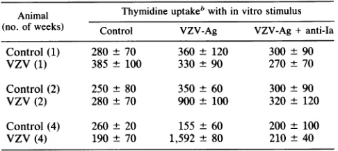
TABLE 1. Proliferative responses of guinea pig lymphocytes toVZVa
Animal Thymidineuptakeb within vitro stimulus (no. ofweeks) Control VZV-Ag VZV-Ag +anti-Ia
Control(1) 280 ± 70 360 ± 120 300 ± 90
VZV (1) 385± 100 330± 90 270± 70
Control (2) 250± 80 350 ± 60 300 ± 90 VZV(2) 280± 70 900± 100 320 ± 120 Control(4) 260 ± 20 155 ± 60 200 ± 100 VZV(4) 190 ± 70 1,592± 80 210 ± 40
a Blood lymphocytes from strain 2 guinea pigs, either control orafter injection with VZV, were culturedwitha 1:200dilution ofVZV antigen. Thymidine uptakewasdetermined after6days of culture.
b Resultsarethe mean±1standarderroroftriplicate cultures fortwostrain 2animalsateachtimepoint.
Cross-priming. Two strain 13 guinea pigs were injected
intraperitoneally with20 mlofmineraloil. Sevendays later, their ascites was tapped and the peritoneal exudate (PE)
cells were recovered by centrifugation. PE cells (2 x 10⁷)
were incubated in siliconized containers with an equal
number ofVZV-infected strain 13 guinea pig fibroblasts in RPMI1640medium with 5%guinea pigserumfor 6h,after which the cellswere washed andirradiatedwith 2,000 rad. Thesecellsweretheninjected intraperitoneallyintostrain2 and 13 guinea pigs. After 20 days, blood and lymph node MNC were recoveredfromtheinjected animals andcultured
withglutaraldehyde-fixed control or VZV-infectedstrain 13 fibroblasts. After 7 days, the MNC were recovered and tested forlysisof L2C cells (controlorVZVinfected).
RESULTS
Timing and phenotype ofVZV-specific immune response in guinea pigs. Guineapigswere injected subcutaneouslywith VZVwhich had been grown inguineapig fibroblasts ofthe
corresponding strain. Blood was obtained at 1-week inter-vals and tested for antibody by Western blot. Serum
anti-bodywasdetectedasearlyas11daysfollowing injection and
included antibody with specificity for gpl. Proliferative re-sponsesby bloodlymphocytesweredetected 3 weeks after
injection (Table 1). VZV-specific proliferation was blocked by the 22C4 antibody, as previously reported for other
antigens (9). Proliferating cells in these cultures were iden-tified by their blastlike light-scattering qualities and were
found to stain with the 155 antibody, which defines a
CD4-like subset ofguinea pigT
lymphocytes
(Table2). Theblasts did not stain with CT6.
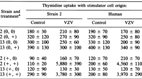
APC requirement for T-cell response to VZV. Blood MNC from VZVand control immunized guinea pigs were stimu-lated with VZV in the form of VZV-infected or control strain-matched fibroblasts in the presence or absence of adherent cells. The results (Table 3) show that proliferative responses required the presence of strain-matched
antigen-presentingcells (APC) and was not influenced by the source of the infected fibroblasts.
As an additional check of the MHC restriction of
VZV-specificresponses,we examined thespecificity of cytotoxic responses. Strain2and 13 guinea pigs were inoculated with VZV and their blood and lymph node lymphocytes were obtained 3to4 weeks later. Thesecellswere stimulated for 7dayswith VZV-infected autologousfibroblasts which had
J. VIROL.
on November 10, 2019 by guest
http://jvi.asm.org/
TABLE 2. Phenotypeof T-cell blasts from VZV-stimulated cultures'
% Cells stained Animal Stimulus No.of weeks Small Blasts
postinjection lymphocytes
CD4 CD8 CD4 CD8
Control Control 4 70 20 4 1
Control VZV-stimb 4 72 18 3 1
VZV VZV-stim 4 74 12 78 1
VZV VZV-stim 8 72 8 77 1
aBlood
lymphocytes
obtained fromguinea pigs4 or8 weeksafter injection with VZVwerestimulated for6days invitro with MHC-matched controlor infectedfibroblasts,after whichthecellswererecovered andstained withthe CD4-likeantibody, 155,ortheCD8-likeantibody,CT6.bstim,Stimulator cells.
been
lightly
fixed withglutaraldehyde.
This degree of fixa-tion does notinterfere withclass I-restricted antigenrecog-nition
by
mouseTcells(17, 24). Culturewascontinued forafurther7
days
after the addition of10U of humanIL-2per ml. Strain 2 effector cells recovered from these cultureslysed
VZV-preincubated,
butnotcontrol,L2Ccells, andthelysis
was inhibited with anti-MHC-II monoclonal antibody(Table 4).
Thelack oflysis
of VZV-L2Ccellsby the strain13effectors
(which
have class I MHC antigens in common)suggests that class I-restricted
cytotoxicity
did not reachdetectable levels in this system. Attempts to demonstrate class I-restricted
cytotoxicity using
VZV-infected strain 2 and 13 andxenogeneic
fibroblastsastargetswereunsuccess-ful because the
IL-2-precultured
guinea
pig
MNClysed
fibroblasts
regardless
ofantigen
specificity.
Cross-priming
to elicit class I-restricted cytotoxicity. Apossible explanation
for our failure to detect classI-re-stricted
VZV-specific cytotoxicity by
MNC fromVZV-inoculated
guinea pigs
wasthatourchoiceof targetcells,
ortheuseofaninvitro culture
period,
favored CD4responses. To address thispossibility,
we used thecross-priming
ap-TABLE 3. Proliferation oflymphocytesfromVZV-injected guineapigs requiresaccessory cells butnot
MHC-matched stimulator cells'
Thymidine uptakewithstimulatorcellorigin: Strainand
treatmentb Strain2 Human
Control VZV Control vzv
2(0, 0) 180 30 210 80 190±70 170±80
2(0, +) 320 120 270 90 320 ± 90 250 ± 80
13(0,0) 300 ± 100 250 ± 60 330 ± 120 200 ± 90 13(0, +) 390± 130 300± 100 400± 130 340±90 2(+0) 90 ± 40 160 ± 70 120 ± 70 210 ± 70 2(+,+) 110±20 5,880 ±390 200±60 4,560 ±110 13(+0) 120 ± 90 130± 30 130± 50 120± 70 13(++) 290 ± 90 3,780 ± 300 200 ±80 3,970 ±290
a Blood
lymphocytes
fromguineapigsofthestrainshown,either controlor4weeksafterVZVinjection,werecultured withglutaraldehyde-fixed MHC-matchedorxenogeneic (human) fibroblasts,either VZV-infectedorcontrol. Thymidine uptake wasdetermined after6daysofculture. Results are the
meancountsper minute + 1 standarderrorfortriplicateculturesfrom three
animals ofeachstrain.
[image:3.612.315.559.88.183.2]bThe first symbol in parentheses indicates VZV injection; the second symbolindicates APC addition(5x 1042,000-R-irradiatedAPC perwell).0, None;+, done.
TABLE 4. Cytotoxicityby guinea piglymphocytes' %Specificlysis
Strain L2Ctarget Fibroblasttarget (stimulus)
L2C- L2C- L2C-VZV Strain 2 Strain 2 Human Human control VZV +anti-Ia control VZV control VZV
2(control) 8 7 7 7 8 12 11
2(VZV) 8 32 12 10 15 11 14
13(control) 7 9 9 2 3 4 3
13(VZV) 9 7 8 4 15 20 20
aBloodlymphocytesfromVZV-injected animalswereculturedfor7days with uninfected (control) or VZV-infected fibroblasts of the corresponding MHC, after which the mediumwasreplaced withfresh medium containing10 U ofrecombinant human IL-2 per ml. After a further 7 days, the cellswere recovered and added in20:1 effector/target ratio to "Cr-labeledcontrol or VZV-infected (VZV) strain2or humanfibroblasts. Percent specific lysiswas determined after 4 h of incubation.
proach of Bevan (3) to elicit class I-restricted responses by injecting antigen onMHC-mismatched APC. We immunized guineapigs with strain 13 PE cells which had been preincu-bated for 4 h with unfixed, VZV-infected or uninfected fibroblasts. Wepreviously found this to be sufficient time for VZV antigens to be processed by human APC (23). Strain 13 PE cells were selectedsothat we could use theL2C cellsas
targets(effector and target cells had tobe matched for MHC to avoid allospecific cytotoxicity). Blood and lymph node MNC from the strain 2 and 13 recipients were obtained 3 weeksafterimmunizationand werestimulated invitro with VZV-infected strain 13 fibroblasts for 1 week, followed by 1
weekof expansion in IL-2. Immunofluorescent staining of the blast cells recovered from these cultures showed that approximately equal numbers bound the 155 and CT6 mono-clonal antibodies, suggesting that at least some class I-re-stricted T cells might be responding. The effector cells recoveredfrom the cultures of strain 2 lymphocytes lysed
VZV-preincubated, but not control, L2C targets (Table 5). This lysis was not blocked by the anti-class II MHC anti-body, although the same antibody had effectively blocked proliferation (Table 1) andVZV-specificlysis (Table 4). The failure of the anti-MHC class II antibodytoblock makes it
unlikely that thelysis observed in thecross-priming combi-nation was restrictedby the strain 2 class II MHC(as might haveoccurred if VZV had been reprocessed invivofrom the strain 13 macrophages onto autologous APC). A simpler
explanation is that the cross-priming strategy had indeed elicited a cytotoxic response which was restricted by the class I MHCantigensshared between strain 2 and 13guinea pigs. The strain 13 effectors, by contrast, lysed the
VZV-TABLE 5. Cross-priming elicits class I-restricted cytotoxicity by guineapig MNC'
Strain of PEcell % Specificlysisof cells
effectors incubation L2C uninfected L2C-VZV L2C-VZV+ anti-Ia
2 Uninfected 10 10 11
2 VZV 10 31 29
13 Uninfected 12 9 10
13 VZV 9 18 16
aGuinea pigsof the strain indicated wereimmunized once with 5 x 10
strain 13 PE cells which had been preincubated with strain 13 fibroblasts, either uninfectedorVZVinfected. Three weekslater,MNCwerestimulated invitro andtestedforlysis of L2C targets.
on November 10, 2019 by guest
http://jvi.asm.org/
[image:3.612.60.300.92.190.2] [image:3.612.59.297.522.654.2] [image:3.612.318.558.614.694.2]1494 HAYWARD ET AL.
infected L2C cells less well than the strain 2 effectors. The lack of suitable target cells preventedus from determining
whether these animals had VZV-specific cytotoxic cells
restricted bythe strain 13 class II MHC.
DISCUSSION
Guineapigs, inbredand
outbred, develop
T-cell responses toVZV, and the 3-weekinterval between inoculation and invitro proliferation by T cells we found is consistent with
previous reports (2, 18). The guinea pig strains we used
(2
and 13)share classIMHCantigensbut differatboth class II
MHC loci. We extend
previous
observationsby showing
that VZV-stimulated
proliferation by guinea
pig
lympho-cytes requires the presenceofstrain-matched APC
(such
asirradiated blood MNC) even when the
stimulating
infectedfibroblasts share MHC classIantigenswith the responders.
This observation, taken together with the ability of
VZV-specific guinea pig T cells to
proliferate
in response to xenogeneic VZV-infected fibroblasts and autologous APC,suggests that the proliferative response is to VZV
antigens
whichare re-presentedonAPC. Therequirement for autol-ogous APC implicates the class II MHC
antigens,
as thesearetheonlyantigensatwhich strains 2 and 13are knownto
differ.TheCD4-likephenotypeoftheproliferatingTcellsis
generally consistentwith thisinterpretation, although guinea pig leukocyte phenotypesare not as securely establishedas
those of humans.
Acriticalquestionwas whetherourfailuretodetect class I MHC-restrictedproliferative responsesin the
convention-allyinoculatedanimals was anartifact of the assay systems
or arosefrom theinvivohandling ofthevirus. The resultsof
the cross-priming experiment are reassuring because they indicate that class I-restricted responses can indeed be detected in our assay system. The mechanism of the
re-sponse to cross-priming is not
clearly
elucidated,
but a plausible explanation forourresults wouldbe that the strain2animals respondedtothe MHC class I-associated VZVon
the strain 13 APC, and that this responsewas more
readily
detected because there were no shared class II
antigens
to permit a class 1I-restricted response. Thecross-priming
experiment couldbeperformed onlyin thedirection ofstrain 13 macrophages into strain 2 animals because no strain 13
lymphoblastoid targetcellswere available.
Our data suggest that where theconditions of immuniza-tion with VZVmitigateagainstaclassII-restricted response
(because the virus-presenting cells do not have class II
antigens in common with the responding cells), class
I-re-stricted responses to VZV become detectable. Recently, VZV-specific human CD8+ T lymphocytes have been
ob-tained from VZV-stimulated cultures by first depleting the
cultured cellsof the CD4+ subsetby sorting. Thesedataare
consistent with the demonstration by Nash and coworkers that CD4 and CD8 responses to herpes simplex virus can
function independently of each other in mice (22). VZV is reportedtoremain latent indorsal root neurones (12) or their
surrounding satellite cells (10). The neurones express only MHC class I antigens, while satellite cells constitutively express class II as well as class I antigens. Whether one or theother T-cell subset has a critical role in the maintenance
of latency in either of these cell types remains to be
determined.
ACKNOWLEDGMENTS
This work was supported by Public Health Service grants
Al22280 and AG07347.
REFERENCES
1. Arvin,A.M.,M.Sharp,S.Smith,C. M.Koropchak,P. S.Diaz,
P. Kinchington, W. Ruyechan, and J. Hay. J. Immunol., in press.
2. Arvin, A. M., S. M. Solem, C. M. Koropchak, E.
Kinney-Thomas,and S. G.Paryani.1987. Humoralandcellular
immu-nity to varicella zoster virus glycoprotein gpl and to a non-glycosylated protein,pl70, in the strain 2guinea pig. J. Gen. Virol.68:2449-2454.
3. Bevan,M.J.1976. Minor HantigensintroducedonH-2different stimulatingcellscross-reactatthecytotoxicT cell levelduring in vivopriming. J.Immunol. 117:2233-2238.
4. Berger, R., G. Florent, and M. Just. 1981. Decrease of the lymphoproliferativeresponsetovaricella-zostervirus
antigen
in theaged. Infect. Immun. 32:24-27.5. Berger, R., D. Luescher, and M. Just. 1984. Enhancement of varicella zoster specific immune responses in the
elderly
by
boostingwithvaricellavaccine. J.Infect. Dis. 149:647. 6. Bowden,R.W.,L.McGavren,A. R.Hayward,and M.J.Levin.
1984.Useof bonemarrowfibroblaststopreparetargetsforan HLA restricted cytotoxicity assay system. J. Clin. Microbiol. 20:696-700.
7. Burger, R., L. Clement, J. Schroer, J. Chiba, and E. M. Shevach.1981.Monoclonal antibodiestoguinea
pig
Iaantigens.
1. Production, serologicand immunologic characterization. J. Immunol.126:32-35.
8. Burger, R., L. Schrod, and H. Schaefer. 1986. Functionally
relevant membraneproteinsof human andguineapigT lympho-cytes. Mol. Immunol. 23:1149-1156.
9. Burger,R.,and E. M.Shevach. 1980. Monoclonal antibodiesto guinea pig Ia antigens. II. Effect on alloantigen-antigen- and
mitogen-induced T lymphocyte proliferation in vitro. J. Exp. Med. 152:1011-1023.
10. Croen, K. D.,J.M.Ostrove,L.J. Dragovic, andS. E. Straus. 1988.Patternsofgeneexpressionand sites oflatencyinhuman nervegangliaare differentfor varicella-zosterandherpes
sim-plexviruses. Proc. Natl. Acad. Sci. USA 85:9773-9777. 11. Diaz, P. S., S. Smith, E. Hunter, and A. M. Arvin. 1989.
T-lymphocyte cytotoxicity with natural varicella zoster virus infection and after immunizationwith live attenuated varicella zostervirus vaccine.J. Immunol. 142:636-641.
12. Gilden, D. H., Y. Rozenman, R. Murray, M. Devlin, and A. Vafai. 1987. Detection ofvaricella-zoster virus nucleic acid in neurons of normal human thoracic ganglia. Ann. Neurol. 22: 337-380.
13. Hayward,A., R. Giller, and M. Levin. 1989. Phenotype, cyto-toxicandhelperfunctions ofTcells from varicellazostervirus stimulated cultures of human lymphocytes. Viral Immunol. 2:175-184.
14. Hayward, A. R., and M. Herberger. 1987. Lymphocyte re-sponsestovaricellazostervirus in the elderly. J.Clin. Immu-nol. 7:174-178.
15. Hayward,A.R.,0.Pontesilli,M.Herberger,M.Laszlo,and M. Levin. 1986. Specific lysisofvaricella-zoster virus-infected B lymphoblastsbyhumanTcells.J. Virol. 58:179-184.
16. Hickling, J. K.,L. K. Borysiewicz,andJ. G. P. Sissons. 1987. Varicella-zoster virus-specific cytotoxic T lymphocytes (Tc):
detection andfrequency analysisof HLA class I-restricted Tc in humanperipheralblood. J.Virol.61:3463-3469.
17. Hosken, N. A., M. J. Bevan, andF. R. Carbone. 1989. Class I-restrictedpresentationoccurswithout internalizationor
proc-essingof exogenousantigenic peptides.J. Immunol. 142:1079-1083.
18. Jenski, L.,and M. G.Myers. 1987. Cell mediatedimmunityto varicellazoster virus infection instrain2guinea pigs.J. Med. Virol.23:23-30.
19. Kurnick, J.T., A. R. Hayward, and P.Altevogt. 1981. Helper
and suppressor inducer activity of human T cells and their cloned progeny maintained in long-term culture. J. Immunol. 126:1307-1311.
20. Myers, M. G., H. L. Duer,and C. K. Hausler. 1980. Experi-mental infection ofguinea pigs with varicellazoster virus. J. Infect. Dis. 142:414-420.
J. VIROL.
on November 10, 2019 by guest
http://jvi.asm.org/
21. Myers, M. G., L. R. Stanberry, and B. J. Edmond. 1985. Varicellazostervirusinfectionof strain 2guinea pigs. J. Infect. Dis.151:106-113.
22. Nash, A. A., A. Jayasuriya, J. Phelan, S. P. Cobbold, H.
Waldmann, and T. Prospero. 1987. Different roles for L3T4+ and Lyt 2+ Tcell subsets in the control ofan acute herpes simplex virusinfection oftheskinandnervoussystem.J.Gen. Virol. 68:825-833.
23. Pontesilli,O.,P.Carotenuto, M.J. Levin, D.J.Suez,andA. R.
Hayward. 1987.Processingandpresentationofcell-associated varicella zoster antigens by human monocytes. Clin. Exp. Immunol. 70:127-135.
24. Shimonkevitz,R., S. Conlon,J.W.Kappler,P.Marrack, and H. Grey. 1984. Antigen recognition by H2 restricted T cells. A tryptic ovalbumin peptide that substitutes for processed
anti-gen.J. Immunol.133:2067-2074.
25. Tan,B. T.G.,F.Ekelaar,J. Luirink, G. Rimmelzwaan, A. J. R. DeJonge, and R.J. Scheper. 1985. Production of monoclonal antibodiesdefining guinea pigT cell surface markers andastrain
13 Ialike antigen: the value ofimmunohistological screening. Hybridoma 4:115-124.
26. Zaia,J. A., P. L.Leary, and M. J. Levin.1985.Specificity of the blastogenicresponseofhuman mononuclearcells to
herpesvi-rusantigens. Infect. Immun. 20:646-651.