JOURNAL OFVIROLOGY, June1991,p. 2884-2894 Vol.65,No.6 0022-538X/91/062884-11$02.00/0
Copyright © 1991,American Society for Microbiology
Identification, Characterization,
and Sequence Analysis
of a
cDNA
Encoding
a
Phosphoprotein of
Human
Herpesvirus
6
CHEOW K. CHANGANDN. BALACHANDRAN*
DepartmentofMicrobiology, Molecular Genetics and Immunology, The University of Kansas Medical Center, Kansas City, Kansas66103
Received8 January1991/Accepted 1 March 1991
Human herpesvirus 6(HHV-6)-specific monoclonal antibody (MAb) 9A5D12 reacted with the nucleus of HHV-6strain GS-infected cells andimmunoprecipitated aphosphorylated polypeptide with anapproximate size of41 kDa, designated HHV-6 P41. A 110-kDa polypeptide was alsoimmunoprecipitated by the MAb. Thesepolypeptides were synthesized early in infection, and the synthesiswasgreatly reduced by phosphono-acetic acid.Polypeptideswithidentical sizeswererecognized bytheMAb fromcellsinfected withanadditional eight HHV-6 strains.A2.1-kb cDNA insertwasidentified from anHHV-6(GS) cDNA libraryconstructed inthe Agtll expression system by usingMAb9A5D12.ThiscDNAinserthybridized specifically with viral DNAfrom HHV-6strains GS andZ-29andwithtwopredominant transcripts withapproximate sizes of 2.5 and 1.2kb from infected cells. The reactivity of the MAb with a fusion protein expressed in the prokaryotic vector suggested that the cDNA encodes a62- to 66-kDa protein. Analysis ofthe nucleotide sequenceofthecDNA insertrevealed a623-amino-acid-residue single open reading frame of 1,871 nucleotides, withan open5'end. Thepredictedpolypeptide is highly basic and containsalong stretchofhighlyhydrophobic residues localized to the carboxy terminus. The amino-terminal half of the predicted HHV-6 protein from the cDNA shows significant homologywiththe UL44 gene product ofhumancytomegalovirus, codingfor the ICP36family of early-late-class phosphoproteins. Two TATA boxes are located at nucleotide positions 668 and 722 of the cDNA. Invitro translation ofRNAtranscribedin vitrofromthecDNA resulted in thesynthesis ofa41-kDa polypeptide only.Thispolypeptide wasreadilyimmunoprecipitated by MAb 9A5D12, and itspartial peptide map wasidentical to that of the 41-kDa polypeptide detected in infected cells. Together, these resultsindicate thattheHHV-6 P41isencoded within agene coding foralargerprotein.
Human herpesvirus 6 (HHV-6) is a newly identified her-pesvirus, initially isolated from peripheral blood lympho-cytes (PBL) ofpatients with lymphoproliferative disorders and AIDS (43). Several related herpesviruses were subse-quentlyisolated from PBL of children with exanthem subi-tum(25, 39, 47, 49) and AIDS (2, 4, 15, 34, 48), from kidney and liver transplant patients (5, 37, 52, 53), and from an infant with fatal fulminant hepatitis (6). Isolation and sero-conversion have strongly implicated its etiological role in exanthem subitum, and the seroprevalence in the normal population is >80% (20, 23, 25, 26, 38, 47, 49). HHV-6 has also been isolated from the cell-free salivaofhealthy adults and human immunodeficiency virus type 1-seropositive in-dividuals (19, 31). Newer virus isolates were initially identi-fied asHHV-6by their hybridization to a9-kb DNA probe (pZVH14) from the HHV-6 prototype strain GS [HHV-6(GS)] (1, 2, 4, 15, 25, 34, 48, 49), and restriction site heterogeneityamongisolates has been reported (1, 4, 24, 25, 34).HHV-6isolates infect CD4+ human Tcells and T-lym-phocyte-derived cell lines and do not infect cultured mono-layer fibroblast or epithelial cells from humans or animals. The various isolates have been shown to differ in growth properties and in the phenotype of T-cell lines that were infected (1, 3, 4, 15, 34, 54).
The DNAof HHV-6(GS) is estimated to beapproximately 170 kb (24), sufficient to code for more than 70 proteins. Knowledge about the structure of the HHV-6 genome, gene
organization,
gene expression, regulation, coding proteins, and theirfunctions is limited. Recently, a sequence of 21,858*
Corresponding
author.bp from the genome of HHV-6 strain U1102 [HHV-6(U1102)] has been determined (29). The sequence has a meancompositionof41% G:C, and 17 openreadingframes havebeenpredicted.Thepredictedgeneproducts ofnineof these open readingframeshave been shown tobe homolo-gous to a set of gene products which are conserved in all other herpesviruses sequenced, and sequence analysis shows that HHV-6 is closely related to
cytomegalovirus
(CMV) (29, 33).
We havepreviously identified severalproteins and glyco-proteins specifictoHHV-6(GS)-infected cells,and monoclo-nalantibodies (MAbs)to HHV-6proteinshavebeen gener-ated (7). Identificationof the genesencodingthese
proteins
is an initial step towards a better understanding of the structure andfunction ofthese proteins. In this report, we describe thecharacterization ofa41-kDa
phosphoprotein
of HHV-6 strain GS(HHV-6P41) that is conserved amongall eight HHV-6 strains examined to date. We also report the characterization ofa cDNA identified from an HHV-6(GS) cDNAlibrary constructed in theXgtll
expression system. The cDNA insertwasusedtomapthe geneonthe genomes ofHHV-6strains GS and Z29 andtodeterminethe number andsizeof transcriptsin infected cells. We havedetermined the nucleotide sequence of the cDNA insert and have characterizedthe cDNAfurtherbyinvitrotranscriptionand translation and bythe expressionof fusion proteinin Esch-erichia coli. Our data suggest that the HHV-6 P41 isencoded withina genecodingfora largerprotein. Furthermore, the amino-terminal half of thepredictedHHV-6protein fromthe cDNA shows strong homology with the human CMV (HCMV)UL44geneproduct, coding for the ICP36family of proteins.2884
on November 10, 2019 by guest
http://jvi.asm.org/
Downloaded from
on November 10, 2019 by guest
http://jvi.asm.org/
Downloaded from
on November 10, 2019 by guest
http://jvi.asm.org/
HHV-6 PHOSPHOPROTEIN GENE 2885
MATERIALS ANDMETHODS
Cells and viruses. Suspension cultures of human T-cell lines HSB-2 (ATCC CCL 120.1. CCRF-HSB-2), Molt-3 (ATCC CRL 1552), and phytohemagglutinin-stimulated
hu-mancord blood lymphocytes (CBL) (3)were used forvirus
propagation. Cells were grown in RPMI 1640 medium
(Sig-ma,St. Louis,Mo.) supplemented with10%heat-inactivated
fetal bovineserumandantibiotics. NineHHV-6strainswere
usedin thisstudy. HHV-6prototypestrain GS[HHV-6(GS)]
was a gift from R. C. Gallo, National Cancer Institute, Washington, D.C., and HHV-6(Z29), originally isolated fromaZairian AIDS patient (34), was agift from P. Pellett,
Centers for Disease Control, Atlanta, Ga. HHV-6 strains DA,OK, DC, Co2, Co3,Co5,and Co6weregifts fromD. V.
Ablashi, National Cancer Institute. HHV-6(DA) was
iso-lated from a patient with chronic fatigue syndrome (2).
HHV-6(OK)was from achild with exanthem subitum (25),
and HHV-6(DC) was isolated from a leukopenia patient.
HHV-6 Co2, Co3, Co5, and Co6 were originally from the
laboratory of G. R. Krueger, University of Cologne, and
were isolated from PBL of patients with unclassified
colla-gen vascular disease (Co2), systemic lupus erythematosus
with facialrash (Co3), chronicfatigue syndrome (Co6), and
ahealthy adult (CoS).
HSB-2 cellswere usedfor routine propagation of HHV-6
strains GS, DA, Co2, Co3, Co5, and Co6; these strains did
not replicate in Molt-3 cells. CBL were used for routine
propagation of HHV-6 strains Z29, OK, and DC. HHV-6(Z29)wasalsogrownin Molt-3 cells. Infectionwascarried
out by mixing
106
uninfected cells per ml withHHV-6-infected cells at a ratio of 5:1 (7). Infected cells were
collected at the peak of cytopathic effect (day 6 to 12 postinfection [p.i.]), and
107
cells weresuspended in 1 mlofmedium and frozen and thawed twice. Cell debris was
removed by centrifugation, and the supernatant fluid was
used as the virus inoculum for radiolabeling procedures.
Infectivity titers were measured as described before(7) and
areexpressedas50% tissue culture-infective dose
(TCID50).
HHV-6(GS) purifiedbycontinuous-flow centrifugationon 10
to60%sucrosegradients (24) was agift from S. F. Josephs,
National Cancer Institute, Washington, D.C.
Antibodies and indirect immunofluorescence assay. The
production and characterization of MAbs and rabbit poly-clonal antibodies against HHV-6(GS)-infected cells have been described earlier (7). High-titer ascitic fluids of MAb 9A5D12 (immunoglobulin G2a [IgG2a] isotype)were used in
these studies. Acetone-fixed, uninfected, and HHV-6-in-fected cells were usedfor the indirect immunofluorescence
assay (IFA). Cells were collected, washed in
phosphate-buffered saline(PBS,pH 7.4),air-driedonslides (5mminner
diameter, 10 circles per slide; Roboz Surgical Instrument
Co., Washington, D.C.), and fixed in cold acetone for 10
min. Fixed cells were incubated with twofold dilutions of
MAbs beginningat 1:10for 30min at37°C. After incubation, slides were washed with PBS and further incubated with a
prestandardized dilution of fluorescein isothiocyanate (FITC)-conjugated goat anti-mouse IgG antibody (HyClone Laboratories, Logan, Utah)for 30 min at37°C. After wash-ing, slides weremounted with50% (vol/vol) glycerol in PBS
andexamined under an Olympus fluorescence microscope.
Radiolabeling procedures, RIP, and SDS-PAGE. Infection
was carriedout at 10TCID5Jcell; 106 uninfected cellswere
mixed with virus andincubated at37°Cfor 2 h. Unabsorbed
virus was removed by centrifugation, and the cells were
further incubated at 37°C. On day 3 p.i., uninfected and
infected cells were washed once with PBS, and
107
cells were labeled for 20 h with 250 ,uCi of[35S]methionine
(specific activity, 1,072Ci/mmol; NEN Du Pont, Wilming-ton, Del.) or with 1 mCi of
32Pi
(H3P04; specific activity, 8,500Ci/mmol; NEN Du Pont). In studies todeterminethe kinetics of HHV-6(GS)protein synthesis, aftervirus absorp-tion for 2 h (time zero), infected cells were labeled for different lengths of time. Instudies withviral DNAsynthesis inhibitor, after virus absorption, HHV-6(GS)-infected cells were incubated for various times withphosphonoacetic acid (PAA; 300 ,ug/ml; Sigma).Radioimmunoprecipitation (RIP) was carried out essen-tially as described previously (7, 9, 10). Cells were solubi-lized with lysing buffer (0.05 MTris hydrochloride, 0.15 M NaCl, 1% sodiumdeoxycholate, 1% Triton X-100, 100 U of aprotinin per ml, 0.1 mM phenylmethylsulfonyl
fluoride),
sonicated, and centrifuged at 100,000 x g for 1 h.
Equal
amounts oftrichloroacetic acid-precipitable
radioactivity
(5
x
105
cpm) from control andvirus-infected cell lysates were mixed with 10 ,u of antibodies and 100 ,lI of protein A-agarose beads (Genzyme, Boston, Mass.), andsamples
were mixedcontinuously at 4°C for 2 h. The
immunoprecip-itates were collected, washed, dissociated by
boiling
in sample buffer, and analyzed by sodiumdodecyl
sulfate-polyacrylamide gel electrophoresis (SDS-PAGE) in 9% acrylamide cross-linked with 0.28%N,N'-diallyltartardiam-ide (DATD). Molecular weight markers (Sigma) were elec-trophoresed in parallel lanes. Gels were stained,
destained,
infused with 2,5-diphenyloxazole, dried on filter paper, and placed in contact with XAR-5 film at -70°C for fluoro-graphy.
Cleveland partial proteolysis ofproteins.
Immunoprecipi-tatedproteins wereelectrophoresed as described
above,
the unfixed gels were dried on a filter paper, andpolypeptide
bands were located by using X-ray film.
Polypeptide
bands wereexcised andrehydrated, andsamplesweretreatedwith 2 and 10 ,ug of V8protease (Sigma), followedby SDS-PAGE in 15% acrylamide (17). Gels were infused with2,5-diphe-nyloxazole, dried on filter paper, andplacedincontact with XAR-5 film at -70°C for fluorography.
Western immunoblot. Protein samples were
separated
by
SDS-PAGE in acrylamide cross-linked with DATD and electrophoreticallytransferredtonitrocellulose sheets
(7,
9).
Standardprestained molecularweightmarkers
(Sigma)
wereincluded in parallel lanes. The nitrocellulose sheets were
treatedovernightwithblocking buffer(10mMTris-HCl
[pH
7.2], 0.15 M NaCl, 5% skimmed
milk)
andreacted for2 hat room temperature with blocking buffercontaining
MAbs. The sheets were washed withwashing
buffer (10 mM Tris-HCl [pH 7.2], 0.15 M NaCl,0.3%
Tween 20) andfinally
incubated for 3 h withalkaline
phosphatase-conjugated
goat anti-mouse IgG antibodies (HyCloneLaboratories).
Bound anti-mouse IgGantibodies were detectedby
using
5-bromo-4-chloro-3-indolylphosphate and nitroblue tetrazolium(Sig-ma) (7, 9).
Construction of cDNA library and
screening
ofAgtll
expression system. cDNA wasconstructed from
polyadenyl-ated RNA obtained from
HHV-6(GS)-infected
HSB-2 cells with a commercial cDNAsynthesis systemaccording
tothe manufacturer's recommendations(Invitrogen,
SanDiego,
Calif.). Internal EcoRI sites were
methylated
with EcoRI methylase and EcoRI linkers were attached to the cDNAs. The cDNA was then ligated into the EcoRI sites ofpredi-gested
Xgtll
arms. The ligated cDNAs werepackaged
into phage heads andamplified by infectionofE.coli
Y1090(r-)
(22). The cDNA library was screened with MAbs
by
theVOL.65, 1991
on November 10, 2019 by guest
http://jvi.asm.org/
2886 CHANG AND BALACHANDRAN
procedures
describedby Huynh etal. (22). Immunoreactivephages
werepicked
andpurified by
foursubsequent
stepsofcloning
andscreening. Phage
DNAcontaining
the cDNA insert wasprepared
from plate lysates as described by Maniatis et al.(35).
This recombinantphage
DNA wasdigested
withEcoRI, and the cDNAwas subcloneddirectly intopUC13 (Pharmacia, Piscataway, N.J.).
Induction offusion protein.
EcoRI-digested
cDNA insert frompUC13
wasligated
intoprokaryotic expression
vectorpGEMEX-1 (Promega, Madison, Wis.)
andtransformedinto E. coliJM109(DE3).
The vectorplasmid
contained thebacteriophage
T7 gene 10 promoter, which controls expres-sionofrecombinant genes(46).
Theamino-terminalportion
of thefusion
protein expressed
from thisconstructcontained theleaderpeptide
ofT7 gene10, withanapproximate
size of 26 kDa. The cDNA insertwasalsodigested with restriction enzymesAsuII, NsiI, andPstI, followedby BamHI, blunt-ended with the Klenowfragment
ofE. coliDNA polymer-ase, andreligated
with T4 DNAligase.
The resultant con-structshad insertsofapproximately
0.7, 1.1, and 1.4 kb and weretermedpCD41A,
pCD41B, andpCD41C, respectively. Bacterial cultures were grown to an OD550 of 0.5, andexpression
offusionprotein
wasinducedby
0.5mMisopro-pyl-p-D-thiogalactopyranoside
(IPTG)
for4h. Bacterial pel-letsfrom20 mlofculturewereresuspendedin 2 ml of 50 mM Tris buffer (pH7.5)
containing
5 mM EDTA and 4 mglysozyme
andincubatedat37°C for15 min, thenlysedwith 150 ,ul of10% Triton X-100-5 mM NaCl at4°C for30 min. Thelysates
weresonicated for 20sandcentrifugedat10,000 rpm.Thefinalpelletswereresuspendedin 10mMTris buffer(pH 7.5),
andprotein
concentrations wereestimated. Equalamounts of
protein
(10 ,ug) were boiled in SDS-PAGEsample buffer,
andproteins
were analyzed by SDS-PAGE and Western blot.DNA and RNA extractionand hybridization. For the iso-lation ofviral DNAfrom infectedcells, 107 HHV-6-infected cellswere washedonce with
PBS, suspended
in 1 ml ofTE buffer(10
mMTris-HCl,
1 mM EDTA[pH7.5]),incubatedat65°C
for 15 min, and thenlysed
in 1% SDS-20 mg ofproteinase
K per ml at 65°C for 20 min (21). After thisincubation,
NaCl was added to a final concentration of 1.0M,
and the samples were kept at 4°C overnight. Sampleswerethen
centrifuged
at12,000rpminaBeckman Ti 80rotor for 1h,
and the supernatantswereextracted with chloroform andphenol
andprecipitated
with ethanol (35). For the isolation ofhost cell DNA, 107 uninfected cells weresus-pended
in 1 ml of TEbuffer,
incubated at65°C for 15 min, and thenlysed
in 1% SDS-20mgofproteinase Kpermlat65°C
for 20 min.Samples
werethen extracted with chloro-formandphenol
andprecipitated
withethanol(21, 35).Viral DNAfrompurified HHV-6(GS)
virions was isolatedby the cesium chloride method described by Josephs et al. (24). TotalunfractionatedRNA wasisolated fromuninfected and HHV-6-infected (96 p.i.) cells by lysingcells in a solutioncontaining
5 Mguanidine isothiocyanate,
0.1M2-mercapto-ethanol,
and0.1% SDS (13, 16). Thelysate was centrifugedthrough
CsClat35,000rpminaBeckman SW55 Ti rotor for 18 h at20°C.
The RNA pellet was recovered by ethanolprecipitation
(13, 16, 35).Viral DNA and host cell DNA from the following sources were used:
(i)
viral DNA frompurified HHV-6(GS) virions; (ii)HHV-6(GS)
viral DNA from infected HSB-2 cells; (iii) viral DNAfromHHV-6(Z29)-infected Molt-3 cells; and (iv) host cell DNA from uninfected HSB-2 and Molt-3 cells. DNAsweredigestedtocompletionwithrestriction enzymes EcoRIand BamHI. Detailedrestrictionmaps of the HHV-6genome have not yet been determined. We have elected to use EcoRI and BamHI enzymes since restriction enzyme bands generated by these two enzymes have been used previously to estimate the size of the HHV-6(GS) genome (24). EcoRI-digested viral DNA from cells infected with herpes simplex virus type 1 (HSV-1), HSV-2, CMV, and Epstein-Barr virus (EBV) were also included in the Southern analysis. Restriction endonuclease fragments of viral and host cell DNA were separated on a 1% agarose gel and transferred to GeneScreen Plus with 5 x SSC(1x SSC is 0.15 MNaCl plus 0.015 M sodium citrate). Radiolabeled cDNA probes were synthesized by nick translation with [a-32P]dCTP (specific activity, 3,000 Ci/mmol; NEN Du-Pont) and the Klenow fragment of DNA polymerase I. Filters containing DNA were hybridized with the labeled probe and washed in aqueous conditions at 65°C as de-scribed by Maniatis et al. (35) and Southern (45).Denaturing agarosegels containing 0.66 M formaldehyde were used for Northern(RNA)blots,and theblotswerehybridizedat65°C in asolution containing 1% SDS, 1 M sodium chloride, 10% dextran sulfate, and denatured salmon sperm DNA (35).
In vitro transcription of cDNA. EcoRI-digested cDNA inserts were cloned into the EcoRI cloning site of the pcDNAIIvector(Invitrogen)containing SP6 and T7 promot-ers orinto the pGEMEX-1 vector (Promega) containing SP6 and T3 promoters. Synthesis of sense and antisense RNA transcripts with SP6, T7, and T3 RNA polymerases and capping of RNAat the 5' end were carried out as described inthe Riboprobe system instruction manual (Promega).
Invitrotranslation andSDS-PAGE. RNAsamples from in vitrotranscription experiments were translated in vitro with [35S]methionine and rabbitreticulocyte lysate preparations (Promega) according to the manufacturer's recommenda-tions. Samples of in vitro-translated products were boiled with sample buffer and analyzed by SDS-PAGE. In vitro-translated products mixed with equal volumes of lysing buffer were usedfor immunoprecipitation with monoclonal and rabbit polyclonalantibodies. Immunoprecipitated sam-pleswere analyzed by SDS-PAGE.
DNAsequence analysis.The cDNAinsert from pUC13 was digested with EcoRI and subcloned directly into the M13mpl8 vector(Pharmacia).Aseries of overlapping clones of both orientations were generated by Erase-A-Base sys-tems (Promega), and sequencing was performed by the dideoxynucleotide chaintermination method (44). Sequence data were analyzed by using the IBI-Pustell GENEric se-quence analysis programs, and hydropathic analysis was conducted with an algorithm of Kyte and Doolittle (28) contained in the software, with a nine-amino-acid window. Amino acid homology analysis was conducted with the FASTA program of Pearson and Lipman (32).
Nucleotide sequenceaccession number. The sequence data presented here have been assigned GenBank accession number M62700.
RESULTS
MAb 9A5D12 recognizes a phosphoprotein specific for HHV-6(GS)-infected cells. MAb 9A5D12 was selected ini-tially based on its specific reactivity with HHV-6-infected cells inenzyme-linked immunosorbent assays (ELISAs) and RIPassays (7).Granular nuclear and occasional cytoplasmic fluorescence were observed by immunofluorescence with acetone-fixed HHV-6(GS)-infected cells (Fig. 1B). The MAb didnotreactwith the surfaces of intact infected cells (data notshown). Uninfected and HHV-6-infected cells collected
on November 10, 2019 by guest
http://jvi.asm.org/
HHV-6 PHOSPHOPROTEIN GENE 2887
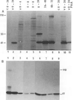
A B
110-
84--78
I,I I, <
U > > u > >
A
10-53-_
41-1 2 3 4 5 6 7 8
FIG. 1. (A) SDS-PAGE analysis of HHV-6(GS) polypeptides recognized by MAb9A5D12.Reactivity of MAb in immunoprecip-itation reactions(lanes 1 to 6). Samples were reduced with 2-mer-captoethanol (lanes1, 2, and 5 to 8) or notreduced (lanes 3 and 4). Lanes 1 and 3, [35S]methionine-labeled uninfected HSB-2 cells. Lanes2 and4, [35S]methionine-labeled HHV-6 strain GS-infected HSB-2cells. Lane 5,32P,-labeledHHV-6(GS)-infected HSB-2 cells. Lane 6,
32Pi-labeled
uninfected HSB-2 cells. Reactivity of MAb with Western-blottedtotal-cellextractsof uninfected(lane 7) and HHV-6-infected (lane8)cells. Sampleswereanalyzedon9% acrylamide cross-linked with DATD, and standard prestained and unstained molecularsize markerswereincluded inparallel lanes. (B) Reactiv-ity of MAb9A5D12with 6(GS)-infected cells in IFA. HHV-6-infected HSB-2cells were collected at 3 days p.i., fixed in acetone, and reacted with MAb 9A5D12 followed by FITC-labeled anti-mouseIgG antibodies.3 days p.i. werelabeled for 20 h with [35S]methionine and used for immunoprecipitation assays. MAb9A5D12 immu-noprecipitated a major HHV-6(GS)-specific polypeptide with anapproximate size of 41 kDa and additional but less intense polypeptides of45, 84, and 110 kDa(Fig.1A, lane 2) (7).Thelessintensepolypeptides werebetter resolved after longer exposure of theautoradiographs(data not shown). No reactivity was seen with uninfected cells (Fig. 1A, lanes 1 and 3). When immunoprecipitated samples were electro-phoresed without reduction by 2-mercaptoethanol, the 41-kDapolypeptideband wasreplaced bya polypeptide of84 kDa, suggestingthat thehigh-molecular-weightform may be adisulfide-linked dimerof the 41-kDapolypeptide(Fig. 1A, lane 4). From cells labeled with
32pi,
a prominent HHV-6-specific phosphorylated polypeptideof 41 kDawas immuno-precipitated (Fig. 1A, lane 5), and no reactivity was seen with uninfected cells (Fig. 1A, lane 6). Several less intense phosphorylated polypeptides of 38, 84, and 110 kDa were alsoimmunoprecipitated. Inphosphoaminoacidanalysisof immunoprecipitated proteins, the 41-kDa polypeptide waspredominantly phosphorylated at serine residues (data not
shown). The 41-kDapolypeptide was also the major phos-phorylatedpolypeptide recognized byhuman sera(8).
In Western blot experiments with HHV-6(GS) proteins, only apolypeptide of41 kDa was recognized by the MAb (Fig. 1A, lane 8). This result demonstrates that the MAb recognized an epitope presents on the 41-kDa polypeptide andfurther suggests that the otherpolypeptidesdetectedby the MAb inimmunoprecipitationreactionsmightresultfrom theirmolecularassociationwith the 41-kDapolypeptide bya
protein-protein interaction. Alternatively, the common
epitope recognized by the MAb in the other
polypeptides
maybesensitivetodenaturing conditions.Apolypeptideof 78 kDa detected in the Western blot reaction from both infected and uninfected cells (Fig. 1, lanes 7 and 8), and
B
z 3 4 i 6 7 8 9 10
12 3 4 i5
110
FIG. 2. (A) Kinetics ofsynthesisofHHV-6proteinP41. Unin-fected HSB-2 cells(C, lanes 1and 9) andHHV-6-infected cells(V, lanes2 to8, 10,and11)labeledwith[35S]methioninewereusedfor immunoprecipitation reactions with MAb 9A5D12. HSB-2 cells were infected with HHV-6 at 10
TCID5dcell,
and after 2 h of absorption, cellswerewashed(timezero)and labeled with [35S]me-thionine between0and4, 4 and8,0and8,0and24,24and48, and 48and72 hp.i. Infected cellswerealso incubated with 300 ,ugof PAA between 0 and 24 h p.i. (lane 11) (B) Reactivity of MAb 9A5D12 with cells infected withdifferentHHV-6 strains. Infected cells labeledwith[35S]methioninewereusedinimmunoprecipitation reactions. Lanes 1 to 9, HHV-6 strains GS, DA, Z-29, OK, DC, Co2, Co3,Co5,andCo6, respectively. HHV-6 strainsGS, DA, Co2, Co3,Co5,and Co6weregrownin HSB-2 cells. HHV-6strainZ-29 wasgrownin Molt-3 cells. HHV-6strains OK andDCweregrown inCBL.absorption ofMAb with uninfected cells didnotremovethe reactivity with thispolypeptide (datanotshown).The
reac-tivity withthe 78-kDapolypeptidewas considered nonspe-cific, since a similar-size protein was also detected by all other MAbs as well as by alkaline phosphatase-conjugated goat anti-mouse IgG second antibodies alone (data not
shown). The MAb did not immunoprecipitate any [3H]glu-cosamine-labeled
polypeptides
(7), and the mobility of the[35S]methionine-labeled
polypeptides immunoprecipitated by the MAbdid notchange aftertreatmentwithglycosidic
enzymes or when extracts from tunicamycin (5 ,ug/ml)-treated cells were used for immunoprecipitation (8). The phosphoprotein recognized byMAb 9A5D12wasdesignated P41.
Kinetics of synthesis ofHHV-6 P41. The kinetics of syn-thesis ofP41 was examined by using HHV-6(GS)-infected cells labeled with[35S]methionine for different timeperiods. Electron microscopic examination of infected cells at 72 h p.i.revealed large
quantities
ofenvelopedvirusparticles
in cytoplasmic vacuoles and in extracellular compartments (data not shown). P41 wasreadily
immunoprecipitated by
theMAbfrom HHV-6(GS)-infected cells labeled between 0 and24hp.i.Ofpotential importance, anadditional
specific
polypeptide
of 110 kDa was also detected with this MAbduring this time
(Fig.
2A, lanes 2 and10),
which wasresolved better afterlongerexposure of the
autoradiograph.
SynthesisofP41 wasundetectable from 0to8h
p.i.
(Fig. 2A,
lanes 3 to5). P41wasfirst detected between 8 and 24h
p.i.,
and the
synthesis
continuedthroughout
the observationVOL.65, 1991
A
41
if
I
4'swil-rl
I.p
191006.0--- NW
on November 10, 2019 by guest
http://jvi.asm.org/
[image:4.612.64.298.77.207.2] [image:4.612.368.508.79.270.2]2888 CHANG AND BALACHANDRAN
period,
to72 hp.i. (Fig. 2A,
lanes 6to8).
Synthesis
of the 110-kDapolypeptide
was also detected first between 8 and 24hp.i.
and continuedthroughout
the observationperiod.
Densitometric
tracings
oftheautoradiographs
andcounting
ofradiolabel in excised
portions
of thegels corresponding
to thedifferentbandsdemonstrated thatsynthesis
ofP41 wasmaximal between 24 and 48 h
p.i.
andsynthesis
of the 110-kDaprotein
was maximal between48 and 72 hp.i. (data
not
shown).
Six timesless110-kDapolypeptide
wasdetected than P41. A hostcell-specific polypeptide
of 53 kDa wasinconsistently
detected from 8to24 hp.i. (Fig. 2A,
lane6).
This
polypeptide
was considerednonspecific,
as it was notdetectedinallextractstakenatsimilartime
points (Fig. 2A,
lanes 2and
10).
PAA hasbeen shown toinhibitthe
replication
ofHHV-6,
presumably by inhibiting
HHV-6 DNAreplication
(2, 14).
To determine the kinetic class ofHHV-6protein
P41,
HHV-6(GS)-infected
cells were labeled with[35S]methionine
be-tween0and 24 h
p.i.
with and withoutPAA(300
,ug/ml).
In the presence ofPAA,
the amounts of radiolabeled P41 and 110-kDapolypeptides immunoprecipitated
weresignificantly
reduced but not abolished
(Fig. 2A,
lane11).
Under theseconditions,
HHV-6glycoproteins
werecompletely
inhibitedby
PAA(data
notshown).
Similarfindings
have beenre-ported previously
forHHV-6(Z-29)
with MAb9A5D12(14).
These data
suggested
that thecontinuedsynthesis
of P41 and the 110-kDapolypeptide
depends
on viral DNAreplication
and these HHV-6
polypeptides probably belong
totheearly-late class ofHHV-6
proteins.
HHV-6(GS) protein
P41 isconserved among HHV-6 strains. Whentestedwith cellsinfected witheight
additionalHHV-6strains,
MAb9A5D12showedgranular
nuclearfluorescenceand
immunoprecipitated
apolypeptide
of41 kDa(Fig. 2B,
lanes2to
9). Polypeptide
bands of45and110kDawerealso detected afterlonger
exposure oftheautoradiographs.
Un-der
nonreducing conditions,
the 41-kDapolypeptide
bandimmunoprecipitated
from the different HHV-6 strains wasalso
replaced
by
an 84-kDapolypeptide
band(data
notshown).
These results demonstrate that HHV-6 P41 iscon-servedamong the HHV-6 strains tested. MAb9A5D12 did not cross-reactwith cells infectedwith HSV-1
(strain KOS),
HSV-2
(strain 333),
CMV(AD169),
orEBV(B95-8)
in IFAand
ELISA, demonstrating
that the MAb isspecific
forHHV-6.
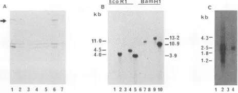
Identification ofacDNA
encoding
the HHV-6phosphopro-tein P41. To
identify
the geneencoding
HHV-6protein P41,
a cDNA
library
ofHHV-6(GS)-infected
cells wascon-structed in the
Agtll
expression
system and screened with MAb 9A5D12. Oneimmunoreactiverecombinantphage
pro-ducing
a,-galactosidase
fusionprotein
wasidentified.Aga-rose
gel electrophoresis
of the insert excised fromXgtll
with EcoRIrevealed acDNA insertof2.1 kb. The cDNAinsert subcloned intopUC13
wasdesignated pCD41.
InaWestern blot assay, MAb 9A5D12recognized
severalpolypeptides
fromthe
lysates
ofinsert-bearing
phage ranging from130to 36 kDa(data
notshown). Rapid proteolysis
of the P-galac-tosidase fusionprotein
may account for the generation of smallerpolypeptides.
Sincethishamperedtheestimation of thesizeoftheprotein
codedby
the cDNAinsert,thepCD41 insert was cloned in theprokaryotic
expression vectorpGEMEX-1
and transformed into E. coli JM109(DE3).Recombinant
protein
was inducedby
theaddition ofIPTG for 4h,
and the Western-blotted bacterial lysates were reacted with MAb 9A5D12. Several proteins were recog-nizedby
the MAbfromtwoof thelysates
withinserts,
and amongthese,
afusionprotein
of 88to92kDawasprominentEco Rl Ba niH 1
kLb k1)
11.0- *0 -13-2
it-10.9 4-5- .
4.0- 0 * -3.9
4.3- 2.5- 1.8-
1.2-1 2 3 4 56 7 8910
FIG. 3. Characterization of thecDNAinsertpCD41identifiedby the MAb 9A5D12fromacDNAlibrary ofHHV-6(GS)-infectedcells constructed in the Xgtll expression system. (A) Western blot analysisof fusionprotein expressed from thecDNAinsert.Bacterial coloniescontaining the pCD41 insert (lanes 1 to6) orthe vector alone(lane 7) in the pGEMEX-1 expression systemwere induced with IPTG for4h.Bacteriallysates obtainedwereelectrophoresed on 12% SDS-PAGE, transferred to nitrocellulose sheets, and re-actedwith MAb 9A5D12. The arrow shows amajor 88-to92-kDa fusion protein band detected in lanes 1 and 6. (B) Southern blot hybridization of pCD41 insert with HHV-6 DNAand uninfected-cell DNAdigested with EcoRI(lanes 1to5) and BamHI (lanes 6to10). Lanes 1 and6,DNAfrom uninfected HSB-2 cells. Lanes 3 and8, DNA from uninfected Molt-3 cells. Lanes 2 and 7, HHV-6(GS) DNAfrom infected HSB-2 cells.Lanes 4and9,HHV-6(Z29)DNA from infected Molt-3 cells.Lanes 5and10,HHV-6(GS)DNAfrom purified virions. The sizes (in kilobases) ofHHV-6-specific DNA fragments hybridizedwiththe32P-labeled 2.1-kb pCD41 insertare indicated. Standard lambda marker DNAs of known sizes were included in parallel lanes. (C) RNA transcripts identified by the cDNAinsert. Uninfected and HHV-6(GS and Z29 strains)-infected-cell total RNAs were subjected to electrophoresis in a formalde-hyde-agarose gel, transferredtoGeneScreen Plus, andprobed with the32P-labeled 2.1-kb pCD41 insert. Each lanewasloadedwith10 ,ugofRNA. Lane 1, uninfected HSB-2 cellRNA. Lane2, HHV-6(GS)-infected HSB-2 cell RNA. Lane 3, uninfected Molt-3 cell RNA.Lane4,HHV-6(Z29)-infected Molt-3 cellRNA.The sizes(in kilobases) of HHV-6-specificRNAsthat hybridized with the 2.1-kb pCD41 insertareindicated.
(Fig. 3A, lanes1and6). However, Coomassie staining of the gel did not reveal any specific protein. No reactivity was seenwithotherinsert-containing (Fig. 3A, lanes2to 5)and non-insert-containing (Fig. 3A, lane 7) bacterial lysates. Nonreactive clones were later found to contain the insert in the wrong orientation. Since the amino terminus of the fusionproteincontainedthe gene 10 protein of T7 approxi-mately 26 kDa, this suggested that the pCD41 insert codes foraprotein of 62to66kDa.
SouthernblotanalysiswithcDNA insertpCD41. To deter-mine the viralspecificity of the cDNA insert and to map the coding geneonthe viralgenome,[oa-32P]dCTP-labeled 2.1-kb pCD41wasexaminedby Southern blot analysis. The pCD41 insert hybridized specifically with the EcoRI- and BamHI-digested DNA fragments from HHV-6 virions (Fig. 3B,lanes 5 and10)andfrom HHV-6-infected cells (Fig.3B,lanes2,4, 7, and 9). No hybridization was seen with DNA from uninfectedcells (Fig. 3B, lanes 1, 3, 6, and 8) or with any of the EcoRI-digested viral DNA fragments from HSV-1, HSV-2, CMV, and EBV (data not shown). Specific hybrid-ization was seen with 3.9-kb EcoRI (Fig. 3B, lane 5) and 10.9-kb BamHI (Fig. 3B, lane 10) DNA fragments from purified virions of HHV-6(GS) and with 4.0-kb EcoRI (Fig. 3B, lane 2) and 11.0-kb BamHI (Fig. 3B, lane 7) DNA fragmentsfromHHV-6(GS)-infected cells. Hybridization to slightly larger sizes of viral DNA fragments from infected cells couldbe due totheretardation of viral DNA mobility by host cellDNA. Nevertheless, these results clearly dem-J. VIROL.
on November 10, 2019 by guest
http://jvi.asm.org/
[image:5.612.324.556.74.165.2]HHV-6 PHOSPHOPROTEIN GENE
1
_TTTTTTTT ITAAAGCACATAAGGCGCGAGTTGGTGCTAGGACTTCGTTCTTGACAGAG
F F F F K A H K A R V G A R T S F L T E 20 63
ATGGAGCGCGGTAGTCGAGATCATCATCGTGATCACCGTGATCATCGGGAACATAGAGAA
M E R G S R D H H R D H R D H R E H R E 40 123
T R E P P T L A F H N K S W K T I N K 8 183
CTTAAAGCGTTTGCAAAACTGTTAAAAGAGAATGCTACAGTGACTTTCACGCCGCAGCCG
60 L K A F A K L L K E N A T V T F T P Q P 80 243
TCGATAATAATTCAGTCTGCTAAAAATCATCTCGTGCAAAAGCTGACTATTCAGGCGGAA
S I I I Q S A K N H L V Q K L T I Q A E 303
TGTTTGTTTTTGTCAGATACGGATCGTTTTTTAACGAAAACCATTAATAATCATATTCCA
C L F L S D T D R F L T K T I N N H I P 363
L F E S F M N I I S N P E V T K N Y I Q
423
CATGATAGTGATCTATATACGAGGGTT TGGTAACGGCTTCCGATACATGTACACAGGCG
H D S D L Y T R V L V T A S D T £ T Q A 483
TCGGTTCCCTGTGTGCACGGACAAGAAGTGGTGCGAGACACCGGGAGATCGCCGTTGAGG
S V P C V H G Q E V V R D T G R S P L R 543
ATTGACCTTGATCATTCGACCGTTTCCGATGTGTTGAAATGGCTTTCACCTGTAACTAAG
I D L D H S T V S D V L K W L S P V T K 603
ACTAAACGCTCTGGTAAATCTGACGCTTTAATGGCGCACATTATAGTACAGGTTAATCCT
T K R S G K S D A L N A H I I V Q V N P
663 ******* /ASUII *
CCGACTATAAAATTCGTGACAGAGATGAATGAACTGGAGTTTTCGAACAGCAATAAGGTT
P T I K F V T E M N E L E F S N S N K V
ATATTTTACGATGTTAAGAACATGCGGTTTAATCTATCTGCCAAAAATTTACAGCAGGCT
I F Y D V K N N R F N L 8 A K N L Q Q A 783
TTAAGTATGTGTGCTGTAATCAAGACGTCGTGTAGTTTACGTACGGTAGCCGCTAAGGAC
L S N C A V I K T S Q S L R T V A A K D 843
TGTAAATTGATATTAACTTCCAAAAGCACGTTGTTGACGGTGGAAGCCTTTTTAACTCAG
C K L I L T S K S T L L T V E A F L T Q
903
GAACAGCTCAAAGAGGAATCTCGCTTTGAACGAATGGGTAAACAAGATGACGGAAAAGGG
100 120 140 160 180 200 220 240 260 280 300 E Q L K E E S R F E R N G K Q D D G K G 320 963
GATAGGAGTCATAAGAACGAAGACGGAAGTGCGTTGGCATCAAAACAGGAAATGCAATAT
D R S H K N E D G S A L A S K Q E N Q Y 340 1023
GAAATAACCAACTACATGGTCCCCGCTAAAAACGGAACTGCGGGCTCTAGCTTATTTAAC
E I T N Y N V P A K N G T A G S S L F N
1083 /NaiI\
GAGAAGGAAGATAGCGAAAGTGACGATTCCATGCATTTTGACTACAGCTCTAATCCCAAT
E K E D S E S D D S M H F D Y S S N P N 1143
CCCGAGGCAGAGATGCGTCGTCTGACAGATAGMTTATTCTCGGTCTTGCJTAAGGGTGCC
360 380 P E A E M R R L T D S F I L G L A K G A 400 1203
GTTATTCCGGGGTTGTATACGTTTAGAATGACAGAGGGGAGGTCCCCCCTCGGACAGATC
V I P G L Y T F R N T E G R S P L G Q I 420 1263
GGCGTTTTGATAACCGTTGCGATTTCCTTTGTTGACTTTTAAAAGATTTGATCCACGC
G V L I T V A I S F L L T F K R F D P R 1323
TTTTATAAACCCATAGGTGATTTCAAGATTGTGTTTCTGTCTTTAATGGCGCCGAAACTG
F Y K P I G D F K I V F L S L M A P K L
1383 /PstI\
CCATCATTATTGTCTGCAGTGGTCATGATCTGCTTGATATTTTCCGAGATGAGGTTGAGA
P S L L S A V V M I C L I F S E M R L R 1443
ATGATTTTAAGTCGTTGTGTCATGATTATGCCGTCTTATTCCCCAGCTGTGTTTACGGGA
M I L S R £ V M I M P S Y S P A V F T G 1503
ATCATGGTGTCTTTGTTTTTTAAGAGTCAAATGTTCGATGATTATTCTGTCTTGATAACG
I M V S L F F K S Q M F D D Y S V L I T 1563
GCTGCGTCTCTCCTGCCGATTACTGTGAGATATGGATGGATGATACGATCGTCTGGATTC
A A S L L P I T V R Y G W M I R S S G F 1623
CTTCTTGGTTTGCAAAAATATCGTCCGATTTTGAAGTCAACGTCGTTTCGTGAAGTTGAT
L L G L Q K Y R P I L K S T S F R E V D 1683
TTGAAATGCCTTGTAAAGTTTACGGTCGAATTTTTGTTATTATTTACGATACTCTGGATC
L K Q L V K F T V E F L L L F T I L W I 1743
GGAAAAATGTTCTTGAGTATGCCAAAATCTAACCATCTTTTTTTCCTGACTGTGGTCAAC
G K N F L S M P K S N H L F F L T V V N 1803
AACGTATTTTTTAAACTAAACGTATTCAAAGCGCTGCGTGCGCGGTGGTGGCGATCTTAT
440 460 480 500 520 540 560 580 600 N V F F K L N V F K A L R A R W W R S Y 620 1863
R D L #
1923
GGTTTAGTTCGATTATGTTGAATTTAAGCAGCGATCTGAAAGACAGATCTTTCTATGCGG
1983
GTGATTATTGAACGGTTTTGTAGTCGTGTGTTGCATGTATTTTGGTGTGTGAT
2043
TAGACAAlTTATCGTGCCTGTTTTGGTGAACACCMTAAAATTTTTTGCAGTATTAAAAA
623
[image:6.612.52.556.84.397.2]AAAAAAAAAA
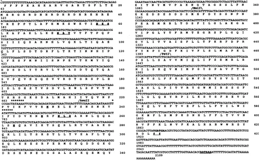
FIG. 4. Nucleotidesequenceof pCD41 and the deduced aminoacidsequenceofthesingle ORF. Thenucleotideposition is numberedon
the left, and the amino acid position is numbered on the right. The termination codons and the polyadenylation signal sequences are
underlined. Asteriskedsequencesindicate possibleTATAboxes, and theunderlined methioninesare the presumedstart sites for in vitro translation. Restriction sites forAsuII,NsiI, and PstI are marked onthenucleotide sequence. The three underlined sequencesindicate potential sites for N-linked glycosylation (NXT/S), and cystine residuesareindicated byunderlining.
onstratetheHHV-6specificity of the cDNA insert identified by MAb 9A5D12. The pCD41 insert also hybridized with 4.5-kb EcoRI (Fig. 3B, lane 4) and 13.2-kb BamHI (Fig. 3B, lane 9) DNA fragments from HHV-6(Z29)-infected Molt-3 cells. Hybridization with different sizes of DNA fragments fromthetwostrains of HHV-6 furthersupportsthe previous reports of restriction site heterogeneity among HHV-6
strains (1, 4, 24, 25, 34).
Northern blotanalysiswith cDNA insert pCD41. To define further the viral specificity of pCD41, equal quantities of totalunfractionatedRNAisolated from HHV-6(GS)-infected and HHV-6(Z29)-infected and uninfected cells were tested
by Northern blotanalysis with a 32P-labeled 2.1-kb pCD41
insert. Hybridizationwas detected with RNA from infected
cells only (Fig. 3C, lanes2 and4), and the sizes of HHV-6 transcriptswerecalculated by comparingtheirmobility with RNA marker molecules (Bethesda Research Laboratories, Gaithersburg, Md.). Two prominent transcripts with esti-mated sizesof 2.5 and1.2 kbwereidentified by pCD41 from
HHV-6 strainGS(Fig. 3C,lane2) and fromstrain Z29(Fig. 3C, lane4). Twoless intense bands of1.8 and 4.3 kbwere
also detected (Fig. 3C, lanes 2 and4). The intensity of the 2.5-kbtranscriptwasabout3.3timesthatof the1.2-kb band.
Even though equal amounts oftotal cellular RNA (10 ,ug)
wereloadedpersample well, thehybridization intensitywas
stronger with HHV-6(GS) RNA than with HHV-6(Z29) RNA. Nevertheless, these data further confirm the
speci-ficity of the pCD41 insert and demonstrate that P41 is conserved between thesetwo HHV-6strains.
DNA sequence analysis of cDNA insert pCD41. DNA se-quenceanalysis revealed that the cDNA insert consisted of
2,109 bp and the sequence hasa meancomposition of 41%
G:C. The sequencewasfurtherverifiedbyrestriction diges-tionanalysiswith thepredictedrestrictionenzymesites. The complete sequence and the predicted amino acid residues
are shown in Fig. 4. Computer analysis revealed a single openreadingframe(ORF)of1,871 nucleotides, beginningat nucleotide 3atthe 5' end,and thestopcondons(TGA TGA)
arelocatedatnucleotidepositions1872to1877atthe 3' end (Fig. 4).The238nucleotides atthe 3' endare untranslated.
There is one potential polyadenylation signal (AATAAA), locatedatnucleotidepositionsbetween2077 and 2082atthe 3' end. The predicted proteinis 623 aminoacid residues in length,with acalculated molecularweightof70,863,and is highly basic, with an estimated pl of 10.99. Hydropathic
analysis of the pCD41 ORF indicates a major stretch of
hydrophobic domains clustered at the carboxy region be-tween amino acids (aa) 402 and 602 (Fig. 5). At least six regionsin the carboxy terminus of theORF showa
hydro-phobic score ofgreater than +1.6. Since sequenceswith a
high probability to be membrane-spanning usually have a
hydrophobic score greater than +1.6 (28), this suggests membrane association of the predicted pCD41 protein. There are nine cysteine and 25 methionine residues in the
VOL.65, 1991 2889
on November 10, 2019 by guest
http://jvi.asm.org/
2890 CHANG AND BALACHANDRAN
B C NMS MA[b9A5Dl2 RaHHV6 D
.4..
,-@
+1-}
11'' '
'1
'
-20
3 l
-4
FIG. 5. Hyd quenceof pCD4 forhydrophobic and Doolittle
all
windowof nine score inwhich arehydrophilic. spanning havea thepCD41sequ NsiI, and PstI represents the 9A5D12, spanni
predicted pro clustered betv potential sites all of which a
the predicted
In vitrn tran
I 1
i.I
I
II9I
hh
45
41 30
1 2 3 4 5 6 7 8 9 1 2 3 4
IgI FIG. 6. Invitrotranscriptionand translation of thecDNAinsert. Sense and antisense transcripts prepared in vitro from the cDNA A A A insertpCD41weretranslated in vitro with rabbitreticulocyte lysates
Asull Nsil Psti and [35S]methionine. Samples were analyzed on 12% SDS-PAGE
I and by RIP followed by SDS-PAGE. (A) In vitro translation of
102 202 302 402 502 602 transcripts from the pCD41 insert cloned in the pCDNAII vector. Lane1, translation withouttheaddition ofRNA. Lane2, antisense Amino Acid RNA transcribed from the Sp6 promoter. Lane 3, sense RNA ropathic analysis of the deduced amino acid se- transcribed from the T7 promoter. (B) In vitro translation of t1. The predicted amino acidsequence wasanalyzed transcripts from the pCD41 insert cloned in the pGEMEX-1 vector.
c andhydrophiliccharacteristicsbyusing the Kyte Lane 1,
translation
without the addition of RNA. Lane 2, sense gorithm (28). Scoreswereaveragedwithinasliding RNA transcribed from the T3 promoter. (C) RIP of invitro-goriThm
(28).tScores wepresentsaerelagvedwidrathin
atranslated
products. Transcripts
from thepCD41
insertclonedintheThe vertical axisrepresentsarelativehydropathic pCDNAII vector were translated in vitro and used for
immunopre-positive valueare hydrophobicandnegative values iiainwtnomlouesrmNM,aes1o3)MA
.Sequenceswith ahighprobability to bemembrane- cipitation with normal mouse serum (NMS, lanes 1 to 3), MAb scoregreaterthan
+h1.6.
Theaminoacidnumberof9A5D12
(lanes 4 to 6), and rabbit polyclonal antibody (RaHHV-6)iencre
ate locationofrestrictionsites for AsuII against HHV-6(GS)-infected cells (lanes 7 to 9). Lanes 1, 4, and 7,are given. The solid line between AsuII and NsiI translation without RNA. Lanes 2, 5, and 8, translation and an-putative antigenic site recognized by the MAb tisense RNA transcribed from theSp6promoter. Lanes 3, 6, and 9, ngahighlyhydrophilic regionoftheprotein, translation with sense RNA transcribed from the
T7
promoter. (D) Product madeby thepCD41sensetranscript is the HHV-6protein P41 immunoprecipitated from HHV-6(GS)-infected HSB-2 cells. ThesenseRNAtranscribed in vitro from thecDNAinpGEMEX-1 vector wastranslatedinvitro,andtheproductswereused forRIP tein, and 20 of the methionine residues are with MAb 9A5D12. The [35S]methionine-labeled 41-kDa in vitro-veen aa 229 and 623 (Fig. 4). There arethree translated polypeptide (lanes3 and4)and the P41 immunoprecipi-fortheaddition of N-linkedoligosaccharides,
tated fromHHV-6(GS)-infected
cells(lanes
1and2)wereseparated
Lre associated with the amino-terminal part of by SDS-PAGE, subjectedtopartialproteolysiswith 10 ,ug(lanes 1 protein within aaposition253. (Fig. 4). and 3) and 2 pLg (lanes 2 and 4) of V8 protease, and analyzedon15% scrintinnandtrnanRntionof enNAinsert
nNnul
SDS-PAGE. Sizesareshown in kilodaltons.Al Vlt'UtXF&aVI"RAUwsW^sLAs"Alsat"MPIR%PI w1-P1 ssss111V L
F%"-result in thesynthesis of a 41-kDa polypeptide. Thepredicted size of the protein encoded by the pCD41 ORF is much larger than that (41 kDa) of the protein recognized by the MAb from the infected cell. Detection of a large fusion protein (Fig. 3)indicatedthat theentirelengthof the cDNA was transcribed in the prokaryotic expression vector pGEMEX-1. Reactivity of the MAb with thefusionprotein expressed in both Xgtll and pGEMEX-1 could be due to a cross-reactive determinant in theprotein codedby pCD41. To determinetherelationship between HHV-6 P41 and the protein coded by pCD41, the cDNA insert was cloned in pcDNAII and pGEMEX-1 vectors. Sense and antisense transcriptswere synthesized by using SP6 and T7 promoters
(pcDNAII)
and sense RNA by using the T3 promoter (pGEMEX-1). Theresulting RNAtranscripts were capped, translated in vitro, and then analyzed by SDS-PAGE. No specific polypeptides were synthesized from RNA tran-scribed from the SP6 promoter (antisense RNA; Fig. 6A, lane 2) orfrom a control in vitro translation without RNA (Fig. 6A, lane 1, and 6B, lane 1), and no large polypeptide predicted to be coded by the pCD41 insert was translated. Instead,aprominent[35S]methionine-labeled
polypeptide of 41 kDa was synthesized from the sense RNA transcribed fromthe T7 promoter (Fig. 6A, lane 3) as well as from the T3 promoter(Fig. 6B,lane 2).To determine the specificity of the polypeptide synthe-sized, the invitro-translatedproducts were immunoprecipi-tated with MAb
9A5D12
and rabbit polyclonal antibodiesagainst HHV-6(GS)-infected cells. Both these antibodies immunoprecipitatedapolypeptide of41kDa translated from the sense RNA (T7 promoter). Longer exposure of the autoradiograph revealed severalless-intense lower-molecu-lar-weight polypeptides, including a 30-kDa polypeptide (Fig. 6C, lanes 5 and 8). No reactivities were seen with translated products from antisense RNA or control RNA (Fig. 6C,lanes3, 6,7, and 9). Normal mouse serum(Fig. 6C, lanes 1 to3) and normal rabbitserum(datanot shown)did notshow anyreactivity. V8 proteasewasusedtodefinethe partial proteolytic peptide mapping ofthe in vitro-synthe-sized 41-kDa
polypeptide
(pGEMEX-1 vectortranscript)
and the HHV-6 P41 from infected cells. Bothpolypeptides showed identicalpeptide profiles (Fig. 6D,lanes1 to4),and similarprofileswerealso seenwith the 41-kDapolypeptide translated from the sense transcript from the pcDNAII vector(datanotshown).These resultsclearly demonstrated that thepCD41 insertpredictedtocode foralargerprotein also encoded the completeHHV-6phosphoproteinP41.
Reactivity of MAb 9A5D12 withfusion proteinsexpressed from deletionconstructsof the cDNA insert.TheSDS-PAGE mobilityof the 41-kDapolypeptide remained unalteredwhen inhibitors ofproteolytic enzymes were included in the in vitro translation reaction mixes (data not shown). This suggested that the 41-kDa polypeptide was notderived by proteolytic cleavage(data notshown). We nextconsidered
J. VIROL.
on November 10, 2019 by guest
http://jvi.asm.org/
[image:7.612.66.301.72.252.2] [image:7.612.324.560.78.175.2]HHV-6 PHOSPHOPROTEIN GENE 2891
the possibility of internal initiation of translation of the transcript leading to the in vitro synthesis of the 41-kDa polypeptide. Careful analysis of pCD41 nucleotide se-quences revealed two TATA boxes, TATAAAA and TATATTT, located at nucleotide positions 668 to 674 (aa 223 to 225) and 722 to 728 (aa 241 to 243), respectively (Fig. 4). In context to these regions, there are three ATG codons, beginning at nucleotide positions 688, 784, and 789, respec-tively (aa positions 227, 248, and 263, respecrespec-tively; Fig. 4). Translation initiation at any of these three codons could result in the synthesis of polypeptides of 45, 42, and 41 kDa, respectively. According to this model, the antigenic epitope recognized by the MAb is predicted to be located after aa 227.
To test this possibility, the cDNA insert in thepGEMEX-1 expression vector was digested with restriction enzymes AsuII, NsiI,andPstI.The restriction sites for these enzymes are located at nucleotide positions 704, 1102, and 1395, respectively, from the 5' end of the insert (Fig. 4). The resulting 234-aa, 371-aa, and 466-aa ORFs are predicted to code for 30-, 42-, and 52-kDa polypeptides, and the deletion constructs were named pCD41A, pCD41B, and pCD41C, respectively. The three enzymes were selected for the fol-lowing reasons. (i) These are the only enzymeswith only one restriction site in the insert, with no other restriction sites either in the polylinker region or in the vector. This kept the 5' end of the fusion protein intact. (ii) The AsuII site is located between the two putative TATA boxes, and a fusion protein induced from this construct would contain only the first 234 amino acids from the amino terminus of the pCD41 ORF. (iii) The amino acids encoded by nucleotides between theAsuIIandNsiIrestriction sites are highly hydrophilic (aa 234 to aa 371, Fig. 5) and contain the two ATG codons at aa positions 248 and 263. Translation initiation at any of these two codons should result in polypeptides of 42 and 41 kDa, which is closer to the observed size of 41 kDa immunopre-cipitated by the MAb.
Fusion proteins from the deletion constructs were induced with 0.5 mM IPTGfor 4 h, and equal quantities of bacterial lysates (10,ug)were analyzed by SDS-PAGE and by West-ern blot. No fusion protein was detected by Coomassie staining or by Western blot of bacterial lysates with the non-insert-bearing control vector (Fig. 7A, lane 1; Fig. 7B, lane 1). Bacterial lysates with thepCD41 insert ligated in the wrong orientation also did not show any reactivity (data not shown). From bacterial lysates with the full-length cDNA insert (2.1 kb, 623 aa), a major fusion protein of >84 kDa together with several minor more rapidly migrating bands were recognized by MAb 9A5D12 (Fig. 7B, lane 2). In contrast, no specific protein was detected in the stained gel (Fig. 7A, lane 2). A prominent specific band of 63 kDa was detected from pCD41A insert-containing bacterial lysate by Coomassie staining (Fig. 7A, lane 3). However, MAb 9A5D12did not react with thisfusion protein in theWestern blot (Fig. 7B, lane 3). In contrast, afusion protein of 74 kDa and several minor bands were recognized by the MAb from bacterial lysates with the pCD41B insert (371 aa of pCD41; Fig. 7B, lane 4), and only faintly stained similar molecular weight bands were observed from the same lysates (Fig. 7A, lane 4). MAb 9A5D12 reacted very strongly with a fusion protein of 78 kDa and several other fastermigrating bands from bacterial lysates with the pCD41C insert (466 aa of pCD41; Fig. 7B, lane 5). Similar proteins were also observed from the Coomassie-stained gel (Fig. 7A, lane 5). These results demonstrate that the first 377 aa in the pCD41 ORF most probably contain the antigenic site recognized by the
A
COOMASSIE STAIN
-78
.-NN_* - -63
WVESTERN BLOT
84-2 3 4
FIG. 7. Reactivity of MAb 9A5D12 with the fusion proteins expressed from cDNA deletion constructs. Deletions in the cDNA insertwere generated by restriction enzymes AsuII, NsiI, and PstI, and the resulting constructs inthepGEMEX-1 vector were named pCD41A, pCD41B, and pCD41C. Bacterial coloniescontaining each of the deletion constructs were induced with IPTG for 4 h, and proteins were extracted and analyzed on 12% SDS-PAGE. Gels were either stained with Coomassie stain (A) ortransferred onto a nitrocellulose sheet and reacted with MAb 9A5D12 (B). Lanes Al and Bi, pGEMEX-1 vector alone. Lanes A2 and B2, pCD41 containing the full-length insert.LanesA3 andB3, pCD41Adeletion construct. LanesA4andB4, pCD41B deletion construct. Lanes A5 and B5, pCD41C deletion construct. The approximate sizes of fusion proteins are indicated in kilodaltons.
MAb and suggest that thesite isprobably locatedbetweenaa positions 235 and 371, which include the two potential start sites (aa 248and 263). This also supported the possibility that internal initiation of translation resulted in the in vitro synthesis ofthe 41-kDa polypeptide.
Predicted ORF ofHHV-6 pCD41 insert shows amino acid homology with the HCMV UL44 gene product. Using the FASTA program of Lipman and Pearson (32), we compared the amino acid sequences ofthe predictedpCD41 ORF with the protein data base libraries and a protein library of all available herpesvirus sequences. The only significant match detected was with the predicted protein product of HCMV geneUL44. Thereis41.8% identity inastretch of 282 amino acids (Fig. 8), with a FASTA score (ktup = 1) of 623. The predicted HCMV UL44 gene product consists of 433 aa, with a predicted molecular weight of 46,234 and anestimated plof 9.9. The HCMV UL44 gene has been shown to encode a group of early-late DNA-binding nonstructural proteins that include phosphorylated and glycosylated species and arereferred to as theICP36protein family of HCMV (11, 12, 30). The homology detected occurred only in the amino-terminal region of the pCD41 ORF, between aapositions 48 and 320. Fourofthe ninecystine residues in the pCD41 ORF are conserved in the HCMV UL44 gene ORF (Fig.
8).
DISCUSSION
In this report, we have characterized in detail a phos-phoprotein encoded in the HHV-6genome of 41 kDa which is specifically recognized by MAb 9A5D12. HHV-6 P41 is one of the most abundant proteins produced early in the replicative cycle. Since PAA reduced the quantity of P41 synthesized, it is tentatively assigned to the early-late class of HHV-6 proteins.
A variety of HHV-6 strains have been isolated from different geographicallocations as well as frompatients with various pathological conditions. These HHV-6 strains show
VOL. 65,1991
5
'4.n.,...-.';;
-78
h-ff.:
..
wgm6mmw .WMWW
IW
I
rl;
on November 10, 2019 by guest
http://jvi.asm.org/
[image:8.612.323.562.82.218.2]2892 CHANG AND BALACHANDRAN
pCD41AA
FFFKAHKARVGARTSFLTEMERGSRDHHRDHRDHREHRETREPPTLAFHMKSWKTINKS
HCMVUL44
:M:D:R:T:R:.L.S.:E.P.P:T
: 2MDRKTRLSEPPT>LARLECPYKTAIQQ 26
LKAFAKLLKENATVTFTPQPSIIIQSAKNHLVQKLTIQAECLFLSDTDRFLTKTINNHIP 120
LRSVIRALKENTTVTFLPTPSLILQTVRSHCVSKITFNSSCLYITD-KSFQPKTINNSTP 86
LFESFMNIISNPEVTKMYIQHDSDLYTRVLVTASDTCTQASVPCVHGQEVVRDTGRSPLR 180
LLGNFMYLTSSKDLTKFYVQDISDLSAKISMCAPDFNMEFSSACVHGQDIVRESENSAVH 146
IDLDHSTVSDVLKWLSPVTKTKRSGKSDA--LMAHIIVQVNPPTIKFV-TEMNELEFSN 236
VDLDFGVVADLLKWIGPHTRVKRNVKKAPCPTGTVQILVHAGPPAIKFILTNGSELEFTA 206
SNKVIFYDVNMRFNLSAKNLQQALSMCAVIKTSCSLRTVAAKDCKLILTSKSTLLTVEA 296
NNRVSFHGVKNMRINVQLKNFYQTLLNCAVTKLPCTLRIVTEHDTLLYVASRNGLFAVEN 266
FLTQEQLKEESRFERMGKQDDGKGDRSHKNEDGSALASKQEMQYEITNYMVPAKNGTAGS 356
FLTEEPFQRGDPFDKNYVGNSGKSRGGGGGGGSLSSLANAGGLHDDGPGLDNDLMNEPMG 326
SLFNEKEDSESDDSMHFDYSSNPNPEAEMRRLTDSFILGLAKGAVIPGLYTFRMTEGRSP 416
LGGLGGGGGGGGKKHDRGGGGGSGTRKMSSGGGGGDHDHGLSSKEKYEQHKITSYLTSKG 386
LGQIGVLITVAISFLLTFKRFDPRFYKPIGDFKIVFLSLMAPKLPSLLSAVVMICLIFSE 476
GSGGGGGGGGGGLDRNSGNYFNDAKEESDSEDSVTFEFVPNTKKQKCG 434
SSGFLLGLQKYRPILKSTSFREVDLKCLVKFTVEFLLLFTILWIGKMFLSMPKSNHLFFL
TVVNNVFFKLNVFKALRARWWRSYRDL 623
FIG. 8. Aminoacid homologybetween the HHV-6 pCD41 ORF
(pCD41AA) and the HCMV UL44 gene product. Amino acid comparison (ktup= 1)wasperformedby using the FASTAprogram
ofLipman and Pearson (32). Identical amino acidsare marked by
twodots,and conservativechanges are markedby singledot. The
positionsof four conserved cystine residuesare indicatedby
bold-facing andunderlining.
DNArestriction site heterogeneity (1, 24, 25,34) and differ considerablyintheirin vitrotropism (1, 3, 4, 15,34, 54)and antigenicproperties (1, 54).Ourpreliminary studiessuggest
that these viruses are antigenically closely related and yet
different. Among the many MAbs tested, MAb 9A5D12 is
oneofthefewthatreacts with allHHV-6 strains tested and hasbeenofconsiderablevalue in monitoring in vitroHHV-6
infection (1, 14, 54). MAb 9A5D12 also showed similar reactivity in our recent testing with 12 additional HHV-6
strains(datanotshown). ThisMAb is thereforeapotentially
ideal diagnostic reagent for the identification of newer
HHV-6 isolates and in the detection of HHV-6 antigen in clinical specimens. Wehave recently used this antibody for thedetectionof HHV-6viral antigens in salivaryglands and in bronchial glands from healthy individuals and patients withvariousdisorders.In these studies, antigen detectionby MAb 9A5D12 was found to be more sensitive than in situ hybridization assays (27). Since human sera also reacted
with a 41-kDa polypeptide of HHV-6(GS) (7, 10), HHV-6
protein P41 may also be useful in the serological
measure-mentofHHV-6 infection.
Sequence analysis of the cDNA insertpCD41 predicteda product ofabout70 kDa, and the MAb recognized afusion protein of about 88 to 92 kDa which included the 26-kDa
portionoftheT7gene10leader protein.The differencefrom the predicted 96-kDa fusion protein may be due to the
migration anomaliesof the protein in SDS-PAGE.
Alterna-tively, this could be due to rapid proteolysis of the fusion
protein, which is suggested by the detection of several
smallerpolypeptides which reactwith the MAb.
Thestabilityofafusion protein isdependentuponthe host
bacterial strain, the expression vector system, and the
incubation conditions. In theXgtll expression system,
pro-teolysis of the
P-galactosidase
fusion protein containing the pCD41 insert appeared to be very rapid. Expression of the fusion protein in the pGEMEX-1 system also appeared to depend on the aminoacid sequencesin the pCD41 ORF.The highlyhydrophobic carboxyterminus of the ORF appears to affect the stability of the fusion protein, as the full-length fusion protein was detected only in the Western blot. Re-moval of part of the carboxy terminus appears to stabilize the fusion protein from the pCD41C insert. Even in this deletion construct, detection of several smaller bandsin the Western blot suggests rapid proteolysis, which increased considerably after 24 h of induction with IPTG (data not shown).Several features of the cDNA identified and characterized in this study merit additional comment. Even though the cDNA is predicted to encode a large protein, evidence presented here indicates that the cDNA insert also encodes the HHV-6 phosphoprotein P41. Localization of MAb reac-tivity to a region carboxy terminal to aa 235 of the pCD41 ORF, together with the absence of other reading frames in the cDNA sequence, absence of high-molecular-weight poly-peptides in the in vitro translation, in vitro translation of a 41-kDa polypeptide from the sense transcript of the cDNA insert, immunoprecipitation of this polypeptide by the MAb, peptide map identical to that of HHV-6 P41, stop and polyadenylation signals at the 3' end of the insert, and the presence of two TATA boxes in the cDNA sequence, strongly suggest that HHV-6 P41 is encoded in a gene with the coding potential for a larger protein.
One of the predominant transcripts detected in the North-ern blot analysis has a calculated size of 1.2 kb, which is sufficient to encode the 41-kDa peptide. Another major 2.5-kb RNA was also detected in Northern blot analysis with the cDNA insert. Since the predicted ORF of the 2.1-kb cDNA is open at the 5' end, the 2.5-kb RNA probably represents the full-length transcript. In addition to the 623 aa predicted from the cDNA insert, the 2.5-kb RNA could code for an additional 100 or more amino acids at the amino terminus. Besides HHV-6 P41, the MAb also immunopre-cipitated a 110-kDa polypeptide in lower quantities from HHV-6-infected cells, with similar kinetics of synthesis (Fig. 2). In preliminary studies, translation of an HHV-6(GS) RNA hybrid selected by pCD41 resulted in the synthesis of a 41-kDa polypeptide and a high-molecular-mass (>80 kDa) polypeptide. Preliminary studies also show partial identity between the 110-kDa and 41-kDa polypeptides. These ob-servations suggest that the 110-kDa polypeptide may be encoded by the 2.5-kb transcript. With a single potential polyadenylation site at the 3' end, it is probable that the 2.5-kb and 1.2-kb RNAs have a common 3' end. Even though the 2.5-kb message is 3.3 times more abundant than the 1.2-kb RNA, the 41-kDa polypeptide was more abun-dantly detected than the 110-kDa polypeptide. The reason for this discrepancy is not known at present; it could be due to the stability of the 110-kDa polypeptide synthesized and/or to the nonaccessibility of the epitope in the 110-kDa polypeptide in immunoprecipitation reactions. Nonreactiv-ityof the MAb with the 110-kDa polypeptide inWesternblot reactions suggests such nonaccessibility of the epitope in the 110-kDa polypeptide under Western blot conditions. Alter-natively, if the 2.5-kb RNA serves as a bicistronic message, then the abundance of 41-kDa polypeptide could be due to its synthesis from the 2.5-kb RNA as well as from the 1.2-kb RNA. Whether the full-length 2.5-kb RNA can serve as a potential bicistronic message for the synthesis of these
on November 10, 2019 by guest
http://jvi.asm.org/
HHV-6 PHOSPHOPROTEIN GENE 2893 polypeptides and theregulation of transcription and
transla-tion of these messages merits further study.
Overlapping in-frame ORFs with coterminal 3' ends may also give rise to these polypeptides, and the detection of TATA boxes in the cDNA sequence provides support for this concept. Overlapping mRNAs with distinct 5' ends from multiple promoters and common 3' ends have been well documented for a number of genes in herpesviruses (11, 12, 18, 30, 36,40-42,50, 51). Usinga cDNA library, Robson and Gibson have identified a 1,050-bp cDNA insert coding for a 40-kDa assembly protein ofprimate CMV (41). This insert hybridized to 1-kb and 1.8-kb transcripts from the infected cells, and the sequence analysis showed a single ORF with an open 5' end (41). These investigators have recently demonstrated that the 40-kDa assembly protein is encoded by a gene at the 3' end of a 2-kb ORF that has three transcriptional start sites. Three proteins of 64, 45, and 37 kDa are shown to be encoded by 2-kb, 1.6-kb, and 1-kb RNA,respectively, and these RNAs have coterminal 3' ends (1Sa).Our studies suggest that HHV-6 phosphoprotein P41 is encoded by a gene at the 3' end of an ORF present in the 2.5-kb RNA. Further support for this suggestion requires primer extension, hybrid arrest, and hybrid selection analy-ses together with the characterization of the full-length genomic DNA coding for these proteins. Such studies are in progress.
MAb 9A5D12 did not react with HSV-1, HSV-2, CMV, and EBV in ELISA and IFAassays, and the HHV-6 pCD41 insert did notcross-hybridize with other herpesviruses under the conditions used. However, a homology search showed that the amino-terminal half of the predicted ORF of the HHV-6 cDNA insert has asignificant match with the HCMV UL44 gene product, belonging to the ICP36 gene family. This finding further supports the concept that HHV-6 is closely related to CMV (29, 33). Comparison of the HCMV UL44 ORF and HHV-6pCD41 ORF with HSV-1, varicella-zoster virus, and EBV sequences has not revealed any significant homologies. The HHV-6 pCD41 ORF shows divergence from the HCMV UL44 ORF in the amino and carboxy termini. Even though the glycine-rich stretches in the carboxy terminus of the HCMV UL44 ORF have been shown to match numerous reading frames as a result of compositional bias (12), no such homology can be detected with the carboxy terminus of HHV-6 pCD41 ORF. The long stretch of multiple hydrophobic domains in the carboxy region of pCD41 ORF is suggestive of membrane associa-tion, and such multiple hydrophobic domains are absent from the HCMV UL44 ORF. The tentative MAb-reactive site located between aa 235 and 371 of the pCD41 ORF shows onlylimited homology with the HCMV UL44 ORF.
Inspite of these differences, there are several similarities between the HHV-6 pCD41 ORF and the HCMV UL44 gene product. The HCMVICP36 (UL44) coding sequences have beenmapped to the 5' 1.5 kb of the 4.5-kb ICP36 RNA, and the 3' end contains multiple potential ORFs (12, 30). The HCMV UL44 gene has been shown to encode a group of early-late DNA-binding nonstructural proteins that include bothphosphorylated and glycosylated species. One of these is a 52-kDa phosphoprotein, translated from a 4.5-kb RNA (12, 30). HCMV UL44 gene expression has been shown to occur from different TATA boxes, in both early and late replicative cycles of HCMV (12, 30). Similar to HHV-6 P41, HCMV UL44 gene products are also localized to the nucleus of HCMV-infected cells (12, 30). Whether the 41- and 110-kDa polypeptides of HHV-6 also have DNA-binding properties remains to be determined, and the highly basic
natureof the HHV-6 pCD41 ORF is certainly suggestive of such a property. Sequence homology, similar pattern of synthesis, phosphorylation, and nuclearlocalization suggest a closeevolutionaryrelationship between HHV-6protein(s) coded by the pCD41 ORF and the HCMV UL44 gene product. Studies are under way to define the antigenic cross-reactivity between theseproteins and itsrelevance in theserological measurement ofinfections by these viruses.
ACKNOWLEDGMENTS
This study was supported by Public Health Service grants A124224 and BRSG-S07RR05373-28from the NationalInstitutesof Health. C. K. Chang is a Wesley FoundationScholarsupportedby a grant from the WesleyFoundation ofWichita, Kansas.
We thank D. C. Morrisonforcritical reading of the manuscript. We are grateful to R.C. Gallo forprovidingHHV-6strainGS andto P. Pellett for providing HHV-6 strain Z-29.
REFERENCES
1. Ablashi, D. V., N. Balachandran, S. F. Josephs, C. L. Hung, G. R. F. Krueger, B. Kramarsky, R. C. Gallo, and S. Z. Salahuddin. Submitted forpublication.
2. Ablashi, D. V., S. F. Josephs, A. Buchbinder, K. Hellman, S. Nakamura, T. Liana, P. Lusso, M. Kaplan, J. Dahlberg, S. Memon, F. Imam, K. L. Ablashi, P. D. Markham, B. Kramar-sky, G. R. F. Krueger, P. Biberfeld, F. Wong-Staal, S. Z. Salahuddin, and R. C. Gallo. 1988. Human B-lymphotropic virus (human herpesvirus-6). J. Virol. Methods 21:29-48. 3. Ablashi, D. V., P. Lusso, C. L. Hung, S. Z. Salahuddin, S. F.
Josephs,T. Llana, B.Kramarsky, P. Biberfeld, P. D.Markham, and R. C. Gallo. 1988. Utilization of humanhematopoieticcell lines forthepropagation andcharacterization of HBLV (human herpesvirus-6). Int. J. Cancer42:787-791.
4. Agut, H., D. Guetard, H. Collandre, C. Dauguet, L.Montagnier, J.M. Miclea, H. Baurmann, and A. Gessain.1988.Concomitant infections by humanherpesvirus6, HTLV-1 andHIV-2.Lancet ii:712.
5. Asano, Y., T.Yoshikawa,S.Suga, T. Yazaki, S.Hirabayashi,Y. Ono, K. Tsuzuki, and S. Oshima. 1989. Human herpesvirus 6 harbouringin kidney. Lancet ii:1391.
6. Asano, Y., T.Yoshikawa,S. Suga, T. Yazaki, K.Kondo,and K. Yamanishi. 1990. Fatal fulminant hepatitis in an infant with humanherpesvirus-6 infection. Lancet 335:862-863.
7. Balachandran, N., R. E. Amelese, W. W. Zhou, and C. K. Chang. 1989. Identification ofproteins specific for human her-pesvirus6-infected human Tcells. J. Virol. 63:2835-2840. 8. Balachandran, N., C. K. Chang, and S.Tirawatnapong.
Submit-tedforpublication.
9. Balachandran, N., D. E. Oba, and L. M. Hutt-Fletcher. 1987. Antigenic cross-reactions among herpes simplex virus types 1 and 2, Epstein-Barr virus, and cytomegalovirus. J. Virol. 61: 1125-1135.
10. Balachandran, N., S. Tirawatnapong, B. Pfiffer,D. V. Ablashi, and Z. Salahuddin. 1991. Electrophoretic analysis of human herpesvirus-6 polypeptides immunoprecipitated from infected cells with human sera. J. Infect. Dis. 163:29-34.
11. Chang, C. P., C. L. Malone, and M. F. Stinski. 1989. A human cytomegalovirus early gene has three inducible promoters that are regulated differentially at various times after infection. J. Virol.63:281-290.
12. Chee, M. S., A. T. Bankier, S. Beck, R.Bohni,C. M.Brown,R. Cerny, T.Horsnell, C. A. HutchinsonIII, T. Kouzarides, J. A.
Martignetti, E. Preddie, S. C. Saatchwell, P. Tomlinson, K. M. Weston, and B. G. Barrell. 1990.Analysisof theprotein-coding content of the sequence of human cytomegalovirus strain AD169. Curr. Top. Microbiol. Immunol. 154:127-169. 13. Chirgwin, J. M., A. E. Przybyla, R. A. MacDonald, and W.J.
Rutter. 1979. Isolation of biologically active ribonucleic acid from sources enriched by ribonuclease. Biochemistry 18:5294-5299.
14. DiLuca, D., G.Katsafansas,E.Schirmer, N.Balachandran, and
VOL.65, 1991