Proof of concept studies for siRNA delivery by non-ionic
surfactant vesicles: in vitro and in vivo evaluation of protein
knockdown
Mohammad A. Obeid1,2*, Christine Dufès2, Sukrut Somani2, Alexander B. Mullen2,
Rothwelle J. Tate2, Valerie A. Ferro2
1Faculty of Pharmacy, Yarmouk University, Irbid, Jordan.
2Strathclyde Institute of Pharmacy and Biomedical Sciences, University of Strathclyde, 161
Cathedral Street, G4 0RE Glasgow, United Kingdom.
*Corresponding author E-mail Address: m.obeid@yu.edu.jo
Tel: +962798270457
Abbreviations
NISV: Non-ionic surfactant vesicles; CN: cationic niosomes; PBS: Phosphate buffered saline;
Chol: Cholesterol; T85: Tween 85; PDI: Polydispersity index; ZP: Zeta potential; siRNA:
Graphical abstract
Abstract
RNA interference (RNAi) is an effective and naturally occurring post-transcriptional gene
regulatory mechanism. This mechanism involves the degradation of a target messenger RNA
(mRNA) through the introduction of short interfering RNA (siRNA) that is complementary to
the target mRNA. The application of siRNA-based therapeutics is limited by the development
of an effective delivery system, as naked siRNA is unstable and cannot penetrate the cell
membrane. In this study, we investigated the use of cationic niosomes (CN) prepared by
microfluidic mixing for siRNA delivery. In an in vitro model, these vesicles were able to
deliver anti-luciferase siRNA and effectively suppress luciferase expression in B16-F10 mouse
melanoma cells. More importantly, in an in vivo mouse model, intratumoral administration of
compared with naked siRNA. Thus, we have established a novel and effective system for the
delivery of siRNA both in vitro and in vivo, which shows high potential for future application
of gene therapeutics.
Key Words
1. Introduction
Short interfering RNA (siRNA) has gained substantial interest as a promising therapeutic agent
as it has the ability to silence specific upregulated genes through a RNA inhibitory (RNAi)
mechanism and therefore hindering corresponding protein expression. However, the efficacy
of siRNA therapy is significantly hampered by poor cellular membrane penetration, rapid
degradation by RNase enzymes, non-specific tissue distribution, and short circulating time [1,
2]. Therefore, therapeutic application of siRNA requires the use of an efficient delivery vehicle
that can carry, protect, and efficiently deliver siRNA into target cells [3]. In this regard,
lipid-based nanoparticles, including liposomes, have been widely investigated as possible siRNA
carriers because of their advantages such as high loading efficiency and biocompatibility [4,
5]. Through their membrane bilayer structure, lipid nanoparticles can protect siRNA that is
embedded in the aqueous core or adsorbed on the surface of the nanoparticles [6]. Although
liposomes have been demonstrated to be successful in siRNA delivery, they have limitations
for widespread use such as stability and high production costs. Non-ionic surfactant vesicles
(NISV, niosomes) are one type of lipid-based nanoparticle that have a membrane bilayer
structure similar to liposomes [7, 8]. NISV consist of non-ionic surfactants, in addition to
cholesterol and charging species in place of the phospholipids used in liposomes [9]. These
non-ionic surfactants are composed of a hydrophilic head and a hydrophobic tail that will
spontaneously arrange in a bilayer structure upon hydration [10]. The use of non-ionic
surfactants in NISV improves the stability of these particles and decreases the production cost
compared to liposomes. Cholesterol is another component in the NISV structure that modulates
the mechanical strength and water permeability of the bilayer structure [11]. Other additives in
NISV include charged molecules which enhance the stability of the formulated vesicles by
considerable interest as a drug delivery system and there are many successful reports for their
application in cancer [12, 13], diabetes [14, 15], and transdermal drug delivery [16, 17].
Nevertheless, there are limited reports about the use of NISV as a delivery vehicle for siRNA
[18], which has significant potential and needs to be investigated and developed in order to
mediate efficacious gene silencing for therapeutic applications.
Previously, we have reported the in vitro efficacy of cationic niosomes (CN) formulation in
suppressing green fluorescent protein (GFP) expression in copGFP-A549 cells by anti-GFP
siRNA [19]. In the present study, to confirm the gene silencing results observed on
copGFP-A549 cells and to further explore the in vivo efficacy, the biological activity of the CN
formulation was tested on a different cell model using another protein reporter. B16-F10 mouse
melanoma cells stably expressing luciferase enzyme were used. The CN formulation were
loaded with either AllStars Alexa Fluor® 488 (AF488)-labelled Negative Control siRNA to
confirm cellular uptake of siRNA or with anti-luciferase siRNA (siLUC) to confirm the
effectiveness of these formulations in delivering siRNA and suppressing luciferase enzyme
expression. An in vivo experiment was then carried out to assess the luciferase suppression in
an animal model.
2. Materials and methods
2.1. Materials
Polyoxyethylenesorbitan trioleate (Tween 85), cholesterol (Chol),
dimethyldioctadecylammonium bromide (DDAB), resazurin powder, serum-free and
antibiotic-free Roswell Park Memorial Institute medium (RPMI 1640), L-glutamine,
penicillin–streptomycin were purchased from Sigma–Aldrich (Irvine, UK) (all at cell culture
grade). Foetal bovine serum (FBS) was purchased from Biosera (East Sussex, UK). Sodium
were purchased from Life Technologies (Loughborough, UK). Mouse melanoma
B16-F10-luc-G5 luciferase expressing cells and D-luciferin were obtained from Caliper life Science
(Hopkinton, USA). Sterile, RNase-free phosphate buffered saline 1M and sterile, RNase-free
water were purchased from LONZA (Slough, UK). AllStars AF488-labelled Negative Control
siRNA and HiPerFect transfecting reagent were purchased from Qiagen (Manchester, UK).
The anti-luciferase siRNA (siLUC) duplex sequence (Sense:
rGrArGrGrCrUrArArGrGrUrGrGrUrGrGrArCrUrUrGrGrACA, Antisense:
rUrGrUrCrCrArArGrUrCrCrArCrCrArCrCrUrUrArGrCrCrUrCrGrA) were synthesised by
Integrated DNA Technologies (Leuven, Belgium). ONE-Glo™ luciferase assay system was
purchased from Promega Corporation (Southampton, UK).
2.2. Formulation of the cationic niosomes (CN)
CN composed of T85:Chol:DDAB (at a molar ratio of 40:40:20) were prepared by microfluidic
mixing as described previously [20] using sterile RNase-free 5 % (w/v) glucose as an aqueous
medium. Briefly, a specific volume of the aqueous medium was mixed with the lipid phase in
ethanol at a volumetric flow rate of 3:1 (aqueous: lipid) in the microfluidic micromixer at a
total flow rate of 12 mL/minute (9 mL/minute for the aqueous phase and 3 mL/minute for the
lipid phase) at 50ºC. The mixing process was carried out using a NanoAssemblrTM (Benchtop,
Precision NanoSystems Inc., Vancouver, Canada).
2.3 Characterisation of the CN
Particle size, polydispersity index (PDI) and zeta potential (ZP) were measured with a Zetasizer
Nano-ZS (Malvern Instruments, UK). These measurements were carried out at 25°C at a 1/20
dilution in the same medium used for the particle preparation. All samples were prepared in
2.4. Cell viability assay
To evaluate the cytotoxicity of the CN, B16-F10 cells were seeded into a 96-well plate at a
density of 1x10⁴ per well in 100µl RPMI 1640 medium supplemented with 10% (v/v) FBS, 1
% (v/v) L-glutamine and 1 % (v/v) penicillin-streptomycin and incubated at 37 °C, 5 % CO2
and 100 % humidity. Twenty-four hours later, cells were treated with CN at concentrations
from 9.77-1250 µg/ml. Ten-percent dimethyl sulphoxide (DMSO) was used as positive control
and untreated cells as negative control. The treated cells were incubated for a further 24 h and
then 20 µl of resazurin (0.1 mg/ml) was added to each well and incubated for a further 24 h.
The quantity of resorufin produced resulting from metabolism of resazurin by viable cells, was
measured on a SpectraMax M5 plate reader (Molecular Devices, USA) at 560 nm - 590 nm.
Cell viability was expressed as a percentage of the untreated control cells. The results were
expressed as a mean and standard deviation obtained from three experiments.
2.5. In vitro cellular uptake
Cellular uptake of CN by B16-F10 cells was quantified by fluorescence-activated cell sorter
(FACS). B16-F10 cells were seeded in 12-well plates at 1x10⁵ cells/well in 1100 μL of RPMI
1640 culture media, supplemented with 10% (v/v) FBS, 1% (v/v) L-glutamine and 1% (v/v)
MEM NEAA (without antibiotics), 24 h before experiments at 37oC, 5% CO
2 and 100%
humidity. CN/siRNA complexes (termed nioplexes) were prepared as follows: an appropriate
volume of siRNA (from 10 µM stock) was mixed with the CN formulation (from a 625 µg/ml
stock) with pipetting up and down to ensure optimal mixing. The nioplex samples were
incubated at room temperature for 30 min to allow the formation of transfection complexes.
Cells were then treated with CN encapsulating AF488-labelled Negative Control siRNA at a
positive control HiPerFect transfecting reagent and the experiments were done with the use of
siRNA alone, CN alone and untreated cells as controls. Cells were incubated for 72 h and then
the media was removed, cells were trypsinised and diluted with PBS to 1 ml. The cell
suspension was then centrifuged at 1200 rpm for 5 min and then the pellet re-suspended in 1
ml of FACS buffer (10%, v/v, FBS in PBS). Quantitative cellular uptake was measured using
a FACSCanto flow cytometer, BD Biosciences (UK) using FACS Diva software. Upon
acquisition, the cells were gated using forward scatter versus side scatter (FCS vs SSC) to
eliminate dead cells and debris. Cells (10,000) were collected for each sample and the data
were analysed with FACS Diva software. The results are presented as a mean and standard
deviation obtained from three samples.
2.6. In vitro luciferase gene silencing assay
B16-F10 cells were plated in a 96-well plate (7500 cells/well) in 75 µl RPMI medium
containing 10% (v/v) FBS, 1% (v/v) L-glutamine and 1% (v/v) MEM NEAA (without
antibiotics) at 37oC, 5% CO
2, 100% humidity for 24 h prior to transfection. Cells were then
treated with CN or the positive control HiPerFect, containing siLUC at final concentrations of
0, 10, 25, 50, 100, and 200 nM per well. siLUC alone (naked siLUC) and particles alone (mock
transfection) were used as controls. The expressed luciferase level in the cells was measured
after 24, 48, and 72 h of transfection using a ONE-Glo™ luciferase assay system following the
manufacturer’s protocol. Briefly, at each time point, 100 µl of ONE-Glo reagent from the assay
kit was added to the cells in each well and incubated at 25 °C for 3 min. The bioluminescence
expressed in Relative Luminescence Unit (RLU) was measured using an in vivo imaging
system (IVIS) (IVIS Spectrum®, PerkinElmer, UK). Luciferase activity of a sample was
expressed as the percentage of luminescence intensity compared to untreated cells. The
% expression = (RLUluc/RLUctl)*100
Where RLUluc is the mean of RLU for luc in treated cells and RLUctl is the mean of RLU for
untreated cells. The results were reported as the mean and standard deviation of four different
experiments.
2.7. In vivo silencing study
2.7.1. Animals
Female BALB/c nude mice, 42-49 days old (average weight of 20 g), were purchased from
Charles River Laboratories (UK). Mice were housed in groups of three at 19°C to 23°C with a
12-h light-dark cycle. They were fed a conventional diet (Rat and Mouse Standard Expanded,
B&K Universal, UK), with mains water provided ad libitum. The in vivo experiments described
below were performed in accordance with the UK Home Office regulations.
2.7.2. Determination of the most tolerable dose of CN/siLUC
Two female BALB/c nude mice were injected intraperitoneally (i.p.) every day respectively
with increased concentrations of empty nanoparticles (0.2 ml, starting from 39 µg/ml
nanoparticles) and siLUC nanoparticles (0.2 ml, from 625 nM siLUC) in order to determine
the maximum tolerable dose of formulations. The mice were monitored daily for any changes
in body weight as a surrogate marker of toxicity.
2.7.3. In vivo luciferase gene silencing study
BALB/c female nude mice were injected subcutaneously with 1x10⁶ B16-F10-Luc cells. Seven
days later, when the tumours became vascularised and palpable, a single dose of siLUC (625
nM) loaded in CN (0.2 ml, at 39 µg/ml) was injected intratumorally. Naked siLUC, particles
alone, and untreated mice were used as controls. Three mice were used in each group. The light
emitted as a result of luciferase gene expression was visualised in mice using quantitative
D-luciferin (150 mg/kg body weight) after 4, 12, 24, and 48 h of treatment and were
anesthetised by isoflurane inhalation. Ten min post-injection, bioluminescence was measured
for 2 min using the IVIS Spectrum®. Data were analysed using Living Image® software
(PerkinElmer, UK). The resulting pseudo-colour images represent the luciferase expression
within the animal. Identical illumination settings were used for acquiring all images. Luciferase
expression in the treated mice was expressed as a percentage of luminescence intensity
compared to the untreated mice.
2.8. Statistical analysis
Results were expressed as means ± standard deviation of three readings. Statistical significance
was assessed by one-way analysis of variance (ANOVA) and Tukey multiple comparison test
and t-test was performed for paired comparisons using Minitab® software, State College, PE.
Differences were considered statistically significant for p values < 0.05.
3. Results
3.1 CN characterisation
CN prepared by microfluidic mixing, using sterile RNase-free 5 % (w/v) glucose for particles
preparation, were characterised by DLS. The average particles size was 61.37 ± 0.16 nm with
a PDI value of 0.18 ± 0.01 indicating homogeneous distribution. Moreover, the average ZP
was 55.80 ± 6.55 mV. Further physicochemical properties were reported previously [19].
3.2. Cell viability assay
Toxicity of the CN was assessed on B16-F10 cells using various concentrations of CN
(9.77-1250 µg/ml) to quantify cell viability and determine the concentration that caused 50% cell
death (EC50). Figure 1 shows the dose-response curves for the cells treated with the CN and
the calculated EC50. The cell viability decreased significantly by increasing the CN
minimal viability. The EC50 value was 84.03 ± 7.39 µg/ml. However, the CN formulation was
not toxic at or below 40 µg/ml in which the cells were 100% viable. Therefore, all subsequent
experiments that included siRNA transfection were carried out so the final NISV concentration
was below 40 µg/ml.
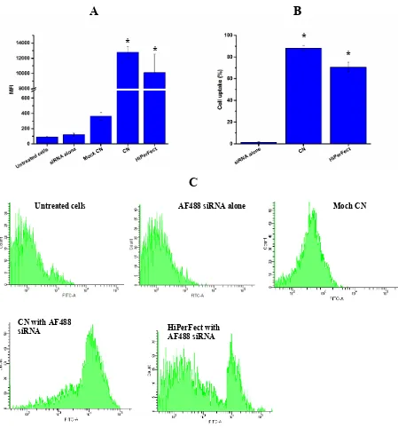
3.3.
B16-F10 cellular uptake
To evaluate the cellular uptake, B16-F10 mouse melanoma cells were treated with CN
formulation loaded with AF488-labelled negative control siRNA. The treated cells were
analysed by FACS for quantitative cellular uptake (Figure 2). As can be seen in Figure 2A,
B16-F10 cells did not show any siRNA uptake after being treated with siRNA alone, which
can be confirmed by the very low MFI (Figure 2B) and the histogram curve (Figure 2C)
compared to untreated cells. The cellular uptake for cells treated with AF488-labelled negative
[image:11.595.94.464.214.495.2]control siRNA encapsulated in the CN formulation was 88.28 ± 2.29% (Figure 2A). This
cellular uptake was significantly (p< 0.05) higher than the cellular uptake achieved by
HiPerFect (70.77 ± 4.35%) and the MFI was significantly higher for cells treated with
AF488-labelled negative control siRNA encapsulated by CN or HiPerFect compared to naked siRNA
(Figure 2B). When the CN formulation alone without AF488-labelled negative control was
used, the MFI values were low, indicating no auto-fluorescence (Figure 2B) and the histogram
Figure 2 FACS results for (A) MFI, (B) percentages of cellular uptake, and (C) flow cytometry histograms of B16-F10 cellular uptake when treated with CN or HiPerFect loaded with AF488-labelled negative control siRNA. Images are representative of three independent images from each sample. The data represents means ± standard deviation (n = 3). *p <0.05 significant difference from cells treated with naked siRNA. CN: cationic niosomes, Moch CN: empty cationic niosomes.
A
B
Untreated cells AF488 siRNA alone Moch CN
CN with AF488
siRNA HiPerFect with
AF488 siRNA
C
*
* *
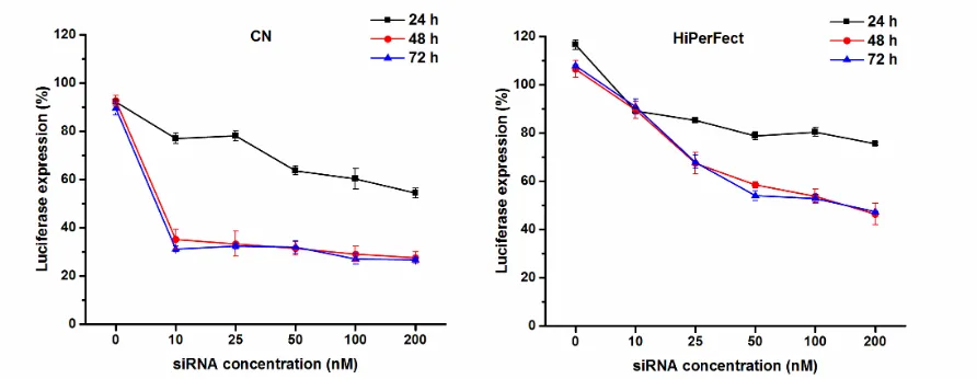
3.4. In vitro luciferase gene silencing study
We examined the extent of gene silencing by siLUC loaded in the CN formulation. To evaluate
the gene knockdown efficiencies, B16-F10 cells stably expressing luciferase were incubated
with CN formulation, loaded with various concentrations of luc siRNA, for 24, 48, and 72 h.
The specificity of the siLUC was confirmed using scrambled negative control siRNA.
Luciferase expression was measured by quantifying the luciferase luminescence intensity at
each time point in the cells treated with CN loaded with various concentrations of siLUC
(Figure 3). The luciferase enzyme knockdown was compared to untreated cells. CN
demonstrated significant (p< 0.05) transfection efficiency, which was concentration- and
time-dependent. After 24 h incubation, the percentage of luciferase expression (compared to
untreated cells) decreased significantly (p< 0.05) from 77.01 ± 2.22% to 54.42 ± 2.06% by
increasing the siLUC concentration from 10 – 200 nM. After 48 h incubation, all the siLUC
concentrations induced the same level of luciferase expression inhibition resulting in a
luciferase expression of around 30% (Figure 3). Longer incubation times with CN formulation
did not result in higher luciferase inhibition. This significant luciferase inhibition, achieved by
the CN formulation, was much higher than the inhibition demonstrated by HiPerFect. With
HiPerFect, the luciferase inhibition was dose-dependent at all time points with the highest
knockdown achieved after 48 and 72 h with no significant difference between both time points.
After 24 h incubation, the highest luciferase inhibition achieved when the cells were transfected
with 200 nM siLUC using HiPerFect was 75.54 ± 0.56% luciferase expression. This was
significantly (p <0.05) higher than the luciferase expression when the cells were treated with
the CN formulation using the same siLUC concentration (luciferase expression 54.42 ± 2.06%).
Longer incubation time for cells transfected with HiPerFect resulted in higher luciferase
inhibition, where the maximum effect seen after 48 h using 200 nM of siLUC resulted in
3). No effect was noticed for CN formulation at 0 nM siLUC (mock transfection) at all time
points. Similarly, no effect was seen when using siLUC alone at all time points (data not
shown). This indicates that the decrease in luciferase expression using siLUC was indeed
caused by the sequence specific gene silencing of siLUC.
3.5. In vivo silencing study
3.5.1. Determination of the most tolerable dose of CN/siLUC
In a pilot study, empty particles at a concentration of 39 µg/mL was found to have no effect on
the weight of the animal, whereas at concentrations higher than 156 µg/mL, there was a
decrease in weight (Table 1). This concentration of empty particles was therefore selected and
used to encapsulate various concentrations of siLUC. A siLUC concentration at 625 nM was
[image:15.595.76.522.254.427.2]found to be well tolerated by the mouse while concentrations higher than 1250 nM caused
weight loss (Table 2). Therefore, in the following in vivo experiments, CN/siLUC nioplexes
[image:16.595.72.524.182.357.2]were prepared using 39 µg/ml of CN encapsulating 625 nM of siLUC.
Table 1 Change of the mouse weight when given increasing doses of empty particles of CN.
Empty particles
Days 1 2 3 4 5
Particle concentration (µg/ml) 39 156 315.5 - -
siRNA concentration (nM) 0 0 0 - -
Animal weight (g)
[image:16.595.72.528.438.580.2]19.2 19.3 18.7 18.6 18.4
Table 2. Change of the mouse weight when given increasing doses of siLUC encapsulated in CN at concentration of 39 µg/ml.
Particles + siLUC
Days 1 2 3 4 5
Particle concentration (µg/ml) 39 39 39 - -
siRNA concentration (nM) 625 1250 2500 - -
Animal weight (g) 19 19.3 18.8 18.6 18.8
3.5.2. In vivo luciferase gene silencing study
To investigate whether CN could release its encapsulated siRNA and inhibit gene expression
in tumours, CN loaded with siLUC (39 µg/ml of CN encapsulating 625 nM of siLUC) were
injected intratumorally in nude mice bearing subcutaneous B16-F10-Luc melanoma.
anaesthetised animals. Mice injected with siLUC alone, CN alone, or left untreated were used
as controls. Figure 4 shows the average bioluminescence measured for each group. A
representative mouse whose emitted light was closest to the average for that group (3 mice per
group) is shown (Figure 5).
Luciferase expression in mice injected with siLUC encapsulated in CN was significantly (p
<0.05) decreased, 4h after injection, by about 50% (Figure 4). The maximum luciferase
expression knockdown (by 70%) was obtained 12 h after injection. This inhibition was
reversible and the luciferase expression returned to normal after 24 h. In contrast, the
bioluminescence signals in the mice treated with naked siLUC increased over the same time
period, suggesting no inhibition of luciferase expression at any time point. Luciferase
expression did not appear to be affected by the CN formulation, as the bioluminescence signal
increased over time following treatment with empty CN. These results suggest that the
inhibition of luciferase expression resulted from siLUC delivery by the CN into the cytoplasm
of the cells and the subsequent RNAi mechanism. This demonstrates that siRNA can be
4. Discussion
Numerous barriers have to be overcome for the effective delivery of siRNA into the cytoplasm.
The main obstacle to the development of effective therapeutics with siRNA is a suitable
delivery system. First, the delivery system has to remain stable over time, to be of minimal
toxicity, to be taken up by the cells, and to escape the endosome compartment with the
subsequent delivery of the siRNA for its interaction with the RNAi machinery in the cytoplasm
[22]. NISV possess attractive properties as a drug delivery system such as biodegradability,
biocompatibility, stability, and ease of manufacture. Limited research is available about the use
of NISV for siRNA delivery. Previous work for siRNA delivery combined non-ionic surfactant
with phospholipids in a formulation called spanosomes [23]. In our previous study, effective
various CN formulations were prepared and their efficacy in siRNA transfection and GFP
inhibition in copGFP-A549 cells evaluated [19]. In order to confirm the gene silencing
observed in copGFP-A549 cells, one formulation was taken forward in this study and the
biological activity of the selected CN was tested on a different cell model, the B16-F10 cells,
which could then be investigated in vivo in a rat model. CN cytotoxicity was evaluated on
B16-F10 cells to make sure that any observed luciferase knock-down was a result of the siRNA used
rather than as a result of any vesicle-related toxicity. Cytotoxicity results revealed that CN were
not toxic at or below 40 µg/ml. Next, the transfection efficiency of the CN was examined by
analysing the B16-F10 cellular uptake with FACS after treating these cells with CN
encapsulating AllStars AF488-labelled Negative Control siRNA. CN were able to deliver
siRNA as confirmed by the high cellular uptake percentages. This high cellular uptake can be
attributed to the positive charge on the surface of CN, which enhanced the interaction with the
negatively charged cellular membrane. The cellular uptake using CN was compared with
fluorescence signal, indicating that the above mentioned cellular uptake was a result of the
siRNA delivery by the CN formulation.
After evaluating cellular uptake, luciferase gene silencing mediated by siLUC loaded in the
CN formulation was evaluated. Luciferase is a bioluminescence producing enzyme widely used
for monitoring siRNA delivering efficacy by monitoring bioluminescence changes after
anti-luciferase siRNA treatment [24, 25]. After evaluating cellular uptake mediated by the CN
formulation, the endosome release after uptake and the subsequent inhibition of the target
luciferase enzyme need to be proven. To examine both the extent of gene silencing and the
optimal incubation time, B16-F10 cells were transfected by various siLUC concentrations
(10-200 nM) using the CN formulation and the changes in bioluminescence intensity was
monitored at various time points. Naked siLUC showed negligible gene silencing effects when
compared to siLUC with CN. Results indicate that the time of incubation and the siRNA
concentrations had effects on the degree of gene knockdown. Increasing the incubation time
meant higher exposure of the particles to the cells in order to increase the cellular uptake. Here,
the efficacy rose by increasing the incubation time until 48 h, where further incubation did not
result in greater knockdown for all siRNA concentrations. This suggests that the observed
downregulation effect as a result of siLUC delivery by CN was stable for at least 72 h.
Moreover, these experiments were carried out in the presence of serum, which indicates that
the CN were able to protect the siLUC from degradation in the presence of serum proteins.
Together, these results demonstrate that by using the CN formulation, siLUC is protected
against degradation, internalised by the cells, and enabled escape of the endosomes to the
cytoplasm where bioactivity was displayed.
CN were able to induce high luciferase suppression by about 68% after 48 h incubation using
10 nM siLUC. CN appeared to be an effective transfection reagent when compared with the
knockdown efficacy across all the siRNA concentrations. These results for luciferase
suppression were consistent with the results reported previously on inhibition of GFP
expression in A549 cells by siGFP delivered by various CN formulations [19]. This high
luciferase suppression by siLUC delivered by this particular CN can be explained by the CN
endosomal escape ability after uptake to the cytoplasm where the RNAi mechanism occurs
[26]. CN were able to escape the endosome at a high rate, release the siLUC into the cytoplasm,
initiating luciferase RNA interference, and inhibit the luciferase expression. This ability is due
in part to the presence of T85 as a non-ionic surfactant in the CN formulation. T85 is believed
to have fusogenic properties that enhances endosome escape by promoting instability in the
endosome compartment and therefore releasing siRNA into the cytosol [27, 28]. The possibility
of the downregulation of gene expression being due to cytotoxic effects of the formulations
can be excluded as the non-toxic concentration of CN formulation was used. In addition, no
luciferase downregulation was observed when cells were transfected with empty particles of
CN alone (0 nM siLUC).
High siRNA concentrations can result in off-target effects which is one of the side effects
associated with siRNA therapeutics [29]. Previous reports of siRNA delivery systems targeting
luciferase, were able to achieve high luciferase suppression only at high siRNA concentrations,
which increases the possibility of siRNA off-target effects. For example, Takemoto et al. were
able to achieve 80% luciferase silencing with 100 nM siRNA using a siRNA-grafted polymer
delivery system [30]. With chitosan nanoparticles, Ragelle et al. were able to achieve the
maximum of 71% luciferase suppression using 200 nM of anti-luciferase siRNA [31]. Li et al.
were able to induce around 70% luciferase gene silencing using targeted cationic liposomes
using 250 nM anti-luciferase siRNA [32]. In our CN study, comparable high luciferase
highlighting the efficiency of this CN formulation. Based on the in vitro results, the use of CN
for in vivo RNA therapeutic applications was then evaluated.
After determining the maximum dose that could be used for the in vivo experiments, mice
bearing luciferase-expressing tumours were intratumorally injected with siLUC encapsulated
in CN. After 4h, the luciferase expression decreased by about 50.77 ± 20.35 %, indicating that
the nioplexes were taken up by the cells where the siLUC released into the cytoplasm and
incorporated in the RNA induced silencing complex (RISC) followed by luciferase expression
knockdown. Twelve hours after the treatment, luciferase expression was significantly
decreased by more than 70%. This luciferase suppression was reversible and, 24 h after
injection, the luciferase expression was fully recovered. This is in agreement with what has
been reported in the literature about the reversibility of the RNAi mechanism [33, 34]. These
results provide an insight into the possible required dosing intervals to maintain the target gene
suppression by siRNA in a therapeutic application. In mice injected with naked siLUC, there
was no luciferase suppression and the luciferase expression increased with time as the tumour
size naturally increased, suggesting that the tumour cells did not take up naked siRNA due to
their hydrophilic properties [35]. Moreover, mice injected with particles alone showed an
increase in the luciferase expression over time, suggesting that the empty particles had no effect
on both luciferase expression and tumour growth.
In the work of Minakuchi et al., significant luciferase suppression was achieved with an
atelocollagen delivery system using a single injection via the same route of administration and
the same tumour type that was used in this study [36]. However, despite the larger doses used
by them, based on the tumour size (2.5 μg siRNA/50 μl/50 mm3 tumor), the luciferase
expression was also reversible after 2-3 days [36]. Filleur et al. investigated the use of naked
results, which was similar to the results reported here in which naked siLUC did not induce
any gene suppression [37].
These in vivo results demonstrate the efficacy of CN in delivering and releasing siRNA into
tumour cells. Although further experiments are required such as i.v. treatment and
biodistribution studies, CN-mediated siRNA delivery possess the potential for in vivo delivery
of siRNA into tumour tissues. These results are proof of concept of the ability of CN to
effectively deliver siRNA into mouse melanoma cells. These CN formulations can be explored
with different types of cancer cells in the expectation of similar outcomes. Moreover, these
intratumor injection results could be used as a model for localised treatment of siRNA
therapeutics delivered by CN and these formulations can also be explored further for topical
applications in treatment of different diseases.
5. Conclusions
We successfully formulated CN nanoparticles that could act as an effective siRNA delivery
system to deliver siRNA and suppress luciferase expression both in vitro and in vivo. With
these CN formulations, the suppression of over-expressed genes in different cancer types can
be investigated through siRNA delivery. We were able to achieve more than 70% of luciferase
knockdown through CN both in vitro and in vivo, which is a promising delivery system in the
field of nucleic acids delivery. In conclusion, we have developed CN to efficiently and safely
deliver siRNA to tumour cells and demonstrated specific inhibition of luciferase gene
expression. To our knowledge, our results present the first evidence that combine in vitro and
in vivo gene silencing data of siRNA delivery by NISV. This suggests that NISV might be used
Acknowledgments
The authors would like to acknowledge the Jordanian Ministry of Higher Education and
Scientific Research and Yarmouk University in Jordan for funding this work.
Conflict of Interest
The authors confirm that there is no conflict of interest with this manuscript.
References
1. Ghildiyal, M. and P.D. Zamore, Small silencing RNAs: an expanding universe. Nature Reviews Genetics, 2009. 10(2): p. 94-108.
2. Siomi, M.C., Short interfering RNA-mediated gene silencing; towards successful application in human patients. Advanced drug delivery reviews, 2009. 61(9): p. 668-671.
3. Resnier, P., et al., A review of the current status of siRNA nanomedicines in the treatment of cancer. Biomaterials, 2013. 34(27): p. 6429-6443.
4. Obeid, M.A., et al., Lipid-based nanoparticles for cancer treatment, in Lipid Nanocarriers for Drug Targeting. 2018, Elsevier. p. 313-359.
5. Al Qaraghuli, M.M., et al., Where traditional drug discovery meets modern technology in the quest for new drugs. Annals of Pharmacology and Pharmaceutics, 2017. 2(11): p. 1-5.
7. Obeid, M.A., et al., The effects of hydration media on the characteristics of non-ionic surfactant vesicles (NISV) prepared by microfluidics. International Journal of Pharmaceutics, 2016. 8. Obeid, M.A., et al., Delivering natural products and biotherapeutics to improve drug efficacy.
Therapeutic delivery, 2017. 8(11): p. 947-956.
9. Marianecci, C., et al., Niosomes from 80s to present: the state of the art. Advances in colloid and interface science, 2014. 205: p. 187-206.
10. Moghassemi, S. and A. Hadjizadeh, Nano-niosomes as nanoscale drug delivery systems: an illustrated review. Journal of Controlled Release, 2014. 185: p. 22-36.
11. Pozzi, D., et al., Effect of cholesterol on the formation and hydration behavior of solid-supported niosomal membranes. Langmuir, 2009. 26(4): p. 2268-2273.
12. Tavano, L., et al., Doxorubicin loaded magneto-niosomes for targeted drug delivery. Colloids and Surfaces B: Biointerfaces, 2013. 102: p. 803-807.
13. Pawar, S. and P. Vavia, Glucosamine anchored cancer targeted nano-vesicular drug delivery system of doxorubicin. Journal of drug targeting, 2016. 24(1): p. 68-79.
14. Huang, Y., et al., PEGylated synthetic surfactant vesicles (Niosomes): novel carriers for oligonucleotides. Journal of Materials Science: Materials in Medicine, 2008. 19(2): p. 607-614. 15. Pardakhty, A., J. Varshosaz, and A. Rouholamini, In vitro study of polyoxyethylene alkyl ether niosomes for delivery of insulin. International journal of pharmaceutics, 2007. 328(2): p. 130-141.
16. Manosroi, A., et al., In vitro and in vivo skin anti-aging evaluation of gel containing niosomes loaded with a semi-purified fraction containing gallic acid from Terminalia chebula galls. Pharmaceutical biology, 2011. 49(11): p. 1190-1203.
17. Rungphanichkul, N., et al., Preparation of curcuminoid niosomes for enhancement of skin permeation. Die Pharmazie-An International Journal of Pharmaceutical Sciences, 2011. 66(8): p. 570-575.
18. Paecharoenchai, O., et al., Nonionic surfactant vesicles for delivery of RNAi therapeutics. Nanomedicine, 2013. 8(11): p. 1865-1873.
19. Obeid, M.A., et al., Formulation of non-ionic surfactant vesicles (NISV) prepared by microfluidics for therapeutic delivery of siRNA into cancer cells. Molecular Pharmaceutics, 2017.
20. Obeid, M.A., et al., Comparison of the Physical Characteristics of Monodisperse Non-ionic Surfactant Vesicles (NISV) Prepared Using Different Manufacturing Methods. International Journal of Pharmaceutics, 2017.
21. Contag, C.H., et al., Visualizing gene expression in living mammals using a bioluminescent reporter. Photochemistry and photobiology, 1997. 66(4): p. 523-531.
22. Khalil, I.A., et al., Uptake pathways and subsequent intracellular trafficking in nonviral gene delivery. Pharmacological reviews, 2006. 58(1): p. 32-45.
23. Zhou, C., et al., SPANosomes as delivery vehicles for small interfering RNA (siRNA). Molecular pharmaceutics, 2011. 9(2): p. 201-210.
24. Fan, F. and K.V. Wood, Bioluminescent assays for high-throughput screening. Assay and drug development technologies, 2007. 5(1): p. 127-136.
25. Reynolds, A., et al., Rational siRNA design for RNA interference. Nature biotechnology, 2004.
22(3): p. 326.
26. Pozzi, D., et al., Mechanistic evaluation of the transfection barriers involved in lipid-mediated gene delivery: interplay between nanostructure and composition. Biochimica et Biophysica Acta (BBA)-Biomembranes, 2014. 1838(3): p. 957-967.
27. Huang, Y., et al., Polysorbate cationic synthetic vesicle for gene delivery. Journal of Biomedical Materials Research Part A, 2011. 96(3): p. 513-519.
29. Jackson, A.L. and P.S. Linsley, Recognizing and avoiding siRNA off-target effects for target identification and therapeutic application. Nature reviews Drug discovery, 2010. 9(1): p. 57-67.
30. Takemoto, H., et al., Polyion complex stability and gene silencing efficiency with a siRNA-grafted polymer delivery system. Biomaterials, 2010. 31(31): p. 8097-8105.
31. Ragelle, H., et al., Chitosan nanoparticles for siRNA delivery: optimizing formulation to increase stability and efficiency. Journal of Controlled Release, 2014. 176: p. 54-63.
32. Li, S.-D., S. Chono, and L. Huang, Efficient gene silencing in metastatic tumor by siRNA formulated in surface-modified nanoparticles. Journal of Controlled Release, 2008. 126(1): p. 77-84.
33. Dickins, R.A., et al., Tissue-specific and reversible RNA interference in transgenic mice. Nature genetics, 2007. 39(7): p. 914-921.
34. Kanasty, R., et al., Delivery materials for siRNA therapeutics. Nature materials, 2013. 12(11): p. 967-977.
35. Whitehead, K.A., R. Langer, and D.G. Anderson, Knocking down barriers: advances in siRNA delivery. Nature reviews Drug discovery, 2009. 8(2): p. 129-138.