PROGNOSTIC SIGNIFICANCE OF BUNDLE BRANCH
BLOCKS IN ACUTE
CORONARY SYNDROME
Submitted in partial fulfilment of
Requirements for
M.D. DEGREE BRANCH I GENERAL MEDICINE
OF
THE TAMILNADU DR.M.G.R. MEDICAL UNIVERSITY,
CHENNAI
MADRAS MEDICAL COLLEGE
CHENNAI - 600 003.
This is to certify that this dissertation entitled "PROGNOSTIC
SIGNIFICANCE OF BUNDLE BRANCH BLOCKS IN ACUTE
CORONARY SYNDROME" Submitted by Dr. MOORTHY .P
appearing for Part II M.D. Branch I General Medicine Degree
examination in March 2010 is a bonafide record of work done by him
under my direct guidance and supervision in partial fulfillment of
regulations of the Tamil Nadu Dr.M.G.R. Medical University, Chennai. I
forward this to the Tamil Nadu Dr.M.G.R. Medical University, Chennai,
Tamil Nadu, India.
Prof. K. SIVA SUBRAMANIAN, MD., Professor of Medicine,
Institute of Internal Medicine, Madras Medical College, Government General Hospital, Chennai - 600 003.
Prof.C.Rajendiran,MD.,
Director and Professor,
Institute of Internal Medicine, Madras Medical College, Government General Hospital, Chennai - 600 003.
Prof.J.Mohanasundaram,MD.,Ph.D.DNB
Dean,
I solemnly declare that the dissertation titled is done by me at
Madras Medical College & Govt. General Hospital, Chennai during
2007-2010 under the guidance and supervision of
Prof. K. Sivasubramanian.
The dissertation is submitted to The Tamilnadu Dr.M.G.R.
Medical University towards the partial fulfillment of requirements
for the award of M.D. Degree (Branch I) in General Medicine.
Place : Dr. Moorthy. P. Postgraduate Student,
Date; M.D. General Medicine,
Institute of Internal Medicine,
Madras Medical College,
At the outset I would like to thank my beloved Dean, Madras Medical
College, Prof. J. Mohanasundram, M.D., Ph.D.DNB., for his kind
permission to use the hospital resources for this study.
I would like to express my sincere gratitude to my beloved Professor
and Director, Institute of Internal Medicine Prof. C. Rajendiran, M.D., for his
guidance and encouragement.
With extreme gratitude, I express my indebtedness to my beloved Chief
Prof. K. Sivasubramanian, M.D., for his motivation, advice and valuable criticism, which enabled me to complete this work.
I am extremely thankful to Assistant Professors of Medicine
Dr.G. Rajan, M.D., and Dr.S. Gopalakrishnan, M.D., for their kind co-operation and guidance.
I thank all Professors, Assistant Professors, and Post-graduates of the
department of cardiology for their valuable support in helping in my study.
I would always remember with extreme sense of thankfulness for the
co-operation and criticism shown by my Postgraduate Colleagues.
I am immensely grateful to the generosity shown by the patients who
participated in this study. If at all, this study could contribute a little to relieve
CHAPTER
NO. TITLE
PAGE NO.
1. INTRODUCTION 1
2. AIM OF STUDY 2
3. REVIEW OF LITERATURE 3
4. MATERIALS AND METHOD 32
5. OBSERVATIONS 39
6. DISCUSSION 61
7. SUMMARY 67
8. CONCLUSION 68
9 ETHICAL COMMITTEE CLEARANCE
CERTIFICATE
10 BIBLIOGRAPHY
ACS - Acute coronary syndrome
AMI - Acute myocardial Infarction
BBB - Bundle Branch Block
CAD - Coronary Artery disease
ECG - Electrocardiography
CK-MB - Creatine kinase - MB
UA - Unstable angina
NSTEMI - Non ST Segment Elevation Myocardial Infarction
STEMI - ST Segment Elevation Myocardial Infarction
ACC - American College of Cardiology.
AHA - American Heart Association
PCI - Percutaneous Coronary Intervention
CABG - Coronary Artery Bypass Graft
EF - Ejection Fraction
AV - Atrio Ventricular
LBBB - Left Bundle Branch Block
RBBB - Right Bundle Branch Block
HERO - Hirulog Early Reperfusion and Occlusion
ABBB - Acute Bundle Branch Block
CCU - Coronary Care Unit
CHF - Congestive Heart Failure
BP - Blood Pressure
HR - Heart Rate
AWMI - Anterior Wall Myocardial Infarction
IWMI - Inferior Wall Myocardial Infarction
PWMI - Posterior Wall Myocardial Infarction
RVMI - Right Ventricular Myocardial Infarction
VAT - Ventricular Activation Time
VSR - Ventricular Septum Rupture
P.(Value) - Probability of the Test
LV - Left Ventricular
PTCA - Percutaneous Transluminal Coronary Angioplasty
LAHB - Left Anterior Hemi Block
CHB - Complete Heart Block
SK - Streptokinase
DD - Diastolic Dysfunction
VPC - Ventricular Premature Contraction
VT - Ventricular Tachycardia
INTRODUCTION
Presence of new onset bundle branch block is associated with increased
mortality in patients with acute coronary syndrome (ACS). Development of
new bundle branch block despite prompt fibrinolytic therapy may signify an
extensive and ongoing AMI. It is associated with overall poor prognosis, and
high risk for short term mortality.
Presence of complete Bundle Branch Block (BBB) Left or Right in AMI
patients represents an independent and very important predictor of in-hospital
complication and poor survival on long term.8
Earlier studies suggested that patients with BBB have move co-morbid
conditions and are less likely to receive therapies such as thrombolytics,
aspirin, β blockers and have an increased in hospital mortality rates.5
In our study we evaluated the prognostic significance of different types
of BBB present during the course of AMI in the hospitalized patients and we
AIM OF STUDY
1. To estimate the prevalence of BBB in patients with Acute coronary
Syndrome.
2. To compare the clinical characteristics in patients with ACS with or
without BBB.
3. To assess the prognostic significance of BBB in patients with ACS
depending on its form of presentation.
4. Presence of BBB in ACS could be used for risk stratification and
REVIEW OF LITERATURE
Coronary heart disease, or atherosclerotic CAD is the number one killer
disease worldwide.
Most patients with coronary heart disease have some identifiable risk
[image:10.595.96.530.297.656.2]factor.
Table 1 Risk Factors and Interventions for Coronary Heart Disease2
Class Risk Factors Intervention
1. Smoking
Dyslipidemia
High blood Pressure
Preventive medications
Smoking cessation
Lipid management
Blood pressure management
Aspirin, angiotensin- converting
enzyme inhibitor, beta blocker
2. Diabetes, prediabetes
Physical inactivity
overweight, obesity
Unhealthy diet, alcohol
Diabetes management
Activity management
Weight management
Improved diet
3. Menopause,
Micronutrients
Psychological factors
Novel biochemical and genetic
markers
Fig.1 Development and Progression of atherosclerosis3
Acute coronary syndromes comprise the spectrum of unstable cardiac
ischemia from unstable angina to acute myocardial infarction. Acute coronary
syndromes are now classified based on the presenting ECG as either "ST
elevation" or "non-ST elevation".
Acute coronary syndromes without ST segment elevation1
• Non-ST-segment elevation acute coronary syndrome is further divided
both, may be elevated) on unstable angina if Cardiac biomarkers are
normal.
• The symptoms consist of substernal chest pain or discomfort that may
radiate to the jaw, left shoulder or arm. Dyspnea, nausea, diaphoresis or
syncope may either accompany the chest discomfort or may be the only
symptoms of acute coronary syndrome.
• About 1/3 of the patients with myocardial infarction have no chest pain
per se- these patients tend to be older, female, have diabetes, and be at
higher risk for subsequent mortality.
• In patients without ST segment elevation it is the presence of abnormal
CK-MB or troponin values that are associated with myocyte necrosis
and the diagnosis of myocardial infarction.
• Many patients with acute coronary syndrome will exhibit ECG changes
during pain - either ST segment elevation, ST segment depression (or) T
wave flattening or inversion. Dynamic ST segment shift is the most
Table Braunwald Clinical Classification of UA/ NSTEMI2
Class Definition Death or MI to One
year * (%)
Severity
Class I New onset of severe angina or accelerated angina; no rest pain 7.3
Class II Angina at rest within past month but not within preceding 48 hr (angina at rest, subacute) 10.3
Class III Angina at rest within 48 hr (angina at rest, subacute) 10.8†
Clinical Circumstances
A. (Secondary Angina) Develops in the presence of extra cardiac condition that intensifies myocardial ischemia 14.1
B. (Primary Angina) Develops in the absence of extra cardiac condition 8.5
c. (Post Infarction Angina) Develops within 2 wk after acute myocardial infarction 18.5‡
Intensity of treatment Patients with unstable angina may also be divided into three groups depending on where unstable angina occurs (1) in the absence of treatment for chronic stable angina (2) during treatment for chronic stable angina. or (3) despite maximal antischemic drug therapy. There three groups that may be designated subscripts 1, 2, and 3, respectively
ECG Changes Patients with unstable angina may be further divided into those with or without transient ST -T
wave change during pain
* Date from TIMI III Registry: Scirica BM, et al: Am J Cardiol 90:821-826,2002. † p = 0.057
‡p = < 0.001
UA/ NSTEMI - unstable angina/ non-ST elevation myocardial infraction.
From Braunwald E: Unstable angina: A Classification, circulation 80:410-A, 1989.
Table Short - Term Risk of Death or Nonfatal Myocardial Ischemia in Patients with Unstable Angina2
Feature High Likelihood (Any of the
Following)
Intermediate Likelihood (Absence of High-Likelihood Features and Presence
of Any of the following
Low likelihood (Absence of High-or Intermediate - Likelihood Features but May
have any of the following
History • Accelerating tempo of ischemic
symptoms in preceding 48 hours
• Prior MI, peripheral or cerebrovascular disease, or CABG; prior aspirin use Character of Pain • Prolonged ongoing (>20 minutes)
rest pain
• Prolonged (>20 min) rest angina, now resolved, with moderate or high likelihood of coronary artery disease
• Now onset or progressive Canadian
Cardiovascular system class III or IV angina the past 2 weeks without prolonged (>20 min) rest pain but with moderate or high likelihood of coronary artery disease
• Rest angina (<20 min) or relieved with rest or sublingual NTG
Clinical findings • Pulmonary edema, most likely
due to ischemia
• New or worsening mitral
regurgitation murmur
• S3 or new/ worsening rales
• Hypotension, bradycardia,
tachycardia
• Age > 75 years
• Age > 70 years
Electrocardiogram • Angina at rest with transient ST segment changes >0.05 mV
• Bundle branch block, new or
presumed new
• Sustained ventricular
tachycardias
• T wave inversions >0.2 mV
• Pathological Q waves
• Normal or unchanged ECG during an episode of chest discomfort
Cardiac markers • Elevated (e.g., TnT or Tnl >0.1 ng/ml
• Slightly elevated (e.g., TnT>0.01 but < 0.1 ng/ml)
• Normal
Table American College of Cardiology/ American Heart Association Recommendations for Antischemic Therapy2
Class Indication Level of
Evidence
Class I (Indicated) Bed rest with continuous ECG monitoring for ischemia and arrhythmia detection in patients with ongoing rest pain C NTG, sublingual tablet or spray, followed by intravenous administration, for the immediate relief of ischemia and
associated symptoms
C Supplemental oxygen for patients with cyanosis or respiratory distress; finger pulse oximetry or arterial blood gas
determination to confirm adequate arterial oxygen saturation (SaO2, greater than 90%) and continued need for
supplemental oxygen in the presence of hypoxemia
C
Morphine sulfate intravenously when symptoms are not immediately relieved with NTG or when acute pulmonary congestion and/ or severe agitation is present
C A beta blocker, with the first dose administered intravenously if there is ongoing chest pain, followed by oral
administration in the absence or contraindications
B In patients with continuing or frequently recurring ischemia when beta blockers are contraindicated, a
nondihydropyridine calcium antagonist (e.g., verapamil or diltiazem) as initial therapy in the absence of severe LV dysfunction or other contraindications.
B
An ACEI when hypertension persists despite treatment with NTG and a beta blocker in patients with LV systolic dysfunction or CHF and in ACS patients with diabetes.
B Class IIa (good
supportive evidence)
Oral long-acting calcium antagonists for recurrent ischemia in the absence of contraindications and when beta blockers and nitrates are fully used
C
An ACEI for all post -ACS patients B
Intraaortic balloon pump (IABP) counter pulsation for severe ischemia that is continuing or recurs frequently despite intensive medical therapy or for hymodynamic instability in patients before or after coronary angiography
C Class IIb (weak
supportive evidence)
Extended - release form of nondihydropyridine calcium antagonists instead of a beta blocker Immediate - release dihydropyridine calcium antagonists in the presence of a beta blocker
B B Class III (not
indicated)
NTG or other nitrate with 24 hr of sildenafil (Viagra) use
Immediate - release dihydropyridine calcium antagonists in the absence of a beta blocker
C A ACEI = angiotensin -converting enzyme inhibitor; ACS =acute coronary syndrome; CHF = congestive heart failure; ECG - electrocardiographic; LV = left ventricular; NTG = nitroglycerin; SaO2 = oxygen saturation in arterial blood.
Acute Myocardial Infarction with ST segment elevation1
STEMI results in most cases from an occlusive coronary thrombus at the
site of preexisting athreosclerotic plaque.
STEMI presents as sudden but not instantaneous development of
prolonged (>30 minutes) anterior chest discomfort (sometimes felt as gas or
pressure).
Sometimes painless, masquerading as acute CHF, syncope, stroke, or
shock.
ECG - ST segment elevation (or) left Bundle Branch Brock.
Immediate reperfusion treatment is warranted.
Primary PCI within 90 minutes to hospital presentation superior to
thrombolysis.
Thrombolysis within 30 minutes of hospital presentation and 6-12 hours
Table Hemodynamic subsets in acute myocardial infarction1
Category CI or SWI PCWP Treatment Comment
Normal >2.2, < 30 <15 None Mortality rate < 5%.
Hyperdynamic >3.0, >40 <15 β-Blockers Characterized by tachycardia; mortality rate < 5%
Hypovolemic <2.5, <30 <10 Volume expansion Hypotension, tachycardia, but preserved left ventricular function by
echocardiography; mortality rate 4-8%.
Left ventricular failure <2.2, <30 >15 Diuretics Mild dyspnea, rales, normal blood pressure; mortality rate 10-20%.
Severe failure <2.0, <20 >18 Diuretics,
Vasodilators
Pulmonary edema, mild hypotension; inotropic agents, IABC may be required; mortality rate 20-40%
Shock <1.8, <30 >20 Inotropic agents,
IABC
IABC early unless rapid reversal occurs; mortality rate>60%
Cl, cardiac index (L/min/m2); SWI, stroke work index (g-m/m2, calculated as [mean arterial pressure-PCWP]. stroke volume index. 0.0136); PCWP, pulmonary capillary wedge pressure (in mm Hg; pulmonary artery diastolic pressure may be used instead); IABC, intra-aortic balloon counterpulsation.
TREATMENT OF STEMI1
ACC/AHA Guideline recommendations for selected medical treatments. Medication Acute Therapies ACS Acute Therapies AMI Discharge Therapies
Aspirin (ASA) IA IA IA
Clopidogrel in ASA - allergic patients IA IC IA
Clopidogrel, intended medical management
IA - IA
Clopidogrel or IIb/IIIa inhibitor, up front (prior to catheterization)
IA
Clopidogrel, early catheterization/ percutaneous coronary intervention (catheterization/ percutaneous coronary intervention (cath/ PCI])
IA (prior to or at time of PCI)
IB IA
Heparin (unfractionated or low - molecular - weight)
IA IA2 -
β-Blockers IB IA IB
Angiotensin - converting enzyme (ACE) inhibitors
IB3 IA/IIaB4 IA
GP IIb/IIIa inhibitors for intended early cath / PCI
Eptifibatide/ tirofiban IA - -
Abciximab IA IIaB5 -
GP IIb/IIIa inhibitors for high-risk patients without intended early cath/ PCI
Eptifibatide/ tirofiban IIaA - -
Abciximab IIIA - -
Lipid-lowering agent6 - - IA
Smoking cessation counseling - - IB
1 Class I indicates treatment is useful and effective, IIa indicates weight of evidence is in favor of usefulness/ efficacy, class IIb indicates weight of evidence is less well established, and class III indicates intervention is not useful/effective and may be harmful. Type A recommendations are derived from large-scale randomized trails, and B recommendations are derived from smaller randomized trials or carefully conducted observational analyses. ACC/AHA - American College of Cardiology/ American Heart Association.
2 As a class IIb, low-molecular-weight heparin (best studied is enoxaparin with tenecteplase) can be considered an acceptable alternative to unfractionated heparin for patients less than 75 years old who are receiving fibrinolytic therapy provided significant renal dysfunction is not present.
3 for patients with persistent hypertension despire treatment, diabetes mellitus, congestive heart failure, or any left ventricular dysfunction.
4 IA for patients with congestive heart failure or ejection fraction < 0.40, IIa for others, in absence of hypotension (systolic blood pressure < 100 mm Hg); angiotensin receptor blocker (valsartan or candesartan) for patients with ACE inhibitor intolerance.
5 As early as possible before primary PCI.
Pharmacological dissolution of thrombus in infarct-related artery2
This figure shows a schematic view of a longitudinal section of an
infarct - related artery at the level of the obstructive thrombus. Following
rupture of a vulnerable plaque (bottom center), the coagulation cascade is
activated, ultimately leading to the deposition of fibrin strands and platelet
aggregates obstructs flow (normally moving from left to right) in the infarct -
related artery; this would correspond to TIMI grade 0 on angiography.
Pharmacological reperfusion is a multipronged approach consisting of
fibrinolytic agents that digest fibrin, antithrombins that prevent the formation
of thrombin and inhibit the activity of thrombin that is formed, and antiplatelet
therapy. STEMI = ST-elevation myocardial infarction; TIMI = Thrombosis in
Reperfusion Therapy1
The current recommendation is to treat patients with STEMI who seek
medical attention within 12 hours of the onset of symptoms with reperfusion
therapy either primary PCI (or) thrombolytic therapy.
Primary Percutaneous Coronary Intervention1
Immediate coronary angiography and primary PCI (including stenting)
of the infarct - related artery have been shown to be superior to thrombolysis
when done by experienced operators in high - volume centers with rapid time
[image:20.595.95.527.394.747.2]from first medial contact to intervention ("door to balloon").
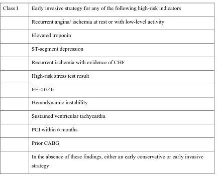
Table Indications for catheterization and percutaneous coronary intervention1
Class I Early invasive strategy for any of the following high-risk indicators
Recurrent angina/ ischemia at rest or with low-level activity
Elevated troponin
ST-segment depression
Recurrent ischemia with evidence of CHF
High-risk stress test result
EF < 0.40
Hemodynamic instability
Sustained ventricular tachycardia
PCI within 6 months
Prior CABG
Class IIa Early invasive strategy for patient with repeated presentations for ACS despite therapy
Class III Extensive comorbidities in patients win whom benefits of revascularizatin are not likely to outweigh the risks Acute chest pain with low likelihood of ACS
Acute MI after fibrinolytic therapy (2004 ACC/ AHA AMI Guideline)
Class I Recurrent ischemia (Spontaneous or provoked) Recurrent MI
Cardiogenic shock or hemodynamic instability
Class IIa LV EF ≤ 0.40, CHF (even transient), serious ventricular arrhythmias
Class IIb Routine PCI as part of invasive strategy after fibrinolytic therapy
MI, myocardial infarction; CHF, congestive heart failure; EF, ejection fraction, PCI, percutaneous coronary intervention, CABG, coronary artery bypass grafting; ACS, acute coronary syndrome; ACC/ AHA, American College of Cardiology/ American Heart Association, AMI, acute myocardial infarction; LV EF, left ventricular ejection fraction.
Stenting - generally in conjunction with the platelet glycoprotein IIb/IIa
antagonist abciximab - is standard for patients with acute myocardial
infarction. In the subgroup of patients with cardiogenic shock, early
catheterization and percutaneous or surgical revascularization (CABG) are the
Thrombolytic Therapy1
Table Thrombolytic therapy for acute myocardial infarction
Streptokinase Alteplase; Tisseu Plasminogen
Activator (t-PA) Reteplase Tenecteplase (TNK-t-PA)
Source Group c streptococcus Recombinant DNA Recombinant DNA Recombinatn DNA
Half-life 20 minutes 5 minutes 15 minutes 20 minutes
Usual dose 1.5 million units 100 mg 20 units 40 mg
Administration 750,000 units over 20 minutes followed by 750,000 units over 40 minutes
Initial bolus of 15 mg, followed by 50 mg infused over the next 30 minutes and 35 mg over the following 60 minutes
10 unit as a bolus over 2 minutes, repeated after 30 minutes
Single eight adjusted bolus, 0.5 mg/ kg
Antticoagulation after infusion
Aspirin, 325 mg daily; there is no evidence that adjunctive heparin improves outcome following streptokinase
Aspirin, 325 mg daily heparin, 5000 units as bolus, followed by 1000 units per hour infusion, subsequently adjusted to maintain PTT 1.5-2 times control
Aspirin,325 mg; heparin as with t-PA
Aspirin, 325 mg daily
Clot selectivity Low High High High
+ +++ + + +
Bleeding + + + +
Hypotension +++ + + +
Allergic reactions ++ 0 0 +
Reocclusion 5-20% 10-30% - 5-20%
Contraindications to thrombolytic therapy include previous hemorrhagic
stroke, other strokes or cerebrovascular events within 1 year, known
intracranial neoplasm, active internal bleeding (excluding menstruation), or
suspected aortic dissections.
Post thrombolytic management1
• Cardiac care unit monitoring
• Asprin, clopidogrel
• Anticoagulation - Unfractionated heparin, Low molecular weight
heparin
• Analgesia
• Morphine sulfate 4-8 mg, or meperidine, 50-75 mg
• β-Adrenergic Blocking Agents
• Nitrates
• ACE Inhibitors
• Angiotensin Receptor Blockers
• Aldosterone Antagonists
Complications1
• Postinfarction Ischemia
• Arrhythmias
• Myocardial Dysfunction
• Mechanical Defects
• LV Aneurysm
• Pericarditis
Secondary Prevention
1. Life-Style Modification
2. Nitrates
3. Anticoagulants
4. Beta Blockers,ACE Inhibitors
5. Hormone therapy
Conduction System of Heart, Coronal Section13
• The sinuatrial (SA) node in the wall of the right atrium near the superior
end of the sulcus terminalis extends over the anterior aspect of the
opening of the superior vena cava. The SA node is the "pacemaker" of
the heart because it initiates muscle contraction and determines the heart
rate. It is supplied by the sinuatrial nodal artery, usually a branch of the
• Contraction spreads through the atrial wall (myogenic induction) until it
reaches the atrioventricular (AV) node in the interatrial septum just
superior to the opening of the coronary sinus. The AV node is supplied
by the atrioventricular nodal artery, usually arising from the right
coronary artery posterior at the inferior margin of the interatrial septum.
• The AV bundle, usually supplied by the right coronary artery, passes
from the AV node in the membranous part of the interventricular
septum, dividing into right and left bundle branches on either side of the
muscular part of the interventricular septum.
• The right bundle branch travels inferiorly in the interventricular septum
to the anterior wall of the ventricle, then though the septomarginal
trabecula to the anterior wall of the ventricle, then through the
septomarginal trabecula to the anterior papillary muscle; excitation
spreads throughout the right ventricular wall through a network of
subendocardial branches form the right bundle (Purkinje fibers)
• The left bundle branch lies beneath the endocardium on the left side of
the interventricular septum and branches to enter the anterior and
posterior papillary muscles and the wall of the left ventricle; further
branching into a plexus of subendocardial branches (Purkinje fibers)
allows the impulses to be conveyed throughout the left ventricular wall.
The bundle branches are usually supplied by the left coronary, except
the posterior limb of the left bundle branch, which is supplied by both
• Damage to the cardiac conduction system (often by compromised blood
supply as in coronary artery disease) leads to disturbances of muscle
contraction. Damage to the AV node results in "heart block" because the
atrial excitation wave does not reach the ventricles, which begin to
contract independently at their own, slower rate. Damage to one of the
branches results in "bundle branch block", in which excitation goes
down the unaffected branch to cause systole of that ventricle; the
impulse then spreads to the other ventricle, producing later,
asynchronous contraction.
Arrhythmias in AMI2
A leading hypothesis for a major mechanism of arrhythmias in the acute
phase of coronary occlusion is reentry caused by in homogenecity of the
electrical characteristics of ischemic myocardium. Thus all forms of
bradycardia and tachycardia can depress the cardiac output in patients with
STEMI.
The role of arrhythemias in complicating the course of patients with
STEMI and the prevention and treatment of these arrhythmias in this setting are
Table Cardiac Arrhythmias and their management during acute myocardial infarction2
Category Arrhythmia Objective of Treatment Therapeutic Options
1. Electrical instability Ventricular premature beats Correction of electrolyte deficits and increased sympathetic tone
Potassium and magnesium solutions, beta blocker
Ventricular tachycardia Prophylaxis against ventricular fibrillation, restoration of hemodynamic stability
Antiarrhythmic agents; cardioversion/ defibrillation
Accelerated idioventricular rythm Observation unless hemodynamic function is compromised
Increased sinus rate (atropine, atrial pacing) antiarrhythmic agents
Nonparoxysmal atrioventricular
junction tachycardia
Search for precipitating causes (e.g., digitalis intoxication); suppress arrhythmia only if hemodynamic function its
compromised
Atrial overdrive pacing; antiarrhythmic agents; cardioversion relatively contraindicated if digitalis intoxication present
2. Pump failure/ excessive sympathetic stimulation
Sinus tachycardia Reduce heart rate to diminish myocardial
oxygen demands
Antipyretics; analgesics; consider beta blocker unless congestive heart failure present; treat latter if present with anticongestive measures (diuretics, after load reduction)
Atrial fibrillation and / or/ atrial flutter
Reduce ventricular rate; restore sinus rhythm
Verapamil, digitalis glycosides; anticongestive measures (diuretics, afterload, reduction); vardioversion; rapid atrial pacing (for atrial flutter)
Paroxysmal supraventricular
tachycardia
Reduced ventricular rate, restore sinus rhythm
Vagal maneuvers; verapamil, cardiac glycosides, beta-adrenergic blockers; cardioversion; rapid atrial pacing 3. Bradyarrhythimas and
conduction disturbances
Sinus bradycardia Acceleration of heart rate only if
hemodynamic function is compromised
Atropine; atrial pacing Junctional escape rhythm Acceleration of sinus rate only if loss of
atrial "kick" causes
Atropine; atrial pacing Atrioventricular block and
intraventricular block
Insertion of pacemaker
Ischemic injury can produce conduction block at any level of the
atrioventicular (AV) or intraventricular conduction system. Such blocks can
occur in the AV node and the bundle of his, producing, various grades of AV
block, in either main bundle branch, producing right or left bundle branch
block. and in the anterior and posterior divisions of the left bundle, producing
left anterior or left posterior (fascicular) divisional blocks, Disturbances of
conduction can, of course, occur in various combination. Clinical features of
proximal and distal AV conduction disturbances in patients with STEMI are
Table Atrioventricular (AV) Conduction Disturbances in Acute Myocardial Infarction2
Proximal Distal
Site of block Intranodal Infranodal
Site of infarction Inferoposterior Anteroseptal
Compromised arterial supply RCA (90%), LCU (10%) Septal perforators of LAD Pathogenesis Ischemia, necrosis, hydropic cell swelling excess
parasympathetic activity
Ischemia, necrosis, hydropic cell swelling
Predominant type features of third-degree AV block
First – degree (PR > 200 msec) Third type I second – degree
Mobitz type II second-degree Third degree
Common premonitory features of third-degree-AV block
(a) First second degree AV block (b) Mobitz I pattern
(a) Intraventricular conduction block (b) Mobitz II pattern
Features of escape rhythm following third-degree block
(a) Location (b) QRS width (c) Rate
(d) Stability of escape rhythm
(a) Proximal conduction system (His bundle) (b) <0.12/sec
(c) 45-60 min but may be as low as 30/min (d) Rate usually stable; asystole uncommon
(a) Distal conduction system (bundle branches) (b) 0.12/ sec
(c) Often < 30 / min
(d) Rate of often unstable with moderate to high risk of ventricular asystole
Duration of high-grave AV block Usually transient (2-3 days) Usually transient but some form of AV conduction disturbance and / or intraventricular defect may persist Associated mortality rate Low unless associated with hypotension and /or
congestive heart failure
High because of extensive infarction associated with power failure or ventricular arrhythmias
Pacemaker therapy (a) Temporary (b) Permanent
(a) Rarely required, may be considered for bradycardia associated with left ventricular power failure, syncope, or angina
(b) Almost never indicated because conduction defect is usually transient
a. Should be considered in patient with anteroseptal infarction and acute bifascicular b lock.
b. Indicated for patients with high-grade AV block in His-Purkinje system and those with transient advanced AV block and associated bundle branch block
* Some studies suggest that a wide QRS escape rhythm (>0.12 sec) following high-grade AV block in inferiori infarction is associated with a worse prognosis. LAD – left anterior descending coronary artery; LCX- left circumflex coronary artery; RCA – right coronary artery.
Modified from Antma EM. Rutherford JD: Coronary Care Medicine: A Practical Approach. Boston, Martinus Nijhoff, 1986; and Dreifus LS, Fisch, C, Griffin JC, et al. guidelines for implantation of cardiac pacemakers and antiarrhythmia devices. J Am. Coll Cordial. 18:1, 11991. Reprinted with permission from the American College of Cardiology.
Intraventricular Block2
The right bundle branch and the left posterior division have a dual blood
supply from the left anterior descending and right coronary arteries, whereas
the left anterior division is supplied by septal perforators originating from the
left anterior descending coronary artery. Not all conduction blocks observed in
patients with STEMI can be considered to be complications of infarcts because
almost half are already present at the time the first ECG is recorded, and they
may represent antecedent disease of the conduction system. Compared with
patients without conduction defects, STEMI patients with bundle branch blocks
have more comorbid conditions; are less likely to receive therapies such as
thrombolytics, aspirin, and beta blockers; and have an increased in-hospital
mortality rate. In the prefibrinolytic era, studies of intraventricular conduction
disturbances (i.e., block within one or more of the three subdivisions (fascicles)
of the His-Purkinje system (the anterior and posterior divisions of the left
bundle and the right bundled) had been reported to occur in 5 to 10 per cent of
patient with STEMI. More recent series in the fibrinolytic era suggest that
intraventricular blocks occur in about 2 to 5 per cent of patients with MI.
(Investigators performing primary PCI for STEMI have reported an association
between new-onset bundle branch block and abnormal myocardial perfusion
even if epicardial flow is restored.
ISOLATED FASCICULAR BLOCKS. Isolated left anterior divisional block is unlikely to progress to complete AV block. Mortality is
increased in these patients, although not as much as in patients with other forms
and, in general, a larger infarct is required to block it. As a consequence,
mortality is markedly increased. Complete AV block is not a frequent
complication of either form of isolated divisional block.
RIGHT BUNDLE BRANCH BLOCK. This conduction defect alone can lead to AV block because it is often a new lesion, associated with
anteroseptal infarction. Isolated right bundle branch block is associated with an
increased mortality risk in patients with anterior STEMI even if complete AV
block does not occur, but this appears to be the case only if its is accompanied
by congestive heart failure.
BIFASCICULAR BLOCK. The combination of right bundle branch block with either left anterior or posterior divisional block or the combination
of left anterior and posterior divisional blocks (i.e., left bundle branch block)C
is known as bidivisional or bifascicular block. If new block occurs in two of the
three divisions of the conduction system, the risk of developing complete AV
block is quite high. Mortality is also high because of the occurrence of severe
pump failure secondary to the extensive myocardial necrosis required to
produce such an extensive intraventricular block. Patients with intraventricular
fibrillation late in their hospital stay. However, the high rate of mortality in
these patients occurs even in the absence of high-grade AV block and appears
to be related to cardiac failure and massive infarction rather than to the
conduction disturbance.
Preexisting bundle branch block or divisional block is less often
associated with the development of complete heart block in patients with
Bidivisional block in the presence of prolongation of the P-R interval
(first-degree AV block) may indicate disease of the third subdivision rather than of
the AV node and is associated with a greater risk of complete heart block than
if first-degree AV block is absent.
Complete bundle branch block (either left or right), the combination of
right bundle branch block and left anterior divisional (fascicular) block, and
nay of the various forms of trifascicular block are all more often associated
with anterior than with inferoposterior infarction. All these forms are more
frequent with large infarcts and in older patients and have a higher incidence of
other accompanying arrhythmias than is seen in patients without bundle branch
block.
TEMPORARY PACING2. Just as is the case for complete AV block, transvenous ventricular pacing has not resulted in statistically demonstrable
improvement in prognosis among patients with STEMI who develop
intraventricular conductions defects. However, temporary pacing is advisable
in some of these patients because of the high risk of developing complete AV
block. This includes patients with new bilateral (bifascicular) bundle branch
block (i.e., right bundle branch block with left anterior or posterior divisional
block and alternating right and left bundle branch block); first-degree AV block
adds to this risk. Isolated new block in only one of the three fascicles even with
P-R prolongation and preexisting bifascicular block and normal P-R interval
poses somewhat less risk; these patients should be monitored closely, with
insertion of a temporary pacemaker deferred unless higher degree AV block
Noninvasive external temporary cardiac pacing is possible routinely in
conscious patients and is acceptable to many but not all patients despite the
discomfort. Used in a standby mode, it is virtually free of complications and
contraindications and provides an important alternative to transvenous
endocardial pacing. Once it is clinically evident that continuous pacing is
required, external pacing, which is generally not well tolerated for more than
minutes to hours, should be replaced by a temporary transvenous pacemaker.
ASYSTOLE. The presence of apparent ventricular asystole on monitor displays of continuously recorded ECGs may be misleading because the
rhythm may actually be fine ventricular fibrillation. Because of the
predominance of ventricular fibrillation as the cause of cardiac arrest in this
setting, initial therapy should include electrical counter-shock, even if
definitive electrocardiographic documentation of this arrhythmia is not
available. In the rare instance in which asystole can be documented to be the
responsible electrophysiological disturbance, immediate transcutaneous pacing
(or stimulation with a transvenous pacemaker if one is already in place) is
indicated.1
PERMANENT PACING. The question of the advisability of permanent pacemaker insertion is complicated because not all sudden deaths in
STEMI patients with conduction defects are caused by high-grade AV block. A
high incidence of late ventricular fibrillation occurs in CCU survivors with
anterior STEMI complicated by either right or left bundle branch block.
conduction and infranodal pacemakers could be responsible for late sudden
death.
Long-term pacing is often helpful when complete heart block persists
throughout the hospital phase in a patient with STEMI, when sinus node
function is markedly impaired, or when type II second- or third-degree block
occurs intermittently.1 When high-grade AV block is associated with newly
acquired bundle branch block or other criteria of impairment of conduction
system function, prophylactic long-term pacing may be justified as well.
Additional considerations that drive a decision to insert a permanent pacemaker
include whether the patient is a candidate for an implantable
cardioverter-defibrillator or has severe heart failure that might be improved with
biventricular pacing.
1. Wong C.K. et al5.
Insights form the Hirulog and Early Reperfusion or Occlusion
(HERO)-2 trial. (European Heart Journal doi:10.1093eurheartj/ ehi6(HERO)-2(HERO)-2 ).
Patients with any type of BBB at randomization had worse baseline
characteristics than those with normal intraventricular conduction. However,
only patients with RBBB accompanying anterior AMI at randomization (and
not patients with RBBB accompanying inferior AMI or patients with LBBB at
randomization) had a higher mortality rate after adjustment for baseline
charac-teristics. This finding persisted after further adjustment for the presenting
Approximately 1% of patients who had ST-elevation AMI with normal
intraventricular conduction at randomization developed new BBB (most
commonly RBBB accompanying anterior AMI) within 60 min after
commencing fibrinolytic therapy. New BBB was associated with higher 30-day
mortality.
The higher mortality and the higher incidence of RBBB seen in patients
with anterior AMI may be explained by septal ischaemia from a more proximal
left anterior descending artery occlusion (before the large septal branch) and
the course of the right bundle branch traversing the septum towards the apex.
Higher peak enzyme levels were observed in these patients. In contrast, the left
bundle has a more varied distribution from a true bifasclcular system to a
network of fibres, and more extensive ischemia or necrosis is required to
produce complete LBBB. Thus, new LBBB was far less likely to develop
within 60 min than new RBBB, but when new LBBB did develop, the
mortality rate was as high as that of patients with RBBB accompanying
anterior AMI.
In the current American and European guidelines, new or presumed-new
LBBB within 12 hours after the onset of symptoms suggestive of AMI is a Class I
indication for fibrinolytic therapy. New or presumed-new LBBB was an inclusion
criterion in the HERO-2 trial. The 300 HERO-2 patients with LBBB at
randomization had worse pre-infarction characteristics (older age and previous
AMI), worse presenting features (higher pulse rate and Killip class), and nearly
twice the unadjusted 30-day mortality rate of patients with normal conduction.
features, their 30-day mortality rate was no higher than that of patients with
normal intraventricular conduction12-an interesting observation which
corraborates the findings of a GUSTO-1 substudy in 131 patients with LBBBJ.
2. Taporan Daniela et al. Study8 says,
Patients with BBB and AMI are less likely to receive thrombolytic
therapy, associate more frequently severe heart failure and have an increased
risk for in-hospital death. No clinically significant differences in the
development of recurrent ischemia, angina or mechanical complications were
seen between patients with BBB and without BBB.
3. Acute Med Dkayama 2009 Feb 63(1):25-33 et al Study14 Says
New permanent RBBB was a strong independent predictor for an
adverse short term prognosis in patients with inferior MI as well as in patients
with anterior MI. New permanent RBBB during inferior MI is a strong
independent predictor for increased in hospital mortality, regardless of the
infarction location.
4. Vojnosaint Preg et al., Study15 2009 Jan 66(1) : 74 says
The patients with ABBB in AMI are at risk group of patients that
commonly exhibit both early and remote complications accompanies by high
mortality . That is the reason why this sub-group of AMI patients should
5. Barsheshet et al (Am. J. Cardiol: 2008 Aug 15:102(4) 507-8. )16
Study says RBBB rather than LBBB is an independent predictor of
mortality in hospitalized patients with systolic heart failure. This prognostic
marker could be used for risk stratification and selection of treatment.
6. Lerecouverex et al Arch Mal Coeur Vaies 2005 Dec 98(12) 1231-8
study says, RBBB is associated with worse prognosis.
7. Biagini et al17 (J Am Coll Cardiology 2005 Sep 6: 46(5) 856-63) Study says
LAHB in AMI increased risk of death. Isolated LAHB should not be
considered a benign electrocardiography abnormality in these patients.
8. Islam MN et al18 Says (Bangladesh Med Res Counc Bul 2002 Apl 28(1) 26-35).
Specific in hospital complication were significantly higher in patients
with RBBB than without RBBB.
9. Vrugada et al19 (Circulation 2002 Jan 1:105 (1): 73-8) study Says
An ECG showing RBBB with STEMI in the right pericardial leads is a
marker of malignant ventricular arrhythmias and sudden death. Recurrence of
Malignant arrhythmias is high after the occurrence of symptoms.
10. Melgarejo Moreso. A et al10., (Chin Cardiol 2001 May:24 (5): 37-6) Study say
In the patients with AMI, the classification of BBB according not only
to location but also to time of appearance is of practical interest. New BBB is
11. Sergia Rocha et al9., (ESC congress 2007), Austria Study says.
RBBB in AMI - is associated with poor prognosis.
12. Gunnarson.G et al., Scand Cardiovasc Dec 2000 34(6) 875-9.
New onset BBB associated with poor programs.
13. EB Sgarbossa et al12 J Am Coll Cardiol 1998: 31:105-12 study says
Bundle Branch block at hospital admission inpatient in patients with
acute AMI predicts in hospital complications and poor shortterm survival.
14. Eugene Brancwold2 8th Edition page 1281
Bifasicular blocks ACS, is associated with risk of developing complete
AV block is quiet high. Mortality is also high because of the occurrence of
severe pump failure secondary to the extensive myocardial necrosis required to
produce such an extensive intraventricular block.
Regarding prognosis significance of RBBB (Page 1281)
Patients with Intraventricular conduction defects particularly right
bundle branch block account for the majority of patients who develop
ventricular fibrillation late in their hospital stay. However the high rate of
mortality in these patients occurs even in the absence of high grade AV block
and appears to be related to cardiac failure and massive infarction rather than to
MATERIALS AND METHODS
Study Design
This study is a single centre prospective analytical study carried out in
the coronary care unit, Department of Cardiology, Madras Medial College
during the period of January 2009 to December 2009.
Total number of patients were 150. All the patients in the study were
hospitalized. No out patients were included. A detailed informed concent was
obtained from the patients Our Institutional ethical committee clearance was
obtained. Standard approved protocol were used for treating all the patients.
The results were tabulated and analysed using chi-square test.
Inclusion and Exclusion Criteria
Patients presented with ACS in the coronary care unit were included and
observed. Serial ECGs of all the patients admitted with ACS in CCU were
studied. CK-MB was measured in some of the cases. Since facilities for
measuring. Troponin was not available we were not able to measure it.
Patients demographics, clinical variables like prior MI, angina, CHF,
Cardiac risk factors like DM, SHT, smoking dylipidemia chest pain on
admission, Killip class, use of thrombolytic therapy, reasons for not using
thrombolytic therapy were recorded.
Patients were followed until discharge from the hospital and at the end
dysfunction, arrhythmias, recurrent angina, CHF, 2°, 3° heart block,
mechanical complications, cardiac arrest and death were recorded. These
variables are compared between ACS patients with BBB and without BBB.
Patients were excluded if presented with
1. Pre existing BBB
2. Non Specific Intraventricular conduction defects.
Study Protocol
Preliminary history with detail questioning regarding.
• Time of onset of chest pain, time of admission.
• Patients clinical status on admission including BP, HR, Killip class were
recorded.
• Serial ECGs were studied to record the AMI location such as AWMI,
IWMI others PWMI, RVMI.
• Presence of new onset of BBB, arrythmias
• History of thrombolysis and treatment of complication during the course
in the hospital.
• Previous history of MI, angina, CHF.
• Presence of cardiovascular risk factors like DM, HT, Smoking
Definitions4
RBBB
ECG Changes in completed and Incomplete RBBB
• A tall R wave (rSR') or notched tall R wave (rR') in lead V1 and V2 and
a deep, wide and prominent S wave in leads V5, V6 and standard leads 1
and a VL is seen in complete right bundle branch block. In incomplete
bundle branch block, a wider rSr, or rSR' is common in V1 - V2.
• QRS (rSR') duration is > 0.12 sec. in complete RBBB but it remains in
between 0.10 to 0.12 sec. in incomplete RBBB.
• VAT is prolonged in V1 - V2 (>0.03 sec.) in complete RBBB but is
normal in complete RBBB.
• ST segment is slightly depressed and T is inverted in V1 and V2, while
ST is slightly elevated with concavity upwards in leads V5 - V6 and T is
upright in these leads. These changes are seen both in complete and
incomplete RBBB.
LBBB
• ECG changes in complete and incomplete LBBB. Precordial leads V1
and V5 are best for diagnosis of LBBB.
• The leads V5, V6 and standard lead 1 show wide slurred R wave or RsR'
disappears. There is a slurring of S wave (if seen) in V5-V6 due to
activation of posterobasal region.
• QRS interval is > 0.12 second resulting in QS pattern in V1-V2. In
incomplete bundle branch block, QRS remains between 0.10 to 0.12 sec.
• VAT is > 0.09 second in complete LBBB. It is less increased in
incomplete LBBB.
• ST segment depression with convexity upwards and T wave inversion
occurs in leads I and V5-V6, while on the other hand, leads V1-V2 may
show mild elevation of ST segment with concavity upwards and T is
upright in these leads. This change is seen in both complete and
incomplete LBBB.
• Horizontal heart position will reveal similar morphology of QRS in
leads, I, aVL and V5-V6. Vertical heart position will show similar
morphologies of QRS in leads II, aVF and V5-V6.
• In LBBB (complete or incomplete), there is always an absence of q
wave in leads, I, V5-V6 due to septal activation from right to left. The
presence of q wave in these leads suggests an associated myocardial
infarction.
LAHB
a. Left anterior fascicular or hemiblock (LAH)
• A rS complex in leads II, III and aVF. The S wave is deeper in lead III
than lead II.
• A qR complex is present in leads I and aVL.
• A slight increase in QRS duration in these leads but less than 0.11 sec.
• VAT is slightly prolonged.
• ST segment depression and T wave inversion in leads registering
positive QRS deflection. T wave is upright with slight elevation of ST
segment in leads registering negative QRS deflection,
• Precordial leads will have RS complex with slurring of S wave in V5-V6.
B. Left posterior fascicular or left posterior hemiblock (LPH)
• Mean right QRS axis deviation (+120° clockwise to + 180°)
• Prominent S waves in leads I and aVL (rs complex), a tall R waves in
leads II, III and aVF (qR or qRs complex); the R wave will be thaller in
lead III than II and may be slurred.
• Procordial leads show rS complex in V1 and V2 and Rs in V5 and V6
with transition zone in V3 through V4.
• The ST segment depression and T wave inversion appears as a
secondary change in leads registering positive QRS deflection. T is
Bifascicular or Bilateral Bundle Branch Block
a. Right bundle branch block (RBBB) with left anterior fascicular or hemiblock (LAH)
• Pattern of right bundle branch block (rSR' or rR' or slurred R) in V1 and
V2 with wide S in leads I, V5, V6. This is due to terminal QRS
orientation to the right and interiorly.
• Left axis deviation is > - 30°. This is due to initial QRS vector being
oriented superiorly and to the left on frontal plane.
B. Right bundle branch block with left posterior fasciular block
• Typical features of RBBB as discussed above
• Right axis deviation > + 120°.
C. Trifascicular block
1. Right bundle branch block, left anteriro fascicular block and AV conduction
delay (commonest type).
• Pattern of RBBB as described above.
• Left axis deviation > - 30°.
• Prolonged P-R interval > 0.20 sec.
2. Right bundle branch block, left posterior fascicular block and AV
• Pattern of RBBB as described above.
• Right axis deviation > + 120°.
• Prolonged PR interval > 0.20 sec.
3. Right bundle branch block plus alternating left anterior and left posterior
fascicular block.
• Pattern of RBBB as described. It is fixed block.
• Sometimes, left anterior fascicular block and, left posterior fascicular
block alternates with RBBB.
• Follow up
All the patients presented will new onset BBB were followed up till
discharge from hospital and for 30 days after the onset of AMI. During this
period complication like ventricular dysfunction, arrhythmias, recurrent angina.
CHF, Heart block, Mechanical complications like VSR, cardiac arrest, and
death were recorded.
Standard guidelines for the treatment of ACS by American heart
Association were followed. The treatment modified according to the
OBSERVATIONS
Gender and BBB
Total BBB No BBB P. Value
Total 150 Total 34 Total 116
Male 111 Male 21 Male 88
Female 39 Female 11 Female 28
0.337
Comparasion of BBB and No BBB in ACS
34
116
BBB No BBB
77.33%
Age and BBB
Total BBB No BBB
AGE
Male Female Male Female Male Female
P Value <40 yrs 41-50 yrs 51-60 yrs >60 yrs 12 30 40 29 0 11 8 20 1 8 10 4 0 3 3 5 11 22 30 25 0 8 5 15 0.413 1 0 11 0 8 3 22 8 10 3 30 5 4 5 25 15 0 5 10 15 20 25 30 No.of Patients
<40 41-50 yrs 51-60 yrs >60 yrs
Years
Comparision of BBB and No BBB According to Age Group and Gender
Killip class and BBB
Total BBB (n=34)
No BBB
(n=116)
P Value
I – 104
II – 21
III – 19
IV – 6
I – 7
II – 12
III – 11
IV – 4
I – 97
II – 9
III – 8
IV – 2
0.001 **
Comparision of Killip Class in BBB and No BBB
7 12 11 4
97
9 8
2 0
20 40 60 80 100 120
I II III IV
Killip Class
No.of Pa
tie
n
ts
BBB No BBB
Arrythmias and BBB
Total BBB No BBB
Brady arrhythmias Taccy arrhythmias Brady arrhythmias Taccy arrhythmias Brady arrhythmias Taccy arrhythmias P Value Atrial Atrioventicular Ventricular – 2 2 1 - 4 – 2 2 – – 2 – – – 1 – 2 0.008 ** 0 1 2 0 4 2 0 0.5 1 1.5 2 2.5 3 3.5 4 No.of P a tients
Atrial Atrioventricular Ventricular
Arrythmias
Comparision of Arrythmias between BBB and No BBB
Complete Heart Block and BBB
Total BBB (n=34) No BBB (n=116) P Value
Total – 11
AWMI – 3
IWMI & Others – 8
Total – 5
AWMI – 2
IWMI & Others – 3
Total – 6
AWMI – 1
IWMI & Others – 5
0.024 *
Death – 5 Death – 4 Death – 1 0.035 *
5
6
4.4 4.6 4.8 5 5.2 5.4 5.6 5.8 6
No.of Patients
BBB No BBB
Complete Heart Block
Comparison of Complete Heart Block in BBB and No BBB
BBB No BBB
P value 0.024
4
1
0 0.5 1 1.5 2 2.5 3 3.5 4
No.of P
a
tients
BBB No BBB
Death
Comparison of Death due to complete Heart Block in BBB and No BBB in Patients with ACS
BBB No BBB
MI location and BBB
Total BBB (n=34) No BBB (n=116) P Value
AWMI – 74
IWMI & Others – 69
UA – 7
AWMI – 20
IWMI & Others – 13
Both – 1
AWMI – 54
IWMI & Others – 55
UA – 7
0.208
Comparison of Systolic LV dysfunction between BBB and No BBB
Total BBB No BBB P Value
Adequate – 20
Mild – 31
Moderate – 55
Severe – 30
Adequate – 0
Mild – 5
Moderate – 12
Severe – 11
Death – 6
Adequate – 18
Mild – 26
Moderate – 43
Severe – 19
Death – 10
0.022 *
0 18
5 26
12 43
11 19
0 5 10 15 20 25 30 35 40 45
No.of P
a
tients
Adequate Mild Moderate Severe
Systolic LV Disfunction
Comparision of Systolic LV Disfunction in BBB and No BBB
BBB No BBB
Diastolic Dysfunction and BBB
Total BBB (n=34) No BBB (n=116) P Value
No DD – 27
Grade - I – 68
Grade - II – 30
Grade - III – 11
No DD – 2
Grade - I – 11
Grade - II – 14
Grade - III – 3
No DD – 26
Grade - I – 57
Grade - II – 16
Grade - III – 8
0.001 **
2 26
11 57
14 16
3 8
0 10 20 30 40 50 60
No.of P
a
tients
No DD Grade - I Grade - II Grade - III
Diastolic LV Disfunction
Comprision of Distolic LV Disfunction in BBB and No BBB
BBB No BBB
P Value 0.001
Recurrent Angina
Total BBB (n=34)
No BBB (n=116) P Value
Total – 21
AWMI – 11
IWMI – 10
Total – 5
AWMI – 2
IWMI – 3
Total – 16
AWMI – 9
Thrombolysis
Total BBB No BBB P Value
Total – 93
0-3 hrs – 32
3-6 hrs – 46
>6 hrs – 15
Not Thrombolysed-50
UA – 7
Yes – 22
0 - 3 hrs – 5
3 - 6 hrs – 11
>6 hrs – 11
Not Thrombolised- 7
Total – 116
0-3 hrs – 27
3-6 hrs – 35
>6 hrs – 35
Not Thrombolysed-43
UA – 7
< 0.001 **
Heart Failure
Total BBB No BBB P Value
Total – 27 Total – 8 Total – 21 0.004 **
8
21
0 5 10 15 20 25
No.of P
a
tie
n
ts
BBB No BBB
Heart Failure
Comparision of Heart Failure in BBB and No BBB
Cardiac Arrest
Total BBB No BBB
Total – 17 Total – 7
Revived – 1
Total – 10
Death in Hospital
Total BBB No BBB P Value
Total – 16 Total – 6
(17.6%)
Total – 10
(8.6%) 0.134
6
10
0 1 2 3 4 5 6 7 8 9 10
No.of Patients
BBB No BBB
Death in Hospital
Comparision of Death in Hospital in BBB and No BBB
BBB No BBB
(8.6%)
Risk Factors
Total BBB No BBB P Value
DM – 53
HT – 50
Dyslipidemia – 6
Smoking – 39
DM – 13
HT – 14
Dyslipidemia – 3
Smoking – 10
DM – 40
HT – 36
Dyslipidemia – 3
Smoking – 29
0.607
13 40
14 36
3 3
10 29
0 5 10 15 20 25 30 35 40
No.of P
a
tients
DM HT Dyslipedemia Smoking
Risk Factors
Comparision of Risk Factors in BBB and No BBB
BBB No BBB
History of Previous MI
Total BBB (n=34) No BBB (n=116) P
Value
BP
Total BBB (n=34) No BBB (n=116) P Value
Hypotension – 10 Hypotension – 7 Hypotension – 7 0.010 **
Hypertension – 31
Normal BP – 109
Hypertension – 6
Normal BP – 25
Hypertension – 25
Normal BP – 84 0.620
7 7
0 1 2 3 4 5 6 7
No.of Patients
BBB No BBB
Hypotension
Comparision of Hypotension in BBB and No BBB
BBB No BBB
P Value 0.010
Tachypnoea
Total BBB No BBB P Value
Total – 21 Total – 7 Total – 14 0.126
History of Surgery
Total BBB No BBB
PTCA with stent – 1
CABG – 2
PTCA with stent– 0
CABG – 0
PTCA with stent– 1
Mechanical Complications
Total BBB No BBB
Total VSR – 1 Total VSR – 0 Total VSR – 1
Mortality in various type of BBB
RBBB LBBB Bifasicular
Block
P Value
Total – 20
Death – 3
Total – 9
Death – 0
Total – 5
Death – 3 0.017 *
20 3
9 0
5 3
0 5 10 15 20 25
No.of P
a
tients
RBBB LBBB Bifasicular Block
Comparision of RBBB, LBBB, Bifasicular Block
Comparison of LBB and RBBB
Characteristics AMI With LBBB
AMI with RBBB
Mean Age (Years) 55.25 65.3
Male Gender (%) 77.8% 65%
Female Gender (%) 22.2% 35%
Cardiovascular history (%)
• Previous Myocardia infarction
• Angina
• Congestive Heart Failure
37.5% 50% 40% 20% 10% 26%
Cardiovascular Risk Factors (%)
• Diabetes Mellitus
• Hypertension
• Current Smoker
• Dyslipedemia 37.5% 25% 12.5% 0 45% 45% 35% 10%
Clinical Status on Admission
Killip Class (%)
• I (No congestive heart failure)
• II (Rales, Jugular venous distension)
• III (Pulmonary edema)
• IV (Cardiogenic shock)
22.2% 44.4% 33.4% 0% 25% 35% 30% 10%
AMI Location at admission (%)
• Anterior
• Inferior
• Others (Posterior & RVMI)
37.5% 25% 37.5% 40% 25% 35%
Ejection fraction (%) (mean) 42% 44%
Arrhythmias 0% 20%
0
20
0 2 4 6 8 10 12 14 16 18 20
Per
centage of Patients with
Arrhyt
hmias
LBBB RBBB
Arrhythm ias
Com paration of Arrhythm ias in AMI w ith LBBB and RBBB
LBBB RBBB
37.5 40
25 25
37.5 35
0 5 10 15 20 25 30 35 40
P
e
rc
e
n
ta
ge
of P
a
tie
n
ts
Anterior Inferior Others (Posterior &
RVMI)
AMI Location
AMI Location at admission AMI with LBBB and RBBB