STUDY OF HEMATOLOGICAL PROFILE IN
RHEUMATOID ARTHRITIS PATIENTS
Dissertation Submitted for
MD Degree (Branch I) General Medicine
March- 2009
The Tamilnadu Dr.M.G.R.Medical University
Chennai – 600 032.
CERTIFICATE
This is to certify that this dissertation titled “STUDY OF
HEMATOLOGICAL PROFILE IN RHEUMATOID ARTHRITIS
PATIENTS” submitted by DR. R.ARUL to the faculty of General
Medicine, The Tamil Nadu Dr. M.G.R. Medical University, Chennai
in partial fulfillment of the requirement for the award of MD degree
branch I General Medicine, is a bonafide research work carried out by
him under our direct supervision and guidance.
DR.D.D.VENKATRAMAN, M.D DR.A.AYYAPPAN, M.D.
Professor of Medicine, Professor and Head,
Chief, V Medical Unit, Department of Medicine,
Department of Medicine, Madurai Medical College,
Madurai Medical College, Madurai.
Madurai.
.
DECLARATION
I, Dr.R.Arul, solemnly declare that the dissertation titled “Study
of hematological profile in rheumatoid arthritis patients” has been
prepared by me.
This is submitted to The Tamil Nadu Dr. M.G.R. Medical
University, Chennai, in partial fulfillment of the rules and regulations
for the award of MD degree (branch I) General Medicine.
Place: Madurai
ACKNOWLEDGEMENT
At the outset, I wish to thank our Dean Dr. S.M.SIVAKUMAR,
M.S., for permitting me to use the facilities of Madurai Medical College
and Government Rajaji Hospital to conduct this study.
My beloved Head of the Department of Medicine,
PROF.A.AYYAPPAN, M.D. has always guided me, by example and
valuable words of advice and has always given me his moral support and
encouragement throughout the conduct of the study and also during my
post graduate course. I owe my sincere thanks to him.
I also owe my sincere thanks to my unit chief and my guide
PROF. D.D.VENKATRAMAN, M.D., for his guidance and advice
throughout the study.
Knowledge and kindness abounds my beloved teachers, Dr.
M.Kamaraj M.D., Dr. Daniel. K .Moses M.D., Dr.S.Vadivelmurugan
M.D., Dr.M.Muthiah M.D., Dr. V.T.Premkumar M.D.,
Dr. P.Thirumalaikolundusubramanian M.D., Dr.Nalini Ganesh
M.D., Dr. P.Selvaraj M.D., I owe them a lot and sincerely thank them.
I offer my heartfelt thanks to my Assistant Professors
Dr.M.selvarani M.D., Dr.R.R.Saravanan M.D., Dr.K.Muralidharan
suggestions throughout the study and also for making my stay in the unit
both informative and pleasurable.
I profusely thank the Pathology Department for their cooperation
and support.
My family and friends have stood by me during my times of need.
Their help and support have been invaluable to the study.
My patients, who form the most integral part of the work, were
always kind and cooperative. I cannot but pray for their speedy recovery
and place this study as a tribute to them and to the numerous others likely
affected.
CONTENTS
S NO. CONTENTS PAGE NO
1. INTRODUCTION 1
2. REVIEW OF LITERATURE 2
3. AIMS AND OBJECTIVES 29
4. MATERIALS AND METHODS 30
5. RESULTS AND ANALYSIS 37
6. DISCUSSION 52
7. CONCLUSION 62
8. SUMMARY 64
9. APPENDIX
BIBLIOGRAPHY
PROFORMA
GLOSSARY
MASTER CHART
ETHICAL COMMITTEE APPROVAL FORM
INTRODUCTION
Rheumatoid Arthritis (RA) in this commonest form of chronic
inflammatory joint disease. It is a symmetrical, non supportive
polyarticular disease unique to modern man.1
Rheumatoid arthritis affects the synovial joints, but it is not
confined to them and the many visceral manifestations have led to the
classification of RA as a systemic disorder of the immunological
mechanism.
Of the systemic lesions, anemia and a focal subcutaneous
granuloma are the most characteristic. Some cases of RA, particularly
those that are seropositive, are fulminating and rapidly progress to severe
REVIEW OF LITERATURE
HISTORICAL REVIEW
Historical review revealed that Hippocrates and other Greek and
Roman writers gave possible descriptions of the disease (Short, 1974).
There are suggestive descriptions in the Sanskrit writings of Charaka
Samhita of 100 AD (Sturrack et al. 1977) and by the 17th century English
Physician, Thomas Sydenham (short 1974).
Sydenham described the diseases state which he called
“Rheumatoid Polyarthritis”. But it was clearly described by
landre-Beauvais (1800). Garrod (1859) used the term “Rheumatoid Arthritis”
and a clinical description of the disease entity began to emerge. The
modern concept of the disease evolved by the pain-staking work of
Nicholes and Richardson (1906), Jones, Milard Smith (1930), Lewis
Faning, Short, Bauer and Reynold (1957). Short’s text book (1957) set
the stage for the modern era of investigation and inquiry in the field of
clinical rheumatology. American Rheumatism Association carefully
drafted the description and the criteria for the diagnosis of Rheumatoid
DEFINITION
Rheumatoid Arthritis is a chronic, symmetrical, inflammatory,
deforming polyarthritis affecting small and large peripheral joints with
associated systemic disturbance such as vasculitis and nodules.
Characteristically the course of the disease is prolonged with
GENERAL ASPECTS OF RHEUMATOID ARTHRITIS
EPIDEMIOLOGY
Rheumatoid Arthritis occurs throughout the world and in all ethnic
groups. Its prevalence is approximately 1 percent of the population
(range 0.3 to 2.1 percent); women are affected approximately three times
more often than men. The prevalence increases with age and sex
differences diminish in the old age group. The onset is more frequent
during the fourth and fifth decade of life, with 80% of all patients
developing the disease between the ages of 35 and 50. (2)
AETIOLOGY
Rheumatoid Arthritis is a disease determined by genetic and
environmental factors, namely an infectious agent which is systemically
distributed in the patient, but has a particular predilection for synovial
joints.
GENETIC FACTORS
Family and mono and dizygotic twin studies have shown that there
is a genetic factor in Rheumatoid Arthritis. Severe Rheumatoid Arthritis
is found at approximately four times the expected rate in first degree
Moreover 30% of monozygous twins are concordant for
Rheumatoid Arthritis whereas only 5% of dizygous twins are concordant.
In Rheumatoid Arthritis the important observation was the finding
that HLA-D4 and HLA-DR4 are significantly elevated, being found in
60-80% of patients compared to 20% of control subjects. (Panayi et al,
1978; Stastny, 1978). This is true for all populations except Israel & India
where the association is with HLA-DR1 (Schiff et al, 1982).
Environmental Factor
Increasing urbanization and industrialization in Europe from 18th
century onwards led to the spread of rheumatic diseases. Such a
demographic change with a change in the prevalence of Rheumatoid
Arthritis seems to have been observed in South Africa.
The urban factor involves alteration in diet, living conditions and
exposure to new environmental agents both microbial and chemical.
Indirect evidence for such environmental factor comes from the work of
Wasmuth et al (1972) who showed that the probability of the
non-consanguineous spouse of a proband with Rheumatoid Arthritis
developing the disease was related to the length of time the two
INFECTION-PATHOGENESIS
Rheumatoid Arthritis is characterized by persistent cellular
activation, autoimmunity and the presence of immune complexes at sites
of articular and extra-articular lesions.
Evidence suggesting RA as autoimmune disease1
1) Hypergammaglobulinemia
2) Presence of variety of auto antibodies especially Rheumatoid
Factor.
3) Presence of circulating and deposited immune complexes.
4) Cryoglobulins.
5) Plasma cells in bone marrow
6) Central lymphoid organs and synovial membrane making
rheumatoid factor.
7) Infiltration of the synovial membrane with mononuclear chronic
inflammatory cells, especially lymphocytes.
The central question concerns the mechanism whereby the
autoimmunity is switched on. There are those who postulate that
initiation of the disease is a genetically determined failure to IgG while
others maintain that external agents or factors activate autoimmunity to
IgG by the breaking of tolerance through a variety of mechanisms such as
macrophages (Carson, 1982). Recent experimental work has
demonstrated that the production of antiglobulins and antinuclear factors
is a very prominent component of a normal immune response although
the physiological function of such auto antibodies is not known. (dresser,
1978; Steele & Cunningham, 1978).1
In Rheumatoid arthritis there is evidence of abnormal immune
reactivity in the bone marrow, lymph nodes and spleen as well as in the
synovial membrane itself.
As a consequence of the persistence of the Rheumatoid Arthritis
Agent and the consequent chronic stimulation of the immune response, a
variety of auto antibodies are formed and in particular rheumatoid factor.
These factors and their ability to form immune complexes are thought to
be responsible for some of the inflammatory lesions especially the
extraarticular features, seen during the course of the disease. Some of the
immune complexes formed locally in the joint while others may be
Disease in the joints
The earliest lesion in Rheumatoid Arthritis involves the blood
vessels running through the synovium, followed by the perivascular
accumulation of lymphocytes and lastly by changes in the synovial lining
cell layer.
This leads to swelling and congestion of the synovial membrane
and the underlying connective tissues, which become infiltrated with CD4
T cells, macrophages and plasma cells. Some 70% of the latter are
producing IgG and IgM rheumatoid factors. Immune complexes are
present in abundance both extra cellularly and intracellularly either as
phagocytosed material or as self associated IgG rheumatoid factors within
the plasma cells (Munthe, 1978).
Effusion of synovial fluid into joint space takes place during active
phases of the disease. Hypertrophy of the synovial membrane occurs
with the formation of lymphoid follicles resembling an immunologically
active lymph node. Inflammatory granulation tissue (Pannus) is formed,
spreading over and under the articular cartilage which is progressively
eroded and destroyed.
Later, fibrous adhesions may form between the layers of pannus
adjacent to inflamed joints atrophy and there may be focal infiltration
with lymphocytes.
Extra Articular Diseases
The most frequent tissue lesion is the subcutaneous granulomata.
It is histologically more characteristic (Moore & Wilikens, 1977). These
are found in 20% cases, usually seropositive.
Subcutaneous nodules have a characteristic histological
appearance. There is a central area of fibrinoid material consisting of
swollen and fragmented collagen fibers, fibrinous exudates and cellular
debris, surrounded by a palisade or radically arranged proliferating
mononuclear cells. The nodules have a loose capsule of fibrous tissue.3
Similar granulamatous lesion may also occur in the pleura, lung,
pericardium and sclera. Lymph nodes are often hyperplastic, showing
many lymphoid, follicles with large germinal centers and numerous
plasma cells in the sinuses and medullary cords. Immunofluorescence
shows that plasma cells in the synovium and lymph nodes synthesize
The 1987 Revised Criteria for the Classification of Rheumatoid
Arthritis4
I. Guideline for Classification
Four of seven criteria are required to classify a patient as having
Rheumatoid Arthritis.
II.Criteria
1. Morning stiffness – stiffness in and around the joints lasting one
hour before maximal improvement.
2. Arthritis of three or more joint areas.
At least three joint areas, observed by a physician
simultaneously, have soft tissue swelling or joint effusions, not just
bony growth. The 14 possible joint areas involved are right or left
proximal interphalangeal, metacarpophalangeal, wrist, elbow, and
knee, ankle, and metatarsophalangeal joints.
3. Arthritis of hand joints
Arthritis of wrist, metacarphophalangeal joint, or proximal
4. Symmetric arthritis
Simultaneous involvement of the same joint areas on both
sides of the body.
5. Rheumatoid Nodules
Sub cutaneous nodules over bony prominences, extensor
surfaces, or juxtaarticular regions observed by a physician.
6. Serum rheumatoid factor
Demonstration of abnormal amounts of serum rheumatoid
factor by any method for which the result has been positive in less
than 5% of normal control subjects.
7. Radiographic changes
Typical changes of Rheumatoid Arthritis on poster anterior
hand and wrist radiograph which must include erosions or unequivocal
bony decalcification localized in or most marked adjacent to the
involved joints.
*Criteria 1-4 must be present for atleast 6 weeks. Criteria 2-5 must
Articular Manifestations of Rheumatoid Arthritis
The onset of the disease is usually insidious especially in the 4th
and 5th decades. Occasionally the onset may be acute as in Still’s disease
with high grade fever, evanescent rash, leucocytosis, lymphadenopathy,
spleenomegaly, pleuritis and pericarditis (Bywaters, 1971; Esdaile et al
1980). More commonly the arthritis is polyarticular and symmetrical
from the outset (Hernandez et al 1980). The test for rheumatoid factor is
usually positive at this state (Jacoby et al 1973). The traditional view is
that small peripheral joints are affected first, but in one large study large
joints were affected first in 52% of patients (Short et al 1957).
Regarding prognosis, as per one study, upto one quarter of the
patients had inactive disease and functional ability and another quarter
had only mild to moderate activity with only slight functional
impairment. The remaining patients, however, had persistent disease
activity and were severely crippled (Short and Bauer 1948; Ragan 1949;
Short et al 1957; Duthie etal 1964).
Patients with persistently high titers of rheumatoid factor have in
general a poor prognosis, whereas those with low titers or intermittently
weakly positive rheumatoid factor tests run a favorable course (Duthie et
The clinical features of the arthritis conform to the cardinal features
of inflammation with the exception-redness is never present. A red
rheumatoid joint always must be suspected as being infected usually with
staphylococcus aureus and demands immediate arthrocentesis. (Kellgren
et al 1958; Karten 1971; Huskisson and Hart 1972).
Joint stiffness is especially troublesome in the morning, attributed
to increased fluid content of the joint tissues (Scott 1960). Stiffness after
sitting for some time is also very characteristic of rheumatoid arthritis,
although also found in other forms of inflammatory joint disease (Wright
and Plunkett, 1966).
The joints most frequently involved are the proximal
interphalangeal joints of the fingers, the interphalangeal joints of the
thumbs, the metacarpophalangeal joints, the wrist, elbow and shoulder
joints, the knee joints and metatarsophalangeal joints in the feet. The
pattern of joint involvement is so often symmetrical (Jacoby et al 1973;
Fleming et al 1976 a, b) typically the distal interphalangeal joints are
spared.4
Involvement of the temporomadibular joints and the cervical spine
(Fleming et al 1976). Severe disease is generally associated with early
wrist and metatarsophalangeal joint involvement.
Clinical Features of the Joints of Hand and Wrist in Rheumatoid
Arthritis
Dorsal Surface
1. Synovial Hypertrophy of the wrist joint.
2. Extensor tendon and extensor pollicis longus involvement.
3. Prominence of the ulnar styloid.
4. Wasting of the dorsal interossei
5. Involvement of the metacarpophalangeal joints with ulnar deviation.
6. Involvement of the proximal interphalangeal joints with ‘Buttonhole’
and ‘Swan-neck’ deformities.
7. The typical z-shaped deformity of the thumb.
Volar Surface
1. Swelling along flexor sheaths and ‘trigger finger’
2. Heaping up of the flexor retinaculam, with compression of median
and ulnar nerves.
3. Wasting of intrinsic muscles due to median or ulnar nerve
Systemic Manifestations of Rheumatoid Arthritis
The commonest non-articular manifestation of Rheumatoid
Arthritis in the granulomatous nodule, which is characteristically found
near the olecranon process in 20 to 30% of patients with ‘definite’ or
‘classical’ Rheumatoid Arthritis as defined by the American Rheumatism
Association. (Ropes et al, 1959). It can also occur over scalp, sacrum,
scapula, Achilles tendon, fingers and toes.
Vasculitis – Arterial inflammation is often clinically silent, but
when severe and extensive may cause dermal infarction, peripheral
neuropathy, perforation or gangrene of the bowel and even myocardial
infarction. (Webb & Payne, 1970; Lind – say et al, 1973).
Approximately 5 to 10% of all Rheumatoid Arthritis patients, especially
females will develop leg ulcers at some time of their illness.
In respiratory system, pulmonary involvement is important.
Caplan syndrome consists of nodular opacities varying in size from 0.5 to
5 cm throughout both lungs in a patient with Rheumatoid Arthritis or in a
subject with minimal or no Rheumatoid Arthritis but with positive tests
for rheumatoid factor (Miall et al, 1953; Caplan et al, 1962). Interstitial
fibrosis, pleurisy, pleural effusion and pleuropulmonary nodules can also
Heart involvement in Rheumatoid Arthritis include granulomatous
infiltration of myocardium and conducting system, valvular insufficiency
(Carpenter et al, 1967) pericarditis and myocardial failure.
In eye keratoconjunctivitis sicca occurs in about 10% of patients
with Rheumatoid Arthritis (Bloch et al, 1965). Episcleritis and Scleritis
also occur.
Sjogren syndrome comprises the triad-dry eyes of
keratoconjunctivitis sicca, xerostomia with or without salivary gland
enlargement and in one-half to two-third of patients a connective tissue
disease, usually Rheumatoid Arthritis. (Bloch et al, 1965; Whaley et al,
1973 a, b).
Neurological manifestations may result from atlantoaxial or
midcervical spine subluxations, entrapment neuropathy of median, ulnar,
radial or anterior tibial nerves, mononeuritis multiplex and peripheral
neuropathy.
Osteoporosis, muscle weakness and wasting may occur adjacent to
inflamed joints.
Hematological involvement in rheumatoid arthritis
The most common extra articular abnormality seen in patients with
rheumatoid arthritis is anemia. This is usually a mild anemia that is either
symptomatic or accompanied by slight fatigue. (Katyrin et al 1987).
Many patients with rheumatoid arthritis have an anemia which
responds poorly to standard hematinic therapy. (Mowat et al 1971).
Fortunately the degree of anemia is usually low and there is correlation
between the severity of disease and depth of anemia (Grennan et al
1975).
Anemia in rheumatoid arthritis:
1. Anemia of chronic disease
2. Iron deficiency anemia due to
(a) poor diet
(b) Chronic blood loss due to treatment
Salicylate-chronic blood loss
Phenacetin - Methhemoglobinemia
Heinz body hemolytic anemia
Phenyl butazone-pancytopenia
Gold-thrombocytopenia and pancytopenia
(c) Significant bleeding in to the joint
Anemia of chronic disease is present in 27% of outpatients
followed for rheumatoid arthritis (5) and 50% of newly diagnosed patients
admitted in wards. Anemia of chronic disease is normocytic or
normochromic or slightly hypochromic. Bone marrow is usually normal
except for slight under hemoglobinisation of mature precursors. (6), Iron
stores are normal but sideroblasts are decreased.
Anemia of chronic
Disease Iron deficiencyanemia FedeficiencywithInflammation
MCV 72-100fl <85fl <100fl
MCHC <36 <32 <32
Serum iron Decreased decreased Decreased
TIBC Below mid normal
Range
increased Less than upper
limit of normal
Transferrin sat. >20% <15% 15%
Serum ferritin >35ng/ml <35ng/ml <200ng/ml
Serum soluble transferring Receptor conc.
Normal increased Increased
Stainable iron in
bone marrow + __ __
The pathogenesis of anemia in rheumatoid arthritis is complex.
(Mowat et al 1971)
Chronic administration of anti rheumatic drugs can lead to GI
There is evidence that the incidence of macrocytic anemia is higher
than expected in patients with rheumatoid arthritis largely owing to folate
deficiency (Gough et al 1964)
The pathogenesis of anemia in rheumatoid arthritis
(1)Shortened red cell survival (Dinant etal 1978, Richmond etal
1978)
(2)Reduced serum and red cell folate concentration (Omer 1968)
(3)Impairment of compensatory increase in iron absorption in
response to anemia(Boddy & will 1969)
(4)Improved erythropoietin release in response to anemia (Ward et al
1969)
(5)Increased ineffective erythropoiesis(Dinanat etal 1978)
(6)Synovial iron deposition (Muirden and senator 1967)
(7)An abnormal pattern of iron handling characterized by rapid serum
iron release and defective release of stored iron from
reticuloendothelial system(Owen& Lawson1966, Bennet etal
1974)
(8)Suppression of proliferative capacity of erythroid bonemarrow by
immune mechanisms by both humoral (Dainak etal 1980,
Goldberg et al 1980)and cell mediated mechanism (Abodu et al
Microcytosis <80 fl present in 2 to 8% anemia of chronic disease
Most recent series states that it is present in 20 to 40%(7,8)of patients with
rheumatoid arthritis.
Hypochromia (MCH < 26) is more common than microcytosis. It
is present in 50-100% of patients with rheumatoid arthritis .Hypochromia
is observed even though hematocrit remains normal .
MCV<72fl is rare (9)
RDW is typically elevated (9)
Hypochromia typically precedes microcytosis in anemia of chronic
disease but typically follows the development of microcytosis in iron
Algorithm for evaluation of anemia of chronic disease:
No NO
↑ . NO ↑ ↓yes
Mechanism for anemia of chronic disease:
(1) Shortened red cell survival 10,11
(2) Impaired marrow response
(a)Impaired erythropoietin production
(b)Impaired marrow progenitor response to erythropoietin
(3)Impaired mobilization of RE iron stores Serum Ferritin
(<50 µg/dl)
yes
Iron deficiency Anemia N0
Serum ferritin>200
Serum sTfr Assay
Stainable iron in bone marrow
Anemia of chronic disease
YES
Iron deficiency Anemia
NO
Anemia of chronic disease
Shortened red cell survival
Anemic patients with rheumatoid arthritis show inverse correlation
between IL-1 level and red cell survival. (12, 13) There is a decrease in
erythropoietin availability resulting in selective hemolysis of youngest
red cells. Another mechanism described is increased generation of peroxy
nitrite in red cells may enhance membrane rigidity and shortened red cell
survival. The mean survival of RBC survival is 80 to 90 days compared
to 100 to 120 days in normal persons. in rheumatoid arthritis.13,14
Impaired erythropoietin production
There is an inverse correlation between serum erythropoietin level
and hemoglobin, as hemoglobin decreases erythropoietin increases. For
any given anemic individual with RA patients, erythropoietin level was
lower than that found in equally anemic individuals with iron deficiency.
Thus erythropoietin response to anemia was blunted in RA(15, 16, 17, 18).
Impaired Erythropoietin response is due to cytokines like IL1, TNF α,
Impaired marrow response to erythropoietin:
Although erythropoietin levels of patients with ACD are not high
as those in equally anemic patients with iron deficiency, these values are
still higher than normal individuals who are not anemic. Other factors
like TNFα, IL-1, IFNγ also inhibits erythroid colony formation.
TNF α was found to inhibit colony formation by erythroid colony
forming units from BM mononuclear cells in a dose dependent fashion19).
However colony formation by highly purified CFU-E generated from
peripheral blood cells (300CFU-E) was not affected; indicating that
TNFα action was indirect, which is likely to be mediated by marrow
stroma. Inhibition was mediated by soluble factors released from stroma,
including IFNγ 20. Similarly inhibitory effect of IL-1 on CFU- E colony
formation was also shown to be indirect and dependent on IFNγ released
from T lymphocytes. IFN γ induces apoptosis in CFU-E a process that
requires Fas activation .Ceramide , a product of sphingomyelin
hydrolysis, a mediator of apoptotic effects of TNFα,IL1,IFN γ and is
frequently implicated in Fas mediated events.It is either endogenous
ceramide produced by exposure to bacterial sphingomyelinase or
exogenous bacterial sphingomyelinase or exogenous cell permeable
ceramide which significantly inhibits bonemarrow CFU- E derived
colony formation. Exposure of marrow cells to IFN y lead to significant
another potential 2nd messenger in cytokine effects, directly inhibits
erythroid colony formation in vitro .Cytokines may alter the expression
of hematopoeisis growth factor receptors during erythroid development
as well. Exposure to 2500 u/ ml IFNγ in vitro resulted in a decrease in
erythroid receptors for EPO and stem cell factor but not insulin like
growth factor 1 probably mediated at the gene translation level.
Impaired mobilization of Reticuloendothelial iron store
Impaired mobilization of reticuloendothelial iron store is also
from cytokine effects.IL1 increase the translation of ferritin messenger
RNA 21, 22) and additional ferritin could act as a trap for iron that might
otherwise be available for erythropoiesis. The acute phase reactant α1AT
is inhibiting erythropoiesis impairing transferring binding to transferring
receptor and subsequent internalization of Tfr
Increase serum concentration of soluble transferring receptor may
able to identify iron deficiency. 23,24 .Concentration of free protoporphyrin
in erythrocytes tend to be elevated in patient with anemia of chronic
disease. FEP increases more slowly in anemia of chronic disorder than it
does in iron deficient and it does not become clearly abnormal with
significant anemia has developed. Hepcidin is a acute phase reactant, anti
microbial protein , regulator of iron storage and transport and has been
underlined the role of hepcidin, a peptide produced by hepatocytes, in the
pathogenesis of ACD. Interleukin-6 produced at sites of inflammation or
infection stimulates hepcidin production, an acute-phase protein, which
causes sequestration of iron in reticuloendothelial cells and also decreases
intestinal iron absorption [25]. Hence blocking hepcidin production may
help in effective treatment of ACD.
Eosinophilia
Normally eosinophils are present upto 5% of total WBC.
Eosinophilia occurs in 40% in rheumatoid arthritis patients .(26)
Large granular lymhocytosis syndrome
The subset of patients with rheumatoid arthritis has increased
number of large granular lymphocytes in peripheral blood, bone marrow
and liver. The lymphocytes contain many azurophilic granules in
cytoplasm and may account for 90% mononuclear cells . In LGL, 1/3 rd
have Rheumatoid arthritis . LGL syndrome in patients with rheumatoid
arthritis has same HLA DR 4 association as seen in Felty syndrome. (27)
Platelet abnormalities
Thrombocytosis is a common finding in acute and severe
activity such as ESR, WBC count, plasma viscosity, liver enzymes,
rheumatoid factor and acute phase reactants (Farr et al 1983).
Thrombocytopenia is not a feature of rheumatoid disease except in
Felty syndrome or as a result of gold and D penicillamine therapy.
Shortened platelet survival is found in a minority of patients with
rheumatoid disease but these patients usually have normal or increased
platelet count (Farr et al 1980; kelton et al 1983).
In patients with platelets greater than 10,00,000, an alternative
causes of the thrombocytosis should be considered.
NEUTOPENIA
In 1924 Felty described the clinical association of leucopenia,
splenomegaly, and rheumatoid arthritis. Other features include
lympadenopathy, weight loss, anemia, skin pigmentation, chronic leg
ulceration. It occurs in about 1% of patients with rheumatoid arthritis.29
The pathogenesis of neutropenia is multifactorial. The following
have been described.
1. Antineutrophil antibodies
2. Antigen-antibody complexes
3. Myelosuppression by a humoral inhibitor as well as a mononuclear
suppressor cell (Goldberg & pinals 1980;logue etal 1981; backnall et
Hyperviscosity syndrome:
Patients with high titres of rheumatoid factor have slightly
increased serum viscosity. Occasionally this may be marked and give
rise to hyperviscosity syndrome. (Pope et al 1975; Cryer et al 1981)
The clinical features consist of dyspnoea, dizziness, ataxia
diplopia, visual blurring, epistaxis, skin hemorrhages, menorrhagia and
rectal bleeding. Examination of fundus reveals venous engorgement , blot
hemorrhages, soft exudates and in severe cases papilloedma.
Paraproteinemia
It is typified by monoclonal gammopathies and it has poor
prognostic significance when it appears in rheumatoid arthritis patients30
Hematological malignancies:
There are conflicting reports on the risk of lymphoma and
leukemia in patients with rheumatoid arthritis. The bulk of evidence
suggests that the risk is increased. In review of 46,101 patients with
rheumatoid arthritis, 130 cases of leukemia, lymphoma and myeloma
were observed as compared to 59.6 cases expected in general population,
There is an increased risk of hematologic malignancies in patients
with Sjogrens syndrome and probably in those with rheumatoid arthritis.
In Sjogrens syndrome the incidence of non Hodgkin’s lymphoma is
more than 40 times of what is expected in the general population.
Coagulation abnormalities:
Conn et al 197632 found that elevated level of fibrinogen and FDP
in patients with rheumatoid vasculitis. C.S Lau etal in 1993 found that
VWF is elevated in patients with rheumatoid vasculitis. Pariaz et al33
states that some patients develop acquired “factor VIII inhibitors” and
acquired hemophilia in rheumatoid arthritis patients .In their study one
AIMS AND OBJECTIVES
(1)To study the hematological status in patients with rheumatoid
arthritis.
(2)To find out the prevalence of anemia in these patients and its
correlation with seropositivity and disease activity which is
measured by DAS 28 score(Disease activity score).
(3)To know the prevalence of iron deficiency anemia and anemia of
chronic disease among anemic patients of rheumatoid arthritis and
its correlation with disease activity i.e DAS 28 score.
(4)To analyse other hematological parameters and its correlation with
DAS 28 score.
Materials and Methods
Setting : Department of Medicine , Govt Rajaji hospital
Design : Cross-sectional study
Period of study : One year study
Ethical approval : Obtained from ethical committee approval headed by Dean, Govt. Rajaji hospital
Consent : Obtained from all patients
Statistical software : EPI Info 2002
Study population : Patients attending Rheumatology OP- randomly
selected
Inclusion criteria:
(1)Patients who satisfied the American Rheumatologic association
criteria 1987, irrespective of hematological signs present or not .
(2)Age group 20 to 60 years irrespective of any sex.
(3) duration of disease upto 5 years
Exclusion criteria:
(1)Previously diagnosed anemia and treated
(2)Previously have any other bleeding disorder not related to
Rheumatoid arthritis
(3)Those who have mixed disorder like SLE and RA ;SS &
(4)previously knowm malignancies, renal failure, hemolytic anemia
(5)any other chronic blood loss like hemorrhoids
Forty four patients of rheumatoid arthritis were selected from
random basis for the study from rheumatology clinic , Govt. Rajaji
hospital, Madurai.
The duration of illness was upto 5 years at different stages of
rheumatoid arthritis. Patients with the age group ranging from 20 to 60
years were studied.
The selected patients were evaluated with detailed history
regarding duration of disease and history of drug intake and type of onset
of symptoms noted.
Presence of joint swelling, tenderness and deformities and number
of tender joints and number of joint swelling noted. Rheumatological
functional class was assigned clinically.
Detailed clinical examination including pallor and rheumatoid
nodule and lymphadenopathy.All systems are examined carefully and
Hemoglobin, Red blood cell count, White blood cell count ,
Hematocrit,Differential count , MCV , MCH, MCHC , Platelet count ,
RDW, MPV, Serum ferritin, Total iron binding capacity, Serum iron,
Peripheral smear, , Bleeding time , Clotting time, ESR, Rheumatoid
factor, C reactive protein , Sugar, urea , creatinine, serum protein ,
albumin and globulin were done in laboratory.
Automated hemogram was done to calculate blood cell counts
through 3 part differential automated coulter cell counter.
Most automated counters measure hemoglobin by modification of
manual hemoglobin cyanide method.Red blood cells and other blood cells
were counted electronically in systems based on light scattering
technology.
A three part differential count assigns cells to categories usually
designated (1) granulocytes or large cells (2) lymphocytes or small cells
(3) mononuclear cells or middle cells
Platelets can be counted using same techniques of electrical or
electro optical deflection as are employed for counting red cells. Platelets
can be counted between two fixed thresholds for example between 2 and
Peripheral smear:
Ideal smear should be tongue shaped with 4cm long and 2 cm wide
was made and stained by Giemsa staining which contain basically
mixture of acidic eosin y and contain basic stain of azure B thionin
ESR is measured by Wintrobes method. The anticoagulation used
is 3.8% trisodium citrate.
CRP is calculated using a latex CRP reagent which consists of
aqueous suspension of polysterene particles sensitized with a globulin
fraction from anti CRP serum.
Rheumatoid factor:
Rheumatoid factor of IgM class can be detected and measured
quantitatively by testing the ability of patient serum to agglutinate carrier
particles coated with IgG. Polysterene particles coated with human IgG
are used in the latex slide test.
Bleeding time:
Clotting time:
It is calculated using tube method
Serum ferritin
Microplates coated with anti human ferritin antibody will bind to
ferritin from serum on the coated plates and non bound component is
removed with washing. Then “antihuman ferritin horseradish peroxidase
conjugate” is pipeted which binds to ferritin and excess is washed off.
With chromogenic substrate of tetramethyl benzidine color develops and
which is titrated by HCL. Amount of color developed is proportional to
the concentration of ferritin which is measured by the optical density
with a 450nm filter with micro plate reader.
DAS 28 score
Disease activity score is a composite score using tender and
swollen joints count , ESR and patients global assessment activity using a
100 mm visual analogue scale.
DAS28=0.56 √(no.of tender joints) + 0.28 √(no.of swollen joints)
Classification
Mild <3.1
Moderate 3.2-5.1
Severe >5.1 ( Minimum score :0; Maximum score : 9 )
Parameters used in Disease activity score:
(1) Total 28 joint count for tenderness
(2) Total 28 joint for swelling
(3) ESR in mm in first hour
(4) Patient assessment of global health using a 100mm visual analogue
scale ranging from 0(very good) to 100 (very poor )
28 joint counts:
(1) Shoulders (2)
(2) Elbows (2)
(3)Wrists (2)
(4)MCP for 4 fingers (8)
(5)MCP thumb (2)
(6) PIP for 4 fingers and thumb (10)
(7) Knees (2)
Data analysis was done using epidemiological information
statistical software. Using the software the frequencies, mean , standard
and kruskol walls chi square test for quantitative variables. p value <0.05
is taken as significant.
RESULTS AND ANALYSIS OF OBSERVED DATA
Table 1
Sex Distribution
Sl. no Sex N0. of patients Percentage
1 male 9 20%
2 female 35 80%
In this study out of 44 cases , 35 are females and 9 are males.
[image:43.612.176.441.321.649.2]The female are 80% and males 20%.
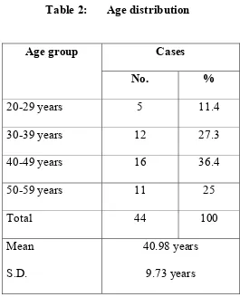
Table 2: Age distribution
Cases Age group
No. %
20-29 years 5 11.4
30-39 years 12 27.3
40-49 years 16 36.4
50-59 years 11 25
Total 44 100
Mean
S.D.
40.98 years
9.73 years
The age of the patients from 20-60 years with an average of 40
AGE DISTRIBUTION
SEX DISTRIBUTION
12
11 5
16
20-29 30-39 40-49 50-59
35
9
Table 3: DAS Score 28
DAS Score 28 Cases
Mild (< 3.1) 2 4.5
Moderate (3.2-5.1) 23 52.3
Severe ( > 5.1) 20 45.5
Total 44 100
Score
Range
Mean
S.D.
2.75 -5.81
4.8
0.78
This table shows that 2 people out of 44 (4.5%) had mild disease
and 23 people (52.3%) has moderate disease. 20 people (45.5%) had
severe disease. DAS 28 score ranges from 2.75 to 5.81 with a mean value
DAS 28 SCORE
MILD 4%
M ODERATE 52% Slice 4
0%
SEVERE 44%
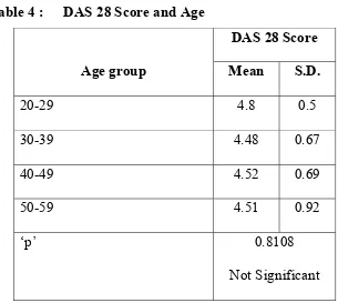
Table 4 : DAS 28 Score and Age
DAS 28 Score
Age group Mean S.D.
20-29 4.8 0.5
30-39 4.48 0.67
40-49 4.52 0.69
50-59 4.51 0.92
‘p’ 0.8108
Not Significant
Analyzing the above data age group doesn’t correlate with disease
activity and DAS 28 score
Table 5
Duration of illness
Duration of
illness(in years)
Range
Mean
S.D.
1 – 5
2.93
1.13
The duration of illness ranged from 1 year to 5 years with an average of
All 44 cases(100% )fulfilled the revised criteria of American rheumatism
association for rheumatoid arthritis
All the cases distal joints involvement( distal interphalangeal joints
are spared) 14 cases had both proximal and distal joint
involvement(31%). Joint deformity were present in 22 cases (50%)
rheumatoid nodules were present in 3cases(6%). Episcleritis was present
[image:48.612.188.423.358.495.2]in 3 cases(6%). Aortic Valve involvement was in 2 cases (4%)
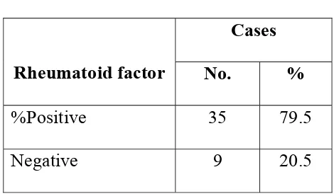
Table : 6
Rheumatoid factor positivity
Cases
Rheumatoid factor No. %
%Positive 35 79.5
Negative 9 20.5
Rheumatoid factor is positive in 35 cases (79.5%) and negative in 9
cases (20.5%).
Serum proteins were normal . There was no reversal of albumin / globulin
ratio and there was no hyperglobulinemia noticed in the study .Serum
27 patients showed radiological evidence of rheumatoid arthritis.
No patients had splenomegaly or significant generalized
lymphadenopathy.
Concomitant usage of NSAIDS 80% and corticosteroids 40% and
methotrexate 4% was present.
Anemia is defined as <11gm in females and <12gm in males as in
[image:49.612.103.447.442.490.2]most of the studies
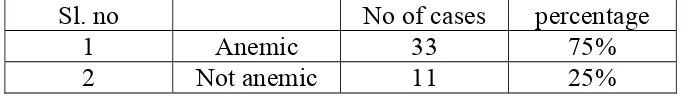
Table No 7
Anemia and rheumatoid arthritis
Among the 44 cases of rheumatoid arthritis 33 cases are anemic
(75%) and not anemic in 11 case (25%)Mean hemoglobin level in
patients was 10.67+1.83
Sl. no No of cases percentage
1 Anemic 33 75%
Table No 8
Anemia and rheumatoid factor positivity:
Anemic 26/33
Not anemic 6/11
P value 0.0305 significant
In patients who are anemic , number of rheumatoid factor
positivity was 87% and in not anemic patients rheumatoid factor
positivity was only 54%.
Mean Hb level in rheumatoid factor positivity was 9.11
gms+2.05s.d
Mean Hb level in rheumatoid factor negativity was 10.23+1.19s.d
When analysing the above values, anemia is one of the indicator of
disease activity and severity of rheumatoid arthritis
Anemia and ESR and Rheumatoid factor positivity
Anemic and non anemic patients were comparatively studied with
their erythrocyte sedimentation rate levels and seropositivity for
rheumatoid factor.
Out of 33 patients 32 patients have elevated ESR out of which
rheumatoid factor positive in 29 patients (90.6%) whereas in 11 non
factor positive (60%). The values suggest that the anemic patients have
more elevation of ESR and percentage of rheumatoid factor positivity is
also more in this group.
Anemia with disease duration
In 33 anemic patients 15 patients had more than 3yr duration (45%)
whereas in non anemic 11 patients only 1 had (9%) had disease more
than 3 year. When analyzing the above, the incidence of anemia
[image:51.612.130.431.345.650.2]correlated with the duration of disease.
Table 9 : DAS 28 Score and duration of disease
Duration of
disease
(in years) DAS 28 Score
Mean S.D.
Mild 2.0 -
Moderate 2.83 1.17
Severe 3.5 1.16
‘p’ 0.0471
Significant
When analyzing the above charts DAS 28 score was correlatedvery well
ANEMIA AND DURATION OF DISEASE
3.45 1.93
0 1 2 3 4
DURATION OF DISEASE
ANA
E
M
IA
Table10: Anemia and DAS 28 Score
DAS 28 Score
Anemia Mean S.D.
Absent 4.32 0.91
Present 5.04 0.58
‘p’ 0.0060
Significant
When analyzing the data incidence of anemia correlated with
activity of disease and anemic patients had higher DAS 28 score than
non anemic patients. P value is significant.
Table 11:Peripheral smear study
Types of anemia and rheumatoid factor positivity
No.o f pts percentag e Rheumatoi d positivity percentag e Microcytic hypochromic
8 18.2 8 100
Normocytic normochromi
c
20 45.5 17 85
Dimorphic 5 11.4 4 80
normal 11 25 6 54
When analyzing the above data 8 patients ( 18.2% )patients show
microcytic hypochromic anemia . Out of 8 patients all shows rheumatoid
PERIPHERAL SMEAR STUDY
ANEMIA AND DAS 28 SCORE
46% 11%
25% 18%
MH NN DA NORMAL
5.04 4.32 5.38 4.32 4.77 4.32 0 1 2 3 4 5 6 D A S 28 S C O R E
ANAEMIA NORM NORM IRON DEFI.
ANAEMIA
anemia. Out of 20 patients 17 patients (85%) were rheumatoid factor
positive. 5 patients shows dimorphic anemia. Out of 5 patients 4 patients
shows (80%) rheumatoid factor positive. But p value is 0.09 not
significant. That means the type of anemia doesn’t correlate with
rheumatoid factor positivity .
Percentage of types of anemia in anemic patients:
Iron deficiency anemia 25%
Anemia of chronic disease 60%
[image:55.612.132.455.329.590.2]Dimorphic anemia 15%
Table 12 : DAS 28 Score and Peripheral Smear Study
DAS 28 Score
Peripheral Smear Study Mean S.D.
Microcytic hypochromic anaemia 4.77 0.35
Normocytic normochromic anaemia 5.38 0.46
Dimorphic anemia 4.71 0.48
Normal 3.8 0.51
‘p’ 0.0001
Significant
When analyzing the above data anemic patients has more DAS 28
score than not anemic patients. Patients with normocytic anemia that
means anemia of chronic disease has high DAS 28 score ( 5.38) than iron
DAS 28 SCORE AND TYPES OF ANEMIA
4.77 5.38 4.71
3.8
0 1 2 3 4 5 6
DAS 28 SCORE MH
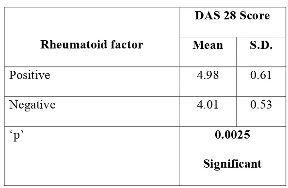
Table 13: DAS 28 Score and Rheumatoid Factor
DAS 28 Score
Rheumatoid factor Mean S.D.
Positive 4.98 0.61
Negative 4.01 0.53
‘p’ 0.0025
Significant
When analyzing the above data rheumatoid factor positive patients have
higher DAS 28 score than rheumatoid factor negative patients
Table 14:
Serum ferritin and DAS 28 score
The table shows that ferritin value correlates with severity of rheumatoid
arthritis.
DAS 28 sore Serum ferritin( mean &S.D )
Mild 50+24.5
Moderate 52.73+34.77
Severe 151.5+88.9
[image:57.612.129.426.106.300.2]DAS 28 SCORE AND RHEUMATOID FACTOR
DAS 28 SCORE AND SERUM FERRITIN
4.98 4.01 0 0.5 1 1.5 2 2.5 3 3.5 4 4.5 5
DAS 28 SCORE
RF + RF
-50 52.73 151.5 1 21 41 61 81 101 121 141 161 DU R A T IO N O F DI S E AS E ( in y e a rs )
Packed cell volume;
Mean value 34 + 5.3
<20% -1 patient -2%
20-30% --9 patients -20%
30-40% -22 patients -50%
[image:59.612.105.508.293.647.2]>40% -5 patients -11%
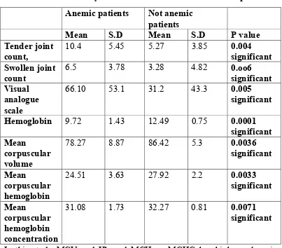
Table 15:
Clinical and laboratory features of anemic and non anemic patients:
In this study MCV and Hb and MCH an MCHC has higher values in
anemic patients than non anemic patients .p value was significant . In
DAS 28 score we are using the variables like swollen joints and tender
Anemic patients Not anemic
patients
Mean S.D Mean S.D P value
Tender joint count,
10.4 5.45 5.27 3.85 0.004
significant Swollen joint
count
6.5 3.78 3.28 4.82 0.oo6
significant Visual
analogue scale
66.10 53.1 31.2 43.3 0.005
significant
Hemoglobin 9.72 1.43 12.49 0.75 0.0001 significant Mean
corpuscular volume
78.27 8.87 86.42 5.3 0.0036
significant
Mean
corpuscular hemoglobin
24.51 3.63 27.92 2.2 0.0033
significant
Mean
corpuscular hemoglobin concentration
31.08 1.73 32.27 0.81 0.0071
joints and visual analogue scale and ESR. ESR already shows the highly
significant correlation with anemia and rheumatoid factor positivity and
disease activity. Swollen joints and tender joints and visual analogue
scale with anemia correlation was highly significant. P value was <0.05
Table 16:
Clinical and laboratory features of iron deficient anemic and
anemia of chronic disease patients
IDA Patients ACD Patients P value
Mean S.D Mean S.D
Tender joint count
5.54 3.57 10.55 7.40 0.006 significant
Swollen joint count
4.01 2.78 7.65 5.43 0.006 significant
Visual analogue scale
41.12 48.08 78.12 45.87 0.002 significant
hemoglobin 8.6 1.81 10.91 0.82 0.0019 significant Mean
corpuscular volume
76.84 5.87 83.97 5.0 0.002 significant
Mean
corpuscular hemoglobin
21.22 3.01 26.93 1.86 0.003 significant
Mean
corpuscular hemoglobin concentration
29.8 2.01 31.83 1.3. 0.0032significant
This table tells that Hb , MCV and MCH and MCHC are lower in
Iron deficiency anemia than anemia of chronic disease. Tender joint
iron deficiency anemia than anemia of chronic disease. P value was
significant in all variables.
WBC count:
Out of 44 patients 13 patients have leucocytosis (29.5%). No
patient had leucopenia.
Neutrophils:
50-70%- 8 patients -18%
>70%-36 patients -82%
Neutrophilia is present in 36 patients.
Lymphocytes:.
<20%-10 patients -23%
20-30%-18 patients -41%
>30%-16 patients -36%
Lymphopenia is present in23% of patients.
Eosinophils:
12patients (27%) have eosinophils >6%. All 12 patients are
rheumatoid factor positive.
Thrombocytes
Table 17:
Platelet count and rheumatoid factor positivity
When analyzing the above data thrombocytosis indicates disease
activity. P value was significant 0.04.
Table 18
Relationship between DAS 28 Score and other parameters
Value for cases with DAS 28 Score Mild Moderate Severe Parameter
mean S.D. mean S.D. mean S.D.
‘p’
PLT 2.75 1.35 3.49 0.93 4.09 0.97 0.0046 Significant
Eosinophil. 1 1.4 2.39 3.25 6.7 5.76 0.0001 Significant
CRP 6.8 5.5 23.13 24.5 53.85 19.9 0.0006 Significant
Ferritin 50 2.3 52.73 34.7 151.5 88.9 0.001 significant
ESR 25 14.1 43.24 12.2 67.25 25.5 0.0001 Significant
When analyzing the above data ESR and CRP, platelet count and
eosinophil count and ferritin are well correlated with disease activity .
that means if ESR and CRP and platelet count and eosinophil and ferritin
was high ,disease activity and DAS28 score was also high. All the other
parameters doesn’t correlate with DAS 28 score.
No. of
patients
Rheumatoid factor positive
percentage
>4 lakhs 14 12 85%
[image:62.612.111.544.335.523.2]Bleeding time ;
Normal in all patients.
Clotting time
Normal in all patients.
No patients had features of hyperviscosity syndrome and no
patient had a features of Felty syndrome and no patient had a feature of
pure red cell aplasia and no lymphoma and leukemia.
DISCUSSION
Rheumatoid arthritis is a chronic, systemic, inflammatory disorder
of unknown etiology that is a pattern of diarthrodial joint involvement. Its
primary site of pathology is the synovium of joints. The rheumatoid
factor positivity and extra articular manifestations commonly accompany
the joint disease , but arthritis is the major manifestation.
In our study we selected 44 cases of rheumatoid arthritis on
random basis as per the American rheumatism association guidelines
1987.
The sex distribution in this study , is predominantly affects
females in a ratio of 4:1.According to Harrison 17th edition, API text
book of medicine that the women are affected 3 times more than male.
In this study males are 20% and females are 80%. Doran Mf, Ponal Gr
et al (35) in his study males are 26.9% and females are 73.1%. Mean age is
58.5 years. In our study is 40.98 years.
Alamonsky, Yougari , Drosos et al (36) in his study the risk of developing
disease is greatest between 40 and 50 years. In our study the risk also is
between 40 to 49 years around 36.4 %.
Navarocaro Gregio et al (37)and Abach, R.R. Buchanan et al (38) in
of disease. In this study patients who had disease more than 3 year have
more DAS 28 score .The p value is significant and this shows that
duration of disease is directly proportional to the severity of disease.
B.Fleeb, L.Andel, J.Sautner et al(39), in their study the mean DAS
28 score was 4.23+1.2 .In this study mean DAS 28 score was 4.8+0.78.
Toshihisomatsui et al (40 )in their study reported 11.6% patients had
mild DAS 28 score and 51.4% has moderate DAS 28 score and 37.4%
patients had severe DAS 28 score. In this study patients with mild DAS
28 was around 4.5% and moderate score was 52.3% and severe 45.5% .
This states that most of the patients are in moderate severity.
Card Richard et al (41) in his study rheumatoid factor positivity was
80% and negativity 20%. In this study RF positivity was 79.5% and
while 20.5% RF was negative . The ratio of rheumatoid factor positivity
to negativity is 4:1
Tracey Houston et al (42)in their study ,mean hemoglobin in
rheumatoid arthritis patients was 9.57 gm% and in this study mean Hb is
10.6gm%.
M. Kar, S. Roy et al (43)in their study, mean hemoglobin level in
rheumatoid positivity patients was 9.57gm% and 10.45gm%. among
rheumatoid negative patients. In this study mean Hb level in rheumatoid
factor positive patients is 9.11+2.05 and mean Hb among rheumatoid
D.J. Borah ,Farhis Iqbal et al (44) in their study ,out of 20 anemic
patients 18 patients were rheumatoid factor positive (90%) and in non
anemic patients out of 11 patients 6 patients (54%) were rheumatoid
factor positive. In this study , out of 33 anemic patients 29 patients are
rheumatoid factor positive ( 87%) and in non anemic 11 patients patients,
6 (54%) are rheumatoid factor positive. This states that anemia is very
well correlated with rheumatoid factor positivity and disease activity.
Agarwal Sumeet et al(45) in their study, mean DAS 28 score in non
anemic patients was 3.83 compared to anemic patients which was 5.13.
In this study in non anemic patients mean DAS 28 score is 4.32 and in
anemic patients 5.04. D J Borah , Farhis Iqbal et al (44) in their study, in
non anemic patients mean DAS 28 score was 4.76 while anemic patients
it was 6.85.This states that anemic patients have more DAS score and
disease activity than non anemic patients.
Distinguishing ACD from iron-deficiency state in the setting of
chronic inflammatory process is difficult. Serum ferritin is a reliable
parameter to predict bone-marrow iron stores in uncomplicated anemic
states.
However, in chronic inflammation, ferritin is increased as a part of
acute-phase reaction and hence determining a cutoff level to differentiate
between iron-replete and depleted state becomes difficult. The value of
from 30 to 70 µg/l [46, 47]. We have taken a value of 50 µg/l as an indicator
of iron-deficiency in our study as it has been shown to have a specificity
of 81% and sensitivity of 100% [23]. Bone-marrow iron staining is
considered as the gold-standard in such conditions. However, the
procedure is invasive, time-consuming and expensive. Two recent studies
have shown that absent bone-marrow stainable iron may not truly be
representative of iron deficiency (48).
Soluble transferrin receptors (sTfR) level has been found to be
useful in distinguishing iron deficiency from ACD as it does not behave
as an acute-phase reactant. A high sTfR level although indicates
iron-deficient erythropoiesis, it does not necessarily indicate iron iron-deficient
state. A serum ferritin of 50 µg/l is as sensitive as sTfR in predicting
iron-deficiency [23]. However a combination of high sTfR ( > 2.5 mg/l)
and low ferritin ( < 50 µg/l) levels or a ratio of sTfR to log ferritin gave
higher specificity ( > 90%) than either of them alone in predicting IDA in
RA [23]. It may also help differentiate patients with mixed anemia from
pure ACD which is probably the most common in patients with RA.
Agarwal Sumeet et al (45),in their study, of rheumatoid arthritis
patients with iron deficiency anemia DAS 28 score was 4.7 and in
patients with anemia of chronic disease DAS 28 score was 5.69 . In this
anemia of chronic disease was 5.38. The p value is significant( 0.001)
.This states that DAS 28 score is higher in ACD than in IDA.
Anemia was defined as hemoglobin level of 11 g/dl in female and
12 g/dl in male patients the prevalence reported in RA patients from
Western countries [49] which varies from 33.3 to 59.1% although the
cutoff hemoglobin value used to define anemia in these studies was
higher than our study.
Most of the above studies used a definition of anemia close to that
suggested by World Health Organization (WHO) while we have used a
definition lower than that. Despite this we find that the prevalence of
anemia is considerably higher. If the WHO criterion had been used (men
Hb 13 g/dl and women 12 g/dl) then the prevalence of anemia in our
cohort would be 84.1%.
This is possibly related to high background prevalence of anemia in
general adult Indian population as well as poor access to medical care
leading to poor disease control of RA.
Anemic patients %
This study 75%
Peters et al (49) 64%
Baer etal(12) 76%
Remacha et al (51) 55%
Hotchberg et al (52) 40%
Baer et al (53) 27%
Types of anemia and rheumatoid arthritis
In this study iron deficiency anemia patients are less (24.5%)
because iron deficient anemia with inflammation(Dimorphic anemia )is
included separately(15%) and there is a probable folic acid and/or Vit
B12 deficiency .
Microcytosis (<80 femtolitre) in this study is 27% patients among
rheumatoid arthritis patients .Alexander et al (8) in their study showed
30% prevalence of microcytosis .
Hypochromia (less than 26pg) is present in 38% of rheumatoid
arthritis patients in this study. Caris J Bastley et al (55) reported 50%
hypochromia in their study.
Kadir et al (56) in his study, mean ESR was 36.6+23.5 .In their
study ferritin was 121.3 +34.2 ,and ESR ,CRP and ferritin showed
significant correlation with disease activity as well as DAS 28 score .
The p value was significant <0.001. In this study mean ESR is
53.5+22.3 and ferritin is 99.5+80.93 and ESR, CRP and ferritin are very
Peter et(49) JK sci (44) Agarwal(45) This study
ACD 65% 60% 51.6% 60.5%
IDA 23% 40% 48.4% 24.5%
well correlated with disease activity and the p value was 0.006 for ESR
and 0.0001 for CRP and ferritin respectively .
Sumeet Agarwal et al (45) and D J Borah , Fahler Iqbal et al (44) in
their study, the variables used in calculating DAS 28 score like tender
joint count , swollen joint count , ESR and visual analogue scale was
correlated significantly. The p value was more significant in anemic
patients than non anemic patients .In this study also all the 4 variables
shows high significance in anemic patients compared to non anemic
patients.
Agarwal et al (45) in their study; tender joint count , swollen joint
count , ESR and visual analogue scale in patients with anemia of chronic
disease showed higher value than iron deficiency anemic patients.
Similar results were obtained in all the variables showing higher
significance in patients with anemia of chronic disease than iron
deficiency anemic patients in this study. The p value is significant.
Hutchuson et al (57) and Dulaguist et al (58) in their study
thrombocytosis in rheumatoid arthritis was 52% and 60% respectively
Alof Selross et al (59) in their study ;thrombocytosis was present in 33%
of patients and it correlated with disease activity. In our study
thrombocytosis is present in 31%. Patients with thombocytosis have
M.Kar , S.Roy et al (43) in their study ;eosinophilia was present in
20.45% of rheumatoid arthritis patients. R J Windchester et al (60) and
Short Bauer et al (61) in their study eosinophilia was present in 40% and
10% of patients of rheumatoid arthritis respectively which correlated
with disease activity . In this study eosinophilia is present in 27%
patients which correlated very well with DAS 28 score and disease
activity with significant p value .
M.Kar S.Roy et al( 43) in their study leucocytosis was present in
20.7% and leucopenia was present in 11.1% patients and lymphopenia
was present in 6% patients. In this study 29.5% patients have
leucocytosis. None of the patient had leucopenia .Lymphopenia is
present in 23% patients. Leucopenia is lacking in this study because
we didn’t take patients with longer duration disease for study group and
lymphopenia is present higher than M.Kar S.Roy study ,probably due
to steroid therapy .
Luis A, Toro Jimenez et al (62) in their study of 214 rheumatoid
arthritis patients 12 patients had M spike with mean age of M spike
being 69 years. IgG gammopathy was present in 50% patients. 1 patient
was diagnosed as multiple myeloma and 1 patient had a primary
leukemia and 4 patients diagnosed as a myelodysplastic syndrome and 4
patients were diagnosed to have monoclonal gammopathy .
In this study no patient has hyperglobulinemia and myelodysplastic
syndrome, leukemia or lymphoma or multiple myeloma and no patient
had a large granular lymphocytosis syndrome.
Agarwal Sachdev et al (63) reported pure red cell aplasia and
immune thrombocytopenia. In this study no patients had
thrombocytopenia and pure red cell aplasia. No patient had decreased red
cell distribution width .
Abach , R,R,Buchnan et al(38) reported a hyperviscosity syndrome
in rheumatoid arthritis. In this study no patient had symptoms
suggestive of hyperviscosity syndrome
Paraiaz , Fayaz etal (33) study none of the patient of rheumatoid
arthritis had evidence of bleeding and DIC. Only isolated abnormalities
of coagulation was present. Protein C and protein S was low in 1case
each while decreased factor VIII level was detected in 5 cases.
Hypofibrinogenemia was demonstrated in 1 case. All patients had
normal factor IX level. In this study no patient had evidence of bleeding