MULTIPLE MILLER’S CLASS I GINGIVAL RECESSION - A 6 MONTHS COMPARATIVE STUDY
Dissertation submitted to
THE TAMILNADU Dr. M.G.R. MEDICAL UNIVERSITY
In partial fulfillment for the Degree of
MASTER OF DENTAL SURGERY
BRANCH II
PERIODONTICS
This dissertation is the result of a lot of effort that has gone in to its making and I wouldn’t be justified if I do not acknowledge the people who stood beside me, helping me accomplish this task.
I extend my sincere thanks to Dr. S. Ramachandran, MDS, Principal,
Ragas Dental College and Hospital, for his support and guidance during my postgraduate course at Ragas Dental College.
I would be failing in my duty if I do not adequately express my deep sense of gratitude and my sincere thanks to my guide Head of the Department, Professor Dr. T.S.S.KUMAR, MDS, Department of Periodontics Ragas Dental college and Hospital, Chennai, for his exceptional guidance, tremendous encouragement, well-timed suggestions and heartfelt support throughout my postgraduate programme which has never failed to derive the best out of me. I would like to profoundly thank him for giving an ultimate sculpt to this study.
Professor, Department of Periodontics, Ragas Dental College, for his valuable advice and encouragement during my postgraduate course. I am deeply grateful to him for his patience, support, and guidance during the study process, without whose intellectual insight, guidance in the right direction, this dissertation would not have been the light of the day.
I extend my sincere heartfelt thanks to Dr. Shiva Kumar, MDS, Reader, Department of Periodontics, Ragas Dental College, for helping me during my post graduate course and encouragement.
I extend my heartfelt thanks to Dr. Avaneendra Talwar, MDS, Reader,
for his guidance and support in all my academic activities,
I extend my thanks to Dr. Ramya Arun, MDS, Lecturer, without whom the study wouldn’t have been completed; I convey my gratitude wholeheartedly for her support and patience.
my course and giving me constant support and encouragement.
My sincere thanks to the Bio-statistician, Mrs.Deepa, from Department of Oral and Maxillopathology, Ragas Dental College and Hospital, Chennai.
I extend my thanks to Mrs. Parvathi, Mrs. Subhulakshmi, Mr.Chellapan, and Mrs.Rosamma for their timely help throughout the tenure.
Last but not the least, even though words wouldn't do much justice, I would like to specially thank my Parents for their blessings, love, and best wishes of my family for being with me and helping me realize all my dreams.
bFGF- Basic fibroblast growth factor
CTG- Connective tissue graft
CAL- Clinical attachment level
CEJ- Cemento enamel junction
DAT- Direct attachment to tooth
ECM- Extra cellular matrix
EGF- Epidermal growth factor
GT- Gingival thickness
GTR- Guided tissue regeneration
PRF- Platelet rich fibrin
PDGF- Platelet Derived Growth Factor
PRP- Platelet Rich Plasma
PD- Probing depth
rCAL- Relative clinical attachment level
RC- Root coverage
RD- Recession depth
SD- Standard Deviation
TGF- Transforming growth factor
VEGF- Vascular Endothelial Growth factor
SL NO INDEX PAGE NO
1. INTRODUCTION
1
2. AIMS AND OBJECTIVES
4
3.
REVIEW OF LITERATURE
5
4. MATERIALS AND METHODS
34
5.
RESULTS
47
6. DISCUSSION
53
7. SUMMARY AND CONCLUSION
59
8. BIBILIOGRAPHY
1 Group-A Test group width of keratinized tissue
2 Group-B Control group width of keratinized tissue
3 Group-A Test group Probing depth
4 Group-B Control group Probing depth
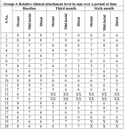
5 Group-A Test group Relative clinical attachment level
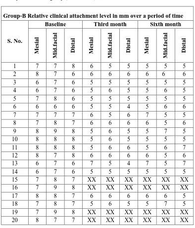
6 Group-B Control group Relative clinical attachment level
7 Group-A Test group Recession depth
8 Group-B Control group Recession depth
9 Group-A Test group Thickness of gingiva 10 Group-B Control group Thickness of gingiva
11 Group-A Intra group comparison of mean clinical parametric values at different time intervals
12 Group-B Intra group comparison of mean clinical parametric values at different time intervals
13 Inter group comparison of mean clinical parametric values at different time intervals
14 Group-A Intra group comparison of mean Gingival Phenotype at different time intervals
15 Group-B Intra group comparison of mean Probing depth at different time intervals
16 Group-B Intra group comparison of mean Relative clinical attachment level at different time intervals
17 Group-B Intra group comparison of mean Recession depth at different time intervals
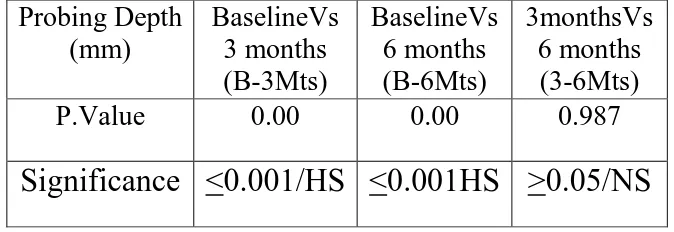
1 Group-A Intra group comparison of mean Probing depth at different time intervals
2 Group-B Intra group comparison of Probing depth at different time intervals
3 Group-A Intra group comparison of mean Relative clinical attachment at different time intervals
4 Group-B Intra group comparison of mean Relative clinical attachment at different time intervals
5
Group-A Intra group comparison of mean Recession depth at different time intervals
6
Group-B Intra group comparison of mean Recession depth at different time intervals
7
Group-A Intra group comparison of mean Width of keratinized Gingiva at different time intervals
8
Group-B Intra group comparison of mean Width of keratinized Gingiva at different time intervals
9
Group-A Intra group comparison of mean Thickness of gingiva at different time intervals
Many surgical techniques have been proposed for the correction of root
exposure. Among these the Connective Tissue Graft (CTG) techniques has
been reported as offering the best results in terms of Root coverage (RC).
However CTG require a second surgical site to harvest the graft, resulting in
discomfort for the patient. The use of platelet concentrate (PRF) avoids need
for second surgical site, and is enriched with growth factors.
Aim:
The aim of the present study to compare and clinically evaluate
Bilaminar technique using Connective Tissue Graft (CTG) and Platelet Rich
Fibrin (PRF) in the management of multiple Miller’s class I gingival recession
situations.
Materials & Methods:
Twenty patients (age group 18-40 years) were selected from the
outpatient department of Periodontics , Ragas Dental college & Hospital,
Chennai-119, with probing depth <3mm and seeking treatment for Miller’s
class I multiple gingival recessions were enrolled into this study. The selected
patients were randomly assigned to one of either group. Group-A (TEST
Width of keratinised gingiva, and Gingival phenotype. The data were
statistically analysed and the significance were co-related.
Results:
Mean Recession depth, Relative clinical attachment loss and Probing
depth were reduced in both the groups, but between groups it was statistically
significant towards control group, from baseline to 6 months’ time period.
There is no any significant relation between and within groups in regarding
Width of keratinised gingiva. But the gingival phenotype showed statistically
significant difference between and within groups.
Conclusion:
From the above study, it was elicited that PRF was not able to achieve
predictable root coverage; only increasing in gingival biotype is seen. Thus in
1
INTRODUCTION
Periodontal plastic surgery not only addresses the functional problem
but also satisfy esthetic needs of the patients. One of the common squeal of
periodontitis is gingival recession. Gingival recession is defined as the
displacement of the gingival tissue margins apical to the CEJ.7 Studies on the
Prevalence and incidence of gingival recession had been analyzed by various
authors, Albander and Kingman52 studied prevalence of gingival recession
among 9,689 subjects aged between 30-90 years. They found prevalence of
1mm or more recession in people aged 30 years and older was 58 % and
increased with age. Prevalence and severity were seen at buccal, than at
interproximal surfaces of teeth.40, 17 Gorman35 found the frequency of gingival recession with age and was greater in men than in women of the same age
group. Cause of gingival recession is multifactorial in nature such as age,74 various anatomical factors such as Fenestrations and Dehiscence,99,64 abnormal tooth Position,33,6 aberrant Frenum,81,92 gingival Biotype51,8 etc.
Even, pathological condition such as trauma from Occlusion, 55, 30 Vigorous tooth Brushing,78, 98 History of periodontal disease,35,49,5 have been implicated.
Many people may exhibit generalized gingival recession without
having any awareness of the condition or others often are anxious about
gingival recession for reasons such as fear of Tooth loss,90 Dentinal
2
1968,93 These classification were based on papilla height,77 width of attached gingiva, vertical and horizontal component of the gingival margin. The
Rationale of treating gingival recession is not only to achieve complete
coverage of the root,3 additionally eliminating plaque trap area, decreasing
hypersensitivity and preventing root caries. Numerous surgical techniques
have been proposed to treat marginal gingival recession,63,43,12 In a broader perspective these treatment modality can be grouped under pediculate and
non-pediculate graft procedures. Based on the surgical technique systemic
review on literature have showed that connective tissue graft (CTG) pioneered
by Edel28 in 1974 later modified by Langer and Langer 63 with pedicle flap (Bilaminar technique) is currently considered to be a better surgical option in
terms of attaining maximum percentage of root coverage. In terms of
predictability for this procedure is 52-97.4% respectively. Especially in
esthetic demanding region connective tissue graft seems to be a better choice
of graft. Karring58 showed that the primary determinant of tissue specificity rests within the connective tissue graft. Even though connective tissue graft is
the gold stranded procedure, the disadvantages are the second surgical site, the
patient discomfort, post-surgical pain, and bleeding from the donor area.42 Numerous alternate viable materials such as guided tissue regeneration
3
Use of different materials for root coverage, obtaining predictable and
esthetic root coverage has become an important goal of periodontal plastic
surgery. A recent innovation of platelet rich fibrin as an autologous source is a
breakthrough in the field of medicine which enhances and stimulates soft and
hard tissue healing.86,29 The clinical application of platelets rich plasma has been extended in medical and dental fields. Platelet concentrate have been
successfully applied in periodontal therapies such as in implant therapy105,
socket preservation,45 GBR procedure,56 GTR procedures either as sole grafting material or in combination with other materials. These Platelet
concentrate contains PDGF, TGF and many other growth factors that
modulate and up regulate the biomimetic of the tissue healing.27 Considering the novel property of platelets the present study was
undertaken to find out the clinical effectiveness and evaluate the platelet rich
fibrin and connective tissue graft in conjunction with coronally advanced flap
in the management of multiple Millers class-I gingival recession.
4
AIMS AND OBJECTIVES
Present comparative clinical study aims to compare and clinically
evaluate Bilaminar technique using Connective tissue graft (CTG) and Platelet
rich fibrin (PRF) in the management of multiple Miller’s class I gingival
5
REVIEW OF LITERATURE
Among the various determinants, the gingival components have an
important bearing on the esthetic nature of the smile in an individual. The
term “Periodontal plastic surgery” includes “Mucogingival surgery”. Until the
early 1980s, mucogingival surgery was predominantly focused on the
functional reconstruction of the gingival complex, with root coverage being a
subordinate consideration. In the late 1980s, new surgical techniques, such as
the epithelialized thick free mucosal graft and the sub-epithelial connective
tissue graft, led to improved and more predictable outcomes of root coverage.
With these then newly developed surgical techniques, clinicians became more
capable of addressing the increased aesthetic demands presented by patients.
Various procedures to improve aesthetics have a different requirement for
success compared with surgery aimed at improving periodontal health. The
patient plays a more important role in determining success in aesthetic
procedures. The surgeon has the responsibility to clearly outline the biological
possibilities, and careful examination of the expected surgical parameters is
essential prior to initiation of mucogingival procedures.
Gingival recession is characterized by the displacement of the gingival
margin apically from the cementoenamel junction, or CEJ.7
Gingival recession can be localized or generalized and be associated
with one or more surfaces. Among the many factors that might predispose a
patient to generalized as well as localized gingival recession, trauma caused by
6
al 1993 & O’Leary 1971.78 Other precipitating factors include anatomical
variations such as abnormal frenulum Donaldson 1973 24tooth malpositioning
Pini Prato et al 1996 82, overhanging restorative and surgical procedures and
aging PiniPrato82 et al 1996. Gingival recessions may results in
hypersensitivity impaired esthetics and root caries Hall39 1989 &
Anson 1999.10
Since the presentation of gingival recession varies widely in the
population, classification systems have been established to better describe it.
Sullivan & Atkins 1968 93 used the descriptive terms “narrow”, ”wide”, ”shallow” and “deep” to classify recession into 4 groups.
Mlinek et al 197372 quantified the gingival recession as “shallow narrow” clefts as being < 3mm in both dimensions.
Miller 1985 71 proposed 4 classes of marginal tissue recession he
classified gingival recession according to the height of the Inter proximal
papillae adjacent to the defect area.
Class I: Marginal tissue recession that does not extend to the
mucogingival junction, with no periodontal loss (bone or soft tissue)
in the interdental area. One hundred percent root coverage can be
7
Class II: Marginal tissue recession that extends to or beyond the
muco-gingival junction, with no periodontal loss (bone or soft
tissue) in the interdental area. One hundred percent root coverage
can be anticipated.
Class III: Marginal tissue recession that extends to or beyond the
muco-gingival junction. Loss of interdental soft and hard tissue
apical to the cemento enamel junction but coronal to the level of
recession, Partial root coverage can be anticipated.
8
Class IV: Marginal tissue recession that extends to or beyond the
mucogingival junction. Loss of interdental soft and hard tissue,
apical to the area of recession.
Norland and Tarnow et al 1998 77 classified gingival recession based
on their papillary height.
Management of gingival recession include different surgical
techniques, they can be broadly classified as
1. PEDICLE GRAFTS
A. Rotational flaps
- Laterally positioned flap
- Obliquely rotated flap
9 B. Advanced flaps
- Coronally positioned flap
- Semilunar flap
2. FREE SOFT TISSUE GRAFTS
A. Epithelialized (Classical Gingival Graft)
B. Non-epithelialized soft tissue graft.
3. COMBINATION GRAFTS
A. One Stage procedure
- Connective tissue graft plus pedicle graft
- Biodegradable membrane barrier plus pedicle graft
B. Two Stage procedure
- Coronally positioned previously placed soft tissue graft
- Non-biodegradable membrane barrier plus pedicle graft.
In any surgical technique it is important to differentiate between
success and predictability with regard to root coverage procedures. Success
10
predictability describes the percent of the treated teeth in which complete root
coverage is achieved.
Connective tissue graft:
The connective tissue graft was first used by Edel28 1974, Broome
and Taggart15 1976 and Donn25 1978 used CTG to increase the width of
keratinized gingiva.
B. Langer and L. Langer63 initially introduced sub-epithelial
connective tissue graft technique in 1985 and outlined the indications and
procedure for the same. Nelson75 in 1987 modified it to further increase
clinical predictability. He attained gain in clinical predictability is by use of
the Bilaminar flap design to ensure graft vascularity (from the bed and the
overlying flap) and a high degree of gingival cosmetics from the secondary
intention healing of the connective tissue graft.
Nelson 1987 75 in his controlled clinical study has reported mean root
coverage of 88%, though Raetzke 198583 reported the root coverage of
60%-83%, Harris 199243 showed much higher (97%) root coverage in his study.
Also it provides excellent aesthetics with good gingival colour match and
minimal likelihood of keloid formation. Contrary to the free gingival graft,
here the donor site wound is less extensive and haemorrhagic and perhaps less
annoying to the patient.
Wennstrom in 1996102, in a literature review of connective tissue
11
This was the highest among all root coverage procedures analysed. Root
coverage achieved using the connective tissue graft procedure is extremely
stable, and thus this procedure is taken as a “Gold Standard” while evaluating
the efficacy of other techniques.
Yong-Moo Lee et al 2002,103 In a, 3-year longitudinal study
evaluation of sub-pedicle free connective tissue graft for gingival recession
coverage. The results indicate that the connective tissue graft with a partial
thickness coronal advancement pedicle is a predictable method for root
coverage and, provided with optimal maintenance care, the clinical outcomes
gained by this technique can be well maintained.
Cetiner D et al 200418 described a technique, EXPANDED MESH
CONNECTIVE TISSUE GRAFT (eMCTG) for the treatment of multiple
gingival recessions in 52 buccal gingival recessions to evaluate the
effectiveness and predictability. The results demonstrated that the use of
eMCTG technique allowed the treatment of multiple adjacent recessions with
adequate wound healing and highly predictable root coverage. This procedure
can be applied favourably in treating multiple gingival recessions in one
surgery.
Harris R.J et al 200746 in a controlled comparative study, the clinical
root coverage achieved with two connective tissue grafts that were removed
from the same donor site at different interval and used in sub epithelial grafts
12
in recession, probing depth, width of keratinized tissue and attachment level.
The second connective tissue graft produced greater mean root coverage than
the first connective tissue grafts.
Harpreet Singh Grover et al in 201141, in a case report sub epithelial
connective tissue graft is used to assess the increasing gingival biotype. The
results emphasized complete root coverage along with more favourable
phenotype.
Surgical technique for Connective Tissue Graft:
Broome & Taggart 197615 used a Brasher-Rees Knife for securing the
connective tissue graft after reflection of the primary partial thickness flap.
Langer & Langer 198563 described the SUBEPITHELIAL
CONNECTIVE TISSUE GRAFT technique for covering gingival recessions
of both single and multiple adjacent teeth. He used a bilaminar procedure with
the combination of connective tissue and epithelium taken from the inside of a
palatal and placed under a partial thickness flap over a denuded root. The
results of the study showed high success rate of 85%.
Edel 199428 employed a trap door approach with three incisions to
harvest connective tissue graft without epithelium.
A different version of connective tissue grafts called as “envelope
13
Nelson 198775, Harris 199243 used bilaminar flap design to assure
graft vascularity and a high degree of gingival cosmetics from the
secondary-intention healing of the connective tissue graft. This seems to avoid the tire
patch look often associated with free gingival grafts. Nelson 1987 described
the SUBPEDICLE CONNECTIVE TISSUE GRAFT (SCTG) technique
which combined a connective tissue graft with a full thickness double papilla
graft to cover the denuded root. It was further modified by Harris (1992) by
using a connective tissue graft with a split thickness double papilla graft.
Bruno J.F.199416 presented some modifications of the original Langer
& Langer technique for root coverage on areas of wide denudation. He
suggested that mesiodistal length of the incision can be extended to provide
easy access to the denuded root without the use of vertical incisions.
Blanes R.J & Allen E.P 199913 described a surgical technique for the
treatment of adjacent soft tissue marginal recession. The technique combined a
tunnel procedure with double lateral pedicle flaps to cover a connective tissue
graft. These techniques proposed to compensate for the lack of blood supply
usually associated with the tunnel technique in deep or adjacent wide
recession. They observed 95% root coverage with these procedure.
Huzeler M.B & Weng D,50 1999 described and demonstrated a new
and simplified surgical approach to harvest subepithelial connective tissue
14
parallel to the gingival margin was used to access the donor site for graft
preparation and harvesting grafts of variable sizes and thickness were obtained
Since no band of epithelium was removed with the connective tissue graft, the
palatal donor site could heal with primary intention. No stents or hemostatic
agents were necessary to cover the donor site post-operatively and suturing
was reduced to a minimum.
Surgical technique comparative reviews:
Ouhayoun J.P.et al 199480 comparative clinical study of two techniques
of subepithelial connective tissue grafts for root coverage which differed with
respect to the use of epithelial collar of the graft. After a six months follow-up,
they demonstrated that both the procedures could accomplish root surface
coverage in class I and class II recessions with reasonable esthetic results.
They suggested that removal of epithelial collar gives better esthetic results.
Cordioli G et al 200120 retrospective clinical study of 1-1.5 years to
compare 2 techniques of sub-epithelial connective tissue graft in the treatment
of Millers class-1 and class-2 gingival recessions to evaluate root coverage
and mucogingival changes. The treatment outcome in terms of keratinized
tissue width seems to be correlated with the pre-surgical gingival dimensions
and the height of the grafted tissue that is left exposed coronal to the flap
15
Da Silva R C et al 200421 in a randomized clinical trial compared the
coronally positioned flap alone or in conjunction with a sub-epithelial
connective tissue graft in the treatment of gingival recession in 11
non-smoking subjects. The results indicate that both surgical approaches are
effective in addressing root coverage. When desired gain in gingival
dimensions is needed, then the combination technique should be used.
Tolga S.Tozum. et al 200596 Comparisson of two techniques Langer
and Langer & modified tunnel of sub-epithelial connective tissue graft for the
treatment of gingival recession suggest that the use of SCTG in combination
with a tunnel procedure may results in an increased amount of root coverage
and clinical attachment gain compared to the Langer and Langer technique.
CTG vs Other Grafts Materials:
Harris R.J.200447 conducted a study to evaluate the short and long
term root coverage results obtained with an acellular dermal matrix and
sub-epithelial graft and concluded that sub-sub-epithelial graft is a better procedure to
produce more predictable and stable long term root coverage results.
McGuire M.K.200668 Done a comparative study of recombinant
human platelet-derived growth factor-BB plus beta tricalcium phosphate and a
collagen membrane to sub-epithelial connective tissue grafting for the
16
tissue response to rhPDGF-BB+β-TCP and a collagen membrane and
comparable clinical outcomes to connective tissue graft.
Suichi S et al 200689 Treated Millers class III recessions with enamel
matrix derivative (emdogain) in combination with subepithelial connective
tissue grafting. Soft tissue coverage of the root surfaces was achieved
clinically and radiographs showed improvements in the interproximal bone
defects
Joly J.C et al 200754 In a comparative clinical study over a 6 months
period among 10 patients isolated gingival defects using a coronally
positioned flap associated with sub-epithelial connective tissue grafts or an
Acellular dermal matrix graft were analysed. The results revealed statistically
significant greater gain in clinical attachment level, recession depth, and
gingival thickness in sub- epithelial tissue graft and no differences were found
in keratinized gingiva and probing depth.
E.P.Rosetti et al 200726 done an 18 months comparative study
between sub-epithelial connective tissue graft and guided tissue regeneration
for the treatment of gingival recession. It was concluded that gingival
recessions treated with sub-epithelial connective tissue graft group were
superior for gingival recession height (GR), root coverage (RC) and
keratinized tissue width (WKT) clinical parameters, while guided tissue
17
Jung S. Han et al 200857 Comparative clinical study to investigate
the changes in gingival dimensions and root coverage using the same surgical
procedure but varying the amount of the connective tissue graft left uncovered.
Both procedures resulted in successful root coverage with an increase in the
width of keratinized tissue. Conclusion of the study showed that a portion of
the graft exposed resulted in a greater increase in keratinized tissue, and
complete coverage of graft resulted in greater root coverage.
Keceli H.G 200860 in a comparative clinical study, between
connective tissue graft +platelet rich plasma with connective tissue graft alone
in the treatment of gingival recession were studied, the results showed no
difference between connective tissue graft and connective tissue graft with
platelet rich plasma.
Sandro Bittencourt et al 200988in his clinical study, to find the root
coverage outcomes for Millers class 1 recessions obtained at 6 months using
semilunar coronally positioned flap (SCPF) or with sub-epithelial connective
tissue grafts, were resulted in 89.25% and 96.83% respectively. And there
were no significant differences between the two groups with regard recession
depth, recession width, width of keratinised gingiva, probing depth and
clinical attachment loss.
Katrin Nickles et al 201059 In a 10 year study for Millers class 1 and
18
using bio-absorbable barriers, the results at 6 months were less significant
compared to respective baseline, at the end of 12 months showed the results
between these two groups as, Connective tissue graft caused more
post-surgical discomfort but it resulted in a better outcome than GTR as perceived
by patients. The long term stability of root coverage is more significant than
GTR.
Nevins M.L 201076 A case series of tissue engineered bilayered cell
therapy (LCT) for the treatment of oral mucosal defects. A bilayered construct
of allogenic viable neonatal cell comprised of a lower fibroblast layer and an
upper keratinocyte layer which appear to promote healing by providing the
wound with extracellular matrix and expressing cytokines. It is not
commercially available.
Giulio Rasperini et al 201134 clinical outcomes of a connective
tissue graft alone or in combination with enamel matrix derivative (EMD) in
the treatment of Miller’s class 1/11 recessions. The mean recessions reduction
was 3.9+0.8mm for EMD treated sites, and 3.6+1.5mm for the CTG alone.
Corresponding root coverage was obtained in 62% of test sites compared to
47% in the control group.
Mauro Pedrine Santamaria et al 201167 In an experimental study,
the connective tissue graft with resin glass ionomer for the treatment of
19
and the result provided statistically significant gains in clinical attachment
level and shallow probing depths. The percentage of cervical lesions height
covered was 74.0%+22.90. Thus showed resin modified glass ionomer filling
did not interfere with coverage achieved by the connective tissue graft.
Histologic review:
Harris R.J 199944 In a histologically study in humans using a
connective tissue graft combined with a partial thickness double pedicle graft.
He observed two different healing patterns. The first was characterized by a
long junctional epithelial attachment that extended well beyond the original
gingival margin and occasionally almost to the original bone level with
minimal connective tissue adjacent to the tooth. The other pattern was a short
junctional epithelium that stopped at the previously exposed root surface.
There was predominately connective tissue adjacent to the tooth with some
isolated areas of epithelium. Also new bone or cementum was seen. He
concluded that though the procedure was successful clinically, it produces no
true regeneration but heals only through repair.
Guiha et al 200137 done an animal study for histological evaluation
of healing and revascularization of the sub-epithelial connective tissue graft.
The vascularization of the connective tissue originates from the periodontal
plexus, and the overlying flap. The attachment of the graft to the root surface
20
connective tissue attachment. There is little potential for new cementum and
new bone formation.
Fabricia Ferreira Suaid et al 200831 In an Histometric study in dogs
comparing the healing process for treating gingival recessions using platelet
rich plasma with connective tissue graft and connective tissue graft alone and
the results obtained were, a greater length of new cementum was observed in
the sites treated with Platelet rich plasma and Connective tissue graft (
2.18+0.78mm) compared to the control group (1.19+0.62mm). No statistically
significant differences were observed in the remaining parameters. Thus
combination of PRP with CTG was more effective in promoting new
cementum formation than the graft alone.
Antonio Scarano et al 200911 To evaluate clinically, histologically
and ultra-structurally the Acellular dermal matrix used in treatment of
increasing the width of keratinized gingiva. Results obtained were clinically
gained keratinized gingiva of 2.92+0.65mm observed after 3 months. After 6
weeks it was difficult to find, the acellular dermal matrix pre-existing collagen
fibres.
Michael K et al 200969 On examination of the histologic and micro
computed tomographic outcomes of the treatment of gingival recession defects
either a connective tissue grafts or 0.3 mg/mL recombinant human platelet –
21
months, sites treated with rhPDGF-BB+B-TCP showed connective tissue
fibres perpendicularly inserting into newly formed cementum and alveolar
bone. In CTG sites a long junctional epithelium was seen coronal to the
osseous crest and connective tissue fibres ran parallel to the adjacent root
surfaces, with no evidence of insertion into cementum or bone.
Systematic Review:
In recent years, many systematic reviews were published focussing on
the effect of root coverage procedures for the treatment of localised gingival
recession like Chambrone19 in 2008, Oates79 in 2003, Roccuzzo84 in 2002.
These authors reported that different surgical techniques and flap designs had
been described and used in an attempt to correct gingival recession producing
statistically significant improvements in gingival recession and clinical
attachment level. Since the common occurrence of recession areas involving
localised or adjacent teeth, evidence based information associating the results
achieved by different surgical techniques can be considered as an important
22
23
25 Platelet concentrate:
Since 1990, a greater understanding of wound, soft tissue and bone
healing has revealed that there are several components within blood
constituents, e.g fibrin, fibronectin, vitronectin, PDGF, TGF-B that are part of
the natural healing process, which can be altered or accelerated by
concentrating these factors.9
Platelet Biology:
Platelets are the end products of megakaryocytes and are formed in bone
marrow. They have no nucleus and cannot replicate, thus the life span of
platelets is 5 to 9 days. During activation, the alpha granules within platelets
fuse with the platelet plasma membrane and release some of their protein
contents to the surrounding called as degranulation. The alpha granules in
platelets contain more than 30 bioactive proteins, many of which have a
fundamental role in hemostasis and or tissue healing Anitua E et al in 20049.
These proteins include PDGF (αα, ββ, αβ isomers), TGF-β (both β1, β2
isomers), platelet factor 4, interleukin-1, platelet derived angiogenesis factor,
VEGF, epidermal growth factor, platelet derived endothelial growth factor,
epithelial growth factor, insulin like growth factor, osteocalcin, osteonectin,
fibrinogen, vitronectin, fibronectin, and thrombospomdin-1 Harrison P et al
in 199348 platelets begin actively secreting these proteins within 10 minutes
26
secreted within 1 hour. After the initial burst of PRP-related growth factors,
the platelets synthesize and secrete additional growth factors for the remaining
several days of their life span, Marx RE et al in 200466
Samir Mehta et al in 200886 in his study platelet activation in response
to tissue damage and vascular exposure results in the formation of a platelet
plug and blood clot to provide hemostasis and
the secretion of biologically active proteins.
The composition of this naturally occurring
hematoma is 95% red blood cells, 4% platelets
and 1% white blood cells. But blood clot
enriched with platelets reveals dramatic
difference in its composition compared to
natural clot with 95% platelets, 4% red blood
cells, and a similar amount of white blood cells.
Wound healing can be enhanced and speeded by the use of a platelet
gel that is harvested from the patient’s own plasma a few minutes before it is
used. The platelet rich plasma (PRP) consists of
1.Concentrated fibrin
2.Stem cells
27
Keceli H.G 200860 compared connective tissue graft +platelet rich
plasma with connective tissue graft alone in the treatment of gingival
recession. No difference could be found between connective tissue graft and
connective tissue graft with platelet rich plasma.
Sanchez et al 200387 have elaborated on the potential risks associated
with the use of PRP. It has been discovered that use of bovine thrombin used
in preparation of PRP may be associated with the development of antibodies to
the factors V, XI and thrombin, resulting in coagulopathies. Bovine thrombin
preparations have been shown to contain factor V, which could result in the
stimulation of the immune system when challenged with a foreign protein.
David M Dohan et al in 200622 In a laboratory analysis, 10ml blood was
collected from 15 healthy volunteers without anticoagulant, PRF was prepared
according to PRF protocol, compared PRP and PRF, showed no significant
difference between the cytokine measurement. Secondly, the values obtained
in PRF clot exudates are all significantly higher than those measured in plasma
and sera samples. Among cytokines measured IL-1B, IL-16 and TNF-A to
anti-inflammatory cytokines, such as IL-4. Were identical for PRP and PRF.
Only the VEGF was an exception, with particularly high serologic
concentrations in PRF.
Griffin TJ et al in 200436 in a case report by using platelet concentrate
28
showed complete root coverage was achieved, optimal esthetic results, with
excellent soft tissue contour and texture were observed.
Mark P Kraver 201165 et al Trapping the platelets and leukocytes
inside the fibrin clot helps in many ways.
1. Transforming growth factor beta, is a protein that assists in cellular
differentiation and proliferation
2. Platelet derived growth factor helps bring in mesenchymal stem cells
into the areas as well as differentiate and proliferate endothelial cells
3. Insulin like growth factors helps healing cells from dying so fast and
continue healing longer
4. Growth factors will work on type 1 collagen to form fibroblasts and
osteoblasts
5. Release cytokines to attract the healing response of the body
6. Leukocytes will enhance the body’s own inflammatory process to heal
quicker
7. Fibrin network increases the blood flow into the area with vascular
endothelial growth factor
8. Neutrophil degrades the site for wound remodeling and bring in the
macrophages to clean up the site.
The newer extract known as platelet rich fibrin (PRF) is a second
29
healing. Its advantages over the better known platelet rich plasma(PRP)
include ease of preparation, application, minimal expense, lack of biochemical
modification ( no bovine thrombin or anticoagulant is required). PRF is a
strictly autologous fibrin matrix containing a large quantity of platelet and
leukocyte cytokines.
PRF was prepared by technique introduced by Dr.Joseph Choukroun in
France, here patients own blood 10ml is withdrawn without any anticoagulant
or chemicals is immediately centrifuged at 2700rpm for 12 minutes. PRF is
formed in test tube as gel between lighter clear platelet poor plasma and the
packed red blood cells. It is then lightly pressed to extract the growth factors
used to rehydrate grafting materials. It is also can be used as a filler for bone
grafts and as healing membrane liner to accelerate the healing process up to
two times faster.
Fu-Mei Huang et al 201032 PRF was prepared by choukroun’s
technique from 6 healthy volunteers. Human Dental Pulp Cells (DPC’s) were
derived from healthy individuals undergoing extraction for third molars. Cell
proliferation resulting from PRF was evaluated by colorimetric assay. Western
blot was used to evaluate the expression of osteoprotegerin (OPG). Alkaline
phosphatase (ALP) activity was examined by substrate assay. And the results
concluded that PRF did not interfere with cell viability of DPCs. DPCs were
observed to attach at the edges of PRF by phase contrast microscopy. PRF was
30
found to increase OPG expression in a time –dependant manner. ALP activity
was also significantly up-regulated by PRF.
Tsai CH et al 200997 He reported that PRF can stimulate cell
proliferation of osteoblasts, gingival fibroblasts and periodontal ligament cells
but suppress oral epithelial cell growth in vitro. These cell type-specific
actions of PRF may be beneficial for periodontal regeneration.
Yu-Chao Chang et al 2011104 PRF prepared by Choukrons technique
was used as sole grafting material in periodontal intrabony defects and
parameters such as probing depth, clinical attachment level, radiographic bone
level between baseline and 6 months were analysed and the results concluded
that from a clinical and radiologic point of view at 6 months after surgery, the
use of PRF as the sole grafting material seems to be an effective modality of
regenerative treatment for intra-bony defects, showing reduction in probing
depth and gain in clinical attachment level, and an increase of 1.6 and 1.3 fold
compared with each preoperative radiography.
Joseph Choukroun, et al in 200656 Nine sinus floor augmentations
were performed. In 6 sites, PRF was added to FDBA particles (test group), and
in 3 sites FDBA without PRF was used (control group). Four months later for
the test group and 8 months later for the control group, bone specimens were
harvested from the augmented region during the implant insertion procedure.
31
reveal the presence of residual bone surrounded by newly formed bone and
connective tissue. After 4 months of healing time, histologic maturation of the
test group appears to be identical to that of the control group after a period of 8
months. Moreover, the quantities of newly formed bone were equivalent
between the 2 protocols.
Sofia Aroca et al 200991Comparing modified coronally advanced flap
with or without PRF was used in Millers class 1 and 11 gingival recession, and
studied at 1,3 and 6 months and results obtained were mean recession
coverage with PRF is 52% and compared to without PRF is 70% but the
gingival thickness were increased in PRF used group.
Rosano G et al in 201185 In this case report, a regenerative technique
using autologous PRGF fibrin plug for preservation of soft tissue architecture
around an implant immediately placed into an extraction site in the anterior
maxilla, and the results showed a pleasant gingival contour at the facial aspect
after a single stage surgery.
David M et al 201022To assess the three dimensional architecture and cell composition of a Choukroun’s PRF, after centrifugation blood analyses
were performed on the residual waste plasmatic layers after clotting PRF clots.
PRF clots and membranes were processed for examination of light microscopy
and scanning electron microscopy. Results were shown that approximately
97% of the platelets and >50% of the leukocytes were concentrated in the clot.
32
of the membrane beyond the red blood cell base. The fibrin network was dense
and mature.
E.Lucarelli et al 201027 Platelet rich fibrin were analysed for its
physical properties, fibrin, and mesenchymal stem cells action.
Macroscopically the PRF is a translucent yellow white disk. PRF is easy to
handle and does not tear when manipulated with forceps. Confocal
microscopy was used to observe the fibrin network, PRF consisted of a very
compact, coarse, fibrin network. The fibres are organised in twisted parallel
strands and bundles, frequently reaching considerable diameters up to 1.1um.
Mechanical testing showed that PRF with a tear elastic modulus of
937.3+314.6 kPa and stress at break of 1476.0+526.3 kPa, while elongation at
break reaches 146.3%+33.8 kPa. Mechanical properties of samples kept
refrigerated in a saline solution for 18 days were not significantly different
compared to the ones measured after 5 days from preparation. The
concentration of growth factors was greater at day 1 compared to day 2,3, and
7. The results of methylene blue assay performed on the mesenchymal stem
cells showed increase in proliferation of 5%, 10% and 20% was tested up to
72 hours.
Recently studies have demonstrated that the PRF membranes has a
very significant slow-sustained release of key growth factors for at least 7 days
33
environment for a significant time during remodelling. The properties of this
natural fibrin bio material thus offer great potential during wound healing. It
has been clearly demonstrated that fibrin matrix leads directly to angiogenesis.
Fibrin, constitutes a natural support to immunity and reduce inflammatory
process. PRF itself can be recognized as an autologous bio-material. PRF as
membrane and grafting material offers an improved space making effect of the
barrier, which is conducive to cell events leading to periodontal regeneration
and facilitation of mineralized tissue formation due to osteoconductive/
osteoinductive properties possibly inherent in PRF.
Lafzi A et al in 201162 in a randomized clinical trial, among 20
non-smoker patients, coronally advanced flap compared with coronally advanced
flap and PDGF, after 3 months the mean root coverage was 43+34.9% in the
CAF group and 61+23.5% in the CAF+PDGF group. While the PRGF
enhanced the outcomes of CAF especially throughout the first month, it
offered no clinical advantage over CAF alone during subsequent 2 months.
34
MATERIALS AND METHODS
The patients were selected from the outpatient of the Department of
Periodontics, Ragas Dental College and Hospitals, Chennai, were enrolled into
the study groups. Twenty healthy patients in the age group of 18-40yrs (both
male & female) seeking treatment for Millers class-1 multiple gingival
recession were enrolled in to the study. At baseline examination Millers
class-1 multiple gingival recessions were documented with dental casts, clinical
photographs, and clinical parameters were recorded. Clinical parameter for the
recession was measured by using a standard Williams periodontal probe.
Customized acrylic stent used to measure the recession depth, probing depth,
and clinical attachment level, and the gingival phenotype with the standard
reference point for easy reproducibility during recall visits.
Pre-surgical Protocol:
All patients were informed about the type of treatment to be rendered
and their consents were obtained prior to the treatment. Every patient were
educated and motivated for the maintaining oral hygiene. Thereafter each
patient had the initial phase of treatment such as scaling and root planning.
Patients were randomly assigned in to two groups for class-1 recession
coverage using coronally advancement flap with Platelet rich Fibrin
35
Group-A: Test group: class I multiple recessions were treated using
Platelet concentrate, in the form of Platelet rich fibrin as membrane in
conjunction with coronally advancement flap (n=10).
Group-B: Control group: class I multiple gingival recession treated
with connective tissue graft (SCTG) in conjunction with coronally
advancement flap (n=10).
The surgical procedure was carried out identically for both the groups
by the single operator. The clinical parameters, probing depth, clinical
attachment level, width of keratinized tissue, recession depth, phenotype, were
recorded at baseline, 3 months and 6 months and the results were statistically
analysed. All the twenty patients who participated in the study were assessed
throughout the study period (6 months) for the complications and maintenance
care. No postsurgical complications and unevent reactions reported throughout
the study period.
Patient Selection:
Twenty systemically healthy patients in the age group of 18-40 years
(both males and females) were selected for the treatment of class-1 multiple
gingival recessions from the outpatient Department of Periodontics, Ragas
36
The following were the inclusion and exclusion criteria for this study.
Inclusion Criteria
1. Patients in the study groups displayed presence of plaque and bleeding
on probing < 20% of the periodontal sites, throughout the study period
2. Patient who had not undergone any periodontal surgery within 12
months.
3. Multiple tooth class-I Millers recession, involving the anterior esthetic
zone.
4. Probing depth <3mm at the recession site.
5. Radio graphically no evidence of interdental bone loss.
Exclusion Criteria
1. Non co-operative patients.
2. Pregnant and lactating mothers.
3. Any systemic conditions that could affect the outcome of
mucogingival therapy.(Recession management)
4. Patients with known allergy to materials and medications.
5. Patients with known risk factors and risk modifiers.
6. Smokers
37 Armamentarium:
1. Mouth mirror. (No: 5)
2. Williams periodontal probe with marking of 10mm (Equinox)
3. Tweezers
4. Tissue holding forceps (non-toothed)
5. Dappen dish
6. Stainless steel bowl
7. Kidney tray
8. Clear Acrylic stent
9. 20 ml saline irrigation syringes – 2 nos.
10. Normal physiological saline 500ml bottles (0.9%w/v)
11. Chlorhexidine mouth rinse (0.2%)
12. Disposable suction tips
13. Lignocaine hydrochloride with 1:80000 adrenaline (2%)
14. Bard Parker handle
38
16. Periosteal elevator
17. Surgical curettes (area specific gracey currettes 1-14, Hu-Friedy)
18. Curved Goldman fox scissors
19. Castroviejo scissors
20. Castroviejo needle holder
21. 4-0 Vicryl absorbable sutures
22. Periodontal dressing-Coe-pack (Non-Eugenol pack)
23. No.20 Reamer
24. Petri-Dish
25. Vaccutainer
26. Centrifuge- (Electronic digital)
28. IV Teflon
MATERIALS:
Harvesting PRF
PRF was prepared by technique introduced by Dr. Joseph Choukroun
39
anticoagulant or chemicals is immediately centrifuged at 2700rpm for 12
minutes. PRF is formed in test tube as gel between lighter clear platelet poor
plasma and the packed red blood cells. The Vaccutainer is kept in straight
position without shaking, the upper part clear plasma is pipetted out, then the
remaining PRF gel and the bottom part RBC’s are left in tube, then tilting the
tub in approximate 45 degree angle by using the tweezer the PRF gel is
retrieved out, the few RBC’S sticking to the PRF gel is sliced out. Now the gel
is placed on the wet gauze bed in the petridish, the gel is again covered with
wet gauze, with uniform force; it is then lightly pressed to make as membrane.
The membrane obtained is folded and trimmed to required size of the defect,
then placed in the recipient site. The above procedure procuring blood from
patient PRF isolation making as membrane and suturing should be done less
than 20 minutes.
Clinical parameters
All clinical recordings of the recession defect were recorded by a single
examiner at the baseline, 3 months and 6 months. All the measurements were
measured using standard Williams periodontal probe. Customized clear acrylic
stents with reference points were fabricated for each patient to assist in the
standardization of the measurements. The acrylic stents were fabricated so as
to cover the incisal or occlusal 1/3rd of the adjacent tooth surfaces on either side. Grooves were created on the labial aspect of the stent that coincides with
40
with recession to obtain a reproducible clinical recording during subsequent
recall.
Width of keratinized gingiva:
Width of Keratinized gingiva is measured clinically by measuring the
distance between stent reference point to the mucogingival line and
subtracting the distance between the stent reference point to the base of the
gingival sulcus.
Probing depth (PD):
The distance between the base of the sulcus to the most apical point of
the gingival margin, using customized acrylic stent with grooves in the
mesio-buccal, mid-buccal and disto-buccal region. The grooves were used for
standardization and reproducibility during recall visits at baseline, 3 months,
and 6 months period. Post-operative changes at the sites were subtracted from
the pre-operative value to obtain the mean amount of root coverage.
Relative Clinical attachment level (rCAL):
The distance between the stent reference point and the base of the
sulcus using customized acrylic stent with grooves in the mesio-buccal,
mid-buccal and disto-mid-buccal region. The grooves were used for standardization and
41
Post-operative changes at the sites were subtracted to obtain the mean amount
of root coverage.
Recession depth (RD):
The distance between the CEJ and the most apical point of the
gingival margin was measured using standard Williams’s periodontal probe.
Recession depths at the sites were measured using customized acrylic stents
with grooves in the mesio-buccal, mid-buccal and disto-buccal region. The
grooves were used for standardization and reproducibility during recall visits
at baseline, 3 months, and 6 months period. Post-operative changes at the sites
were subtracted to obtain the mean amount of root coverage. Percentage root
coverage was calculated as
Thickness of gingiva:
It was determined at the mid-buccal location at about 2mm from the
marginal gingiva with a no:20 reamer. The reamer was inserted perpendicular
to the mucosal surface, through the soft tissue with light pressure until a hard
surface was felt. The silicon stopper was then placed in tight contact with the
soft tissue surface. After careful removal of the reamer, the penetration depth
42 Surgical procedure:
All patients enrolled in the study groups (test & control) at initial
examinations were assessed for all the clinical parameters. Surgical procedures
were performed by a single operator. Surgery was carried out in the OP
Department of Periodontics under strict aseptic and sterile environment. The
patients were instructed to use pre-procedural rinse with 10 ml of 0.2%of
Chlorhexidine mouth rinse before the surgery.
Local anaesthetic with lignocaine hydrochloride 2% with adrenaline
1:80000 was administered at the recipient sites. Intrasulcular incision was
made at the recipient site extending to the adjacent middle of the papilla, with
two vertical releasing incisions made at the mesial and distal line angles of the
adjacent teeth. A split thickness muco-periosteal flap was elevated up to the
mucogingival junction and a periosteal release incision was made to eliminate
tension within the flap for advancement coronally. The facial portion of the
interdental papilla was de-epithelialized at the coronal 1/3rd aspect to provide
a connective tissue bed for easy suturing. The exposed root surface was
planned with area specific curettes.
Group-A Platelet Rich Fibrin (PRF): The PRF prepared by
Choukron’s technique (i.e) using patients 10ml own blood. The blood
collection was performed quickly and the tubes were immediately centrifuged
43
tube contains middle jelly layer/clot of white translucent PRF, which is then
removed with sterile tweezers, separated from RBC base using scissors and
then carefully placed on wet gauze, and gauze placed over it subjected to
uniform pressure to squeeze out remaining serum/plasma and made into a
membrane. This membrane is folded into required size and placed on the
recipient bed and sutured to stabilize it. The papilla is de-epithelialized for
ease in suturing. Then the recipient flap was coronally advanced and
positioned on to the de-epithelialized papillary area. The flap was stabilized
using simple interrupted sutures (4.0 Vicryl suture material). A non eugenol
pack (Coe-pack) was placed to cover the wound site.
Group-B Connective tissue graft (CTG): Closed approach technique
was used to harvest the CTG from the palate (distal of canine to the mesial of I
molar). Donor site is sutured using silk suture. A pre-fabricated acrylic stent
was used to reduce the post-operative bleeding and discomfort. The harvested
CTG graft was trimmed and shaped to fit the recipient site and was placed
over the denuded root surface. The graft was stabilized at or above the CEJ to
the papilla on either side of the graft using resorbable sutures. Then the
recipient flap was coronally advanced and secured to the de-epithelialized
papilla using similar sutures. Post-operative non-eugenol periodontal (Coe –
44 Post-Operative care:
Verbal and written post-operative instructions were given to all the
subjects. Antibiotics (Amoxicillin 500 mg 9 capsules thrice daily for 3 days)
and analgesics (Ibuprofen 400mgs thrice daily for 3 days), along with 0.2% of
Chlorhexidine gluconate mouth rinse were prescribed for the first one week
post operatively. The subjects were instructed refrain from brushing so to
avoid trauma to the treated area. The recall visits were done at 5 days,
followed by 2 weeks, 1month, 3 months and 6 months post surgically. In
recall visits the patients’ oral hygiene status was monitored and any adverse
events of surgery and healing response of the tissues were recorded. Clinical
parametric measurements were recorded during 3rd month and 6th month
post-operatively.
The instructions given includes,
1. Rest on the day of surgery
2. If bleeding noticed pressure application with sterile cotton for
10 minutes, if still bleeds report to surgeon.
3. Avoid hot/spicy or any hard food
4. Report to surgeon if dressing dislodged at any time
5. Take prescribed medication regularly
45
7. Do not brush that area till speculated time, only clean surgical
site with sterile cotton
8. Report at regular intervals to dentist.
Recall visit:
Out of 20 patients who participated in Miller’s – Class I recession
coverage. One patient in the test group discontinued from the study during at
the end of the first month. Of the remaining 9 patients, 8 patients in the control
group completed the stipulated time period of 6 months. The remaining
patients are having recall of 2 months’ time period. Similarly in the control
group, out of 10 patients, 8 patients completed 6 months follow up period
successfully. The remaining 2 patients completed a follow up of 1 month
Address:
Date:
Phone No:
Chief Complaint:
History of Chief Complaint:
Past Dental History:
Past Medical History:
Parameters
Width of
keratinized gingiva
(mm)
Probing Depth
(mm)
Relative Clinical
Attachment Level
(mm)
Recession depth
(mm)
I have been explained about the nature and purpose of the study in which
I have been asked to participate. I understand that, I am free to with draw my
consent and discontinue at any time without prejudice to me or effect on my
treatment.
I have been given the opportunity to ask questions about the procedure. I have also given consent for taking pre and post-operative photographs. I hereby give consent to be included in the clinical study “Platelet rich fibrin and autogenous sub epithelial connective tissue graft in the treatment of class I gingival recession-A 6 months comparative study”
Signature of the PG Student Signature of the patient
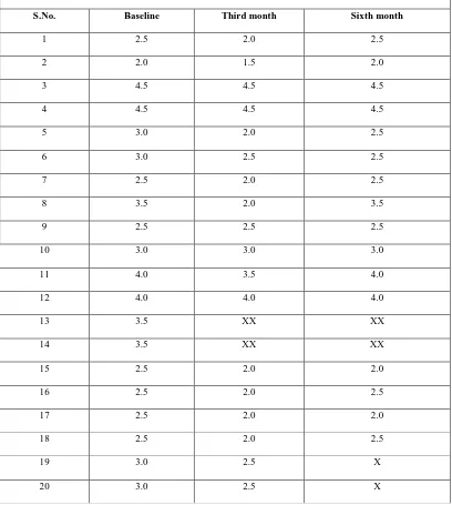
Table 1
Group- A: - Test group (PRF+CAF)
X= Denotes the patient who haven’t completed 6 months post-operative period.
XX= Denotes the patient who discontinued in course of the study period.
Group-A, width of keratinized gingiva in mm Over a period of time
S.No. Baseline Third month Sixth month
1 2.5 2.0 2.5
2 2.0 1.5 2.0
3 4.5 4.5 4.5
4 4.5 4.5 4.5
5 3.0 2.0 2.5
6 3.0 2.5 2.5
7 2.5 2.0 2.5
8 3.5 2.0 3.5
9 2.5 2.5 2.5
10 3.0 3.0 3.0
11 4.0 3.5 4.0
12 4.0 4.0 4.0
13 3.5 XX XX
14 3.5 XX XX
15 2.5 2.0 2.0
16 2.5 2.0 2.5
17 2.5 2.0 2.0
18 2.5 2.0 2.5
19 3.0 2.5 X
Table 2
Group-B:- Control group (CTG+CAF)
X= Denotes the patient who haven’t completed 6 months post-operative period.
XX= Denotes the patient who discontinued in course of the study period.
Group-B, width of keratinized gingiva in mm over a period of time
S.No. Baseline Third month Sixth month
1 3.5 3.0 3.5
2 4.5 3.5 4.5
3 4.5 3.5 4.5
4 4.5 4.0 4.5
5 2.5 2.5 3.0
6 3.5 3.5 2.5
7 3.5 3.5 3.5
8 3.5 3.5 4.0
9 2.5 2.5 3.0
10 3.0 3.0 3.0
11 3.0 3.0 3.0
12 2.5 2.5 2.5
13 2.5 2.0 2.5
14 2.5 2.0 2.0
15 4.5 XX XX
16 4.5 XX XX
17 3.5 3.5 3.5
18 3.5 3.5 3.5
19 2.5 XX XX
Table 3
Group-A:- Test group (PRF+CAF)
Group-A probing depth in mm over a period of time
S.No.
Baseline Third month Sixth month
M esial M id .f ac ial
Distal Mesial
M
id
.f
ac
ial
Distal Mesial
M id .f ac ial Distal
1 2 1 2 2 1 1 2 1 1
2 2 1 2 2 2 1 2 1 1
3 1 1 1 2 1 1 2 2 2
4 1 1 1 2 2 1 2 1 2
5 2 1 1 2 1 1 2 1 2
6 2 1 2 2 1 2 1 1 1
7 2 1 2 2 1 1 1 1 1
8 2 2 2 1 1 1 1 1 1
9 2 2 2 1 1 0 1 1 1
10 2 2 3 1 1 1 1 1 1
11 3 2 2 2 1 2 2 1 2
12 2 2 2 1 1 1 1 1 1
13 2 1 2 XX XX XX XX XX XX
14 1 1 1 XX XX XX XX XX XX
15 2 1 2 2 1 1 2 1 1
16 2 1 1 2 2 2 2 2 1
17 1 1 1 1 1 1 1 1 1
18 1 1 1 1 1 1 1 1 1
19 2 1 1 2 1 2 X X X
20 2 1 2 2 1 2 X X X
X= Denotes the patient who haven’t completed 6 months post-operative period.
Table 4
Group –B:- Control group (CTG+CAF)
Group-B probing depth in mm over a period of ti