Copyright © 1998, American Society for Microbiology
Extent of Antigenic Diversity in the V3 Region of the Surface
Glycoprotein, gp120, of Human Immunodeficiency Virus
Type 1 Group M and Consequences for Serotyping
JEAN-CHRISTOPHE PLANTIER,
1SOPHIE LE POGAM,
1FRANCIS POISSON,
1LAURENCE BUZELAY,
1BERNARD LEJEUNE,
2ANDFRANCIS BARIN
1*
Laboratoire de Virologie, EP CNRS 117,
1and Laboratoire de Biophysique Pharmaceutique
et Biomathe´matiques,
2Universite´ Franc¸ois Rabelais, Tours, France
Received 20 February 1997/Accepted 24 September 1997
Human immunodeficiency virus type 1 (HIV-1) may be studied by molecular or immunological approaches.
Most analyses have been performed by genetic comparison of isolates and have led to the definition of clades
or subtypes within the major (M) group of HIV-1. Five subtypes (A to E) were initially identified by comparison
of genomic sequences. Four new subtypes (F to I) were identified more recently. Amino acid differences in the
immunogenic V3 loop between isolates have also been studied, leading to a phenetic classification of at least
14 clusters (1 to 14) of sequences (B. T. M. Korber, K. McInnes, R. F. Smith, and G. Myers, J. Virol. 68:6730–
6744, 1994). In this study, we compared the antigenicity of the V3 consensus sequences defined by phylogenetic
analysis to the antigenicity of those defined by phenetic analysis. We used a recently developed subtype-specific
enzyme immunoassay (SSEIA) that uses the principle of blocking with an excess of peptide in the liquid phase.
Two SSEIAs were performed, the first with five V3 sequences defined by phylogenetic analysis and the second
with 14 V3 sequences defined by phenetic analysis. A total of 168 HIV-1 sera taken from seropositive
individ-uals from seven different countries or regions were studied. Experimental and statistical data, including
correlation matrix and cluster analyses, demonstrated associations between the genetic subtypes and
phen-etically associated groups. Most of these were predicted by Korber et al. (J. Virol. 68:6730–6744, 1994) by
theoretical analysis. We also found that V3 sequences can be grouped into between three and five antigenically
unrelated categories. Residues that may be responsible for major antigenic differences were identified at the
apex of the V3 loop, within the octapeptide xIGPGxxx, where x represents the critical positions. Our study
provides evidence that there is a limited number of V3 serotypes which could be easily monitored by serological
assays to study the diversity and dynamics of HIV-1 strains.
The diversity of human immunodeficiency virus type 1
(HIV-1) is a major problem in the development of an effective
vaccine against AIDS. Many HIV-1 sequences are now
avail-able, and phylogenetic analysis resulting in a continuously
de-veloping classification into subtypes or clades is possible (45).
HIV-1 isolates are classified into the M group (for major) or O
group (for outlier). The O group contains only a few variants,
all from a limited area of Africa (19, 27, 50). The M group
includes variants responsible for the present AIDS pandemic.
It contains at least five subtypes (A to E), to which have been
added more recently four other subtypes (F to I) (23, 28, 34,
36, 37). Subtypes A, C, D, G, and H are common in Africa (21,
35, 37, 38). Subtype B is the most common in America and
Europe (24, 26, 51). Subtype E occurs mainly in Asia (25, 30,
41), and subtype F has been detected in Brazil and Romania
(3, 28, 34). These distributions are not restrictive. Subtype C is
also present in Asia (India and China), and subtype G is also
present in Russia (7, 12, 29). The African subtypes (A, C, and
D) and the Asian subtype (E) have also been identified in
North America and in European countries (9, 13, 14, 32, 48).
All the subtypes are present in Africa, including B (detected in
West Africa), E (Central African Republic), and F
(Cam-eroon) (1, 35, 38). Analysis of the genetic diversity of HIV-1 is
becoming more difficult due to the increasing frequency of
coinfections and recombinations (15, 20, 44).
Phylogenetic trees have been generated with gag, env, or tat
nucleotide sequences. Shorter DNA sequences encoding the
functionally important V3 region of the envelope protein are
most frequently used to provide reliable subtype designations
(37). The diversity of the immunogenic V3 loop has also been
studied by comparing the amino acids of different isolates,
leading to a phenetic classification of at least 14 clusters of
sequences, each one characterized by a consensus sequence
based on the most common amino acid in a given position (22).
The heterogeneity of HIV-1 strains is studied mostly by
molecular characterization of genomic sequences. This
in-volves sequencing fragments amplified by the PCR or the use
of the heteroduplex mobility assay (10, 11). However, although
these methods allow direct subtype classification, they are
time-consuming and expensive and require highly trained
workers. Serotyping of HIV-1 by antibody (Ab) binding to the
V3 region has been suggested as an alternative approach (8,
40, 49, 51). Such an approach may make it possible to identify
subtypes based on antigenic rather than genetic properties.
This immunological information about antigenic diversity
might be of value in vaccine development. We recently
devel-oped a subtype-specific enzyme immunoassay (SSEIA) which
gave results consistent with those of genotyping (4, 48). This
assay used V3 consensus sequences defined by genetic
classi-fication, so we wanted to compare the antigenicity of these V3
consensus sequences to the antigenicity of those defined by
phenetic analysis. The phenetic clustering of V3 loop amino
acid sequences is not always consistent with phylogenetic
anal-ysis. Our results suggested that a limited number of serotypes
* Corresponding author. Mailing address: Laboratoire de Virologie,
EP CNRS 117, CHRU Bretonneau, 37044 Tours Cedex, France.
Phone: (33) 2 47 47 80 58. Fax: (33) 2 47 47 36 10.
677
on November 9, 2019 by guest
http://jvi.asm.org/
may exist and identified amino acids at the tip of the V3 loop
that may be responsible for serological discrimination.
MATERIALS AND METHODS
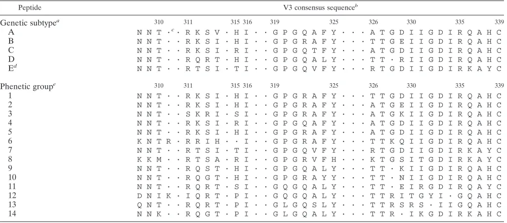
Peptides.Nineteen peptides whose sequences corresponded to sequences in
the V3 region of HIV-1 were synthesized (Table 1). These peptides corre-sponded to the consensus sequences of the five major subtypes of HIV-1 group M (A to E) (36) or to the consensus sequences of the 14 phenetic clusters described by Korber et al. in 1994 (22). The peptides were synthesized by Merrifield’s solid-phase procedure with an automated peptide synthesizer (Ap-plied Biosystems 431A), 9-fluorenylmethoxycarbonyl-protected amino acids, and hydroxymethylphenoacetic-polystyrene resin (31). The resin support and side-chain-protecting groups were removed with trifluoroacetic acid after synthesis, and distilled water, phenol, ethanedithiol, and thioanisole were used as scaven-gers. The peptides were cleaved and purified by reverse-phase chromatography on C8columns (Aquapore octyl, 20mm, 100 by 10 mm; Applied Biosystems). The
purity of the preparations was checked both by observation of a single sharp peak in high-pressure liquid chromatography analysis with C8columns (Aquapore
octyl RP-300, 7mm, 220 by 4.6 mm; Applied Biosystems) and by amino acid analysis. Peptide compositions were as planned.
Sera. A total of 168 HIV-1 Ab-positive sera collected in seven different
countries or regions were used (24 sera per country). The regions were selected so that the widest antigenic diversity was obtained. They were France and West Indies (subtype B), Thailand (subtype E), and four African countries (Burundi, Burkina-Faso, Congo, and Coˆte d’Ivoire) (subtypes A, C, and D).
Immunoassays.We have shown that cross-reactivity between V3 sequences is
both very strong and very frequent when analyzed by indirect enzyme immuno-assays (EIA) (2). We developed an SSEIA that uses the principle of blocking with an excess of peptide in the liquid phase (4). The SSEIA is more discrimi-nating than normal indirect EIA, probably because it is more dependent than indirect EIA on Ab affinity. Two SSEIAs were performed, the first with 5 V3 sequences defined by phylogenetic analysis (SSEIAgen) and the second with 14 V3 sequences defined by phenetic analysis (SSEIAphen).
For the SSEIAgen, wells of polyvinyl microtiter plates (Falcon) were coated with an equimolar mixture of the five V3 peptides (0.5mg/ml each in 0.05 M bicarbonate buffer [pH 9.6]; 100ml per well) by incubation for 20 h at 37°C. The wells were washed twice with phosphate-buffered saline containing 0.5% Tween 20 (PBS-TW), and the unoccupied sites of the wells were saturated with phos-phate-buffered saline containing 2% newborn calf serum by incubation for 45 min at 37°C. Serum samples were diluted 1:100 in 0.01 M sodium phosphate buffer (pH 7.4) containing 0.75 M NaCl, 10% newborn calf serum, and 0.05% Tween 20 (PBS-TW-NBCS). Each sample was tested in seven wells in the presence of various blocking solutions. Preliminary assays were used to select the
optimal concentrations of peptides to be used for both coating solutions and blocking solutions (4). Ten microliters of a 100-mg/ml solution (in PBS-TW-NBCS) of V3 peptide of the A, B, C, D, or E subtype was added to the wells (A to well 1, B to well 2, and so forth). Ten microliters of a 100-mg/ml solution of an equimolar mixture of the five peptides (theoretically 100% blocking) was added to well 6. Ten microliters of PBS-TW-NBCS (theoretically 0% blocking) was added to well 7. One hundred microliters of diluted serum was added to each well and incubated for 30 min at room temperature, and the wells were washed four times with PBS-TW. Peroxidase-conjugated goat F(ab9)2anti-human
im-munoglobulin (TAGO, Burlingame, Calif.; 100ml of a 1:5,000 dilution in PBS-TW-NBCS) was added, and the mixture was incubated for 30 min at room temperature. The wells were washed four times with PBS-TW and incubated with hydrogen peroxide–o-phenylenediamine for 15 min at room temperature. Color development was stopped with 2 N H2SO4, and the absorbance value
(optical density [OD]) was read at 492 nm.
[image:2.612.50.551.81.302.2]The percent inhibition of binding induced by each of the five peptides for each serum sample was calculated with the following formula: {(OD without blocking [well 7]2OD in the presence of peptide)/(OD without blocking [well 7]2OD in the presence of the five peptides (well 6)]}3100.
The SSEIAgen indicates, for each serum sample, the immunodominant sub-type (the peptide with the strongest blocking) as well as a serological profile defined by the five values of inhibition (percent inhibition by peptide A, peptide B, peptide C, peptide D, and peptide E).
The same experimental conditions were used for the SSEIAphen, except that wells were coated with an equimolar mixture of the 14 V3 peptides (0.2mg/ml each in 0.05 M bicarbonate buffer [pH 9.6]; 100ml per well). Each serum sample diluted 1:100 was tested in 16 wells in the presence of various blocking solutions. Ten microliters of a 100-mg/ml solution (in PBS-TW-NBCS) each of peptides 1 to 14 was added to wells 1 to 14 (peptide 1 to well 1, peptide 2 to well 2, and so forth). Ten microliters of a 100-mg/ml solution of an equimolar mixture of the 14 peptides (theoretically 100% blocking) was added to well 15. Ten microliters of PBS-TW-NBCS (theoretically 0% blocking) was added to well 16. The remaining steps were identical to those for SSEIAgen. The percent inhibition of binding induced by each of the 14 peptides for each serum sample was calculated with the following formula: [OD without blocking (well 16)2OD in the presence of peptide]/[OD without blocking (well 16)2OD in the presence of the 14 peptides (well 15)]3100.
The SSEIAphen indicates, for each serum sample, the immunodominant sub-type (the peptide with the strongest blocking) as well as a serological profile defined by the 14 values of inhibition.
Statistical analysis.The antigenic relationship between the various peptides
was studied by calculating the Pearson correlation matrix with Systat statistical software (Deltasoft, Meylan, France). For each SSEIAgen and SSEIAphen, specimens were grouped according to a similar serological profile as defined
TABLE 1. Sequences of synthetic peptides used in the assays
Peptide V3 consensus sequenceb
Genetic subtype
a 310 311 315 316 319 325 326 330 335 339A
N N T ·
c· R K S V · H I · · G P G Q A F Y · · · A T G D I I G D I R Q A H C
B
N N T · · R K S I · H I · · G P G R A F Y · · · T T G E I I G D I R Q A H C
C
N N T · · R K S I · R I · · G P G Q T F Y · · · A T G D I I G D I R Q A H C
D
N N T · · R Q R T · H I · · G P G Q A L Y · · · T T · R I I G D I R Q A H C
E
dN N T · · R T S I · T I · · G P G Q V F Y · · · R T G D I I G D I R K A Y C
Phenetic group
e 310 311 315 316 319 325 326 330 335 3391
N N T · · R K S I · H I · · G P G R A F Y · · · T T G D I I G D I R Q A H C
2
N N T · · R K S I · H I · · G P G R A F Y · · · A T G E I I G D I R Q A H C
3
N N T · · S K R I · S I · · G P G R A F Y · · · A T G K I I G D I R Q A H C
4
N N T · · R K S I · R I · · G P G Q A F Y · · · A T G D I I G D I R Q A H C
5
N N T · · R K S I · H I · · G P G R A F Y · · · A T G D I I G D I R Q A H C
6
K N T R · R R I H · · I · · G P G R A F Y · · · T T K Q I I G D I R Q A H C
7
N N T · · R T S I · T I · · G P G Q V F Y · · · R T G D I I G D I R K A Y C
8
K K M · · R T S A · R I · · G P G R V F H · · · K T G S I T G D I R K A Y C
9
N N T · · R Q S T · H I · · G P G Q A L Y · · · T T · K I I G D I R Q A H C
10
N N T · · R Q G T · H I · · G P G R A Y Y · · · T T · N I I G D I R Q A H C
11
N N T · · R Q R T · S I · · G Q G Q A L Y · · · T T · E I R G D I R Q A Y C
12
D N I K · I Q R T · P I · · G Q G Q A L Y · · · T T R I T G Y I · G Q A H C
13
Q N T · · R Q R T · P I · · G L G Q S L Y · · · T T R S R S · I I G Q A H C
14
N N K · · R Q G T · P I · · G L G Q A L Y · · · T T R · I K G D I R K A H C
aV3 consensus sequences of the five subtypes initially described by Myers et al. (36). bResidues are numbered according to Ratner et al. (42).
cThe dots represent insertions. dPeptide E is similar to peptide 7.
eConsensus sequences of the 14 V3 phenetic groups initially described by Korber et al. (22) and truncated at the four N-terminal amino acids.
on November 9, 2019 by guest
http://jvi.asm.org/
above. We used the cluster analysis of PCSM statistical software (Deltasoft). The serological profiles of all the reactive samples for each assay were entered into a computer. We used agglomerative hierarchical clustering of observations, taking into account the euclidean distance and the average linkage. The results were displayed as dendrograms. The two dendrograms obtained were drawn on x (SSEIAphen) and y (SSEIAgen) axes (see Fig. 2). Each sample was plotted at the intersection of its x and y positions (at the intersection of its positions within each dendrogram).
RESULTS
Antigenicity of the V3 peptides.
Among the 168 sera positive
for the HIV-1 antibody, 154 were reactive with V3 peptides
(Table 2). All the sera that reacted in the SSEIAgen were also
reactive in the SSEIAphen. The nonreactive samples were
negative in both assays. The serum samples collected in France
or the West Indies were reactive to peptide B, most of those
collected in Thailand were reactive to peptide E, and various
reactivities were observed with African samples. Of the 86
reactive African samples, 22 (25.6%) were most reactive with
peptide A, 1 (1.2%) was most reactive with peptide B, 53
(61.6%) were most reactive with peptide C, and 10 (11.6%)
were most reactive with peptide D. The most striking
obser-vation with the SSEIAphen was the limited number of
domi-nant reactivities. Indeed, although 14 peptides were included,
only 6 were bound preferentially by the tested sera. The
dom-inant reactivities involved peptides 1 (1 case), peptides 2 and 5
(59 cases), peptide 4 (76 cases), peptide 7 (7 cases), and
pep-tide 9 (11 cases) (Table 3). All the B-reactive serum samples
were reactive predominantly to peptides 2 and 5, the same
intensity of binding being observed with these two very similar
sequences. Of the 47 B- or peptide 2- or 5-reactive samples, 23
cross-reacted with peptides 1, 3, and 6 but not with other
peptides. An example is shown in Fig. 1 (sample F7). All the
C-reactive samples reacted with peptide 4, with no
cross-reac-tivity. All the D-reactive samples reacted with peptide 9, with
no cross-reactivity. The E-reactive samples reacted with either
peptide 4 (10 cases) or peptide 7 (7 cases). The most diverse
reactivities were observed with A-reactive samples, which
bound preferentially to peptide 1 (1 case), 2 or 5 (12 cases), or
4 (10 cases), with various degrees of cross-reactivity (Table 3).
Representative serological profiles of serum samples are
shown in Fig. 1.
The antigenic relationships among these peptides were
fur-ther analyzed by a statistical approach. The serological profiles
of every serum sample, defined as the percentages of binding
inhibition by each of the 19 peptides, were entered into a
computer, and a Pearson correlation matrix was established
(Table 4). This analysis showed positive correlations between
sequence B and sequences 1, 2, 3, 5, and 6 (r, 0.69 to 0.85),
between sequences C and 4 (r, 0.66), between sequences D and
9 (r, 0.67), and between sequences E and 7 (r, 0.67). No
sig-nificant correlation was observed for A-reactive serum
sam-ples, consistent with the heterogeneity of responses for these
samples. The matrix also yielded negative correlation
coeffi-cients, showing that reactivity to some sequences systematically
excluded reactivity to others. These results show the value of
SSEIA for serological discrimination. For example, reactivity
to peptide 1 strongly correlated with reactivity to peptides 2
(r, 0.87), 3 (r, 0.75), 5 (r, 0.89), 6 (r, 0.89), and B (r, 0.85) and
with no or low reactivity to peptides 4 (r,
2
0.45), 7 (r,
2
0.55),
9 (r,
2
0.37), C (r,
2
0.35), and E (r,
2
0.33).
Cluster analysis.
Groupings among the 154 reactive samples
were identified by cluster analysis. Results are displayed as two
dendrograms corresponding to results obtained with
SSEIA-gen and SSEIAphen (Fig. 2). This analysis demonstrated a
correlation between the two classifications and identified five
serological clusters, two of which overlap. Cluster 1 contains
samples from France and the West Indies reactive with
pep-tides B, 2, and 5. Cluster 2 contains African samples reactive
with peptides D and 9. Cluster 3 contains African samples
reactive mostly with peptides A, 2, and 5. Clusters 4 and 5,
which overlap, contain samples mainly from Africa reactive
mostly with peptides C and 4 and samples mainly from
Thai-land reactive with peptides E and 7, respectively. The
overlap-ping area contains samples with high cross-reactivity with
pep-tides C and E and/or peppep-tides 4 and 7. Cluster 4 also contains
A-reactive samples reactive mostly with peptide 4.
Amino acids responsible for subtype-specific serological
identification.
We compared the amino acid sequences of the
[image:3.612.48.292.99.189.2]19 peptides used in the study. The comparison was based on
both antigenicity in SSEIA, i.e., positive or negative correlation
as described above, and biochemical properties, i.e.,
conserva-tive or nonconservaconserva-tive substitutions. We identified three
ma-jor groups of independent (antigenically unrelated) reactive
sequences from the SSEIA data (Table 5). The first comprised
peptides B, 2, and 5. The second comprised peptides A, C, E,
4, and 7. The third comprised peptides D and 9. The major
difference between the first group and the others is the
pres-ence at the tip of the loop of an arginine residue at position 322
in the first group. This arginine is replaced by glutamine in the
TABLE 2. Reactivity of serum samples to subtype-specific V3
consensus sequences in SSEIAgen according
to geographical origin
Peptide No. of serum samples reactive in a:
WI Fr CI BF Co Bu Th
A
2
9
10
1
1
B
24
22
1
C
20
7
7
19
3
D
2
7
1
1
E
17
NR
b2
7
3
2
aWI, West Indies; Fr, metropolitan France; BF, Burkina-Faso; CI, Coˆte d’Ivoire; Co, Congo; Bu, Burundi; Th, Thailand.
bNR, nonreactive.
TABLE 3. Correlation between SSEIAgen and SSEIAphen
Peptide in SSEIAgen
No. of sera reactive to peptideain SSEIAphen
1 (None) 2 or 5 (None) 2 or 5 (1, 3, or 6) 2 or 5 (4) 4 (None) 4 (2 or 5) 4 (7) 7 (None) 7 (4) 9 (None) Total
A
1
11
1
8
2
23
B
24
23
47
C
56
56
D
11
11
E
8
2
5
2
17
aDominant reactivity (cross-reactivity). Dominant reactivity produces 100% blocking. Cross-reactivity occurs when blocking is between 95 and 100%.
on November 9, 2019 by guest
http://jvi.asm.org/
[image:3.612.53.549.636.718.2]others. The major difference between the second and the third
groups occurs at position 324, where there is a leucine residue
in peptides D and 9 but a more hydrophobic residue,
phenyl-alanine, in peptides A, C, E, 4, and 7 (5). There is another,
smaller difference between the second group and the two
oth-ers at position 315. A histidine residue in the first and third
groups is replaced by an arginine or a threonine residue in the
second group, except in the consensus sequence of subtype A,
which conserves this histidine.
DISCUSSION
Studies on HIV-1 diversity led to the identification of five
major genetic subtypes within group M and at least 14
phen-etically associated groups (22, 36). The analysis of genetic
relationships between isolates is complicated, as many isolates
are mosaic viruses (15, 19, 44). Serotyping of HIV-1 infections
based on V3 seroreactivity would be of great value in the study
of HIV-1 diversity and would provide important information
for both epidemiology and future vaccine composition. This
work studied the antigenic relationships between consensus
sequences defined by two different approaches. The aim was to
investigate the extent of the V3 serotypes and to identify, at
least in part, the molecular determinants of the
serotype-asso-ciated antigenic specificity. Our experimental data confirmed
most of the associations between the genetic subtypes and
phenetic groups that were predicted by theoretical analysis
(22). However, our data showed that only a limited number of
V3 serotypes can be identified due to the immunodominance
of a few sequences. Korber et al. suggested that genetic
sub-type B was associated mainly with phenetic groups 1, 2, 3, 5,
and 6 (22). Our results are consistent with these results but
showed that the consensus sequences of groups 2 and 5 are
immunodominant. Serological discrimination of these two
se-quences was almost impossible due to a single synonymous
substitution (glutamic acid
3
aspartic acid at position 329;
Ta-ble 1). We also found associations between genetic subtype C
and phenetic group 4, genetic subtype D and phenetic group 9,
and genetic subtype E and phenetic group 7. However, our
data also suggested that consensus sequence 7 is
immunodom-inant for subtype E and that consensus sequence 9 is
[image:4.612.51.290.72.334.2]immu-FIG. 1. Serological profiles in the two SSEIAs of typical HIV-1-positive sera from various regions. Shown are two A-reactive sera, BF2 reacting predomi-nantly to peptides 2 and 5 and BF 228 reacting predomipredomi-nantly to peptide 4; a B-reactive sample (F7); a C-reactive sample (Bu 192); a D-reactive sample (Co 119); and an E-reactive sample (Th 4).
TABLE 4. Pearson correlation matrix for the antigenicities of the 19 V3 sequences
Peptide r
afor SSEIAphen peptide
1 2 3 4 5 6 7 8 9 10 11 12 13 14
SSEIAphen
1
1
2
0.87
a1
3
0.75
0.79
1
4
2
0.45
2
0.36
2
0.23
1
5
0.89
0.96
0.79
2
0.36
1
6
0.89
0.88
0.74
2
0.42
0.90
1
7
2
0.55
2
0.58
2
0.46
0.70
2
0.55
2
0.49
1
8
0.14
0.10
0.01
0.02
0.08
0.15
2
0.03
1
9
2
0.37
2
0.44
2
0.41
2
0.03
2
0.42
2
0.34
0.09
0.01
1
10
0.24
0.15
0.21
2
0.09
0.18
0.22
2
0.06
0.45
0.09
1
11
2
0.16
2
0.24
2
0.13
0.14
2
0.21
2
0.15
0.29
0.16
0.41
0.42
1
12
2
0.30
2
0.32
2
0.23
0.30
2
0.30
2
0.27
2
0.41
0.15
0.34
0.19
0.77
1
13
2
0.29
2
0.33
2
0.22
0.30
2
0.31
2
0.26
0.35
0.03
0.34
0.18
0.73
0.73
1
14
2
0.16
2
0.14
2
0.16
0.19
2
0.15
2
0.10
0.28
0.17
0.36
0.22
0.63
0.70
0.70
1
SSEIAgen
A
0.20
0.20
0.08
2
0.11
0.17
0.16
2
0.11
2
0.08
2
0.03
0.00
2
0.11
2
0.11
2
0.01
0.04
B
0.85
0.79
0.69
2
0.58
0.81
0.81
2
0.62
0.06
2
0.39
0.19
2
0.25
2
0.35
2
0.34
2
0.29
C
2
0.35
2
0.19
2
0.07
0.66
2
0.21
2
0.28
0.47
0.07
2
0.32
2
0.13
0.05
0.10
0.11
0.08
D
0.02
2
0.04
2
0.11
2
0.50
2
0.03
0.10
2
0.28
2
0.01
0.67
0.02
0.16
0.09
0.08
0.16
E
2
0.33
2
0.31
2
0.23
0.38
2
0.33
2
0.21
0.67
0.09
2
0.04
2
0.03
0.21
0.28
0.27
0.28
aCorrelations in bold were significant (P,0.05) after Bonferroni correction.
on November 9, 2019 by guest
http://jvi.asm.org/
[image:4.612.56.547.481.717.2]nodominant for subtype D. The phenogram of HIV-1 V3 loop
protein similarities indicated that for subtype A, the
associa-tion between genetic subtype and phenetic group was weaker
(22). Our results are also consistent with this observation.
Indeed, some subtype A-reactive samples bound to peptides
containing the consensus sequences of phenetic group 2 or 5,
whereas others bound to peptides containing the consensus
sequences of phenetic group 4. This may be because most
isolates of subtype A have the sequence GPGQ at the tip of the
V3 loop but others possess the sequence GPGR. Genetic
sub-types C and E appear distantly related, but cross-reactivity
between peptides containing their V3 consensus sequences has
been reported (4, 8, 40). We also found cross-reactivity
be-tween subtypes C and E based on data obtained with consensus
sequences of phenetic groups 4 and 7.
V3 consensus sequences can be grouped into three to five
major antigenically unrelated categories on the basis of our
statistical data, including correlation matrix and cluster
analy-ses. A comparison of protein sequences in these categories
identified residues at positions 322 to 324 that may be
respon-sible for major antigenic differences (Table 5). This result is
consistent with previous data showing that human antibodies
to the V3 region are mostly directed to the central area of V3
(51). The crystal structure of a complex between a V3 peptide
and the Fab fragment of a neutralizing antibody, 59.1, showed
that amino acids at the these three positions are involved in
Fab binding (16). Table 5 shows the sequences responsible for
serotype specificity and the contribution of individual residues
[image:5.612.126.475.71.407.2]to antibody interactions according to Ghiara et al. (16).
Two-thirds of the hydrogen bonds, salt bridges, and Van der Waals
contacts between V3 and Fab involve residues 322 to 324.
Sherefa et al. used substitution peptide analogs to show that
FIG. 2. Result of the cluster analysis. Each sample, represented by a symbol, is located at the intersection between the two dendrograms. The symbols correspond to the dominant reactivities in both assays. Clusters correspond to samples with similar dominant reactivities in both classifications, with only a few exceptions.
TABLE 5. Contribution of individual residues
to serotype specificity
Parameter
Result for sequencea
315 316 319 320 321 322 323 324 325
x I G P G x x x Y
Genetic subtype/phenetic group(s)b
B/2–5 H —d — — — R A F —
A/4 H/R — — — — Q A F —
C R — — — — Q T F —
E/7 T — — — — Q V F —
D/9 H — — — — Q A L —
No. of Van der Waals
contactsc 0 3 1 14 4 14 9 17 0
No. of hydrogen bondsc 0 0 1 0 1 2 1 0 0
No. of salt bridgesc 0 0 0 0 0 1 0 0 0
Peptide surface area
buried (Å2)c 6 52 28 77 23 86 53 93 2
aResidues are numbered according to the BH10 sequence (36). bIncluding only the dominant reactivities.
cData are from Ghiara et al. (16). d—, sequence similarity.
on November 9, 2019 by guest
http://jvi.asm.org/
[image:5.612.306.549.535.692.2]the lack of cross-reactivity between subtype B and subtype C
peptides was due to an arginine-322
3
glutamine-322
substitu-tion (46). They also found that the phenylalanine at posisubstitu-tion
324 was also essential for subtype specificity and showed that
an alanine-323
3
threonine-323 substitution, which would not
cause any major structural changes in the peptide backbone,
might explain the cross-reactivities among peptides A, C, and
4. In another study, Sherefa et al. compared V3 serotyping and
genotyping by V3 sequencing (47). They obtained results
sim-ilar to ours. Amino acids present upstream at positions 315 and
316, which are part of the core of another V3 epitope
recog-nized by human sera, might play a complementary role (6, 17,
18, 43). This fact may explain why some subtype A-reactive
samples bound preferentially to peptide 1, 2 or 5, or 4 with
various degrees of cross-reactivity. Peptides A, 1, 2, and 5 have
a histidine residue at position 315, whereas peptide 4 has an
arginine. Thus, the subtype-specific signature sequences are
located at the tip of the V3 loop and involve the octapeptide
xIGPGxxx. The immunodominance of some peptides within a
“serogroup” may be due to slight modifications within the
flanking residues that affect the conformation (Table 1).
Our study did not include samples from patients in whom
the genotype of the infecting strain had been identified. We
believed that this information was not relevant to the present
analysis, as previous studies have shown a strong correlation
between SSEIAgen and genotyping. In three independent
studies involving 285 samples, the specificities of SSEIAgen
were between 0.96 and 0.98 for subtype A, 0.84 and 0.98 for
subtype B, 0.74 and 0.81 for subtype C, and 0.99 and 1 for
subtype D; the specificity was 1 for subtype E (6a, 48).
Multivariate analysis of HIV-1 neutralization data shows the
existence of a small number of neutralization clusters not
cor-related with the genetic clades (39). The neutralization
epi-topes responsible for this clustering have not been identified
and may be independent of the V3 region, but this previous
study and our study, addressing different questions and using
different approaches, have both suggested the existence of a
limited number of serotypes despite the genetic diversity of
HIV-1.
Surveys of the diversity and dynamics of HIV-1 strains are an
important challenge in the current AIDS pandemic and for the
future. There are at least two reasons for typing: (i)
surveil-lance of diversity, particularly its dynamics for epidemiological
studies, and (ii) understanding the biological significance of
this diversity. Both are essential for public health. Genotyping
is probably the reference method for epidemiological
surveil-lance of HIV-1 diversity, but current molecular methods
can-not be used for large populations. Alternative methods, such as
serotyping with simple immunoassays, would be of great value.
Our study provides evidence for the existence of a small
num-ber of V3 serotypes. The statistical analysis indicated strong
positive and negative correlations between the different
se-quences. This result indicates the discriminative power and
reliability of SSEIA. The biological significance of HIV-1
di-versity is unknown, but immunity in vivo is probably the most
important property. For many viruses, such as poliovirus or
influenza viruses, protection in vivo is associated with
seroneu-tralization in vitro. Immunity in vitro does not correlate with
protection in vivo for HIV-1, but a few authors have studied
the association between neutralization serotypes and
geno-types. Moore et al. clearly showed that genetic clades do not
correlate with neutralization serotypes, indicating that
geno-typing may not be the ideal method for studying HIV-1
diver-sity for vaccine development purposes (33). It is therefore still
unclear what assays or tools might be most adaptable for
pre-dicting the antigenic composition of future vaccines. The
bio-logical relevance of the V3 serotypes is unknown. However, it
is unclear which methods are best suited, and all approaches to
studying diversity require further evaluation.
ACKNOWLEDGMENTS
This work was supported by funds from the Agence Nationale de
Recherche sur le SIDA (Paris, France) and the Institut Universitaire
de France.
We thank J. Hoebeke for helpful discussion of the manuscript and
R. Chout, F. Denis, and V. Vithayasai for providing serum samples.
REFERENCES
1. Ariyoshi, K., R. Cheingsong-Popov, A. Wilkins, T. Corrah, J. Weber, and H.
Whittle.1996. HIV-1 subtype B in West Africa. Lancet 347:328. (Letter.)
2. Baillou, A., D. Brand, F. Denis, S. M’Boup, R. Chout, A. Goudeau, and F.
Barin.1994. High antigenic cross-reactivity of the V3 consensus sequences of
HIV-1 gp120. AIDS Res. Hum. Retroviruses 9:1209–1215.
3. Bandea, C. I., A. Ramos, D. Pieniazek, R. Pascu, A. Tanuri, G. Schochetman,
and M. A. Rayfield. 1995. Epidemiologic and evolutionary relationships
between Romanian and Brazilian HIV-1 subtype F strains. Emerg. Infect. Dis. 1:91–93.
4. Barin, F., Y. Lahbabi, L. Buzelay, B. Lejeune, A. Baillou-Beaufils, F. Denis,
C. Mathiot, S. M’Boup, V. Vithayasai, U. Dietrich, and A. Goudeau.1996.
Diversity of antibody binding to V3 peptides representing consensus se-quences of HIV type 1 genotypes A to E: an approach for HIV type 1 serological subtyping. AIDS Res. Hum. Retroviruses 12:1279–1289. 5. Black, S. D., and D. R. Mould. 1991. Development of hydrophobicity
pa-rameters to analyze proteins which bear post- or co-translational modifica-tions. Anal. Biochem. 193:72–82.
6. Catasti, P., J. D. Fontenot, E. M. Bradbury, and G. Gupta. 1995. Local and global structural properties of the HIV-MN V3 loop. J. Biol. Chem. 270: 2224–2232.
6a.Cheingsong-Popov, R. Unpublished data.
7. Cheingsong-Popov, R., A. Bobkov, M. M. Garaev, P. Kaleebu, P. Callow, A.
Rzhiminova, S. R. Saukhat, N. P. Burdajev, N. D. Kolomijets, and J. Weber.
1993. Identification of human immunodeficiency virus type 1 subtypes and their distribution in the Commonwealth of Independent States (former So-viet Union) by serologic V3 peptide-binding assays and V3 sequence anal-ysis. J. Infect. Dis. 168:292–297.
8. Cheingsong-Popov, R., S. Lister, D. Callow, P. Kaleebu, S. Beddows, J.
Weber, and the WHO Network for HIV Isolation and Characterization.
1994. Serotyping HIV type 1 by antibody binding to the V3 loop: relationship to viral genotype. AIDS Res. Hum. Retroviruses 11:1379–1386.
9. Clewley, J. P., C. Arnold, K. L. Barlow, P. R. Grant, and J. V. Parry. 1996. Diverse HIV-1 genetic subtypes in UK. Lancet 347:1487. (Letter.) 10. Delwart, E. L., E. G. Shpaer, F. E. McCutchan, J. Louwagie, M. Grez, H.
Ru¨bsamen-Waigmann, and J. I. Mullins.1993. Genetic relationships
deter-mined by a heteroduplex mobility assay: analysis of HIV env genes. Science
262:1257–1261.
11. Delwart, E. L., H. W. Sheppard, B. D. Walker, J. Goudsmit, and J. I.
Mullins. 1994. Human immunodeficiency virus type 1 evolution in vivo
tracked by DNA heteroduplex mobility assays. J. Virol. 68:6672–6683. 12. Dietrich, U., M. Grez, H. von Briesen, B. Panhans, M. Geissendo¨rfer, H.
Kuhnel, J. Maniar, G. Manhambre, W. B. Becker, M. L. B. Bechler, and H.
Ru¨bsamen-Waigmann.1993. Human immunodeficiency virus type 1 strains
from India are highly divergent from prototypic African and U.S./European strains but are linked to a South African isolate. AIDS 7:23–27.
13. Fransen, K., A. Buve´, J. N. Nkengasong, M. Laga, and G. van der Groen. 1996. Longstanding presence in Belgians of multiple non-B HIV-1 subtypes. Lancet 347:1403. (Letter.)
14. Gao, F., L. Yue, S. C. Hill, D. L. Robertson, A. H. Graves, M. S. Saag, G. M.
Shaw, P. M. Sharp, and B. H. Hahn.1994. HIV-1 sequence subtype D in the
United States. AIDS Res. Hum. Retroviruses 10:625–627.
15. Gao, F., D. L. Robertson, S. G. Morrison, H. Hui, S. Craig, J. Decker, P. N.
Fultz, M. Girard, G. M. Shaw, B. H. Hahn, and P. M. Sharp.1996. The
heterosexual human immunodeficiency virus type 1 epidemic in Thailand is caused by an intersubtype (A/E) recombinant of African origin. J. Virol.
70:7013–7029.
16. Ghiara, J. B., E. A. Stura, R. L. Stanfield, A. T. Profy, and I. A. Wilson. 1994. Crystal structure of the principal neutralization site of HIV-1. Science 264: 82–85.
17. Gorny, M. K., J.-Y. Xu, V. Gianakakos, S. Karwoswka, C. Williams, H. W.
Sheppard, C. V. Hanson, and S. Zolla-Pazner. 1991. Production of
site-selected neutralizing human monoclonal antibodies against the third variable domain of the human immunodeficiency virus type 1 envelope glycoprotein. Proc. Natl. Acad. Sci. USA 88:3238–3242.
18. Gorny, M. K., A. J. Conley, S. Karwoswka, A. Buchbinder, J.-Y. Xu, E. A.
Emini, S. Koenig, and S. Zolla-Pazner.1992. Neutralization of diverse
hu-man immunodeficiency virus type 1 variants by an anti-V3 huhu-man
on November 9, 2019 by guest
http://jvi.asm.org/
nal antibody. J. Virol. 66:7538–7542.
19. Gu¨rtler, L. G., P. H. Hauser, J. Eberle, A. von Brunn, S. Knapp, L. Zekeng,
J. M. Tsague, and L. Kaptue.1994. A new subtype of human
immunodefi-ciency virus type 1 (MVP-5180) from Cameroon. J. Virol. 68:1581–1585. 20. Hahn, B. H., D. L. Robertson, and P. M. Sharp. 1995. Intersubtype
recom-bination in HIV-1 and HIV-2, p. III-22–III-29. In G. Myers, B. Korber, S. Wain-Hobson, K.-T. Jeang, L. E. Henderson, and G. N. Pavlakis (ed.), Human retroviruses and AIDS 1995: a compilation and analysis of nucleic acid and amino acid sequences. Los Alamos National Laboratory, Los Alamos, N.Mex.
21. Janssens, W., L. Heyndrickx, K. Fransen, J. Motte, M. Peeters, J. N.
Nken-gasong, P. M. Ndumbe, E. Delaporte, J. L. Perret, C. Atende, P. Piot, and G.
van der Groen.1994. Genetic and phylogenetic analysis of env subtypes G
and H in Central Africa. AIDS Res. Hum. Retroviruses 10:877–879. 22. Korber, B. T. M., K. McInnes, R. F. Smith, and G. Myers. 1994. Mutational
trends in V3 loop protein sequences observed in different genetic lineages of human immunodeficiency virus type 1. J. Virol. 68:6730–6744.
23. Kostrikis, L. G., E. Bagdades, Y. Cao, L. Zhang, D. Dimitriou, and D. D. Ho. 1995. Genetic analysis of human immunodeficiency virus type 1 strains from patients in Cyprus: identification of a new subtype designated subtype I. J. Virol. 69:6122–6130.
24. Kuiken, C. L., G. Zwart, E. Baan, R. A. Coutinho, J. A. R. van den Hoek, and
J. Goudsmit. 1993. Increasing antigenic and genetic diversity of the V3
variable domain of the human immunodeficiency virus envelope protein in the course of the AIDS epidemic. Proc. Natl. Acad. Sci. USA 90:9061–9065. 25. Kunanusont, C., H. M. Foy, J. K. Kreiss, S. Rersk-Ngarm, P. Phanuphak, S.
Raktham, C. P. Pau, and N. Young.1995. HIV-1 subtypes and
male-to-female transmission in Thailand. Lancet 345:1078–1083.
26. LaRosa, G. J., J. P. Davide, K. Weinhold, J. A. Waterbury, A. T. Profy, J. A.
Lewis, A. J. Langlois, G. R. Dreesman, R. N. Boswell, P. Shadduck, L. H. Holley, M. Karplus, D. P. Bolognesi, T. J. Matthews, E. A. Emini, and S.
Putney.1990. Conserved sequence and structural elements in the HIV-1
principal neutralization determinant. Science 249:932–935.
27. Loussert-Ajaka, I., M. L. Chaix, B. Korber, F. Letourneur, E. Gomas, E.
Allen, T. D. Ly, F. Brun-Ve´zinet, F. Simon, and S. Saragosti.1995.
Variabil-ity of human immunodeficiency virus type 1 group O strains isolated from Cameroonian patients living in France. J. Virol. 69:5640–5649.
28. Louwagie, J., E. L. Delwart, J. I. Mullins, F. E. McCutchan, G. Eddy, and
D. S. Burke.1994. Genetic analysis of HIV-1 isolates from Brazil reveals
presence of two distinct genetic subtypes. AIDS Res. Hum. Retroviruses
10:561–567.
29. Luo, C. C., C. Tian, D. J. Hu, M. Kai, T. J. Dondero, and X. Zheng. 1995. HIV-1 subtype C in China. Lancet 345:1051–1052.
30. McCutchan, F. E., P. A. Hegerich, T. P. Brennan, P. Phanuphak, P.
Sing-haraj, A. Jugudee, P. W. Berman, A. M. Gray, A. K. Fowler, and D. S. Burke.
1992. Genetic variants of HIV-1 in Thailand. AIDS Res. Hum. Retroviruses
8:1887–1895.
31. Merrifield, R. B. 1963. Solid-phase peptide synthesis. I. The synthesis of a tetrapeptide. J. Am. Chem. Soc. 85:2149–2154.
32. Montpetit, M. 1995. HIV-1 subtype in Canada. AIDS Res. Hum. Retrovi-ruses 11:1421–1422.
33. Moore, J. P., Y. Cao, J. Leu, L. Qui, B. Korber, and D. D. Ho. 1996. Inter-and intraclade neutralization of human immunodeficiency virus type 1: ge-netic clades do not correspond to neutralization serotypes but partially correspond to gp120 antigenic serotypes. J. Virol. 70:427–444.
34. Morgado, M. G., E. C. Sabino, E. G. Shpaer, V. Bongertz, L. Brigido,
M. D. C. Guimaraes, E. A. Castilho, B. Galvao-Castro, J. I. Mullins, R. M.
Hendry, and A. Mayer.1994. V3 region polymorphisms in HIV-1 from
Brazil: prevalence of subtype B strains divergent from North American/ European prototype and detection of subtype F. AIDS Res. Hum. Retrovi-ruses 10:569–576.
35. Murphy, E. E., B. Korber, M. C. Georges-Courbot, B. You, A. Pinter, D.
Cook, M. P. Kieny, A. Georges, C. Mathiot, F. Barre´-Sinoussi, and M.
Girard.1993. Diversity of V3 region sequences of human immunodeficiency
viruses type 1 from the Central African Republic. AIDS Res. Hum. Retro-viruses 9:997–1006.
36. Myers, G., B. Korber, S. Wain-Hobson, R. F. Smith, and G. N. Pavlakis. 1992. Human retroviruses and AIDS 1992. Los Alamos, NM: a compilation and analysis of nucleic acid and amino acid sequences. Los Alamos National Laboratory, Los Alamos, N.Mex.
37. Myers, G., B. Korber, S. Wain-Hobson, R. F. Smith, and G. N. Pavlakis. 1994. Human retroviruses and AIDS 1994. Los Alamos, NM: a compilation and analysis of nucleic acid and amino acid sequences. Los Alamos National Laboratory, Los Alamos, N.Mex.
38. Nkengasong, J. N., W. Janssens, L. Heyndrickx, K. Fransen, P. M. Ndumbe,
J. Motte, A. Leonaers, M. Ngolle, J. Ayuk, P. Piot, and G. van der Groen.
1994. Genotypic subtypes of HIV-1 in Cameroon. AIDS 8:1405–1412. 39. Nyambi, P. N., J. N. Nkengasong, P. Lewi, K. Andries, W. Janssens, K.
Fransen, L. Heyndrickx, P. Piot, and G. van der Groen.1996. Multivariate
analysis of human immunodeficiency virus type 1 neutralization data. J. Vi-rol. 70:6235–6243.
40. Pau, C. P., M. Kai, D. L. Holloman-Candal, C. C. Luo, M. L. Kalish, G.
Schochetman, B. Byers, J. R. George, and the WHO Network for HIV
Isolation and Characterization.1994. Antigenic variation and serotyping of
HIV type 1 from four World Health Organisation-sponsored HIV vaccine sites. AIDS Res. Hum. Retroviruses 11:1369–1377.
41. Pfu¨tzner, A., U. Dietrich, U. von Eichel, H. von Briesen, H. D. Brede, J. K.
Maniar, and H. Ru¨bsamen-Waigmann.1992. HIV-1 and HIV-2 infections in
a high-risk population in Bombay, India: evidence for the spread of HIV-2 and presence of a divergent HIV-1 subtype. J. Acquired Immune Defic. Syndr. 5:972–977.
42. Ratner, L., W. Haseltine, R. Patarca, K. J. Livak, B. Starcich, S. F. Josephs,
E. R. Doran, J. A. Rafalski, E. A. Whitehorn, K. Baumeister, L. Ivanoff, S. R. Petteway, Jr., M. L. Pearson, J. A. Lautenberger, T. S. Papas, J. Ghrayeb,
N. T. Chang, R. C. Gallo, and F. Wong-Staal.1985. Complete nucleotide
sequence of the AIDS virus, HTLV-III. Nature 313:277–284.
43. Rini, J. M., R. L. Stanfield, E. A. Stura, P. A. Salinas, A. T. Profy, and I. A.
Wilson.1993. Crystal structure of a human immunodeficiency virus type 1
neutralizing antibody, 50.1, in complex with its V3 loop peptide antigen. Proc. Natl. Acad. Sci. USA 90:6325–6329.
44. Sabino, E. C., E. G. Shpaer, M. G. Morgado, B. T. M. Korber, R. S. Diaz, V.
Bongertz, S. Calvacante, B. Galvao-Castro, J. I. Mullins, and A. Mayer.
1994. Identification of human immunodeficiency virus type 1 envelope gene recombinants between subtypes B and F in two epidemiologically linked individuals from Brazil. J. Virol. 68:6340–6346.
45. Sharp, P. M., D. L. Robertson, F. Gao, and B. Hahn. 1994. Origins and diversity of human immunodeficiency viruses. AIDS 8:S27–S42.
46. Sherefa, K., M. Saˆllberg, and A. So¨nnerborg. 1994. Evidence of no change in V3 loop antibody recognition pattern in HIV type 1-infected Ethiopians between 1988 and 1993. AIDS Res. Hum. Retroviruses 10:1551–1556. 47. Sherefa, K., A. So¨nnerborg, J. Steinbergs, and M. Saˆllberg. 1994. Rapid
grouping of HIV-1 infection in subtypes A to E by V3 peptide serotyping and its relation to sequence analysis. Biochem. Biophys. Res. Commun. 205: 1658–1664.
48. Simon, F., I. Loussert-Ajaka, F. Danond, S. Saragosti, F. Barin, and F.
Brun-Vezinet.1996. HIV-1 diversity in Northern Paris, France. AIDS Res.
Hum. Retroviruses 12:1427–1433.
49. Smith, J. D., C. B. Bruce, A. S. R. Featherstone, R. G. Downing, B.
Birya-hawaho, J. C. S. Clegg, J. W. Carswell, and J. D. Oram.1994. Reactions of
Ugandan antisera with peptides encoded by V3 loop epitopes of human immunodeficiency virus type 1. AIDS Res. Hum. Retroviruses 10:577–583. 50. Vanden Haeseveld, M., J. L. Decourt, R. J. de Luys, B. Vanderborght, G. van
der Groen, H. van Heuverswijn, and E. Saman.1994. Genomic cloning and
complete sequence analysis of a highly divergent African human immuno-deficiency virus isolate. J. Virol. 68:1586–1596.
51. Zwart, G., T. F. W. Wolf, R. Bookelman, S. Hartman, M. Bakker, C. A. B.
Boucher, C. Kuiken, and J. Goudsmit.1993. Greater diversity of the HIV-1
V3 neutralization domain in Tanzania compared with The Netherlands: serological and genetic analysis. AIDS 7:467–474.