A CLINICO PATHOLOGICAL STUDY OF MENINGIOMAS
Dissertation submitted in partial fulfillment of the requirements for the degree of
M.D. (Pathology) – Branch III
THE TAMILNADU DR.M.G.R.MEDICAL UNIVERSITY
CHENNAI
CERTIFICATE
This is to certify that this dissertation entitled
“
A CLINICO PATHOLOGICAL
STUDY OF MENINGIOMAS
”
is a bonafide work done by
Dr. RADHA. A.
, in partial
fulfillment of the requirements of The TAMIL NADU DR.M.G.R. MEDICAL
UNIVERSITY, Chennai for the award of M.D. Pathology Degree.
DIRECTOR
GUIDE
Prof. Dr.A.V.SHANTI, M.D.,
Director and Head, Institute of Pathology, Madras Medical College, Chennai – 600 003.
Prof. Dr.SHANTHA RAVISANKAR, M.D. DCP.,
Professor of Neuropathology, Institute of Neurology,
Madras Medical College, Chennai – 600 003.
DEAN
Prof.Dr.KALAVATHY PONNIRAIVAN, B.Sc., M.D.,
Dean
DECLARATION
I declare that this dissertation entitled “
A CLINICO PATHOLOGICAL
STUDY OF MENINGIOMAS
” has been done by me under the guidance and supervision ofProf. Dr.SHANTHA RAVISANKAR, M.D. DCP., It is submitted in partial fulfillment of the
requirements for the award of the M.D., Pathology degree by The Tamilnadu Dr.M.G.R. Medical
University, Chennai. This has not been submitted by me for the award of any degree or diploma from
any other University.
ACKNOWLEDGEMENT
My sincere thanks to Prof. Dr. KALAVATHY PONNIRAIVAN, B.Sc. M.D., Dean Madras Medical College, for permitting me to utilize the facilities of the institution.
It is with profound gratitude that I express my heartfelt thanks to Prof. Dr. A.V. SHANTI,
M.D., Director and Head, Institute of Pathology, Madras Medical College, for her valuable guidance at
every stage, constant encouragement and words of advice which have been the motivating forces in bringing forth this piece of work.
I am also extremely grateful to Prof. Dr. SHANTHA RAVISANKAR, M.D.D.CP., Professor of Neuropathology, for her kind guidance with out whom the study would have been impossible.
I wish to thank all the Additional Professors and Assistant Professors of the Department of Pathology for their continuous support.
My sincere thanks to all my fellow post graduates and friends for their support and encouragement.
I also thank the technical staff of the Neuropathology Lab for their cooperation and assistance.
I wish to thank all my family members for their moral support and encouragement.
CONTENTS
1. Introduction 01
2. Aims and objectives 03
3. Review of literature 04
4. Materials and Methods 26
5. Observations and discussions 29
6. Summary and Conclusion 63
7. Annexures
8. Bibliography
INTRODUCTION
“The pathological curiosity of one day becomes in its proper time a common place condition at
a later time”.
Sir Harvey Cushing
Meningiomas were once considered a complex and diverse group of neoplasms in view of their
varied histological patterns. Over the years these morphologically different tumours have been studied
in detail and the mysteries of their origins unraveled.
The term “meningioma” was coined by Sir Harvey Cushing, the most revered neurosurgeon of
the 20th century. But as early as 1614 Felix Plater removed a brain tumor at autopsy which he described
accurately. The neoplasm was in all probability a meningioma. The first brain tumour reported from
the United States also happens to be a meningioma.
In 1835, Professor Peccholi, Professor of Surgery and Operating Medicine from the University
of Siena operated on a dural based tumour at the occiput which he described as a “fungus of the dura
mater”. Over a period of 16 years he operated upon and studied the morphology of several such
tumours and published a comprehensive review of his work in 1847.
Francesco Durante of Sicily, Italy, was the first surgeon to successfully resect a cranial
base meningioma.
But the most celebrated work on meningiomas is by Sir Harvey Cushing co – written by Louise
Eisenhardt in 1938 titled “Meningiomas : Their classification, regional behaviour, life history and
surgical end results”. This works gives incredible details and morphological descripotions of
Various authours over the years have attempted to classify meningiomas. It was understood that
most of these tumours followed a benign course and that morbidity and mortality relating to them stem
from unusual locations and limitations to surgical resectablity.
Cushing5 identified 20 subtypes that included non meningothelial mesenchymal tumours too.
Further refinement down the year divided meningiomas in to benign, atypical and malignant variants.
The 1993 WHO classification28 recognizes two broad categories of meningeal neoplasms :
1. Meningiomas arising from meningothelial (arachnoidal cap) cells
2. Tumours arising from mesenchymal non meningothelial cells which are classified as soft
tissue tumours.
Kustanikul and Brown42 attributed the histological diversity of meningiomas to the
AIMS AND OBJECTIVES
1. To study the incidence of intracranial and spinal meningiomas during the five and a half year
period from January 2001 to August 2006 at the Institute of Neurology Madras Medical
College.
2. To study the distribution of the various sub types of meningiomas with regard to age, sex and
site.
3. Histopathological categorization of meningiomas and grading them as per the WHO 2000
classification of Central nervous system tumours.
4. To correlate the histopathological findings with clinical and radiological findings where ever
REVIEW OF LITERATURE
The pachymeninges or duramater and the leptomeninges – the arachnoid membrane and pia
mater are generally regarded to be mesodermal in origin. The leptomeninges have a common
phylogenetic history, develop embryologically in continuity and preserve this intimate relationship in
their final differentiation and hence are regarded together as “pia – arachnoid”48.
The meninges are composed chiefly of collagen, elastin and reticulin fibres covered by flat cells
considered t be mesothelial. The fine arachnoidal trabeculae that traverse the sub – arachnoid space are
covered by these mesothelial cells too. These cells have pale cytoplasm with thin long cytoplasmic
processes and form several layers.
The cell of origin of meningiomas has long been a matter of hot debate. Endothelial cells,
fibroblasts, primitive mesenchymal stem cells and arachnoidal cap cells were considered potential
candidates at various points of time. This led to the designation of “meningioma” being extended to
diverse neoplasms that shared only the tendency to arise within the histogenetically complex tissues of
the leptomeninges or the duramater. Thus dissimilar entitites like meningeal hemangiopericytomas,
hemangioblastomas, solitary fibrous tumours and other meningeal sarcomas were once yoked under the
term “meningioma”44.
The combination of electron microscopy and immunhistochemistry with in vitro methodology
of cell lines derived form normal leptomeninges and meningiomas has supported the hypothesis that
meningiomas are derived from arachnoidal cap cells. These are specialised elements that populate the
arachnoid membranes and cap the arachnoid villi associated with intra-dural venous sinuses and their
tributaries48.
Meningiomas present a bewildering variety of histological patterns.
A classical study by Cushing and Eisenhardt5 demonstrated nine main types and twenty sub
types.
Later a working classification identified only four main types: syncytial, fibroblastic,
transitional and angioblastic.
Uncertainities regarding the histogeneis of the angioblastic types complicated this classification.
The identification of other variants like papillary and rhabadoid types with less favourable prognosis
necessitated the formulation of a new classification39.
In 1993, a new WHO classification was formulated which identified 11 sub-types in addition to
papillary, atypical and anaplastic meningiomas.
The latest classification has been the WHO 2000 classification28, given below28.
GRADE I
Meningothelial
Fibrous
Transitional
Psammomatous
Angiomatous
Microcystic
Secretory
Lymphoplasmacytic
GRADE II
Atypical
Clear Cell
Chordoid
GRADE III
Anaplastic / Malignant
Papillary
Rhabdoid
EPIDEMIOLOGY
:-Literature states that the frequency of meningioma is 20% of all intra cranial neoplasms in men
and 38% in women. Countries like Africa have a higher frequency at 30%9 in men.
In India meningiomas are the second largest category of brain tumours after gliomas.
A two year report of the population based cancer registries (1999-2000) by the Indian council of
Medical Research states that in Chennai central nervous system tumours comprise 3.71% of the total
tumours reported in males and 1.87% of those in females35.
As many meningiomas are asymptomatic, autopsy study material show a higher incidence of
meningiomas.
AGE – SPECIFIC INCIDENCE RATES
of 0-19 year the rate increases to 18.86 after the age of 80 years.
The ICMR study35 has shown the percentage of brain tumours by the 5 year age group to be
3.33% in males and 1.94% in females35.
Incomplete reporting, inadequate follow up studies and potential selection biases have limited
the estimates of mortality associated with meningiomas in the West and more so in India.
However overall survival rates are :
2 year survival rate : 81%
5 year survival rate : 69%
Population based survival studies from the 1990’s in the United States have put the 5 year
survival rate as ranging from 73% to 94%2.
Table 1
AGE SPECIFIC INCIDENCE RATES
Claus et al8
Age 0 – 19 20 – 34 35 – 44 45 – 54 55 – 64 65 – 74 75 – 84 85+
Rate 0.12 0.74 2.62 4.89 7.89 12.79 17.04 18.86
There is a pronounced female predominance, particularly with reference to the benign category
of meningiomas24.
MALE TO FEMALE RATIO
1 : 1.4 to 1: 2.8
Hormone receptor status appears to be the main reason behind this females predominance. But
the incidence in childhood remain equal among boys and girls with some studies demonstrating a male
predominance.
Furthermore, men are found to have a higher incidence of the more ominous atypical and
anaplastic meningiomas.
RACE :
Meningiomas are more prevalent in Africa than the West., even there African – Americans form
a major part of the population diagnosed with meningiomas9.
RISK FACTORS ASSOCIATED WITH THE DEVELOPMENT OF MENINGIOMAS
The two factors for which the strongest evidence exists with respect to an association with
meningioma risk are
1) Exposure to ionizing radiation
Ionizing Radiation
Ionizing radiation increase the risk of intra cranial tumours particularly meningiomas, probably
by damaging the DNA9.
Mann et al reported the first case of radiation induced meningioma following high dose
radiation to the orbit in a four year old child following the resection of an optic nerve glioma.
Commonly meningiomas occur after radiation therapy for pituitary adenoma, glial tumours and
scalp abnormalities like tinea capitis.
Dental radiographs are another important causative factors of meningiomas. A population based
case control study by the University of Washington Seattle shows that dental X-rays do cause
meningiomas26.
Meningiomas also occur following high dose radiation therapy for other primary intra cranial
tumours.
Factors like tissue vulnerability, radiation type and dose, underlying disease and additional
chemotherapy may influence tumorigenesis of meningiomas after another primary intracranial
neoplasm has been irradiated.
Dore et al26 found that the majority of secondary tumours were within the margin region of the
treatment volume, when the volume received was less than 6 gy.
The average time of tumour induction is twenty one years after high dose therapy compared
with thirty five years following low dose therapy.
A recent case control study of 200 meningioma reported on odds ratio of 2.06 (C.I : 95%)
Hormones
Several factors point to the association of meningioma and hormones.
1) Increased incidence of the disease in women compared to men
2) The presence of progesterone, rarely estrogen and sometimes androgen receptors in meningiomas.
3) The association of breast cancer and meningiomas13.
4) Change in the size of meningiomas during the luteal phase of the menstrual cycle and pregnancy.
Endogenous Hormones
:-Exposure to hormones may be exogenous or endogenous. A population based study of brain
tumours from the late 1980s found that women with natural or surgical menopause (RR 0.59 – 0.12)
had a reduced risk of meningiomas9.
Similarly in the Nurses Health Study48 the relative risk for meningiomas was less for candidates
with late menopause. This study has also observed an increased risk for parous women compared with
nor parous women. But other studies have found pregnancy protective.
Exogenous Hormones :-
Only recently have researchers started addressing the issue of exogenous hormones in the form
of hormone replacement therapy and oral contraceptives. In a case control study inserted within the
Nurses Cohort study9 that included 125 meningiomas an overall positive association with hormone
replacement therapy was found. Another case control study from three Chicago hospitals between 1987
and 1992 reports a protective effect for contraceptives9.
positive association of HRT and meningiomas9.
These slightly contradictory findings emphasize the need for larger well controlled population
based studies.
Head Trauma :
Since the time of Harvey Cushing, trauma has been suggested as a risk factor for meningiomas.
Several small case studies from the 1980’s report a positive association with meningioma.
In a cohort study of 228,055 Danish residents hospitalized for head trauma between 1978-1992
the standardized incidence ratio was 1.2 (95% CI) 9.
Cell Phone Use :
Exposure to the electromagnetic fields from cell phone use and the subsequent meningioma
risk, though of great public interest has not been substantiated. This is partly because cell phone use
began a relatively short while ago and the few conducted studies were of small sample size.
Breast Cancer Association With Meningioma
Common risk factors and shared genetic predisposition may be the reason why meningiomas
are seen commonly associated with breast cancer. Studies have shown that BRCA1 or BRCA2
mutations are not found in sporadic meningiomas.
The relative risk for the association between breast cancer and meningioma observed across
currently existing studies range between 1.5 and 2.0 with the majority being statistically significant.
Contrary to the positive association between glial tumours and allergy, no such association has
been found for meningiomas. Few large population based studies have proven this non – association.
Viruses :
Though several viruses have been implicated, papovavirus DNA has been found in
meningiomas. But a definite role for viruses is yet to be proven9.
CYTOGENETICS :
Consistent cytogenetic abnormalities are found between 60 and 100% of meningiomas,
depending on the various epidemiological factors and the laboratory technique used.
Both chromosomal loss and structural abnormalities are found. The most frequent chromosomal
abnormality is simple monosomy of chromosome 22 in about 70-80% of cases. Less often a deletion of
Ch. 22q occurs instead of simple monosomy. Of the other chromosomes Ch 14, is frequently involved
in non-random loss or structural rearrangements2. Chromosomes 1, 7, 14 and Y and to a lesser degree
18, 19 and 20 are also noted.
A recent cytogenetic study has raised the possibility of chordoid meningiomas being associated
with a characteristic translocation of chromosome arms 1p and 3p : t(1 : 3) (p12 – 13, q11).
In the early stages of development meningiomas are likely to have a normal karyotype or a
simple monosomy. As the neoplasm progresses the karyotype becomes increasingly hypodiploid and
later structural rearrangements may occur13.
The increasing grades of the meningiomas are associated with chromosomal changes of
In a study of 765 meningiomas the grade I meningiomas were found largely to have a normal
karyotype, or monosomy 22. Grade II meningiomas were found to have both numerical and structural
abnormalities. Fibroblastic meningiomas were found to have telomeric associations9.
Meningiomas of the convexity were often grade II or III with loss of chromosome 22 and
complex karyotypes.
The consistent losses of 22q followed by 1p, 1O and 14q strongly suggests the presence of
meningioma tumour suppressor genes on these chromosomal arms. Analysis of polymorphic loci in
DNA has supported the hypothesis that the loss of a tumour suppressor gene from the long arm of
chromosome 22 to be the initiating genetic event in the pathogenesis of meningiomas34.
NF – 2 gene in meningiomas :
Mutations in the NF2 gene are found in upto 60% of benign meningiomas and are probably
inactivating mutations. Most of there mutations are small insertions or deletions or nonsense mutations.
Among benign meningiomas, the frequency of NF2 mutations varies. Both fibroblastic and
transitional variant carry NF2 gene mutations in 70-80% of cases while meningiothelial meningiomas
to so only in 25% of cases.
Chromosome 22q may harbour a second meningioma gene. β - adaptive gene, found in a region of 22q, has been found to be disrupted by translocations in several meningiomas.
Atypical and malignant meningiomas consistently show losses in 1P, 10 and 14q. Loss of
chromosome 10 is associated with morphological signs of malignancy2.
SPECIALISED CATEGORIES OF MENINGIOMAS
Rubenstein et al34 defined radiation induced meningiomas as a separate nosological entity due to
distinct clinico pathological featuers with regard to their presenting symptoms, multiplicity, recurrence
rate and histopathology.
They characteristically occur in younger populations who have received a high dose of ionizing
radiation. This suggests that the effect of radiation on the younger vulnerable meninges is more.
Clinically the hallmarks of radiation induced meningioma are alopecia and an atrophic scalp
that overlies the meningioma.
The average time to tumours induction is 21 years for high doses and 35 years for low doses
indicating that the chromosomal injury caused by higher doses elicit more rapid loss of cell control
mechanisms and earlier expression of the neoplastic phenotype34.
The frequency of atypical meningiomas is more among radiation induced cases. Similarly
multiplicity is far more common. But surprisingly proliferation indicies like chromo deoxy uridine
labeling indices, PCNA and Ki – 67 labelling indices show a slow growth potential2.
Cohan et al have laid down the following criteria to label a meningioma as “radiation –
induced”
1. The tumour must occur within the field of irradiation.
2. The tumour must differ from any pre existing neoplasm.
3. It must occur after a reasonable interval, sufficient to demonstrate that the neoplasm did not
exist prior to radiation.
Studies have long stressed the need for life long clinical and radiological surveillance of cranially
irradiated patients.
ATYPICAL AND ANAPLASTIC MENINGIOMAS :
Various sources put the incidence of meningiomas at 13-26% of all intracranial tumours. A
minority of these tumours possess aggressive qualities. Atypical meningiomas constitute about 4.7 –
7.2% of all meningiomas while anaplastic meningiomas comprise 1-2.8%2.
2% of all benign meningiomas transform into more malignant forms, while 28.5% of all
recurrent benign meningiomas turn out to be atypical or anaplastic30.
They are more common in men than women.
Additional genetic mutations are responsible for atypical and anaplastic features.
Atypical meningiomas carry mutations of Ch 22q, a gain of 1q, 9q, 15q and 17q or lss of 1p, 6q,
10q, 14q or 18q.
Anaplastic meningiomas are fund to have further mutations like amplification of 17q and loss of
9p.
CLINICAL PRESENTATION OF MENINGIOMAS :
Meningiomas produce symptoms by several mechanisms like irritation of the underlying cortex,
compression of the brain or nerves, hyperostosis of the overlying bone, soft tissue invasion and
vascular compromise10. Hyperostosis does not always occur due to direct bony permeation but may be
a consequence of increased sub-periosteal bone formation stimulated by impaired blood supply.
properties.
Characteristic symptoms are produced by site specific meningiomas.
Para Sagittal : Monoparesis of contralateral leg
Sub-frontal : Change in mentation
Olfactory groove : Foster – Kennedy syndrome
Cavernous sinus : Multiple cranial nerve deficits
Occipital lobe : Contralateral hemianopia
Cerebello-pontine angle : Decreased hearing
Spinal cord : Back pain, Brown – Sequard syndrome, paraparesis.
QUALITY OF LIFE :
Though classified clinically and histopathologically as benign, the clinical effects of
meningiomas can be devastating. Upto 30% of patients operated for meningiomas cannot read, write,
drive or even think at the same level as before their diagnosis.
DIAGNOSIS OF MENINGIOMA
No specific laboratory test are available for screening meningiomas. Levels of linolineic acid
may raised in the serum29. Imaging studies are the mainstay of diagnosis.
X – RAYS :
Meningiomas may exhibit the following features on x- rays
- Increased vascular markings
- Intra-cranial calcifications
CT SCANS :
Dural based lesions that are iso-attenuating to hyperattenuating. They enhance homogenously
and intensely after the injection of iodiated contrast material. Perilesional edema is usually marked. The
underlying brain may be compressed. Multiple lesions are difficult to differentiate from metastasis.
MRI – MAGNETIC RESONANCE IMAGING :
On T1 and T2 weighted MRI’s the tumours have variable signal intensity. Enhanced MRI’s are
imperative for a diagnosis of meningioma. Meningiomas enhance intensely and homogenously after
injection of gadolinium gadopentate. Peri lesional edema may be more apparent on MRI than CT scans.
An enhancing tail involving the duramater may be apparent on MRI.
ANGIOGRAPHY :
Angiography features of meningiomas demonstrate the following :
Vascular supply from the external circulation
So called “ mother in law blush” a vascular blush that comes on early and leaves late.
PROGNOSTIC FACTORS IN MENINGIOMAS
The 2000 WHO classification of meningiomas provides a grading system for menigiomas based
on histological criteria that has a pronounced impact on prognosis.
Over the years several authours like Rubenstein, Tallkalnin and Mahmood have attempted to
classify and grade meningiomas but had inconsistent results.
Two well structured studes by Perry et al from the Mayo clinics have a simply worked out and
very reproducible grading system2.
The Mayo Clinic Grading Scheme Pathological Criteria
Atypical Meningioma
≥ 4 Mitosis / 10hpf
Or at least three of the following features
Sheeting
Macronucleoli
Small cell formation
Hyper cellularity (> 53 nuclei/hpf)
Brain invasion
Anaplastic meningiomas
≥ 20 mitosis / 10 hpf
or
like appearance.
HORMONE RECEPTOR STATUS
Some hormones and their receptors have been studied extensively in meningomas due to the
increased growth rates of meningiomas with raised sese hormone levels.
Estrogen Receptors :
Donnell et al1 first described the presence of estrogen receptors in four out of six meningiomas.
They are fund at in a much lower percentage of cases when compared to progesterone receptors.
Ligand binding and enzyme immunoassay does not reveal estrogen receptors but hand shift assays have
found an estrogen like protein. Estrogen receptor related small heat shoed protein (HSP – 27) has also
been demonstrated.
Data from the Brigham and Women’s Hospital1 has shown variations among the estrogen
receptors iso-forms. 68% of cases express receptors alpha mRNA and 32% express beta mRNA. Both
types are capable of binding estrogen and activating genes, but elicit different responses in different
organs. A more detailed sub-typing of estrogen receptors may explain why anti-estrogen drugs like
tamoxifen has yielded inconclusive results.
Progesterone Receptors
Progesterone receptors are found in 40-100% of meningiomas.
PR status is immensely related to the mitotic index and grade and therefore associated with a
between prognosis.
Jay et al have shown that hormonal manipulation may modify the growth in some tumours.
In in vitro growth of meningioma cells, Koper et al suggested that the presence of progesterone
in the culture medium increases the sensitivity of the meningioma cells to mitogenic stimuli like
epidermal growth factor. Mifepristone a progesterone receptor blocking agent can counteract the
effects of progesterone.
Studies by Grunberg et al have shown that mifepristone may play a role in tumour size
reduction particularly in benign meningiomas.
High progesterone receptors expression are found in the following groups :
1. Male patients in the age group above 50 years.
2. Grade I meningiothelial meningiomas when compared to fibrous or transitional variants.
3. Meningiomas with a low MIB – 1
4. Atypical and anaplastic meningiomas show low progesterone receptor indices.
A recent paper by the American Cancer Society further studied the status f progesterone receptors
and MIB – 1 status with relation to age. They have concluded that either index has no definite
MIB – 1 LABELLING INDICES :
Predicting the biological behaviors and propensity for recurrence has roved to be notoriously
difficult in meningiomas. various proliferation indices like MIB – 1 labelling indices have been tried.
MIB – 1 is a monoclonal antibody that detects Ki 67 antigen, a non-histone protein expressed
only in the proliferative phase of the cell cycle i.e., during G1, S, G2 and (Mitotic) M phase. It was
earlier considered to be a reliable marker for higher grade tumours. But larger scale studies have shown
that a high MIB-1 index need not predict recurrence.
Other factors like tumour location, grade of excision of surgery and tumour histology are more
predictive indications of recurrence.
SURGICAL GRADE OF EXCISION
Simpson’s grading schemes29 have categorized the extend of surgical resection for
meningiomas.
Table 2
Grade I Total Macroscopic Resection With Dural Attachment / Bone Involved Sinus
Grade II Total resection and diathermy of attachment
Grade III Total macroscopic resection of tumour only
Grade I resection had the best rate of recurrence when compared to grade III, emphasizing the
need for complete resection.
The highest rate of recurrence has been documented for olfactory groome meningiomas and
basal meningiomas at 41.7% followed by cerebellopontine angle tumours and posterior fossa tumours.
Recurrences are the least among convexity meningiomas.
MOLECULAR BIOLOGY OF MENINGIOMAS
Oncogene expression analysis has revealed a host of growth factor and their receptors in
meningiomas.
Platelet derived growth factors (PDGF – A, PDGF – B and PDGF - β receptor) are found to be increased and they further raise C – fos levels. PDGF – A and PDGF - β receptors are involved in the growth control of meningiomas through autocrine and paracrine mechanisms.
In in vitro studies have revealed other powerful mitogenic agent like epidermal growth factor in
meningiomas. Somatostatin receptors are present in all those tumours that contain EGF leading to
interaction of the two.
Meningiomas also expressed insulin like growth factor 1 and 2 in excess of the amounts found
in normal brain and meninges.
Suramin, a polyanionic compound that interferes with growth factor to cell binding has been
shown to inhibit meningioma growth and holds therapeutic promise.
bFGF (basic fibroblastic growth factor) levels are increased and function as mitogens,
angiogenic agents and differentiators. Intracellular calcium levels appear to play a key role in their
voltage dependent signal transduction methods.
VEGF (Vascular endothelial growth factor) is a potent angiogenic agent that also acts as a
VEGF.
Endothelin, a potent vasoconstrictor also acts as a growth promoting agent in meningiomas.
Down stream mediators of cellular proliferation like the multiple JAK/STAT molecules are
particularly elevated in transitional meningiomas.
All these growth factors are being investigated as potential targets of molecular targeted
MATERIALS AND METHODS
This is a single Institution based study of all the meningiomas received at the Department of
Neuropathology, Madras Medical College, during the period from January 2001 to August 2006.
The total number of central nervous system tumours received during this period were 1946, of
which 342 were meningiomas.
All these 342 cases were diagnosed pre – operatively as meningiomas by imaging studies, either
CT scans or MRI.
Medical records of all the patients were reviewed and supplemental clinical information was
obtained from the treating physician where ever necessary.
The recorded clinical data include the patients age, sex, history of any significant co-morbidity,
imaging findings, location of the tumour, date of surgery, extent of surgical resection and additional
therapy given if any.
All the specimens were received and fixed in 10% buffered formalin and were manually
processed.
Gross features like the overall size, shape, colour, consistency, cystic and necrotic changes,
attached duramater and bone were evaluated. Where ever possible the specimens were bisected along
the longitudinal diameter and a minimum of four bits each measuring 3 – 5 mm in thickness were
taken. After manual processing sections of 3 – 5 micron thickness were cut and stained with
hematoxylin and eosin. In some cases additional special stains like periodic acid Schiff and Verhoeff’s
Van Gieson were performed.
Grading of the meningiomas was done as per the WHO 2000 classification of Central nervous
system tumours into grade I, II and III.
The distribution of these various histological types of meningiomas with reference to age, sex
and site of occurrence were analysed and compared with data from other studies.
Further, recent literature regarding the epidemiology, clinical presentation, etiopathogenesis,
grading and other prognostic factors were reviewed.
PROCEDURE FOR HEMATOXYLIN AND EOSIN STAINING
1. Dewax the section, dehydrate through graded alcohols and bring sections to water.
2. Remove fixation pigments if necessary, stain in haematoxylin for 5 minutes.
3. Wash well in running tap water.
4. Differentiate in 1% acid alcohol for 2- 4 seconds.
5. Wash well in running tap water until sections are blue again for 15 – 20 minutes.
6. Stain in eosin for one minute
7. Wash in water for 5 minutes
OBSERVATION AND DISCUSSION
At the Department of Neuropathology, Madras Medical College, a total of 2702 specimens were
received for histopathological examination during the period extending from January 2001 to August
2006.
Out of these 2702 specimens there were 1946 central nervous system neoplasms, which
encompassed a wide range of lesions including gliomas, meningiomas, oligodendrogliomas,
schwannomas, etc.,
Among them gliomas formed the largest category with a total of 623 cases.
Our study showed that the total number of meningiomas received during this five year period
was 342. They formed the second largest category after gliomas. Other tumour of the meninges too
were reported during this period, which include 9 haemangiopericytomas. These tumours were not
taken up for this study as they have been excluded form the category of meningiomas by the new WHO
2000 classification.
As per the WHO 2000 classification there were 320 grade I tumours, 11 grade II tumours and
11 grade II tumours.
In our study there was a wide age range with the youngest candidate being 5 years of age and
the oldest 78 years of age.
The peak incidence was found in the fifth decade with 104 cases contributing to 30.40% of the
cases. The least incidence was recorded in the eight decade with only a single 78 year old man in the
entire 5 year study period.
The number of cases in the first decade too was low with 10 cases in 5 years.
The frequency of meningiomas at our institute as per our study is 17.47% (342 meningiomas
out of 1946 CNS neoplasms).
This finding is comparable with statistics quoted in literature9 that places the frequency of
meningiomas at 20%.
According to Indian literature meningiomas account for the second largest group after gliomas.
The statistics from our study are comparable with that of a similar hospital based study of meningiomas
conducted at Bombay Hospital by A.B. Shah et al42. They too found that meningiomas were the second
INCIDENCE OF MENINGIOMAS OVER THE STUDY PERIOD
The incidence of meningiomas at our Institute over these five and half years has not been
constant but subject to fluctuations.
Table 3
DISTRIBUTION OF CASES OVER A FIVE AND HALF YEAR PERIOD
Year No. of cases
2001 58
2002 43
2003 54
2004 69
2005 48
2006 (upto August) 65
The highest incidence has been the year 2006 with 65 cases being reported over a period of
eight months. The average number of meningiomas reported per year is about 62 at our Institute
58
43
54
69
48
65
0
10
20
30
40
50
60
70
N
o
. o
f
C
a
s
e
s
1
2
3
4
5
6
[image:35.612.62.597.62.458.2]Years
Table No. 4
AGE DISTRIBUTION OF MENINGIOMAS
Age group (Years) No. of cases
n = 342 Percentage
0 – 10 10 2.92%
11 – 20 18 5.26%
21 – 30 48 14.03%
31 – 40 91 26.60%
41 – 50 104 30.40%
51 – 60 47 13.74%
2001 2002 2003 2004 2005 2006
61 – 70 23 6.72%
71 – 80 1 0.29%
Our study showed a gradually increasing age specific incidence rate from the first decade
onwards. This peaked at the fifth decade with 104 cases forming 30.40%.
The incidence rate thereafter tapered off till the eight decade which showed only a single
reported case.
Literature from the west9 shows that the age specific incidence rate rises from childhood
onwards and reaches a peak in the 8th decade. The findings in our study and in the study at Bombay
Hospital do not corroborate with the same. A.B. Shah et al42 too showed a peak incidence in the fifth
decade with 26% of their cases falling in this age group.
The reasons why in India statistics tend to remain low in the elderly age groups are probably
poor availability of health services to the aged when compared to their western counterparts.
0
20
40
60
80
100
120
1
2
3
4
5
6
7
8
Age (Decades)
N
o
.
o
f
P
a
ti
e
n
ts
Most of the patients in our hospital hail from poor socio – economic groups, particularly from
rural areas. Several elderly patients have associated co – morbid conditions like diabetes mellitus,
hypertension and ischemic heart disease, which increase the risk for anaesthesia. Further many elderly
patients and their families show a general reluctance to undergo a major neurosurgical procedures with
all its attendant risks. All these factors along with a general lack of awareness of the condition may be
Meningioma Age Distribution 10 18 48 91 104 47 23 1 0 20 40 60 80 100 120 N u m b e r o f c a s e s
0 – 10 11 – 20 21 – 30 31 – 40 41 – 50 51 – 60 61 – 70 71 – 80 Age in Decades
Present Study 3 5 22 57 71 67 25 1 0 10 20 30 40 50 60 70 80 N u m b e r o f C a s e s
0 – 10 11 – 20 21 – 30 31 – 40 41 – 50 51 – 60 61 – 70 71 – 80 Age in Decades
SEX – SPECIFIC INCIDENCE RATES
In our study among a total number of 342 meningiomas, there were 212 female patients and 130
male patients. The sex specific incidence rate for women was thus 61.98% and that for men was
38.01%.
This overall female predominance was not seen in the pediatric age group. In the first decade,
out of a total of 10 cases there was a pronounced male predominance with 9 males and one female.
There findings correlate with other studies on childhood meningiomas as in the one by E.J. Rushing et
al10. Of 87 child hood meningiomas studied by them, there were 52 males and 35 females.
AGE AND SEX DISTRIBUTION OF MENINGIOMAS
9
1
8 10 13 35 26 65 44 60 18 29 11 12 1 0 0 10 20 30 40 50 60 70 N u m b e r o f c as e s
0-10 11-20 21-30 31-40 41-50 51-60 61-70 71-80
Age group
Male Female
A study by Claus et al on the epidemiology of meningiomas show that women have a higher
incidence of meningiomas9. The overall incidence rate is reported to be 38% for women compared to
Our study showed the largest number of female patients in the fourth decade with a total of 65
women. This predominance of women can be partly explained by the fact that meningiomas are
hormone dependent. Progesterone specially acts as a potent growth factor1. Our study had two women
who were pregnant when diagnosed with meningioma. Surgery was performed in the post partum
period in both these women. Both women experienced an increase in the size of the tumour during
SEX DISTRIBUTION OF MENINGIOMAS
112
171
0 20 40 60 80 100 120 140 160 180
Male Female
AB Shah et al
130
212
0 50 100 150 200 250
Male Female
CLINICAL FEATURES
:-Our study revealed that the symptoms and signs among various patients varied according to the
site of the tumour.
The most common presenting symptoms were headache associated with vomiting or nausea.
52% of all the cases had these symptoms. New onset seizures were the second most common
symptoms accounting for 20% of all the cases. Children in particular presented with seizures more
often than any other symptoms.
Other commonly observed symptoms were urinary incontinence, memory disturbances,
behavioural abnormalities twitching of the face, derivation of mouth, rarely hemiparesis or hemiplegia.
These symptoms were observed mostly in supra – tentorial meningiomas. Some patients experienced
drowsiness or were even admitted in a comatose condition.
Orbital meningiomas presented with progressive proptosis, reduction of visual acuity and rarely
with total blindness.
Spinal cord meningiomas presented with back pain in 80% of the cases. Some patients
presented with paraparesis, numbness in the lower limbs and difficulty in getting up or walking. Two
patients with spinal cord lesions in the 6th decade presented with pathological features.
Cerebellopontine angle meningiomas manifested with ataxia, nystagmus, or reduction in
hearing.
Three patients presented primarily with bony swellings of the scalp which later were diagnosed
as underlying meningiomas with osseous involvement.
diagnosed during their pregnancy.
In our study four patients had a history of previous head trauma. Three male patients developed
meningiomas at the site of head injury following road traffic accidents after a mean period of five
years. One female patient reportedly developed a meningiomas 3 year after a fall from a height.
There were two patients who had a history of cranial surgery but no case of cranial irradiation
was noted in our study.
Table 5 :- CLINICAL FEATURES IN MENINGIOMAS
Signs Frequency (n =342)
Headache, Vomiting / Nausea 52%
Seizures 20%
Back ache 12%
Paraparesis / paraplegia 4%
Ataxia, deafness 3%
Reduction of vision 1%
Others 8%
52%
20% 12%
4% 3%
1% 8%
Headache, Vomiting Seizures
Back ache
Paraparesis/ paraplegia Ataxia, deafness
Reduction of vision Others
CO – MORBID CONDITIONS
Several elderly patients in our study had co – existing morbid conditions like diabetes mellitus,
ischemic heart disease and hypertension.
Some patients had so – existent tumours while some were treated earlier for other malignancies.
One 12 year old boy had a history of a schwannoma operated about a year before diagnosis of a spinal
cord meningioma.
A 37 year old female had a co – existent neurofibroma, with a meningiothelial meningioma.
Both were operated upon in two different surgeries.
Two women, both in the fifth decade were known cases of carcinoma cervix: one stage III, the
other stage II. Both had undergone surgery and radiotherapy on an average of five years before being
diagnosed with meningiomas.
Kennedy syndrome.
Literature states that meningiomas occur commonly in patient with neurofibromatosis type
(NF2), Gorlins’ syndrome and Down’s syndrome10. Several co – existent tumours have been reported
along with meningiomas like medulloblastomas, primitive neuroectodermal tumours, schwannomas,
astrocytomas, retinoblastomas and rarely cases of leukemia.
The three cases of lympho – plasmacytic meningiomas reported in our study did not have
monoclonal gammopathy or endocrine abnormalities but two of then had anaemia.
Similarly chordoid meningiomas are closely associated with Castleman’s disease but the single
reported case in our study did not4.
The association of meningiomas with breast carcinomas too is well known. We did not see any
case with a history of breast carcinoma1. Follow up studies were inadequate as most cases were lost to
follow up.
IMAGING STUDIES
Out of the total 342 meningiomas in our study imaging study details were available for 237
cases. Of these computerized tomography (CT) scans were done pre – operatively in 181 cases while
magnetic resonance imaging (MRI) scans were done in 56 cases.
Analysis of the results of computerized tomographic imaging of meningiomas revealed that
hyperdense lesions numbered 96 out of the 181 cases done. Most of these cases were fibrous or
meningiothelial meningiomas. No site or age predilection was noted. Isodense lesion on CT scans
numbered 46 while hypodense lesions were 20 in number. Mixed dense lesions were 19 which included
Most of the lesions showed extensive perilesional edema.
In those cases where contrast enhanced CT scans were done, brilliant enhancement with
contrast was noted. Intra tumoural calcification was picked up, in many psammomatous meningiomas.
Magnetic resonance imaging showed that 35 cases were hypointense on T1 weighted MRI and
hyperdense on T2 weighted MRI, while 21 cases were iso intense on T1 weighted MRI hyperdense
images on T2 weighted MRI which 21 cases were iso intense on T1 weighted MRI hyperdense images
on T2 weighted MRI.
Table No.6 : CT SCAN DATA DISTRIBUTION
Image findings No. of cases
n = 181 Percentage
Hyperdense lesions 96 53.03%
Isodense Lesions 46 25.41%
Hypodense Lesions 20 11.01%
Mixed Dense Lesions 19 10.49%
CT SCAN DATA DISTRIBUTION
96 46 20 19 0 10 20 30 40 50 60 70 80 90 100
N
o
. o
f
c
a
s
e
s
Hyperdense Isodense Hypodense Mixed
Density
CT Scan Data
SITE SPECIFIC INCIDENCE OF MENINGIOMA
Of the total number of 342 meningiomas, the vast majority of them were intracranial in location
numbering 303. The remaining 39 meningiomas were intraspinal in location.
The most common location for the intra cranial tumours were the frontal lobes followed by the
parietal lobes.
The distribution of the studied meningiomas among various sites is given below.
Table 7
Location of the tumour No. of cases
n = 342 Percentage
Frontal 63 18.42%
Parietal 62 18.12%
Para Sagittal 13 3.80%
CP Angle 20 5.84%
Sphenoid wing 23 6.72%
Posterior Cranial Fossa 14 4.09%
Falx 24 7.01%
Temporal 20 5.84%
Suprasellar 8 2.33%
Intraventricular 10 2.92%
Orbital 6 1.75%
Clival 1 0.29%
Basal 2 0.58%
Spinal Cord 39 11.40%
Miscellaneous 47 13.74%
63 62 13 26 23 14 24 20 8 10 39 56 0 10 20 30 40 50 60 70 N o . o f c a s e s F ro nt al P ar ie ta l P ar as ag itt al C P a ng le S ph en oi d w in g P os t C F os sa F al x T em po ra l S up ra se lla r IV S p C or d O th er s Site Table 8
COMPARISON OF ANATOMIC LOCATIONS OF INTRACRANIAL MENINGIOMAS
Location of the tumour
PRESENT STUDY A.B. SHAH et al
No. of cases n = 342
Percentage
No. of cases
n = 247 Percentage
Frontal 63 18.42% 69 28%
Parietal 62 18.12% 33 13%
Middle cranial fossa 59 17.25% 36 15%
CP Angle 20 5.84% 24 9%
Suprasellar 8 2.33% 17 6%
Temporal 20 5.84% 16 6%
Falx 24 7.01% 12 5%
Posterior Cranial Fossa 14 4.09% 16 6%
These figures corroborate with other studies. The epidemiological study by A.B. Shah et al42
from Bombay Hospital show similar statistics.
Certain site specific meningiomas carry prognostic and surgical implications.
INTRAVENTRICULAR MENINGIOMAS
Intraventricular meningiomas are considered rare tumours. The origin of these tumours can be
traced to embryological invagination of arachnoidal cells into the choroid plexus. Our study revealed
ten meningiomas located intraventricularly. Bhatoe et al3 reported 12 cases, from the Army Hospital,
New Delhi. All were slow growing tumours and most of them were angiomatous meningiomas.
Out of 10 cases, 8 tumours were located in the lateral ventricles, one tumour in the third
ventricle and one in the fourth ventricle. There were six meningothelial meningiomas, two fibrous
meningiomas, one angiomatous meningioma and one papillary meningioma. They were no different
histologically from dural based examples.
Sphenoid Wing Meningiomas
Sphenoid wing meningiomas are considered challenging to the neurosurgeon due to
involvement of the adjacent bone31. The site of tumour origin, and the presence or absence of the
arachnoidal plane between the tumour and cerebral vessels affect resectability.
Though most are benign meningiomas they occupy the parasellar region with its complex
anatomical boundary zone between orbital and intracranial compartments including the cavernous
sinus, prejudicing radical surgery.
Sphenoid wing meningiomas with osseous involvement are considered a separate entity by most
variant11. The latter form is characterized more by its clinical and radiological appearance rather than
histology. They are more likely to produce hyperostosis or even show direct infiltration of the sphenoid
wing.
Hyperostosis due to meningiomas :
The cause of associated hyperostosis in meningiomas at the sphenoid bone remains a point of
controversy – specifically regarding whether this represents a secondary change of the bone without
tumour invasion or a direct infiltration of the bone by the tumour.
The mechanism by which meningiomas accomplish this extensive invasion of bone may be due
to preceding trauma, vascular disturbances enzymatic reactions, or stimulation.
Impaired blood supply due to tumour growth may induce increased sub periosteal bone
formation. The level of alkaline phosphatase which is known to possess indirect ossifying properties is
found to be three times higher in meningiomas with osseous involvement than those without5.
Pompili et al11 studied and demonstrated that there is true hyperostosis with formation of
additional bone and that the invasion of bone is not merely a lytic process.
In spite of this seemingly aggressive growth pattern, sphenoid wing meningiomas are not
different histologically from those occurring at other sites. In our study there were 23 sphenoid wing
meningiomas.
There were 12 meningothelial meningiomas making up the largest category. There were four
fibroblastic meningiomas, three angiomatous meningiomas, two transitional meningiomas, one
metaplastic variant and one atypical example.
DISTRIBUTION OF HISTOLOGICAL TYPES AMONG SPHENOID WING MENINGIOMAS
Histological Type
Study by F.Roser et al
n = 82
Present study
n= 23
Meningiomas No. of cases Percentage No. of cases Percentage
Meningothelial 58 70.73% 12 52.17%
Fibrous 4 4.87% 4 17.39%
Transitional 5 6.09% 2 8.69%
Atypical 5 6.09% 1 4.34%
Others 10 12.19% 4 17.39%
We found a slightly higher incidence of fibrous and angiomatous meningiomas and a lower
incidence of atypical examples.
SPINAL CORD MENINGIOMAS
Our study showed extra cranial meningiomas located only in the spinal cord. Other sites
reported in the literature include lung, ovary, soft tissue etc., but we did not encounter any such
examples.
Spinal cord meningiomas occurred across all age groups and occurred in both sexes with equal
frequency.
The most common site of occurrence was the thoracic spine with a predilection for the 6th – 8th
thoracic vertebrae.
predominant type with 23 cases making up 58.97%. Meningiothelial meningiomas were the second
most common category with 8 cases.
A study by Walter Reed Army Medical Centre, spinal cord meningiomas demonstrated a
predominance of psammomatous types too. Childhood meningiomas presenting in the spinal cord too
tend to be psammomatous in types.
Table No.10
HISTOLOGICAL VARIANTS OF SPINAL CORD MENINGIOMAS
Types No. of Cases
n = 39
Percentage
Psammomatous 23 58.97%
Meningiothelial 8 20.51%
Fibrous 4 10.25%
Transitional 3 7.69%
Angiomatous 1 2.56%
MULTIPLICITY IN MENINGIOMAS
Multiple synchronous tumours were encountered in three patients in our series. A thirty year old
female with recent onset seizures and loss of consciousness was diagnosed to have multiple
lobe and (L) para – sagittal area. All four tumours showed features of meningiothelial meningioma. A
27 year old male presented with headache and vomiting of short duration. CT scan findings showed
one tumour in the (L) fronto parietal area and one in the (L) sphenoid bone. Histopathological
examination of the frontoparietal mass showed a fibrous meningiomas while the (L) sphenoid
meningiomas turned out to be a psammomatous variant.
An elderly male patient aged 66 years presented with diminution of vision. CT scans showed
bilateral occipital tumours, that were found to be anatomical distinct from each other during surgery.
Both tumour were meningothelial meningiomas.
Multiple meningiomas are usually reported to occur in patient with neurofibromatosis 2 (NF –
2) 24. Though there are various criteria for the diagnosis of NF – 2, all require either a family history of
NF – 2 or presence of a vestibular schwannoma. All three of our patients with multiple meningiomas
had neither. Families with multiple meningiomas have been reported without chromosomal 22q
reported without chromosomal 22q deletions and it is hypothesized that this disorder may result from
alterations in other negative growth regulators important for meningial cell growth and differentiation.
None of our reported patients had other features of neurofibromatosis like café – au – lait spots,
subcutaneous neurofibromas, axillary freckling, Lisch nodules or bony dysplasias.
Aggrawal et al reported a case of a 27 year old female with 20 intra cranial meningiomas
without evidence of neurofibromatosis. It is likely that all these cases belong together.
RECURRENCES IN MENINGIOMAS :
In this study, there were ten cases of recurrent meningiomas. Six of these cases were operated
for the initial tumour at our Institute while four cases were referred from other hospitals. Three
intraventricular location.
The recurrences among the adult patients were more in the men with five cases compared to two
women. All the initial lesions were WHO grade I meningiomas while two lesions had progressed to
become WHO grade II atypical meningiomas during recurrence. There was no site predilection among
the recurrences.
Perry et al studied 44 cases of atypical and anaplastic meningiomas and concluded that sub total
resections, male gender, and age less than 40 year are associated with a greater likelihood of
recurrences2. They also noted a higher incidence of recurrent meningiomas being located in the
convexities.
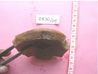
MORBID ANATOMY
Grossly most of the specimens of meningiomas received during the study were fragmented
specimens measuring about 10 cc on an average. The largest specimen was an atypical meningioma
that measured about 8cms in diameter. Some cases were received with the attached duramater and three
cases with the adjacent involved bone. All the bony specimens showed thickening as evidence of
hyperostosis.
The cut surface varied according to the histological type. Most cases were firm and lobulated,
tan to brown – grey in colour. Microcystic meningiomas demonstrated tiny cysts even grossly, while
the psammomatous meningiomas had a gritty quality while cutting. Metaplastic meningiomas with
xanthomatous changes had a pale yellow hue on the cut surface. There was a single case of a cystic
meningioma with a mural nodule that proved to be a meningothelial meningioma on histopathological
examination.
site involved was difficult to access like the sphenoid wing or posterior cranial fossa.
HISTOPATHOLOGICAL VARIANTS OF MENINGIOMAS
The WHO 2000 classification of meningiomas has defined fifteen variants of which nine are
classified as WHO grade I tumours. Three WHO grade II tumours and three WHO grade III tumours
are listed28.
Of the 342 meningiomas studied during a five year period all the variants of the new
classification were seen with the exception of clear cell meningiomas. WHO grade I meningiomas
formed the largest category with 320 cases. Among these, meningothelial meningiomas were the most
common type accounting for 128 cases.
The frontal lobes were the site most frequently involved by them, with 21 cases, followed by
the parietal lobe with 16 cases, and sphenoid wing with 12 cases.
Among the intraventricular meningiomas, meningothelial variant accounted for a majority of
the cases.
From the classic Russell and Rubinstein textbook, Pathology of tumours of the nervous system,
meningothelial meningioma have been the most common category. A review of 936 patients by
Jaackelainen et al showed 43% of the cases to be syncytial meningiomas.
Table 11
Variants No. of Cases Percentage
Meningothelial 128 37.4%
Fibrous
77 22.51%
Transitional 47 13.74%
Psammomatous 33 9.64%
Angiomatous 23 6.72%
Lymphoplasmacytic 3 0.87%
Microcystic
4 1.16%
Secretory
1 0.29%
Metaplastic 5 1.46%
Atypical 10 2.92%
Chordoid 1 0.29%
Papillary 6 1.75%
Rhabdoid 3 0.87%
Anaplastic
1 0.29%
17 16
12 15
7 7 7 6 6 69 0 10 20 30 40 50 60 70 N o . o f c a s e s F ro nt a l P ar ie ta l S ph en o id w in g P ar as a gi tta l S p c or d F al x S up ra se lla r C P a ng le IV O th er s Site
The histology of most of these cases were typical with whorls, sheets and nests of ill – refined
meningothelial cells having abundant cytoplasm and vestibular nuclei with marginated chromatin.
Though a few cases exhibited pleomorphic nuclei, those without increased mitotic activity were
still classified as meningothelial grade I meningiomas.
Fibrous meningiomas were the second most common subtype. The frequency of fibrous
meningiomas were found to increase with age. There were 25 fibrous examples among the 104
meningiomas reported in the 5th decade, and 10 cases out of the reported 47 meningiomas in the 6th
decade.
Shah et al42 have noted a slight increase in the frequency of fibrous meningiomas with age.
Meningothelial and fibrous meningiomas were found in equal frequency in the cerebello –
pontine angles.
chordoid sub types10.
Out of the 6 papillary meningiomas in our series three examples occurred in the first three
decades. One case presented with a recurrence, while the other two were lost to follow up.
A single case of chordoid meningioma was reported in a 46 years old male in the regions of the
clivus. The tumour had eroded the posterior clinodal process and a partial excision was performed.
Through the patient had a microcytic anaemia be did not have hypergamma globulinemia or associated
castheman’s disease. The patient expired two days after surgery.
Kepes et al described the chordoid variant of meningioma for the first time, and also stated that
administration of steroids resulted in complete resolution of symptoms23. Couce et al in a large series of
42 patient observed that none of their cases had systemic manifestations and that the average age of
their patient was 47.4 years4.
Recurrence rate is high among chordoid meningiomas which are classified as WHO grade II
tumours. A possible explanation could be related to the mucoid quality of its stroma which
mechanically facilitates the spread of the neoplastic cells. Recurrent tumours usually have an mucin
rich chordoid pattern.
Three cases of rhabdoid meningiomas WHO grade III were reported. Two were in their sixties
while the other was a young 17 years old female. The latter patient died a day after surgery following a
massive epileptic attack and aspiration.
Rhabdoid meningiomas are WHO grade III and have an aggressive clinical course. Their
histology is similar to rhabdoid tumours elsewhere in the body. There were large areas of necrosis,
extreme cytological atypia and high mitotic counts.
behaviour of these tumours being undetermined as yet.
MENINGIOMA - GRADES
3.52% 2.92%
93.56%
Grade I Grade II Grade III
Atypical and Anaplastic Meningioma
There were 10 atypical meningioma under WHO grade II meningiomas, forming 2.92% of the
total. Two of these were recurrent lesions. They were seen with a slightly higher frequency among
men. No specific site predilection was noted. The age groups commonly involved were the fifth and
sixth decades.
A single case of anaplastic meningioma was reported in a 16 year old girl in the posterior
cranial fossa. The mitotic count was 30/hpf and the course was aggressive with the patient succumbing
to the lesion 2 months after surgery.
Malhotra et al reported an incidence of 3.8% (5 out of 130) papillary meningiomas42. Atypical
meningiomas accounted for 8.3% (32 out of 382) in a series reported by Joseph et al and only one
Perry et al stated the two most use ful criteria for predicting recurrences (Mayo Clinic Series)
were brain invasion and presence of mitotic activity (at least 4/ hpf).
Mc Carthy et al in their large series of 9000 cases determined that important prognostic factors
for benign tumours included age at diagnosis, tumor size, surgical treatment and radiation therapy.
DISTRIBUTION OF THE HISTOLOGICAL VARIANTS OF MENINGIOMAS
128
77
47
33 23
3 4 1 5 10 1 6 3 1
0 20 40 60 80 100 120 140 N o . o f c a s e s M e n in g o th e lia l F ib ro u s T ra n si tio n a l P sa m m o m a lo u s A n g io m a to u s L ym p h o p la sm a cy tic M ic ro cy st ic S e cr e to ry M e ta p la st ic A ty p ic a l C h o rd o id P a p ill a ry R h a b d o id A n a p la st ic Histological variants INFERENCE
study. Meningiomas were more common in women than men and show an increasing trend with age at
least upto the fifth decade. Child hood meningiomas alone show a male preponderance. These tumours
are located supratentorially in most cases.
Among the benign meningiomas meningothelial examples are the most common followed by
fibrous, transitional and other variants. Histological grade, grade of surgical resection and adjuvant
Fig.1, Meningothelial meningioma – whorled cut surface
Fig.3, Transitional Meningioma
[image:64.612.136.479.364.626.2]Fig.5, Hyperostotic Parietal Bone
[image:65.612.157.459.365.627.2]Fig.7, Meningothelial Meningioma – Syncytial Whorls of Meningothelial Cells H&E (x 100)
Fig.8, Meningothelial Meningioma
[image:66.612.119.494.370.630.2]Fig.9, Fibrous Meningioma - Interlacing Fascicles of thin spindle shaped cells H & E ( x 100)
[image:67.612.134.480.381.643.2]Fig.11, Psammomatous Meningioma – Numerous psammoma bodies H & E ( x 100)
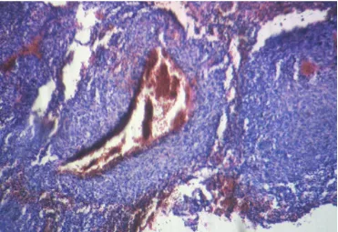
[image:68.612.135.479.368.624.2]Fig.13, Angiomatous Meningioma – Numerous blood vessels interspersed among the meningothelial cells - H & E ( x 100)
[image:69.612.122.493.367.622.2]Fig.15, Angiomatous Meningioma – Tumour vasculature showing lack of elastic laminae. Verhoeff Van Gieson (x 100)
Fig.16, Lymphoplasmacytic meningioma
[image:70.612.128.484.391.653.2]Fig.17, Metaplastic Meningioma – Islands of meningothelial cells separated by mature adipose tissue. H & E (x 100)
[image:71.612.128.486.367.628.2]Fig.19, Microcystic Meningioma – Numerous cystic spaces of various sizes H & E (x 100)
[image:72.612.111.502.354.613.2]