THE STUDY OF THE EXPRESSION OF P53 AND KI-67 IN
GASTRIC CARCINOMAS AND THEIR CORRELATION
WITH CLINICO-PATHOLOGICAL VARIABLES
Dissertation submitted in partial fulfilment of
the requirements for the degree of
M.D. (PATHOLOGY) BRANCH - III
INSTITUTE OF PATHOLOGY AND ELECTRON MICROSCOPY,
MADRAS MEDICAL COLLEGE,
CHENNAI – 600 003.
THE TAMIL NADU
DR. M.G.R. MEDICAL UNIVERSITY CHENNAI
This is to certify that this Dissertation entitled “THE STUDY OF THE EXPRESSION OF P53 AND KI-67 IN GASTRIC CARCINOMAS AND THEIR CORRELATION WITH CLINICO-PATHOLOGICAL VARIABLES” is the bonafide original work of Dr. C. HEMA VANEESWARI, in partial fulfillment of the requirement for M.D., (Branch III) in Pathology examination of the Tamilnadu Dr.M.G.R Medical University to be
held in April 2012.
Prof. Dr. SUDHA VENKATESH,M.D., Prof. Dr. A. SUNDARAM, M.D.,
PROFESSOR OF PATHOLOGY, DIRECTOR,
Institute of Pathology and EM, Institute of Pathology and EM,
Madras Medical College, Madras Medical College,
Chennai – 600003. Chennai – 600003.
Prof. Dr. KANAGASABAI, M.D., DEAN.
Madras Medical College and Government General Hospital,
DECLARATION
I, Dr. C. Hema Vaneeswari, solemnly declare that the dissertation titled
“THE STUDY OF THE EXPRESSION OF P53 AND KI-67 IN GASTRIC CARCINOMAS AND THEIR CORRELATION WITH CLINICO-PATHOLOGICAL VARIABLES” is the bonafide work done by me at
Institute of Pathology, Madras Medical College under the expert guidance and
supervision of Dr. Sudha Venkatesh, M.D., Professor of Pathology, Institute of
Pathology and Electron Microscopy, Madras Medical College. The dissertation
is submitted to the Tamilnadu Dr.M.G.R Medical University towards partial
fulfillment of requirement for the award of M.D., Degree (Branch III) in
Pathology.
Place: Chennai
I express my sincere thanks to Prof. Dr. KANAGASABAI, M.D., Dean, Madras Medical College and Government General Hospital, for permitting me
to utilize the facilities of the Institution.
I take this opportunity to express my heartfelt sincere gratitude to
Dr.A.SUNDARAM, M.D., Professor and Director of Institute of Pathology and Electron Microscopy, Madras Medical College, Chennai for his keen interest,
constant encouragement, valuable suggestions and expert guidance throughout
the study.
I am extremely thankful to Dr. SUDHA VENKATESH, M.D., Professor
of Pathology, Institute of Pathology and Electron Microscopy, Madras Medical
College for her advice, encouragement and suggestions during the study.
I take the opportunity to express my thanks to Dr.P.KARKUZHALI,
M.D., Professor of Pathology, Institute of Pathology and Electron Microscopy,
Madras Medical College for her opinions and encouragement throughout the
study.
I am truly thankful to Dr.GEETHA DEVADAS, M.D., D.C.P.,
Professor of Pathology, Institute of Pathology and Electron Microscopy, Madras
I express my heartfelt thanks to Dr.SHANTHA RAVISHANKAR,
M.D., Professor of Neuropathology, Institute of Neurology, Madras Medical
College for her valuable advice and encouragement during the study.
My thanks to Dr. M. P. KANCHANA, M.D., Professor of Pathology,
Institute of Obstetrics & Gynaecology, Madras Medical College for all her
encouragement and opinions about the study.
I convey my thanks to Dr. K. RAMA, M.D., Professor of Pathology,
Government Kasturba Gandhi Hospital, Madras Medical College for her
suggestions and support during the period of study.
I thank Dr. T. CHITRA, M.D., Professor of Pathology, Institute of Child
Health, Madras Medical College for her help and encouragement during the
course of the study.
I thank Dr. S.PAPPATHI, M.D., D.C.H., Professor of Pathology,
Institute of Pathology and Electron Microscopy, Madras Medical College for
her support during the study.
I put across my thankfulness to Dr. INDIRA, M.D., Professor of
Pathology, Regional Institute of Ophthalmology, Madras Medical College, for
her abets and aids during the study period.
I express my heartfelt sincere thanks to all my Assistant Professors for
Institute of Pathology and Electron Microscopy, Madras Medical College,
Chennai for all their help and support they extended for the successful
ABBREVIATIONS
GI : Gastro - Intestinal
MIB - 1 : Monoclonal antibody directed against Ki-67 protein
WHO : World Health Organization
IL – 1 : Interleukin - 1
CDH - 1 : Cadherin – 1 gene
HNPCC : Hereditary Non – Polyposis Colorectal Cancer
MLH - 1 : MutL Homolog – 1gene
hMSH : human MutS Homolog gene
hPMS : human Protein Homolog gene
OGJ : Oesophago – Gastric Junction
MMP : Matrix Metallo-Proteinase
TIMP : Tissue Inhibitor of Metallo-Proteinase
EGC : Early Gastric Carcinoma
IHC : Immunohistochemistry
PGP 9.5 : Protein Gene Product 9.5
PCR : Polymerase chain reaction
SSCP : Single Strand Conformation Polymorphism
AgNOR : Silver stained Nucleolar Organizer Region
PCNA : Proliferating Cell Nuclear Antigen
MCM : Mini – Chromosome Maintenance
GIST : Gastro – Intestinal Stromal Tumour
HRP : Horse – Radish Peroxide
LI : Labeling Index
CONTENTS
S. NO. TITLE PAGE NUMBER
1 INTRODUCTION 1
2 AIMS AND OBJECTIVES 3
3 REVIEW OF LITERATURE 4
4 MATERIALS AND METHODS 28
5 OBSERVATION AND RESULTS 33
6 DISCUSSION 58
7 SUMMARY 70
8 CONCLUSION 73
ANNEXURES
BIBLIOGRAPHY
INTRODUCTION
Gastric cancer is the third most common cancer in India and the second
most common type of cancer worldwide1. Approximately 800,000 new cases
are diagnosed every year, despite a steadily declining incidence over the
previous 50 years2. There is wide variation in incidence in different continents.
The highest incidence of gastric cancer is in Asia3,4, Central Europe, and South
America. There have also been changes noted in the topographic distribution of
gastric cancer in recent years. The incidence of proximal gastric tumors5,6 has
been on the rise. The widespread use of upper GI endoscopy has led to more
frequent detection of superficial cancers. This trend has had a dramatic impact
on the mortality rate, to the point that gastric cancer is now considered
potentially curable when detected at an early stage7.
Gastric carcinoma exhibits a wide range of morphological phenotypes.
The histological appearances of tumor cannot fully reveal the prognosis. The
prognosis of gastric carcinoma is mainly dependent on the stage of the disease.
Because of the variability of prognosis within a clinical or pathological stage of
gastric cancer, there has been a constant search for specific biological markers
in order to identify subgroups of patients with more aggressive course of
disease8. The immunohistochemical protein expression of p53 and Ki-67 has
been proposed as a potential tool for the evaluation of the biological behavior of
Mutations of the p53 gene have been found in a number of
malignancies10-17. In contrast to the normal p53 protein, the mutated p53 protein
has an increased half-life and hence accumulates within the cell nucleus. This
can be detected immunohistochemically using monoclonal antibodies.
Cell proliferation can be assessed by immuno-histochemical staining with
proliferation markers such as Ki-67 antigen. The monoclonal antibody MIB – 1
reacts with nuclear antigen present throughout the cell cycle of proliferating
cells but absent in quiescent cells18. The level of Ki 67 immuno-reactivity
correlates with the degree of tumour proliferation18.
Patients expressing high levels of p53 and Ki-67 have poorer prognosis
because of an aggressive tumour behavior, independent of the already known
adverse predictors. Thus the routine evaluation of p53 and Ki -67 could be
useful in identifying patients with more aggressive disease and contribute to a
better therapeutic approach.
In this study of 50 cases, an attempt is made to study the expression of
p53 and Ki-67 immunohistochemically and compare it with various
clinico-pathological parameters.
AIMS AND OBJECTIVES
1. To identify the incidence and distribution of gastric carcinoma in patients
admitted in Government General Hospital, Chennai during the year 2010.
2. To study the histo-morphological features of gastric carcinoma including
tumour size, tumour location, macroscopic appearance, histological type,
grade, depth of infiltration, lymph node status, stage , lympho-vascular
invasion, perineural infiltration, lymphocytic response, and necrosis.
3. To study the immunohistochemical expression of p53 in gastric
carcinoma
4. To study the immunohistochemical expression of Ki-67 in gastric
carcinoma
5. To determine the correlation of p53 and Ki 67 expression with known
prognostic factors such as tumor size, histological type, grade, depth of
infiltration, lymph node status, stage, presence of tumor necrosis,
lymphocytic response, lympho-vascular invasion and perineural
infiltration.
REVIEW OF LITERATURE
Gastric carcinomas are a group of malignant tumours of the stomach
arising from the gastric glandular epithelium.
The first case of gastric cancer was reported in the Ebers papyrus in 1600
BC and in the Hippocrates reports related by Galen in the second century AD in
Rome19. At the end of the first millennium AD, a possible description of a
gastric cancer could be read in Avicenna‟s Medical Encyclopedia. Despite this,
in the eighteenth century, gastric cancers were largely unknown because benign
and malignant gastric ulcers were only described later by J. Cruveilhier, in
1835.
The official history of gastric cancer surgery began 40 years later when
Jules Emile Pean, a very famous French surgeon, performed the first gastric
resection for cancer in 187920. The first successful subtotal resection with
gastro-duodenal anastomosis was performed by Theodor Billroth in 1881 in
Vienna21.
Later several classification systems were proposed. One of the earliest
was the Lauren classification proposed in 1965 which divided gastric
adenocarcinomas into 2 types – Intestinal and Diffuse22. This was followed by
the Ming classification in 1977 which divided the adenocarcinomas into 2 types
– expanding and infiltrative, based on the growth pattern23
classification was proposed in 1977 which was based on the histomorphology24 (Annexure II).
Epidemiology:
In 2000, about 880 000 people were diagnosed with gastric cancer and
approximately 650 000 died of the disease world wide2. Japan and Korea have
the highest gastric cancer rates in the world25,26. Age-standardized incidence
rates in Japan are 69.2 per 100,000 in men and 28.6 per 100,000 in women27.
In India there is a wide variation in the incidence of gastric carcinoma.
According to the study conducted by the National Cancer Registry Programme
of India in 2001, the number of new gastric cancer cases was estimated to be
approximately 35,675 (n=23,785 in men; 11,890 in women)28. The incidence
rate of gastric cancer was four times higher in Southern India compared with
Northern India29.The rates in rural population were much lower than those in the
urban population. Among the six registries in Southern India, the highest
incidence in both sexes was reported from Chennai. The age-standardized
incidence rates in Chennai are 13.6 per 100,000 in men and 6.5 per 100,000 in
women28.
The 5 year survival rate of early gastric cancers is higher (upto 95%)
Clinical presentation:
The symptoms associated with gastric cancer are usually non-specific.
Early gastric cancers are usually asymptomatic. Some of them may cause
anorexia, weight loss, fatigue, nausea, vomiting and mild to moderate epigastric
distress. Hematemesis occurs in 10% to 15% cases. Proximal gastric tumours
cause dysphagia and distal gastric tumours may cause gastric outlet obstruction.
Pathogenesis:
Gastric carcinogenesis is a multistep and multifactorial process that in
many cases appears to involve a progression from normal mucosa through
chronic gastritis, atrophic gastritis and intestinal metaplasia to dysplasia and
carcinoma31.
Risk factors:
The risk factors associated with gastric carcinoma include chronic
atrophic gastritis, Helicobacter pylori infection, diets rich in salt (dried and
salted fish) and low in micronutrients (vitamin C), intestinal metaplasia,
smoking, pernicious anemia, bile reflux in patients with post-operative gastric
stumps, Menetrier‟s disease and peptic ulcer disease32,33
. First-degree relatives
of affected patients are almost three times as likely to develop the disease as the
general population. This may be partly attributable to H. pylori infection being
Etiology: Diet:
The most consistent etiological factor associated with gastric cancer is
diet. Intraluminal and intramucosal synthesis of carcinogens like N-
nitrosamines by bacteria34 and excessive salt which acts as an irritant35-37, cause
inflammation and intestinal metaplasia which later leads to malignancy.
Consumption of fresh fruits and vegetables which contain Vitamin C, Vitamin E
and carotenoids38 counteracts the formation of N-nitroso compounds39 and
scavenges oxygen free radicals, thereby playing a protective role.
Helicobacter pylori infection:
Several epidemiological investigations have found a consistent
association between H.pylori seropositivity and risk of gastric cancer40,41. The
development of severe gastritis with atrophy and intestinal metaplasia is
particularly associated with infection by CagA-positive strains of the
bacillus42,43 and these strains have been associated with increased risk of gastric
cancer in some studies44. The sequence of events include development of
atrophic gastritis, intestinal metaplasia, dysplasia and carcinoma. The various
mechanisms proposed are increased epithelial cell proliferation with a resultant
increased risk of mutations45, bacterial overgrowth with increased potential to
generate intraluminal carcinogens46, increased free radicals47 and reduced
gastric antioxidant levels48.
There is evidence that germline truncating mutations in the gene for
E-cadherin (CDH-1), a calcium-dependent cell adhesion protein, are responsible
for a rare autosomal dominant inherited form of gastric carcinoma in young
persons. This condition is characterized by multiple tumours of diffuse or signet
ring cell histological types that do not arise in a background of intestinal
metaplasia49. Affected family members can be identified by mutation-specific
genetic testing and offered prophylactic gastrectomy50. Patients with hereditary
non-polyposis colorectal cancer (HNPCC), which results from germline
mutation of one of the DNA mismatch repair genes hMLH1, hMSH2, hMSH6,
hPMS1 and hPMS2 also have an increased frequency of gastric cancers51. Peutz
– Jegher‟s syndrome also shows an increased risk of gastric cancers52
.
Topography of gastric carcinoma
Carcinomas of the distal stomach are most common in the prepyloric
region, in the pyloric antrum and on the lesser curvature. Tumours arising at the
cardia in the region of the oesophago-gastric junction (OGJ), whose frequency
is increasing are generally smaller than those of the distal stomach. In 1996,
Siewert et al proposed a classification of gastro-oesophageal junction
adenocarcinomas based upon their location relative to the gastro-oesophageal
junction. The tumours whose centre lay between 5 cm proximal to and 5 cm
distal to the gastro-oesophageal junction were considered to be
oesophago-gastric junction tumours. Siewert et al subdivided these gastro-oesophageal
gastro-oesophageal junction, type II if between 1 cm proximal and 1 cm distal
to the junction and type III if 1–5 cm distal to the junction53. This classification
has been internationally recognised and is used by surgeons to plan
management of the tumour.
Early gastric cancer:
Early gastric cancer is defined as a carcinoma which is limited to the
mucosa or the mucosa and submucosa only, irrespective of the lymph node
status. It can be subdivided further after histological examination into two
groups, intramucosal and submucosal carcinoma. The term „early‟ does not
imply a stage in the genesis of the cancer but means that the gastric cancer is
potentially curable54. Early gastric cancer is also known as superficial spreading
carcinoma55, surface carcinoma56 and cancer gastrique au début57. Increasing
numbers of early gastric cancers are being detected mainly due to screening
programs in countries like Japan. The mean age at presentation is somewhat
lower58 and the duration of symptoms is generally longer59.
Advanced gastric cancer:
Advanced gastric cancer is defined as a carcinoma which has spread
beyond the submucosa into the muscularis propria and beyond, irrespective of
the lymph node status. The term „advanced‟ does not indicate a higher stage of
disease but means that treatment of such tumours is difficult and has decreased
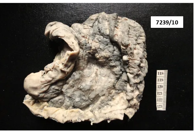
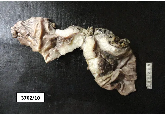
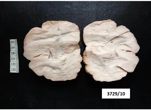
Macroscopic appearance of gastric cancer:
A sub-classification of the gross appearance of early gastric cancer was
devised by the Japanese Gastro-enterological Endoscopic Society on the basis
of macroscopic appearances at endoscopy and in gastrectomy specimens. They
were divided into three main types and three subtypes. (Figure 1)
Type I - Protruded - The tumour projects clearly into the lumen and includes all polypoid, nodular and villous tumours.
Type II – Superficial - This is further subdivided into three groups: Type IIa - Elevated above surrounding mucosa by few
millimetres.This is seen as a well – circumscribed flat
plaque.
Type IIb - Flat. No abnormality is macroscopically visible
Type IIc - Depressed. The surface is slightly depressed below
adjacent mucosa
Type III – Excavated - Ulceration of variable depth into the gastric wall.
EGC is located mainly in the corpus and antrum of the stomach60 .
Lesions are multifocal in up to 14% of cases61.
Figure 1 : Gross classification of early gastric cancer
Macroscopic types of advanced gastric cancer can be understood from the
schema depicted in 1925 by Dr. R. Borrmann, who was a German surgeon and
pathologist. (Figure 2)
Type I – Polypoid / Nodular
Type II – Ulcerative, localized / Fungating
Type III – Ulcerative, infiltrative
Type IV – Diffusely infiltrative
Figure 2 : Borrmann classification of gross types of advanced gastric cancer
Ulcerated tumours occur most frequently in the antrum on the lesser
curve and these ulcers are large with an irregular margin, raised rolled edges
and necrotic shaggy base62. Polypoid, fungating and nodular tumours tend to
occur in the body of the stomach in the region of the greater curvature, posterior
wall or fundus. Infiltrative cancers spread superficially in the mucosa and
submucosa producing plaque-like lesions. It is commonly accompanied by
thickness of the entire stomach wall producing the so-called linitis plastica or
„leather bottle‟ stomach. Many gastric carcinomas secrete considerable amounts
of mucin which gives the gelatinous appearance of colloid carcinomas.
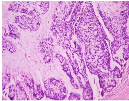
Gastric adenocarcinomas are either gland-forming malignancies
composed of tubular, acinar or papillary structures, or they consist of a complex
mixture of discohesive, isolated cells63. Several classification systems have been
proposed, including Ming, Carniero, and Goseki, but the most commonly used
are those of WHO64 (Annexure II) and Laurén.
WHO CLASSIFICATION: Tubular adenocarcinoma :
Tubular adenocarcinoma is composed predominantly of neoplastic
tubules often showing irregular branching and anastomosis embedded in or
surrounded by fibrous stroma. Individual tumour cells are columnar, cuboidal,
or flattened by intraluminal mucin. The degree of cytological atypia varies from
low to high-grade. A poorly differentiated variant is sometimes called solid
carcinoma. An oncocytic variant of tubular adenocarcinoma has been
described65.
Papillary adenocarcinoma :
These are well-differentiated carcinomas with elongated finger-like
processes lined by cuboidal cells supported by fibro-vascular connective tissue
cores. Some
tumours show tubular differentiation (papillotubular). Rarely, a micropapillary
architecture is present. Typically this tumour grows as a polypoid mass into the
Mucinous carcinoma :
WHO defines carcinomas containing large amounts of extracellular
mucin in more than 50% of the tumour as Mucinous carcinomas. In some such
tumours the cells form glands lined by columnar mucus-secreting cells (well
differentiated type). In others there are disaggregated ribbons or clusters of cells
which appear to be floating in lakes of mucin (poorly differentiated type). There
may also be mucin in the inter-glandular stroma. Scattered signet-ring cells,
when present, do not dominate the histological picture. They most commonly
occur as polypoid, fungating or ulcerative masses.
Signet ring cell carcinoma:
WHO defines this tumour as “Carcinomas composed predominantly of
single cells or small clusters of cells containing intra-cytoplasmic mucus
vacuoles and accounting for more than 50% of the tumour”. The cells contain
nuclei which push against cell membranes creating a classical signet ring cell
appearance due to an expanded, globoid, optically clear cytoplasm. These
contain acid mucin and stain with Alcian blue at pH 2.5. They also contain cells
with no mucin and cells with eosinophilic granular cytoplasm containing neutral
mucin. These tumours are more common in younger patients and in the distal
stomach. Signet ring cell carcinomas tend to infiltrate the wall of the stomach
diffusely and are accompanied by marked fibrosis giving rise to the linitis
plastica appearance on gross examination.
LAUREN CLASSIFICATION :
The histological classification of Lauren22 (1965) divides gastric
adenocarcinoma into two main types - Intestinal and Diffuse. Tumours that
contain approximately equal quantities of intestinal and diffuse components are
called mixed carcinomas. Carcinomas too undifferentiated to fit neatly into
either category are placed in the indeterminate category.
Intestinal carcinoma:
Intestinal-type tumours have a glandular pattern usually accompanied by
tubules papillary formation or solid components. The glands range from well
differentiated to moderately differentiated grade, sometimes with poorly
differentiated tumour at the advancing margin. The glandular epithelium
consists of large pleomorphic cells with large hyperchromatic nuclei often with
numerous mitoses. The adjacent gastric mucosa often shows chronic gastritis,
widespread intestinal metaplasia and sometimes dysplasia. Intestinal-type
tumours are commoner in the elderly and in males.
Diffuse carcinoma:
Diffuse-type carcinomas are predominantly composed of poorly cohesive
diffusely infiltrating small tumour cells with indistinct cytoplasm and
hyperchromatic nuclei. Glandular formation may occur in the more superficial
part of the tumour. Signet ring cells are common and there may be extracellular
accompanying intestinal metaplasia or dysplasia. The diffuse tumours usually
occur at a younger age and there is equal sex incidence.
CLASSIFICATION OF MING :
The Ming‟s classification (1977) divides gastric adenocarcinomas into
two types - Expanding type and Infiltrating23. The expanding type has a pushing
edge and forms discrete tumour nodules. This compares roughly to the intestinal
type of Lauren and occurs in patients over 50 years of age. The infiltrative type
is ill defined and contains widely infiltrative tumour cells with poor
inflammatory cell response and collagenous stroma and is more common under
the age of 50.
CLASSIFICATION OF MULLIGAN AND REMBER :
This classification expands Lauren‟s classification by adding a third type
- pylorocardiac gland carcinoma66. Pylorocardiac carcinomas commonly
present as well demarcated fungating tumours. These tumours are commoner in
men and are characterized microscopically by varying-sized glands showing
tubular or papillary pattern cells that often show striking vacuolation or clear
cell change and stain brilliantly with the periodic acid–Schiff reaction.
THE GOSEKI CLASSIFICATION :
Goseki et al divided gastric cancer into four histological types according
to the degree of tubular differentiation and the amount of intracellular mucin67.
Group I - consists of well differentiated tubules with poor intracellular mucin.
Group III – consists of poorly differentiated tubules & poor intracellular mucin;
Group IV - consists of poorly differentiated tubules & plentiful intracellular
mucin.
CARNEIRO CLASSIFICATION :
Carneiro et al proposed a much simpler system in which the tumours are
divided into glandular, isolated cell carcinomas, solid variety and a mixed type
that consists of a mixture of glandular and isolated cell types63.
The rare variants of gastric carcinoma include Adenosquamous
carcinoma68, Squamous cell carcinoma69, Hepatoid adenocarcinoma70,
Choriocarcinoma71, Medullary carcinoma with lymphoid stroma72, Small cell
carcinoma73, Parietal cell carcinoma74, Gastric carcinoma with rhabdoid
features75 and Carcinosarcoma76.
SPREAD OF GASTRIC CANCER
Gastric cancer may spread directly through penetration of the serosa and
infiltration into organs like pancreas, liver, spleen, transverse colon and
omentum and this is particularly common in signet ring cell carcinomas and
diffuse carcinomas. The incidence of lymphatic spread increases with increasing
depth of invasion into the stomach wall. The nodes commonly involved include
the nodes along the left gastric, common hepatic, coeliac arteries and the
pancreatic and splenic nodes. More distant lymphatic spread may involve
para-aortic and mesenteric nodes. Spread by way of the thoracic duct to the left
Hematogenous spread occurs most commonly to the liver, followed by lung,
peritoneum, adrenal glands, skin and ovaries (Krukenberg tumour). Diffuse
tumours tend to involve unusual sites such as kidney, spleen, uterus and
meninges more often77.
STAGING OF GASTRIC CANCER :
The TNM staging system78 (Annexure III) is widely used in western
countries. It is the best available predictor of prognosis and is recommended.
PROGNOSIS:
The prognosis of gastric carcinoma varies from country to country with
Japan having the best results with an overall 5-year survival rate of 46% for
advanced carcinoma and 89% for early carcinoma79. The overall survival rate
in the Western countries is between 4% and 13%80. This can be explained at
least partly by the greater frequency of superficial carcinomas, aggressive
Japanese surgical approach to treatment with extensive and meticulous lymph
node dissection81. A recent study of untreated early gastric cancer has indicated
a 63% cumulative 5-year risk of progression to advanced cancer82.
PROGNOSTIC FACTORS:
Prognostic factor is defined as any variable that provides information
useful in assessing the outcome at the time of diagnosis of the disease. The
prognostic factors are classified as clinical factors, morphological factors and
genetic / molecular factors. The clinical factors with poor prognosis include
younger age group, larger tumor size, and proximal gastric cancers80. The
year survival rates in tumours of the cardia are under 20%83 and the median
survival is about 7 months only84. The pathological factors play a more useful
role in assessing prognosis which includes the following:
1. Tumour stage: This parameter is the most significant prognostic factor. One of the features that it incorporates is the depth of the
invasion, for the deeper the penetration, the greater the chance of
metastasis. This feature is directly related to the gross appearance of
tumour – large intraluminal neoplasms have lower incidence of
metastasis than those growing primarily within the wall.
2. Microscopic type and grading: The intestinal type tumours in Lauren‟s classification behave relatively better than the diffuse
types85.
3. Regional lymph node involvement: With nodal involvement the 5-year survival rate drops to less than 10% when compared to 50% in
the node negative cases. The number of nodes involved is also
prognostically significant. The overall survival rate declines as the
number of positive node increases86.
4. Tumour size: Small tumour size is associated with a better prognosis
but this is closely linked to the depth of penetration79.
6. Lymphatic invasion is a poor prognostic factor strongly associated with the presence of lymph node metastasis and poor patient survival.
7. Vascular invasion denotes the infiltration of tumor cells into vascular spaces and it predicts the risk of recurrence and visceral metastasis.
Other factors reported to have poor prognosis includes tumour necrosis,
infiltrative tumour margins and positive surgical margins.
Many molecular biomarkers have been identified which play a significant
prognostic role in gastric carcinoma management. DNA aneuploidy has been
reported in approximately 40–50% of gastric carcinomas and it has been found
that aneuploid tumours are significantly associated with both lymph node and
distant metastases and lower survival rates in comparison with diploid cancers87.
Her 2 neu is a transmembrane epidermal growth factor receptor protein also
known as c erb2. Its overexpression is reported to have poorer outcome88.
Mutation of the p53 gene was identified in approximately 25% of gastric
carcinomas and this correlated well with demonstration of p53 protein
overexpression by immunohistochemistry in these tumours. Some studies based
on immunohistochemistry indicate that p53 protein overexpression is associated
with shortened survival89 but some studies failed to confirm this90. E-cadherin is
a transmembrane protein which plays an important role in maintenance of
intercellular connections. Germline mutations of the E-cadherin gene (CDH-1)
are associated with cancers of diffuse type and are highly aggressive91. Other
proliferation indices and loss of Fhit protein are associated with reduced
survival.
p53 :
p53 was identified in 1979 by Lionel Crawford, David P. Lane, Arnold
Levine, and Lloyd Old. The human TP53 gene was cloned in 1985. Its character
as a tumor suppressor gene was revealed in 1989 by Bert Vogelstein. p53 gene
is considered “Guardian of the genome” and represents a tumor suppressor gene
located on the 17p chromosome, coding a protein of 53 kD. The role of p53 is
central in cell – cycle regulation, in DNA repair and in cell apoptosis. The
production of p53 is increased in response to cellular insults or DNA damage
and p53 then induces cell - cycle arrest at the G1/S junction. Therefore, p53 is
essential for control of tumor growth, apoptosis and maintaining genome
stability. Unlike normal p53 protein, which is rapidly removed from the
nucleus, mutant forms have a prolonged half-life, which favors intranuclear
accumulation, becoming detectable immuno-histochemically. Mutations of the
p53 have been observed in a wide variety of human carcinomas, such as
colorectal carcinoma, breast carcinoma, gallbladder carcinoma, esophageal
carcinoma, and gastric carcinoma. Numerous studies have reported the
correlation between the overexpression of p53 and the poor prognosis of
patients with these tumors. The p53 pathway is also involved in regulating the
metallo-proteinase-2 (MMP-2), MMP-13 and the tissue inhibitor of metalloproteinase-3
(TIMP3).
Mutation of the p53 gene was identified in approximately 25% of gastric
carcinomas and this correlated well with demonstration of p53 protein
overexpression by immuno-histochemistry in these tumours. Carcinomas of the
cardia showed mutation of p53 in a considerably higher proportion of cases than
carcinoma of the body or antrum96. Overall prevalence of p53 immunoreactivity
in advanced gastric carcinoma is about 50–60%. Alterations of the p53 gene
have also been demonstrated in precancerous lesions of the stomach. The p53
gene mutation and overexpression of gene protein is more common in
intestinal-type carcinomas than in diffuse tumours97. Some studies based on
immuno -histochemistry indicate that p53 protein overexpression is associated
with shortened survival but few other studies have failed to confirm this89,90.
The most commonly used methods for detection of these mutations are
immunohistochemistry, flow-cytometry, polymerase chain
reaction-single-strand conformation polymorphism (PCR – SSCP) and genomic sequencing.
Although sequencing is the most unambiguous method, it is technically
cumbersome. Therefore, both immune-detection and PCR- SSCP have been
widely used as alternative methods.
Immuno-histochemically, a positive reaction is considered in the presence
of brown immunostained nuclei.
p53-negative (-): Absence of immunostaining in < 10% of the tumour nuclei
p53-positive (+): Presence of immunostaining in > 10% of the tumour nuclei
Ki-67:
Ki-67 also known as MKI67 is a protein encoded by the MKI67 gene98
which was discovered by Gerdes. The Ki-67 protein was originally defined by
the prototype monoclonal antibody Ki-67, which was generated by immunizing
mice with nuclei of the Hodgkin lymphoma cell line L428. The name is derived
from the city of origin (Kiel, Germany) and the number of the original clone in
the 96-well plate.
Ki-67 is a nuclear protein that is necessary for cellular proliferation and
ribosomal RNA transcription99. It is present during all active phases of the cell
cycle (G1, S, G2, and M), but is absent from resting cells (G0). The protein is
predominantly localized in the peri-nucleolar region in the G 1 phase, in the
later phases it is also detected throughout the nuclear interior, being
predominantly localized in the nuclear matrix. In mitosis, it is present on all
chromosomes98. Ki-67 is an excellent marker to determine the growth fraction
of a given cell population. The fraction of Ki-67-positive tumor cells (the Ki-67
labeling index) is often correlated with the clinical course of various tumours
like carcinomas of the prostate, brain and the breast. For these types of tumors,
the prognostic values for survival and tumor recurrence have repeatedly been
MIB-1 is a commonly used monoclonal antibody that detects the Ki-67
antigen. It is used in clinical applications to determine the Ki-67 labeling index.
One of its primary advantages over the original Ki-67 antibody and the reason
why it has essentially replaced the original antibody for clinical use is that it can
be used on formalin-fixed paraffin-embedded sections, after heat-mediated
antigen retrieval. Ki-67 labeling index is calculated by the percentage of
tumours cells showing distinct brown staining of the nucleus with strong
intratumoural heterogeneity. The other methods of detection of Ki-67 are by
Western blot analysis and immunofluorescence.
The various other markers of proliferation include AgNOR staining,
PCNA and Topoisomerase II. The novel markers being evaluated for
identifying cell proliferation include Fen-1, MCM proteins (mini-chromosome
maintenance), mitosin, polo – like kinase and claspin.
IMMUNOHISTOCHEMISTRY:
Albert Coons et al in 1941 first labeled antibodies directly with
fluorescent isocyanate. Nakane and Pierce et al in 1966, introduced indirect
labeling technique in which unlabeled antibody is followed by second antibody
or substrate. Various stages of development of Immunohistochemistry include
peroxidase – antiperoxidase method (1970), alkaline phosphatase labeling
(1971), avidin biotin method (1977) and two layer dextrin polymer technique
(1993)92.
ANTIGEN RETRIEVAL:
Antigen retrieval can be done by the following different techniques to
unmask the antigenic determinants of fixed tissue sections.
1. Proteolytic enzyme digestion
2. Microwave antigen retrieval
3. Pressure cooker antigen retrieval
4. Microwave and trypsin antigen retrieval
PROTEOLYTIC ENZYME DIGESTION:
Huank et al in 1976 introduced this technique to breakdown formalin
cross linkages and to unmask the antigen determinants. The most commonly
used enzymes include trypsin and proteinase93. The disadvantages include over
digestion, under digestion and antigen destruction.
MICROWAVE ANTIGEN RETRIEVAL:
This is a new technique most commonly used in current practice.
Microwave oven heating involves boiling formalin fixed paraffin sections in
various buffers for rapid and uniform heating. Antibodies against Ki67 and
PRESSURE COOKER ANTIGEN RETRIEVAL:
Miller et al in 1995 compared and proved that pressure cooking method has
fewer inconsistencies, less time consuming and can be used to retrieve large
number of slides than in microwave method94.
PITFALLS OF HEAT PRETREATMENT:
Drying of sections at any stage after heat pretreatment destroys
antigenicity. Nuclear details are damaged in poorly fixed tissues. Fibers and
fatty tissues tend to detach from slides while heating. Not all antigens are
retrieved by heat pretreatment and also some antigens like PGP 9.5 show altered
staining pattern.
DETECTION SYSTEMS:
After addition of specific antibodies to the antigens, next step is to
visualize the antigen antibody reaction complex. The methods employed are
direct and indirect methods.
In the direct method, primary antibody is directly conjugated with the
label. Most commonly used labels are flouro-chrome, horse radish peroxidase
and alkaline phosphatase. Indirect method is a two-step method in which
labeled secondary antibody reacts with primary antibody bound to specific
antigen. The use of peroxidase enzyme complex or avidin biotin complex
further increases the sensitivity of immunohistochemical stains92.
In 1993, Pluzek et al introduced enhanced polymer one step staining, in
which large numbers of primary antibody and peroxidase enzymes are attached
to dextran polymer back bone. This is the rapid and sensitive method95.
Dextran polymer conjugate two step visualization system is based on
dextran technology in Epos system. This method has greater sensitivity and is
MATERIALS AND METHODS
This study is a retrospective descriptive study of gastric adenocarcinomas
conducted in the Institute of Pathology, Madras Medical College and Rajiv
Gandhi Government General Hospital, Chennai during the period between
January 2010 and December 2010.
A total of 9,541 cases were submitted to our department during the
period January 2010 – December 2010 for histopathological examination.
Among the 660 gastric specimens, 571 were endoscopic biopsies and 89 were
gastrectomies. Among the 660 specimens, 297 were non neoplastic, 4 were
benign and 359 were malignant tumours. A total of 275 endoscopic biopsy
specimen and 84 gastrectomy specimens were reported to be malignant tumors.
Out of the 89 gastrectomies, 78 gastrectomies were done to treat gastric
carcinoma, 3 were done to treat GIST, 3 were done to treat Non – Hodgkin‟s
lymphoma, 3 were done to treat giant bleeding benign ulcers, 1 was done to
treat morbid obesity and 1 was done as revision gastrectomy to rule out stump
carcinoma.
SOURCE OF DATA:
The gastric adenocarcinoma cases reported in gastrectomy specimens
received in the Institute of Pathology, Madras Medical College between January
2010 to December 2010 from the Department of Surgery, Surgical
General Hospital. A total of 84 gastrectomy specimens (Subtotal, Total, Radical
and Palliative gastrectomy) were received during this period.
Inclusion criteria
All the gastric carcinoma cases reported in gastrectomy specimens
irrespective of the age and sex were included for the study.
Exclusion criteria
Non neoplastic lesions and benign tumors of stomach.
Gastric carcinomas reported in endoscopic biopsies.
Gastrectomies performed for reasons other than treating carcinomas.
METHOD OF DATA COLLECTION:
Detailed history of the cases regarding age, sex, history, type of
procedure, history of neo adjuvant therapy, details of gross characteristics and
nodal status were obtained for all the 78 gastrectomy cases reported during the
period of study from Surgical pathology records. Hematoxylin and Eosin
stained 4 µ thick sections of the paraffin tissue blocks of gastrectomy specimens
were reviewed. The following clinical and pathological parameters were
evaluated: Age (<55 and >= 55), gender, tumour size (<5 and >=5cm), tumour
location (Eso-cardiac, body, antrum, pangastric), macroscopic appearance
(Borrmann Type I, Type II, Type III and Type IV). Carcinomas were classified
histological types (tubular, papillary, mucinous, signet ring cell and diffuse).
Regarding the depth of invasion, the carcinomas were classified into 4 groups:
T1 (invasion of mucosa and submucosa), T2 (invasion of muscularis propria
and subserosa), T3 (invasion of serosa) and T4 (invasion of adjacent organs),
and according to grade the carcinomas were divided into 3 groups: G1 (well
differentiated), G2 (moderately differentiated) and G3 (poorly differentiated)
according to the recommendations of the American Joint Committee on Cancer
(2002). Lymph node metastasis was assessed and the patients were divided into
3 groups: N0 (No lymph node metastasis), N1 (metastasis in 1-6 nodes) and N2
(metastasis in 7 – 15 nodes). Carcinoma staging was done according to the
standards of the American Joint Committee on Cancer (2002) and TNM
classification of gastric carcinomas (Annexure – III). The tumours were further
evaluated for the presence of necrosis, lymphocytic response, perineural
invasion and lympho-vascular invasion by tumor and were graded as present or
absent. 50 cases of gastric adenocarcinomas of varying grades were randomly
selected from the total cases and their representative formalin fixed paraffin
embedded tissue samples were subjected to immunohistochemistry for a panel
of 2 markers – p53 and Ki-67.
IMMUNOHISTOCHEMICAL EVALUATION:
Immuohistochemical analysis of a panel of markers including p53 and
polymer HRP system based on non-biotin polymeric technology. 4 µ thick
sections from formalin fixed paraffin embedded tissue samples were transferred
onto gelatin coated slides. Heat induced antigen retrieval was done. The antigen
was bound with mouse monoclonal antibody (Biogenex) against p53 protein
and Ki – 67 protein and then detected by the addition of secondary antibody
conjugated with horse radish peroxidase-polymer and diaminobenzidine
substrate. The step by step procedure of Immunohistochemistry is given in
Annexure IV.
Antigen Vendor Species(clone) Dilution Positive control
P53 BIOGENEX Mouse Ready to use Stomach
Ki - 67 BIOGENEX Mouse Ready to use Stomach
INTERPRETATION & SCORING SYSTEM:
The immunohistochemically stained slides were analyzed for the
presence of reaction, cellular localization, percentage of cells stained and
intensity of reaction. Nuclear staining was assessed for both p53 and Ki 67. P53
immuno-reactivity was assessed as being positive when tumours exhibited
intense nuclear staining and was categorized into 2 groups: Positive expression -
(at least 10% positive tumour cell nuclei) and Negative expression - (less than
10% positive tumour cell nuclei)
A distinct nuclear immuno – reactivity for Ki -67 was considered
positive. The Ki-67 labeling index was determined by observing 1000 cancer
cell nuclei in areas of the section with highest labeling frequency. The Ki- 67
labeling index for the 50 tumours ranged from 3.9% to 75.3% with a mean
labeling index of 25.4%. The mean Ki – 67 labeling index of 25.4% was chosen
as the cut off point for separating the cases into 2 groups: High Ki – 67 labeling
index (LI > 25.4%) and Low Ki – 67 labeling index (LI < 25.4%).
STATISTICAL ANALYSIS :
The statistical analysis was performed using statistical package for social
science software version 11.5 which consisted computing the frequency counts
and percentages for qualitative variables and mean for the quantitative variables.
The expression of p53 and the Ki – 67 labeling index was correlated with
clinico – pathological factors like age, gender, tumour site, tumour
configuration, size, Lauren‟s type, histological types, histological grade, depth
of infiltration, lymph node status, stage, lympho-vascular invasion, perineural
invasion, lymphocytic response and necrosis using the Pearson‟s Chi –Square
test. The expression of p53 and the Ki – 67 labeling index were also correlated
with each other using the McNemar‟s test. T – test was used to detect the
association between the mean Ki- 67 labeling index in the p53 positive and
OBSERVATION AND RESULTS
In the study period of 12 months from January2010 to December 2010, a
total of 9,541 specimens were received in the Institute of Pathology, Madras
Medical College for histological examination. Total numbers of gastric
specimens received were 660, of these gastric tumors accounted for 359 with a
percentage of 3.76 %. The total number of non- neoplastic, benign and
malignant cases was 297, 4 and 359 respectively. Thus the distribution of
non-neoplastic lesions was 45 %, of benign tumors were 0.6% and of malignant
tumors were 54.39% among the gastric specimens.
Among the 660 gastric specimens, there were 89 gastrectomies. Of the 89
gastrectomies, 78 were done to treat gastric carcinoma, 3 were done to treat
GIST, 3 were done to treat Non – Hodgkin‟s Lymphoma and the remaining 5
for non – neoplastic conditions. Thus the distribution of non-neoplastic lesions
was 5.7% and malignant tumours was 94.3% among the gastrectomy
specimens.
Gastric cancers had a peak incidence in the age group of 51-60 years. The
youngest age of presentation of gastric cancer is at 23 years in this study.
Among the 78 cases, 57 (73%) cases were reported in males and 21 (27%) cases
TABLE 1 AGE AND SEX WISE DISTRIBUTION OF GASTRIC CANCERS
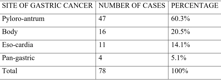
Among the 78 cases, 47 (60.3%) of cases involved the pyloro-antrum, 16
(20.5%) involved the body, 11 (14.1%) involved the eso-cardia and 4 (5.1%)
[image:47.595.105.491.114.386.2]cases were pan-gastric. (Table 2 and Chart 2)
TABLE 2 DISTRIBUTION OF SITE OF INVOLVEMENT IN GASTRIC CARCINOMA
SITE OF GASTRIC CANCER NUMBER OF CASES PERCENTAGE
Pyloro-antrum 47 60.3%
Body 16 20.5%
Eso-cardia 11 14.1%
Pan-gastric 4 5.1%
Total 78 100%
AGE GROUP
NUMBER OF
CANCERS PERCENTAGE
MALES FEMALES
21 – 30 years 0 2 2.5%
31 – 40 years 5 4 11.6%
41 – 50 years 11 6 21.8%
51 – 60 years 24 2 33.5%
61 – 70 years 11 7 23%
More than 70
years 6 0 7.6%
Total cases 57 (73%) 21 (27%) 100%
[image:47.595.86.510.561.716.2]Based on the gross morphology, the gastric tumours were divided into 4
groups according to Borrmann‟s classification & the distribution is shown in
Table 3 & Chart 3
TABLE 3 DISTRIBUTION OF GASTRIC CARCINOMA ACCORDING TO GROSS MORPHOLOGY
GROSS NUMBER OF CASES PERCENTAGE
Borrmann Type - I 12 15.3%
Borrmann Type - II 32 41%
Borrmann Type - III 25 32%
Borrmann Type - IV 8 10.2%
Early gastric cancer – Type III 1 1.2%
Total 78 100%
Among the study samples, 46 cases (58.9%) had tumor less than 5 cm in
size and 32 cases (41%) were 5cm or more in size. (Table 4 & Chart 4)
TABLE 4 - DISTRIBUTION OF SIZE IN GASTRIC CARCINOMA
SIZE OF TUMOUR NUMBER OF CASES PERCENTAGE
<5 cm 46 58.9%
>=5 cm 32 41.1%
Total 78 100%
The distribution of histological subtypes of gastric carcinoma is shown in
TABLE 5 DISTRIBUTIONS OF HISTOLOGICAL SUBTYPES OF GASTRIC CANCERS
Histological subtypes Number of cases Percentage
Tubular carcinoma 42 53.9%
Papillary carcinoma 5 6.5%
Mucinous carcinoma 11 14.2%
Signet ring cell carcinoma 6 7.6%
Diffuse carcinoma 13 16.6%
Squamous cell carcinoma 1 1.2%
Total number of cases 78 100%
77 of the gastric adenocarcinomas were grouped into 2 according to
Lauren‟s classification out of which 58 (75.4%) belonged to Intestinal type and
19 (24.6%) belonged to Diffuse type (Table 6 and Chart 6).
TABLE 6 DISTRIBUTION OF GASTRIC CANCER ACCORDING TO LAUREN’S CLASSIFICATION
LAUREN‟S TYPE NUMBER OF CASES PERCENTAGE
Intestinal type 58 75.4%
Diffuse type 19 24.6%
Total 77 100%
The gastric carcinomas were graded according to AJCC recommendation
[image:49.595.123.475.140.348.2]differentiated (G1), 38 cases (48.7%) were moderately differentiated (G2) and
29 cases (37.1%) were in poorly differentiated (G3). (Table 7 & Chart 7)
TABLE 7 DISTRIBUTION OF HISTOLOGICAL GRADE IN GASTRIC CARCINOMAS
GRADE NUMBER OF CASES PERCENTAGE
G1 11 14.2%
G2 38 48.7%
G3 29 37.1%
TOTAL 78 100%
In this study, 1 case (1.2%) showed invasion upto the submucosa (T1), 36
cases (46.2%) showed infiltration into the muscularis propria or subserosa (T2),
36 cases (46.2%) showed infiltration into the serosa and 5 cases (6.4%) showed
infiltration of adjacent organs (T4) (Table 8 and Chart 8).
TABLE 8 DISTRIBUTION OF GASTRIC CARCINOMAS ACCORDING TO DEPTH OF INVASION
DEPTH OF INVASION
NUMBER OF
CASES PERCENTAGE
T1 1 1.2%
T2 36 46.2%
T3 36 46.2%
T4 5 6.4%
This study showed that 39 cases (50%) had up to 6 nodes with metastatic
carcinomatous deposit (N1), 5 cases (6.4%) had 7 to 15 involved nodes (N2)
while 34 cases (43.6%) had no node involvement (N0). (Table 9 & Chart 9)
TABLE 9 DISTRIBUTION OF LYMPH NODE METASTASIS IN GASTRIC CANCERS
Lymph node
status Number of cases Percentage
N0 34 43.6%
N1 39 50%
N2 5 6.4%
Total 78 100%
In the present study, 17 cases (21.8%) belonged to stage I, 35 cases
(44.9%) belonged to stage II, 21 cases (26.9%) belonged to stage III and 5 cases
(6.4%) belonged to stage IV. (Table 10 and Chart 10)
TABLE 10 DISTRIBUTION OF GASTRIC CARCINOMAS
ACCORDING TO STAGE
STAGE NUMBER OF
CASES PERCENTAGE
I 17 21.8%
II 35 44.9%
III 21 26.9%
IV 5 6.4%
Total 78 100%
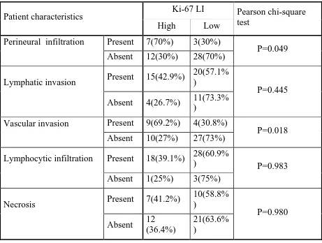
In this study, among the 78 cases, 52 cases (66.6%) had lymphatic
invasion as against 26 cases (33.4%) without lymphatic invasion. 15 cases
(19.3%) showed vascular invasion while 63 cases (80.7%) cases had no
vascular invasion, 17.9 % of the cases had perineural infiltration, 85.8% of the
cases had lymphocytic infiltration , 23.1% of the cases had necrosis. (Table 11
& Chart 11)
TABLE 11 DISTRIBUTION OF OTHER PROGNOSTIC FACTORS IN GASTRIC CARCINOMA
Patient characteristics Present Absent Total
Lymphatic invasion 52 (66.6%) 26 (33.4%) 78 (100%)
Vascular invasion 15 (19.3%) 63 (80.7%) 78 (100%)
Perineural infiltration 14 (17.9%) 64 (82.1%) 78 (100%)
Lymphocytic infiltration 67 (85.8%) 11 (14.2%) 78 (100%)
Necrosis 18 (23%) 60 (77%) 78 (100%)
RESULTS OF IMMUNOHISTOCHEMICAL STUDIES
Of the total 78 cases, 50 cases of varying grade and stage were selected in
a random manner and subjected to immunohistochemical analysis with a panel
of 2 markers – p53 and Ki-67.
Of the 50 cases, there were 39 males (78%) and 11 females (22%). The
ages ranged between 28 and 75 with a mean of 55.04. There were 18 cases
tumour was located in the pyloro – antrum in 27 cases (54%), body in 12 cases
(24%), eso-cardia in 7 cases (14%) and were pan-gastric in 4 cases (8%). 10
cases (20%) belonged to Borrmann Type I, 20 cases (40%) belonged to Type II,
14 cases (28%) belonged to Type III and 6 cases (12%) belonged to type IV.
The tumours ranged in size from 2 to 12 cm with an average of 5.72.
There were 27 cases (54%) with tumour size <5 cm and 23 cases (46%) with
size >5cm. 31 cases (62%) were of the tubular type, 4 cases (8%) were of the
papillary type, 6 cases (12%) were mucinous carcinomas, 3 cases (6%) were of
the signet ring cell type and 6 cases (12%) were of the diffuse type. 41 cases
(82%) belonged to Lauren‟s Intestinal type and 9 cases (18%) belonged to the
Diffuse type.
Among the final study group, 9 (18%) cases were of G1, 25 (50%) cases
were of G2 and 16 (32%) cases were of G3. 23 (46%) cases belonged to T2, 24
(48%) cases belonged to T3 and 3 cases (6%) belonged to T4. Of the 50 cases,
35(70%) showed lymphatic invasion, 13 cases (26%) showed vascular invasion,
10 cases (20%) showed perineural invasion, 46 (92%) cases showed
lymphocytic response and 17(34%) showed necrosis. Nodal metastasis was
present in 1-6 nodes (N1) in 23 cases (46%), 7-15 nodes (N2) in 5 cases (10%)
and absent in 22 (44%) cases. 12 (24%) cases belonged to stage I, 19 (38%)
cases belonged to stage II, 16 cases (32%) belonged to stage III and 3 cases
(6%) belonged to stage IV. (Table 12)
TABLE 12 - DISTRIBUTION OF GASTRIC CARCINOMA AMONG THE VARIOUS CLINICOPATHOLOGICAL GROUPS FOR THE IHC STUDY (50 CASES)
Clinico-pathological factor No. of cases
Age
<55 18 (36%)
>55 32 (64%)
Sex
Males 39 (78%)
Females 11 (22%)
Site
Pyloro -antrum 27 (54%)
Body 12 (24%)
Eso- cardia 7 (14%)
Pan - gastric 4 (8%)
Borrmann I 10 (20%)
II 20 (40%)
III 14 (28%)
IV 6 (12%)
Size <5cm 27 (54%)
>5cm 23 (46%)
Histological type
Tubular 31 (62%)
Papillary 4 (8%)
Mucinous 6 (12%)
Signet ring cell 3 (6%)
Diffuse 6 (12%)
Lauren Intestinal 41 (82%)
Diffuse 9 (18%)
Grade G1 9 (18%)
G2 25 (50%)
G3 16 (32%)
Depth T2 23 (46%)
T3 24 (48%)
T4 3 (6%)
Lymphatic invasion P/A 35 (70%) / 15 (30%)
Vascular invasion P/A 13 (26%) / 37 (74%)
Perineural invasion P/A 10 (20%) / 40 (80%)
Lymphocytic response P/A 46 (92%) / 4 (8%)
Necrosis P/A 17(34%) / 33 (66%)
Lymph nodes N0 22 (44%)
N1 23 (46%)
N2 5 (10%)
Stage I 12(24%)
II 19 (38%)
III 16 (32%)
In this study, 32 cases (64%) expressed positive reaction for p53 and 18
cases (36%) were p53 negative. With the mean Ki-67 as 25.4%, the cases were
divided into two groups – High Ki-67 labeling index which was present in 19
cases (38%) and Low Ki-67 labeling index which was present in 31 cases
(62%). (Table 13 & Chart 12)
TABLE 13 - DISTRIBUTION OF p53 EXPRESSION AND Ki -67 LI IN GASTRIC CARCINOMA
IHC PARAMETER P53 Ki-67 LI RESULT POSITIVE NEGATIVE HIGH LOW
32 (64%) 18 (36%) 19 (38%) 31(62%) TOTAL(%) 50 (100%) 50 (100%)
CORRELATION OF p53 WITH VARIOUS CLINICOPATHOLOGICAL FACTORS
p53 positivity was noted in 50% patients with age less than 55 and in
71.9% patients with age more than 55. (Table 14 & Chart 13)
TABLE 14 CORRELATION OF AGE WITH p53 EXPRESSION
Age (yrs) p53 positive (%) p53 negative (%) Total Pearson chi square test
<55 9(50%) 9(50%) 18(100%)
P=0.215 >55 23(71.9%) 9(28.1%) 32 (100%)
Total 32 (64%) 18 (36%) 50 (100%)
p53 positivity was obtained in 66.7% of men and 54.5% of women,
noting a slight predominance in males. (Table 15 & Chart 14)
TABLE 15 CORRELATION OF GENDER WITH p53 EXPRESSION
Gender p53 positive (%) p53 negative (%) Total Pearson chi square test Male 26(66.7%) 13(33.3%) 39(100%)
P=0.701 Female 6(54.5%) 5(45.5%) 11(100%)
Total 32 (64%) 18 (36%) 50 (100%)
In the present study, p53 positivity was observed in 74.1% of tumours of
the pyloro – antrum, 66.7% of tumours of the body, 14.3% of tumours of the
eso – cardia and 75% of pan – gastric tumours. The association with respect to
site was found to be significant with increased expression seen in tumours of the
pyloro – antrum and in pan – gastric tumours. (Table 16 and Chart15)
TABLE 16 CORRELATION OF TUMOUR SITE WITH p53 EXPRESSION
Site p53 positive (%) p53 negative (%) Total Pearson chi square test
P - antrum 20(74.1%) 7(25.9%) 27(100%)
P=0.030 Body 8(66.7%) 4(33.3%) 12(100%)
Eso – cardia 1(14.3%) 6 (85.7%) 7 (100%) Pan - gastric 3 (75%) 1 (25%) 4 (100%) Total 32 (64%) 18 (36%) 50 (100%)
Among the various gross types, p53 positivity was noted in 6 cases (60%)
Borrmann type III and 5 cases (83.3%) of Borrmann type IV. (Table 17 and
Chart16)
TABLE 17 CORRELATION OF GROSS TYPE WITH p53 EXPRESSION
Gross p53 positive (%) p53 negative (%) Total Pearson chi square test Type I 6(60%) 4(40%) 10(100%)
P=0.555 Type II 11(55%) 9(45%) 20(100%)
Type III 10(71.4%) 4 (28.6%) 14 (100%) Type IV 5 (83.3%) 1 (16.7%) 6 (100%) Total 32 (64%) 18 (36%) 50 (100%)
In the present study, p53 positivity was noted in an increased frequency
(70.4%) in cases with tumour size <5cm compared to the 56.5% of cases with
size >=5cm. (Table 18 and Chart 17)
TABLE 18 CORRELATION OF TUMOUR SIZE WITH p53 EXPRESSION
Size p53 positive (%) p53 negative (%) Total Pearson chi square test <5 cm 19(70.4%) 8(29.6%) 27(100%)
P=0.471 >=5 cm 13(56.5%) 10(43.5%) 23(100%)
Total 32 (64%) 18 (36%) 50 (100%)
Among histological forms, 64.5% of tubular carcinomas, 25% of
papillary carcinoma, 33.3% of signet ring cell carcinomas and 66.7% of diffuse
carcinomas showed p53 positivity. In this study, 100% of mucinous carcinomas
showed p53 positivity. (Table 19 and Chart 18)
TABLE 19 CORRELATION OF HISTOLOGICAL TYPE WITH p53 EXPRESSION
His.type p53 positive
(%)
p53 negative
(%) Total
Pearson chi square test
Tubular 20(64.5%) 11(35.5%) 31(100%)
P=0.123
Papillary 1(25%) 3(75%) 4(100%)
Mucinous 6 (100%) 0 (0%) 6 (100%)
Signet 1 (33.3%) 2 (66.7%) 3 (100%)
Diffuse 4 (66.7%) 2 (33.3%) 6 (100%)
Total 32 (64%) 18 (36%) 50(100%)
When Lauren‟s classification was taken into account, a greater frequency
of p53 positivation with Intestinal type cancers (65.8%) in comparison with
diffuse type carcinomas (55.6%) was observed. (Table 20 and Chart 19)
TABLE 20 CORRELATION OF LAUREN’S HISTOLOGICAL TYPE WITH p53 EXPRESSION
Lauren type
p53 positive (%)
p53 negative
(%) Total
Pearson chi square test
Intestinal 27(65.8%) 14(34.2%) 41(100%)
P=0.560
Diffuse 5(55.6%) 4(44.4%) 9(100%)
Total 32 (64%) 18 (36%) 50
(100%)
An increasing percentage of cases showing p53 positivity with increasing
moderately differentiated tumours (G2) and 75% of poorly differentiated
tumours (G3) showing positivity for p53 was observed. (Table 21 and Chart 20)
TABLE 21 CORRELATION OF TUMOUR GRADE WITH p53 EXPRESSION
Grade p53 positive (%) p53 negative (%) Total Pearson chi square test G1 4(44.4%) 5(55.6%) 9(100%)
P=0.311 G2 16(64%) 9(36%) 25(100%)
G3 12 (75%) 4 (25%) 16 (100%) Total 32 (64%) 18 (36%) 50 (100%)
According to the T stage, a progressive increase in the number of p53
positive cases from T2 to T3 was noted. p53 positivity was identified in 52.2%
of T2 carcinomas, 75% of T3 carcinomas and 66.7% of T4 carcinomas.(Table
22 and Chart 21)
TABLE 22 CORRELATION OF T STAGE WITH p53 EXPRESSION
T stage p53 positive (%) p53 negative (%) Total Pearson chi square test T2 12(52.2%) 11(47.8%) 23(100%)
P=0.264 T3 18(75%) 6(25%) 24(100%)
T4 2 (66.7%) 1 (33.3%) 3 (100%) Total 32 (64%) 18 (36%) 50 (100%)
In this study, a progressive increase in the percentage of p53 positive
59.1 % of N0 cases, 60.9% of N1 cases and 100% of N2 cases.(Table 23 and
Chart 22)
TABLE 23 CORRELATION OF N STAGE WITH p53 EXPRESSION
N stage p53 positive (%) p53 negative (%) Total Pearson chi square test N0 13(59.1%) 9(40.9%) 22(100%)
P=0.208 N1 14(60.9%) 9(39.1%) 23(100%)
N2 5 (100%) 0 (0%) 5 (100%) Total 32 (64%) 18 (36%) 50 (100%)
p53 positivity was noticed in 50% of stage I cases, 63.2% of stage II
cases, 75% of Stage III cases but only 66.7 % of Stage IV cases. (Table 24 and
Chart 23)
TABLE 24 CORRELATION OF TNM STAGE WITH p53 EXPRESSION
Stage p53 positive (%) p53 negative (%) Total Pearson chi square test I 6(50%) 6(50%) 12(100%)
P=0.505 II 12(63.2%) 7(36.8%) 19(100%)
III 12 (75%) 4 (25%) 16 (100%) IV 2 (66.7%) 1 (33.3%) 3 (100%) Total 32 (64%) 18 (36%) 50 (100%)
In this study, a significant increase in the number of p53 positive cases in
the presence of lymphatic invasion was observed. A higher percentage of p53
positive cases were found in tumours having perineural infiltration, vascular