PROGNOSTIC FACTORS AND OUTCOMES
AFTER LIVER RESECTION FOR
HEPATOCELLULAR CARCINOMA IN
NON-CIRRHOTIC, NON-FIBROTIC LIVER
Dissertation submitted to
THE TAMILNADU DR.M.G.R.MEDICAL UNIVERSITY
In partial fulfillment of
the requirements for the degree of
M.Ch (SURGICAL GASTROENTEROLOGY & PROCTOLOGY)
BRANCH – VI
INSTITUTE OF SURGICAL GASTROENTEROLOGY & LIVER TRANSPLANTATION
GOVT.STANLEY MEDICAL COLLEGE & HOSPITAL, THE TAMILNADU DR. M.G.R. MEDICAL UNIVERSITY
CHENNAI, INDIA
CERTIFICATE
This is to certify that this Dissertation entitled “PROGNOSTIC
FACTORS AND OUTCOMES AFTER LIVER RESECTION FOR
HEPATOCELLULAR CARCINOMA IN NON-CIRRHOTIC,
NON-FIBROTIC LIVER” is the bonafide original work of Dr. M.SATISH
DEVAKUMAR, in partial fulfillment of the requirement for M.Ch.,
(Branch VI) Surgical Gastroenterology & Proctology examination of
the Tamilnadu Dr.M.G.R Medical University to be held in AUGUST
2013. The period of study is from August 2010 to December 2012.
Prof.S.GEETHALAKSHMI,M.D.,Ph.D., Prof. G. MANOHARAN MS, MCh, Dean Professor and Head, Govt.Stanley Medical College Hospital Institute of Surgical gastroenterology Chennai-1 and liver transplantation.
DECLARATION
I, Dr. M. SATISH DEVAKUMAR, solemnly declare that the
dissertation titled “PROGNOSTIC FACTORS AND OUTCOMES
AFTER LIVER RESECTION FOR HEPATOCELLULAR
CARCINOMA IN NON-CIRRHOTIC, NON-FIBROTIC LIVER”
is the bonafide work done by me at Govt. Stanley Medical College
& Hospital during August 2010 to December 2012 under the expert
guidance and supervision of Prof. G. MANOHARAN, MS., MCh.,
Professor and Head, Institute of Surgical Gastroenterology & Liver
transplantation, Stanley Medical College,Chennai-600001.
The dissertation is submitted to the Tamil Nadu Dr. M.G.R
Medical University, towards partial fulfillment of requirement for
the award of M.ch., Degree (Branch - IV) in Surgical
Gastroenterology & Proctology.
Place: Chennai
ACKNOWLEDGEMENTS
I owe my sincere thanks to Prof.S.GEETHALAKSHMI, M.D.,
Ph.D., Dean, Government Stanley Medical College and Hospital,
Chennai for permitting me to utilize all the needed resources for this
dissertation work.
I am deeply grateful to Prof.G.MANOHARAN, M.S., M.Ch.,
Professor and Head, Institute of Surgical Gastroenterology & Liver
transplantation, Stanley Medical College for his unstinted support and
advice rendered throughout my study. I thank him for being a constant
source of encouragement, inspiration, not only in this study but in all my
professional endeavors.
I am extremely thankful to Prof. P. RAVICHANDRAN, M.S.,
M.Ch., and Prof. S. JESWANTH, M.S., M.Ch., for their advice,
constant encouragement and valuable suggestions during the study
I express my heartfelt sincere thanks to all my Assistant
Professors, Dr. R. RAVI, Dr. U.P. SRINIVASAN, Dr. P. SENTHIL
KUMAR, Dr. R. SUKUMAR, Dr. R. KAMALKANNAN and
Dr. P. ANBALAGAN for their unstinted encouragement, guidance and
I also sincerely thank Ethical Committee, SMC, Chennai for
approving my study and Statistician, Mr. V. Periannan for guiding me
in the statistical analysis of the study.
I extend my sincere thanks to my subjects but for them the
project would not have been possible.
I am greatly indebted to all my friends, Postgraduate colleagues
who have been the greatest source of encouragement, support,
enthusiasms, criticism, friendly concern and timely help.
TABLE OF CONTENTS
S.no TITLE Page no
1 INTRODUCTION 1
2 AIM OF THE STUDY 4
3 REVIEW OF LITERATURE 5
4 MATERIALS AND METHODS 31
5 RESULTS 39
6 DISCUSSION 52
7 CONCLUSION AND SUMMARY
BIBLIOGRAPHY
APPENDIX
PROFORMA
MASTER CHART
INTRODUCTION
Hepatocellular carcinoma (HCC) is a major health burden
and its incidence is increasing globally.1 HCC ranks the 6thmost
common cancer worldwide with more than 1 million new cases
diagnosed every year.2 Regardless of etiology, more than 80% of HCC
occur in patients with cirrhosis worldwide.3 However, in India, more
than 40% of HCC occurs in non-cirrhotic liver.
The presence or absence of underlying liver disease is important
because the clinical presentation, treatment approaches and prognosis
differs depending upon whether HCC develops in cirrhotic or
non-cirrhotic liver.4 In patients with HCC in cirrhotic liver, tumors are more
likely to be detected early as part of routine screening and hence are
treated by more minor intervention or by liver transplantation.
Conventional liver resection is rarely performed in patients with HCC in
cirrhotic livers, owing to the risk of post-hepatectomy liver failure.5 In
contrast, patients with non-cirrhotic liver present at later stage with large
tumors and are usually treated by major liver resection due to high rate
of recurrence in transplanted liver.6,7
Although multiple treatment modalities have been established for
gives the best chance of long term survival and cure.8 In general, the
role of liver transplantation for non-cirrhotic patients with HCC is not
clear because they usually fall outside the current transplantation
criteria, issue of organ shortage, associated with high rate of recurrence
in transplanted liver and high mortality rate of up to 25% among patients
on the transplant wait list. Moreover, transplantation for HCC in
non-cirrhotic liver has no additional benefit of improving the patient’s long
term liver function.9Several studies have shown that hepatic resection
can offer optimal therapy for non-cirrhotic patients with HCC. However,
intrahepatic recurrence after curative resection of HCC is unacceptably
high in non-cirrhotic patients. Therefore, in order to improve the
surgical outcomes in these patients, it is of paramount importance to
identify the prognostic factors for survival and the risk factors for
intra-hepatic tumor recurrences after curative resection for HCC in
non-cirrhotic liver.10
The alternative treatment options, such as trans-arterial
chemoembolization (TACE) and radiofrequency ablation (RFA), are
limited by lack of complete tumor eradication and are clearly not
suitable for such large tumors in non-cirrhotic HCC patients.
The significant improvements in diagnostic imaging, surgical
mortality in the experienced centers.11Despite these advances, the long
term prognosis of patients with HCC is dismal. Numerous publications
have addressed the prognostic factors and long term survival after liver
resection for HCC, but most of these studies have included patients with
and without cirrhosis.12-16 The prognostic factors for survival or the risk
factors for tumor recurrence in patients undergoing liver resection for
HCC in non-cirrhotic liver is not well documented.17Data analyzing the
outcomes of liver resection for HCC in patients with non-cirrhotic liver
AIM OF THE STUDY
The aim of the present study was:
1. To compare the clinic-pathological and technical factors between
patients with HCC in non-cirrhotic, non-fibrotic liver and that in
cirrhotic liver.
2. To evaluate the peri-operative and short-term outcomes after liver
resection for HCC in non-cirrhotic liver.
3. To identify predictive factors and outcome after liver resection for
REVIEW OF LITERATURE
Hepatocellular Carcinoma (HCC) is the most common primary
malignant tumor in the adult liver.18 HCC is the sixth most common
cancer with 749,000 new cases per year and accounts for 7% of all new
cancer diagnosed worldwide.19It is a deadly malignancy and the 3rdcause
of cancer related death with 692,000 deaths worldwide per year.18
The global distribution of HCC presents marked geographical
variation. According to age-adjusted incidence rate (AAIR) per 100,000
population per annum, the different geographical regions of HCC can be
divided into three incidence zones: Low (<5), intermediate (between 5
and 15), high (>15).20 Most Asian countries, including India are in the
intermediate or high incidence zone for HCC.21
Several registries have shown that the HCC incidence rates have
changed during the past several years. Hepatitis B virus vaccination and
improvements in health standards have decreased the incidence rate in
some high risk areas such as Taiwan and Japan. In contrast, HCC
incidence is increasing in the United States and India, probably
reflecting the different timing in the appearance of risk factors.18 In
India, the four population based registries at Chennai, Bangalore,
incidence of liver cancer (Figure 1). The annual percent change (APC)
was 1.6 for Chennai and Delhi, 2.0 for Bangalore and 2.6 for Mumbai.
The average incidence of HCC in India is 2.7% for males and 1.3% for
females.22,23
Worldwide, up to 90% of HCC are associated with
cirrhosis, 24,25-28 however in India, up to 40% of HCC occurs in
non-cirrhotic liver.
DEMOGRAPHIC CHARACTERISTICS:
Most studies regarding non-cirrhotic liver have demonstrated
equal sex distribution (male/female ratio: 1.3-2: 1) when compared with
[image:18.595.133.450.232.425.2]cirrhotic counterpart with male predominance (male/female ratio: 3.2 to Figure 1: The incidence of HCC in the four population
8:1).26,27 The mean age at presentation in non-cirrhotic is about 10 year
lower thanin cirrhotic patients.11,29
ETIOLOGICAL FACTORS:
1. Chronic viral hepatitis:
Hepatitis B virus (HBV) or C virus (HCV) chronic infections are
the major risk factors in non-cirrhotic HCC, as seen in cirrhotic patients
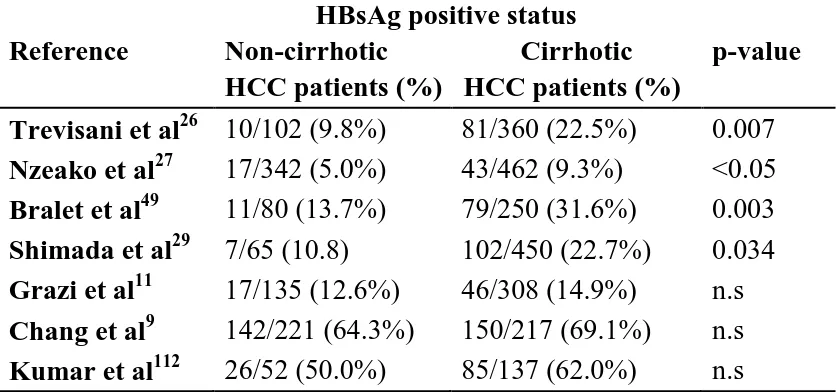
with HCC. Table-1and 2 shows the prevalence of HBV surface antigen
(HBsAg) positive and anti-HCV antibody positive status in cirrhotic and
non-cirrhotic patients with HCC coming from different geographical
areas.
Table 1: The reported prevalence of HBsAg positive status in non-cirrhotic and non-cirrhotic patients with hepatocellular carcinoma (HCC) in the literature.
Reference
HBsAg positive status Non-cirrhotic Cirrhotic HCC patients (%) HCC patients (%)
p-value
Trevisani et al26 10/102 (9.8%) 81/360 (22.5%) 0.007
Nzeako et al27 17/342 (5.0%) 43/462 (9.3%) <0.05
Bralet et al49 11/80 (13.7%) 79/250 (31.6%) 0.003
Shimada et al29 7/65 (10.8) 102/450 (22.7%) 0.034
Grazi et al11 17/135 (12.6%) 46/308 (14.9%) n.s
Chang et al9 142/221 (64.3%) 150/217 (69.1%) n.s
Kumar et al112 26/52 (50.0%) 85/137 (62.0%) n.s
[image:19.595.82.502.498.694.2]Table 2: The reported prevalence of anti-HCV positive status in non-cirrhotic and non-cirrhotic patients with hepatocellular carcinoma (HCC) in the literature.
Reference
Anti HCV antibody positive status
p-value Non-cirrhotic
HCC patients (%)
Cirrhotic HCC patients (%)
Trevisani et al26 15/28 (53.6%) 104/138 (75.4%) 0.03
Bralet et al49 2/80 (2.5%) 68/250 (27.0%) 0.0001
Shimada et al29 22/42 (52.4%) 243/332 (73.2%) 0.007
Grazi et al11 33/135 (24.4%) 182/308 (59%) <0.001
Chang et al9 39/216 (18.1%) 57/208 (27.4%) 0.02
Kumar et al112 1/52 (1.9%) 22/137 (16.1%) n.s
HCV: Hepatitis C virus, HCC: hepatocellular carcinoma, n.s: not statistically significant
HBV can directly cause hepatic carcinogenesis, independent of
the development of cirrhosis.30 This is possible because HBV is a DNA
virus and hence can integrate into host DNA leading to host DNA
microdeletions and produce HBx protein that modifies the host growth
control genes.18More recently, many studies have shown that the
mutations in basal core promoter region, HBeAg seropositivity, HBV
genotype C and a high viral load (104-5copies/ml) are independent
predictors of HCC development even in the absence of cirrhosis.31-33
Hence in the future, identification of these factors could be applied to
predict HCC risk in HBs Ag carriers irrespective of the presence of
[image:20.595.80.502.158.341.2]The main mechanism of HCC development in chronic HCV
infection is due to sustained viral replication leading to
necro-inflammatory process. This results in high proliferative rate and
increased risk for DNA mutation. Accumulation of these genetic
changes over time confers a survival advantage with abnormal
phenotype. Hence, nearly all HCV related HCC occurs against a
background of advanced liver fibrosis or cirrhosis.5,34,35 However,
specific HCV gene products (core, NS3, NS4B and NS5A)36,37 has
produced malignant transformation in murine fibroblast culture
suggesting that HCV also has a direct carcinogenic potential.38,39,40
The detection of HBV and HCV infections by serology
underestimates their etiological role because HBV-DNA and HCV-RNA
fragments have been identified in livertissue and/or serum of 18% to
33% of patients respectively, in non-cirrhotic HCC individuals with
negative serological markers.41
2. Alcohol intake:
Numerous studies have addressed that fact that heavy alcohol
intake has been less common in non-cirrhotic HCC patients (Table 3).
potential of alcohol is almost always due to development of
cirrhosis.42.43
Table 3: The reported prevalence of alcohol intake in non-cirrhotic and cirrhotic patients with hepatocellular carcinoma (HCC) in the literature.
Reference Alcohol intake
Non-cirrhotic Cirrhotic HCC patients (%) HCC patients (%)
p-value
Trevisani et al26 15/91 (16.5%) 105/350 (30.0%) 0.01
Nzeako et al27 25/342 (7.3%) 130/462 (28.1%) <0.0001
Bralet et al49 11/80 (13.7%) 73/250 (29.2%) 0.009
Shimada et al29 44/63 (69.8%) 232/433 (53.6%) 0.022
Grazi et al11 29/135 (21.5%) 67/308 (21.7%) n.s
Chang et al9 98/223 (43.9) 99/222 (44.6%) n.s
Kumar et al112 5/52 (9.6%) 25/137 (18.2%) n.s
HCC: hepatocellular carcinoma, n.s: not statistically significant
3. Toxic substances:
Non-cirrhotic HCC can also occur due to the exposure of
following genotoxic agents:
i. Aflatoxin B1, a toxin produced by the fungus Aspergillus
flavus can contaminate many food products, such as cereals,
nuts, spices, figs and dried fruit. This contamination is
common in developing countries like India and other parts of
Asia and Africa with a high HCC incidence.44A study by Qian
aflatoxin metabolites with a 4-fold increase in HCC risk.
Infact, HBV infection increases the HCC risk in
aflatoxinexposure to 60-fold when compared to the general
population. Ming et al46 has shown that HBV infection
sensitizes hepatocytes to the aflatoxin-induced 249ser-p53
mutation.
ii. Industrial carcinogens (such as azo-dyes, organic solvents,
vinyl chloride, pesticides, nitrosamines, arsenic, aromatic
amines) and substances derived from tobacco (benzopyrene)
have been shown to havehepato-carcinogenic potential in
animal studies. Hence, these substances could also play a role
in human carcinogenesis.
iii. Thorotrast (Radioactive elements) may also cause HCC, albeit
much less frequently than angiosarcoma and
cholangiocarcinoma.47
iv. Finally, tissue iron overload may act as a co-carcinogen factor,
as suggested by the frequent observation of mild parenchymal
iron excess in the non-tumorous liver tissue of most patients
4. Inherited diseases:
The development of non-cirrhotic HCC may also be attributed to
rare inherited disorders such as:
i. Metabolic diseases such as hereditary hemochromatosis,
alpha-1-antitrypsin (AT) deficiency, porphyria,
hyper-citrullinemia and type I glycogen storage diseases.24,51
ii. Congenital diseases like congenital hepatic fibrosis and
Alagille’s syndrome.
iii. Hepatic vascular abnormalities, which are associated with
inherited coagulation disorders such as Budd-Chiari
syndrome, hepato-portal sclerosis and nodular regenerative
hyperplasia.24
5. Steroid hormones:
Many case reports have described the development of HCC in
non-cirrhotic patients with anabolic steroids taken for therapeutic
purpose.52,53 Although many studies have reported an higher risk for
HCC in patients taking oestrogen for 8 years or more, a direct
etiological role of contraceptive steroids is not yet established.54,55
promotors of carcinogenesis and also produce cellular and vascular
changes thus amplifying an already developing tumor.
6. Genetic mutations:
Zuckmann-Rossi et al56 postulated that the monoclonal mutations
of both oncogenes and anti-oncogenes in hepatocellular adenoma are
associated with the malignant transformation. Particularly, the gain of
function mutation of β-catenin, which is found in 15-20% of
hepatocellular adenomas are most likely to be associated with malignant
transformation.57
Familial clustering of HCC has also been reported and a recent
study by Hassan et al58 has demonstrated that family history of liver
cancer significantly increases the HCC risk irrespective of the HBV and
HCV status. But, this study does not analyze separately for non-cirrhotic
patients. However, it can be assumed that the possible relationship
between a positive family history and HCC risk is the end result of
environmental, genetic, behavioural and clinical risk factors.58
The molecular mechanisms of carcinogenesis involved in
non-cirrhotic liver are beginning to be explored. Recently, Chaubertet al59
reported a germ line mutation in tumor suppressor gene, p10INK4 (MTSI)
cirrhotic HCC patients. Although these studies support the existence of
familial form of carcinogenesis, the confirmation of hereditary
component in HCC risk awaits further studies of familial clusters.
HEPATO-CARCINOGENETIC PATHWAY:
Different authors have postulated different hepato-carcinogenetic
pathway in non-cirrhotic HCC. This hypothesis is based on different
scenario of genetic alteration found in non-cirrhotic HCC, that is, a
tendency towards a low incidence of p53 mutation and a high rate of
β-catenin mutation, p14 inactivation and global gene methylation in
non-cirrhotic HCC.60
Two recent studies by Chiappiniet al61 and Togni et al62 have
found the presence of microsatellite instability (MSI), a sign of defect in
DNA mismatch repair, in non-cirrhotic HCC patients with non-alcoholic
and non-virally infected livers.
PATHOLOGY:
The tumor bearing portion of liver in patients with
non-cirrhotic HCC usually show steatosis, steato-hepatitis, varying degree of
fibrosis, iron excess or other metabolic disorder and hence only few
is seen in 25-40% of cases.25,63 This figure reduces to 6-20% in patients
with non-fibrotic liver.29,49
Most (50-85%) non-cirrhotic patients with HCC present as a
large, solitary mass (about 10cm in most series).26, 64-67Multinodularity is
not as common (18% vs. 34%) as seen in cirrhotic HCC26. This is
possibly due to both the delay in presentation and distinct mechanism of
hepato-carcinogenesis in non-cirrhotic HCC.
According to World Health Organization (WHO) histological
classification, the trabecular type is the most common type (40-75%) in
both non-cirrhotic HCC, and cirrhotic HCC.25-27,49,56 The scirrhous type
and mixed hepatocellular/cholangiocellular carcinoma tend to occur
more commonly in a non-cirrhotic background68,69, whereas
Fibrolamellar HCC is almost exclusively limited to non-cirrhotic liver
(115 vs. 1.5%).27
Two larger studies by Nzeakoet al27 and Shimada et al29 have
shown that encapsulation is more common in non-cirrhotic HCC,
however, this finding has not been confirmed in other studies.
A recent series by Trevisaniet al26 have demonstrated that the
and/or metastases) is more common in non-cirrhotic HCC when
compared to cirrhotic HCC.
Although the more advanced stage of tumor at presentation in
non-cirrhotic patients can be attributed to delayed diagnosis, a greater
biological aggressiveness cannot be excluded, because some authors
have reported poor tumor differentiation and early portal vein invasion
in non-cirrhotic HCC.11,29 At present, the available data on this issue is
controversial, as other authors have reported opposite results.9,26,27
DIAGNOSIS:
According to the current international guidelines for the
management of HCC, routine ultrasonographic surveillance is now
being performed in all patients with chronic liver disease. This has
resulted in early detection of tumor, usually before the manifestation of
clinical symptoms. In contrast, in otherwise healthy person or with
undiagnosed liver disease, HCC are generally detected late, usually after
the appearance of symptoms such as abdominal pain or discomfort in
right upper quadrant, jaundice or toxic syndrome (anorexia, weight loss,
malaise, asthenia, fever).35 Trevisani et al26 have reported that non
cirrhotic patients with HCC has a symptomatic presentation in about
suggests that the delay in presentation and diagnosis accounts for most
differences in pathological differences between non-cirrhotic and
cirrhotic patients.
Most studies have demonstrated that the serum α-fetoprotein
(AFP) level rarely exceeds 40ng/dl in non-cirrhotic when compared to
cirrhotic patients with HCC (30-67% vs. 60-85%).26,27,64,65 This suggests
the likely role of cirrhosis as independent promoter of moderate AFP
elevation. However, the prevalence of level of AFP considered
―diagnostic‖ for HCC is similar in both the groups.26,64
PREOPERATIVE EVALUATION:
The preoperative investigations for HCC should focus on three
main issues:
1. Tumor status
2. Underlying liver status
3. Patient performance status.
1. Tumor status:
Assessment of tumor extent is best achieved with cross sectional
imaging such as dynamic contrast enhanced Magnetic resonance
(Figure: 10-12). There are no data at present to support the value of PET
scanning in HCC. A chest X-ray, CT chest and Bone scan are
recommended to rule out extra-hepatic disease. Many studies have
shown that the most common sites of metastatic spread of HCC are
lung, bone, peritoneum and adrenals. Although these sites of spread may
be detected by standard imaging techniques, peritoneal disease requires
staging laparoscopy.
2. Underlying liver status:
Blumgart et al18 have demonstrated that a healthy, non-cirrhotic
liver may tolerate a resection of up to 80% due to the enormous
regenerative capacity of liver. However, such favorable responses
cannot be taken up for granted for extended hepatic resections. The risk
of clinically significant liver insufficiency can be avoided only if there is
50% reduction in functioning liver parenchyma even in non-cirrhotic
liver. Although helpful, the Child-Pugh score and Model for end-stage
liver (MELD) score are not adequate to select patients for major hepatic
resection.
Most centers in Asia perform indocyanine green clearance
(ICG-15) at 15 minutes as defining criterion for selection of liver
insufficiency is the pre-operative evaluation of the post-operative future
liver remnant (FLR). CT is used to assess the ratio of future remnant
(FLR) and total liver volume (TLV). The consensus panel recommends
that this ratio should be at least 30-40% in non-cirrhotic patients.
In patients with estimated FLR/TLV below the recommended values,
pre-operative portal vein embolization (PVE) should be considered.
ROLE OF PRE-OPERATIVE PORTAL VEIN EMBOLIZATION
(PVE):
PVE produces atrophy in portion of liver to be resected and
compensatory hypertrophy in the portion of liver to be preserved. By
reducing the risk of liver failure, complication rate and hospital stay,
PVE increases the safety of resection and expand the indication for liver
resection in otherwise poor candidates for hepatectomy.18
A recent meta-analysis by Abulkhiret al70 have reviewed 37
published series of preoperative PVE in 1088 patients (265 patients with
HCC, the reminder with cholangiocarcinoma or liver metastases). In 2-4
weeks, there was significant hypertrophy of FLR that was independent
of technique and 85% of patients underwent planned liver resection. The
reasons for not operating in remaining 15% of patients were inadequate
(n=35) and other reasons such as refusal for surgery, poor medical
condition, altered treatment approach for variety of reasons (n=35). In
patients who underwent laparotomy, 27 patients were found
unresectable, mainly due to advanced or unresectable disease. Of the
remaining patients who underwent liver resection, transient liver failure
was seen in only 2.5% (n=23), and death due to acute liver insufficiency
in 0.8% (n=7).
ROLE OF PRE-OPERATIVE TACE:
The role of TACE in pre-operative setting remains conflicting. In
a RCT by Wu et al, there was no significant difference in overall
survival or disease free survival. In addition, they have reported higher
incidence of extra-hepatic recurrence with pre-operative TACE possibly
due to easier tumor cell dislodgement during surgery.71
Similarly, Shimada et al72 have found pre-operative TACE to be a
poor prognostic factor in univariate analysis, but not in multivariate
analysis. The potential disadvantages of pre-operative TACE are
impairment of pre-operative liver function, delayed in planned surgery
and difficult surgery due to development of collateral vessels and severe
inflammatory changes around tumor. At present, there is no sufficient
STAGING:
Numerous staging systems have been proposed for clinical
classification of HCC. The standard classification in any cancer is based
on TNM staging, however in HCC, the 7th TNM classification has
several limitations.73 First, microvascular invasion is assessed by
pathology, hence only available in patients (~ 20%) undergoing surgery.
Second, it does not include information regarding liver function status or
patient performance status. The more popular Child-Pugh classification
and Okuda staging serve only to class prediction in HCC patients. The
five more comprehensive staging that have been broadly tested are: 3
European [ the Barcelona Clinic Liver cancer (BCLC)74 staging, the
cancer of liver Italian program (CLIP) 75classification, the French
classification76] and 2 Asian [the Chinese University Prognostic Index
(CUPI)77 score and the Japan Integrated staging with biomarkers
(bm-JIS)78 ]. Overall, the most external validated staging systems are BCLC,
CUPI, CLIP and bm-JIS with only two include prognostic variables
(BCLC and CUPI) and only one (BCLC) assign treatment allocation to
specific prognostic subgroups. Hence the current EASL-EORTC
TREATMENT AND SURVIVAL:
The various treatment options for non-cirrhotic patients with HCC are:
1. Hepatic resection
2. Liver transplantation
3. Trans-arterial embolization
4. Radio-embolization
5. Radio-frequency ablation
6. Systemic chemotherapy
1. HEPATIC RESECTION:
Regardless of etiology, the optimal management in HCC is
complete resection of tumor. Numerous studies have shown that the
patients with non-cirrhotic HCC undergo resection more than the
reported (12-28%) incidence in patients with underlying cirrhosis.80,81
In spite of large tumor size, the preserved liver function in non-cirrhotic
patients allows major hepatic resections to be performed quite safely.
In fact, despite major hepatic resection, the post-operative morbidity
and mortality are rather low in these patients.7,11,66,67,82Moreover, the
overall survival and recurrence free survival are better in non-cirrhotic
The overall survival and disease-free survival of patients resected
for non-cirrhotic HCC are depicted in Table16. The 5-year overall
survival rate ranges between 25% and 81% with best figures reported in
in non-cirrhotic HCC. Similarly, the 5-year disease-free survival ranged
from 24% to 58% with best value achieved in non-cirrhotic patients.82
The independent factor for overall survival in patients with
non-cirrhotic HCC is the rate of tumor recurrence, which varies between
27% and 73% in different series, as outlined in table-16.82 Most (2 out
of 3) recurrences occur in the initial two post-operative years, but can be
delayed to 10 years or more. Up to 40% recurrences are amenable to
second hepatectomy, but long-term survivals are noted in patients with
early recurrence. These results highlight the importance of follow-up
during the first two post-operative years and the need for prolonged
surveillance for aggressive management of recurrences.7,10,65,67,82,83,85
2. LIVER TRANSPLANTATION:
The role of orthotopic liver transplantation (OLT) in treatment of
patients with non-cirrhotic HCC is a subject of controversy.
A systematic review conducted by Houbenet al6 included all published
series of OLT for non-cirrhotic HCC performed between 1966 and 1998
5-year survival rate of 11%. This dismal figure suggest the advanced
tumor stage at the time of OLT, which resulted in recurrences in about
50% of patients, mostly (>75%) in the first 2 years, indicating the likely
possibility of extra-hepatic micrometastases.6
The specific selection criteria for transplantation in non-cirrhotic
HCC are lacking. The commonly adopted criteria in non-cirrhotic HCC
patients are actually proposed for cirrhotic patients, which is
inappropriate because most non-cirrhotic HCC are outside the Milan
criteria at the time of diagnosis and those fulfilling these criteria are
potentially resectable tumors. In addition, the tendency towards OLT in
non-cirrhotic HCC is limited by the report that the outcome after
resection in non-cirrhotic HCC within Milan criteria is comparable to
that expected in cirrhotic patients transplanted within Milan criteria.
Pragmatically, OLT could be reserved as a salvage treatment for
non-cirrhotic patients with post-resection tumor recurrence and those with
high risk of post-hepatectomy liver failure.9,85
3. TRANS-ARTERIAL EMBOLIZATION:
The basic physiological principle that makes TAE feasible is that
most of blood supply (90% to 100%) to liver tumors is derived from the
selective ischemic damage of the tumor, while sparing the normal liver
parenchyma supplied mainly by the portal vein. Moreover the
pharmacokinetic advantage of selective loco-regional drug
administration further enhances the theoretic benefit.
Basically there are three types of trans-arterial therapy:
1. Trans-arterial embolization (TAE) with bland particles.
2. Trans-arterial chemo-embolization (TACE) with or without
lipiodol.
3. Trans-arterial chemotherapy (TAC) alone or with lipiolol.
A meta-analysis by Camma et al86 have failed to demonstrate any
significant survival differences between TAE and TACE, in spite of
a trend towards longer survival with TACE.
Indications for TACE:
1. Unresectable large hypervascular liver tumor.
2. Intermediate BCLC stage HCC (Okuda staging 1 to 2,
Performance score 0 and large or multinodular HCC)
4. As neoadjuvant therapy to downsize tumor before resection or
bridging therapy in patients awaiting liver transplantation.
Contraindication to TACE:
1. Extensive tumor involvement of more than 50% to 70% of liver
and class C cirrhosis.
2. Main portal vein occlusion.
3. Active gastrointestinal bleeding, extra-hepatic spread, hepatic
encephalopathy and biliary obstruction.
4. Anaphylactoid reaction to contrast agents, cardiac or renal
insufficiency, uncorrectable coagulopathy and severe peripheral
vascular disease.
The mean diameter of tumor treated by TACE was 5.2cm in
randomized controlled study by Llovet et al87 and 5cm in GETCH88
(Grouped Etude et de Traitement du CarcinomeHepatocellulare ) study.
In addition to tumor size, factors such as good liver function,
performance status – 2, Okuda stage II were associated with survival
benefit. However, tumor size >5cm is a strong negative factor affecting
The survival benefit from TACE was not demonstrated in 3 early
randomized controlled trails (RCT) by Pelletier et al89 (1990), Grouped
Etude et de Traitement du Carcinome Hepatocellulare88 (GETCH
study-1995) and Camma et al86 (2002). However, 2 newer RCT’s from
Barcelona (Llovetet al87, 2002) and Hongkong (Lo et al90, 2002)
demonstrated significant survival advantage with TACE. A recent
meta-analysis of all RCTs published from 1978 to 2002, American
Association for study of liver diseases (AASLD-2005, Bruix and
Sherman et al35) and European association for study of liver
(EASL-2001, Bruix et al5) have recommended TACE as first line non-curative
therapy for non-surgical patients with large or multifocal HCC without
vascular invasion or extra-hepatic spread.
A recent advancement in TACE is the usage of drug eluting beads
(DEBs) which slowly releases chemotherapeutic drug thereby reducing
the systemic toxicity. Early results are promising but most series are
associated with short-term follow-up only.91
4. RADIO EMBOLIZATION or SELECTIVE INTERNAL
RADIATION THERAPY (SIRT):
Radio-embolization is a method of delivering localized radiation
external radiation. 90Yttrium is the most commonly used radioactive
element for radio-embolization. It is a pure β-emitter with tissue
penetration ranging from 2.5 to 11mm. Radio-embolization was
first studied by Nolan and Grady92 (1969) using 90Yttrium oxide
(90Y2O3) contained in metal particle of 50-100 µm in size. Their study
was limited by small number of patients but showed a favorable
response with decrease in tumor size. The subsequent 90Y study was
published by Mantravadiet al93 (1982) concluding that patients with
hypervascular tumor are more likely to be benefit from
radio-embolization. Based on animal safety studies, Shepherd et al94
conducted phase I studies suing 90Y glass microspheres and reported that
doses of up to 150 Gy were tolerable with minimal toxicities. In view of
the encouraging results, a phase II study performed by same authors
showed a 20% response rate with doubling of survival duration (635 vs.
323 days) in patients who underwent radio-embolization.
Analysis from these studies and other trials have demonstrated
that tumor-liver ratio <2, low Okuda stage, low AFP favored longer
survival whereas elevated bilirubin was associated with post-treatment
liver dysfunction.95 The role of radio-embolization in HCC patients is
evolving and further trials are needed to document their safety and
5. RADIOFREQUENCY ABLATION:
Radiofrequency ablation (RFA) destroy tumor by generating heat
within a lesion. During RFA, a high frequency alternating current
changes the direction of ions around an alternating electrode. This
produces frictional heating of tissues and when tissue temperature
increases above 600 C, loss of cellular structure and protein denaturation
occurs, resulting in cell death. RFA can be performed by percutaneous,
laparoscopic or open (laparotomy) approach.
Clinical trials of RFA for HCC have shown promising results
andLivraghiet al96 showed a local response rate of only 2.8% at a
median follow-up of 31 months. Poon et al97 demonstrated a complete
response rate of 91% for large (>3cm) HCC and showed rate of local
recurrence, distant recurrence were independent of tumor size. Two
randomized control trials by Chen et al98 and Lu et al99 have
demonstrated therapeutic equivalence of RFA, when compared with
resection in small HCC, until more prospective studies have been
performed, resection remains the initial choice in these patients.
6. SYSTEMIC CHEMOTHERAPY:
The role of systemic chemotherapy is very limited in patients with
A recent retrospective study by Edelineet al101 reported that combination
chemotherapy with Epirubicin, cisplatin, and 5-flurouracil/ capecitabine
was associated with objective response in 25% of cases with
unresectable HCC.
Lau et al102 have shown that 10% of advanced HCC patients
underwent successful resection after tumor downsizing with
combination chemotherapy composed of Cisplatin, interferon alfa,
Adriamycin and 5-flurouracil (PIAF) regimen. The 3-year survival rate
was 53% for those who underwent resection following PIAF regimen. In
the multivariate analysis of 149 patients who received PIAF by the same
author, non-cirrhotic patients with normal bilirubin level had a shown a
better response to combination chemotherapy.
New molecular drug, Sorafenib which is an oral multi-kinase
inhibitor that targets Raf kinase and receptor tyrosine kinase has shown
promising results in phase III randomized placebo controlled (SHARP
trial) trial103 involving 602 patients with advanced HCC in terms of
improved survival and median time to progression. This result has
suggested sorafenib as first line therapy for patients with advanced
HCC, although no patient is reported to have tumor downsized to
undergo resection. Hence, the optimal management of patients with
MATERIALS AND
MATERIALS AND METHODS
STUDY DESIGN:
Prospective study
STUDY PERIOD:
September 2010 to December 2012
INCLUSION CRITERIA:
All consecutive patients with histologically proven HCC in
non-cirrhotic, non-fibrotic liver undergoing liver resection will be included
in the study
EXCLUSION CRITERIA:
1. Patients undergoing liver resection with palliative intent were
excluded from the study.
2. Histological proven HCC in cirrhotic or fibrotic background.
3. Patient refuses to give informed consent to be included in the
METHODOLOGY:
PATIENTS:
Between August 2010 and December 2012, 76 patients with HCC
underwent liver resection in the Institute of Surgical gastroenterology
and liver transplantation at Government Stanley Medical College and
Hospital. Of the 76 patients who had liver resections for HCC, 30
patients had no underlying parenchymal liver disease (no cirrhosis or
fibrosis) and these 30 patients were included in the study
PREOPERATIVE EVALUATION:
Routine pre-operative evaluation include recording of detailed
demographic profile, history of presenting symptoms, physical
examination and routine laboratory investigations like complete
hemogram, renal function test with electrolytes, liver function test
including albumin. Data regarding underlying liver disease (such as past
or current hepatitis B virus or hepatitis C virus infection, alcoholic or
non-alcoholic steatohepatitis, iron overload and hemochromatosis) and
risk factors for the development of HCC such as chronic alcohol intake (
≥ 40 g/day), tobacco consumption (≥ 20 pack years), diabetes mellitus,
substance exposure) were also recorded. Serum alpha fetoprotein and
upper gastro-intestinal endoscopy were done in all patients.
Pre-operative investigations performed to assess the extent of
disease included Chest X-ray, abdominal ultrasonography, contrast
enhanced computerized tomography (CT) and/or MRI in all patients and
CECT thorax in selected patients. The diagnosis of HCC was based on
either pre-operative imaging and serum AFP level or rarely biopsy.
Liver function status was assessed by the Child-Pugh grading. Patient
performance status at the time of diagnosis was determined according to
the Eastern Co-operative Oncology Group (ECOG).
SURGERY:
Prior to surgery, all patients were discussed in a multidisciplinary
meeting in order to ensure an optimal management strategy. Staging
laparoscopy was done immediately before surgery in all patients. In
addition to evaluation of peritoneal deposits, staging laparoscopy
allowed assessment of future liver remnant size and severity of cirrhosis,
when major liver resection was planned.
The operation was performed through a Maakuchi’s incision or
bilateral subcostal incision with an upward midline extension
cases to detect any additional tumor and relationship of tumor to
vasculo-biliary structures. Selective vascular inflow and outflow
control was obtained followed by parenchymal transection under low
central venous pressure (CVP) anaesthesia, using a combination of
kellyclasia, harmonic, ultrasonic dissector and water jet.
Anatomic resections were defined according to the Brisbane
terminology described by International Hepato-Pancreatico-Biliary
Association (IHPBA) and Non-anatomical resections were defined as
atypical or wedge resection with tumor free margin of at least 1cm. A
major hepatic resection was defined as resection of 3 or more segments
of liver according to Couinaud’s classification of liver segments and
minor hepatic resection was defined as resection of 2 or less segments of
liver.
Intra-operative parameters like type of liver resection
(Major/minor, anatomic/non-anatomic), duration of surgery, blood loss,
number of blood transfusion and intra-operative complications were
recorded. All patients received prophylactic broad-spectrum antibiotics.
POST-OPERATIVE CARE:
All patients were monitored in the intensive care unit during the
oxygenation and tissue perfusion. Intravenous albumin and diuretics
were administered in selected patients to reduce ascites. Early
ambulation was encouraged and oral intake was resumed once the bowel
activity was restored. Liver function test with prothrombin time and INR
were routinely done in 1st, 3rd and 5th operative days to detect
post-operative liver insufficiency. All post-post-operative complications were
recorded and were classified as: Infectious (Anastomotic leak,
Intra-abdominal abscess, Wound infection, Pneumonia, Septicemia) and
Non-infectious (Pulmonary embolism, Renal failure, Cardiac events
including Acute coronary syndrome and Acute MI). Length of hospital
as well as post-operative stay were recorded.
HISTOPATHOLOGY:
All the resected specimens were analyzed by the experienced
pathologists. A standard histo-pathological assessment of tumoral and
non-tumoral tissue was performed.
The macroscopic features of tumor such as size, number, tumor
capsule and vascular invasion were recorded. Tumors were classified
using World Health Organization criteria (such as trabecular,
pseudoglandular, compact, scirrhous or mixed) and graded using
moderately-differentiated (grade II) and poorly differentiated (grade III).
A clear resection (R0) margin was defined as negative of 1mm from the
inked margin or 1mm of liver tissue between capsule and margin.
Macroscopic vascular invasion was defined as presence of tumor
thrombi in right or left main branches of the hepatic veins or the portal
veins. Microscopic vascular invasion was defined as presence of tumor
emboli within central vein, capsular vessels or portal vein and hepatic
vein radicles.
Non-tumoral tissue was assessed in area distant to tumor to avoid
inflammatory or fibrotic changes caused by tumor itself. Fibrosis and
chronic activity were graded using the METAVIR grading system.
Steatosis was graded according to the percentage of steatotic
hepatocytes into mild (5% to 20%), moderate (20% to 50%) and severe
(>50%). Iron overload was graded according to Modified Searle scale
into mild or grade I (iron barely visible at low magnification but
confirmed at high magnification, moderate or grade II (iron visible at
low magnification and only in zone 1, and severe or grade III (iron
visible at low magnification occupying most of acinus). Precancerous
lesions in the non-tumoral liver were defines by the presence of liver
cell dysplasia, clear cell foci or free foci in otherwise
FOLLOW UP:
All patients were followed up in the out-patient department, every
month during the first 3 months, then every 3 months for initial 2 years
and every 6months thereafter. In each visit, physical examination, liver
function test, serum AFP and abdominal ultrasound was performed.
CT scan was performed at month 6 and then every year.
The diagnosis of recurrence was based on clinical, laboratory
(elevated serum AFP level) and radiological (abdominal ultrasound/CT,
chest X-ray) findings. The number and pattern of recurrence
(intrahepatic, extra-hepatic or both) were also recorded. Patients with
isolated and resectable intra-hepatic recurrence underwent re-resection.
All others were treated with TACE, systemic therapy or best supportive
care.
The follow up period of this study was closed in February 2013 to
ensure that every patient had at least 2 months of observation following
surgery. The above described protocol was approved by our institutional
STATISTICAL ANALYSIS:
All clinico-pathological and follow-up data were prospectively
collected and entered with regular update of any tumor recurrence for
each patient after each follow-up. Categorical variables were expressed
as percentages and compared using chi-square test or Fisher exact test.
Continuous variables were expressed as mean ± SD and compared using
the student-t test. Survival and recurrence were expressed as median ±
SEM. Patient survival and recurrence were calculated using the
Kaplan-Meier test and compared using the log-rank test. Clinico-pathological
variables found to bear prognostic significance in univariate analysis
were entered into Cox multivariate proportional hazard model to
determine which of these factors possessed independent predictive
value. p-value< 0.05 was considered statistically significant and analysis
was carried out using the Statistical Package for the Social Sciences
RESULTS
This prospective study was conducted in the Institute of Surgical
gastroenterology and liver transplantation, Government Stanley Medical
College and Hospital from August 2010 to December 2012. During the
study period, a total of 76 patients underwent liver resection for
hepatocellular carcinoma. Of the 76 patients who had liver resections
for HCC, 30 patients (39.47%) had no underlying parenchymal disease
(no cirrhosis or fibrosis) and were included in the study.
The clinical characteristics of 30 patients with non-cirrhotic
HCC are shown in table-4. The mean age at presentation was 48.23
years (range: 13-77). The age distribution (Figure 2) of 30 patients
shows that the majority of patients (67%) are in the fifth and sixth
decades. The gender distribution (Figure 3) shows that there were 19
males (63%) and 11 females (37%) with male: female ratio of 1.7: 1.
Table 4: Clinical characteristics of 30 patients with HCC in non-cirrhotic liver studied.
Variables n %
Risk factor
Hepatitis B virus infection Hepatitis C virus infection Chronic alcohol intake Tobacco smoking Diabetes mellitus Obesity Unknown 4 0 9 9 8 3 12 13 0 30 30 26.7 10 40 Symptoms Abdominal pain
Weight loss and anorexia Abdominal mass Fever Jaundice Hepatomegaly 30 28 7 4 2 17 100 93 23 13 6 57
ECOG performance status
0 1 2 26 3 1 87 10 3 Pre-operative biopsy Present Absent 3 27 10 90 ECOG: Eastern Co-operative Oncology Group.
The potential risk factors for the development of HCC were
present in 18 patients (60%). Four patients (13%) presented with current
or past hepatitis B viral infection and no patient was positive for
anti-HCV antibody. Tobacco and alcohol consumption were found in 9
and overweight in 3 patients (10%). In contrast, 12 patients (40%) did
not reveal any underlying risk factor for the development of HCC.
Abdominal pain was the most common presentation in the
non-cirrhotic HCC patients, which was noted in all 30 patients (100%),
followed by anorexia and weight loss in 28 patients (93%), abdominal
mass in 7 patients (23%), fever in 4 patients (13%) and jaundice in 2
patients (6%). None of the patient was asymptomatic at the time of
presentation. Hepatomegaly was observed in 17 patients (57%) and none
of the patients had ascites. Three patients had undergone pre-operative
biopsy and none of the patient received any type of pre-operative
treatment.
The pre-operative laboratory investigations done in 30 patients
with non-cirrhotic HCC were listed in table-5. The mean hemoglobin
level was 10.7 g/dl ± 1.6 (range: 8.5-14). Serum bilirubin was elevated
in 2 patients (6%). The mean serum bilirubin value was 1.1 mg/dl ± 0.83
(range: 0.25-3.6). Serum aspartate transaminase (AST) was elevated in
19 patients (63%) with mean value of 84.6 U/L ±91.45 (range: 21-498).
Serum albumin was low in 17 patients (57%) with mean value of 3.42
g/d ± 0.55 (range: 2.5-4.9). Serum alpha-fetoprotein level was normal in
20 patients (67%) and elevated above 1000ng/ml in only 2 patients
Table 5: Pre-operative laboratory data in 30 patients with HCC in non-cirrhotic liver studied.
Laboratory parameter (unit)
Normal range
Mean ± Standard deviation (range)
Hemoglobin (gm/dl) 14-18 10.73 ± 1.6 (8.5-14)
Total leucocyte count (cells/cu.mm)
4000-11000
8346.3 ± 2428 (2200-15000)
NLR 1-8 x 10⁹/L 2.77 ± 1.58 (1.09-7.33)
ESR (mm/hr) 0-20 41.6 ± 26.49 (8-120)
Platelet (cells/cu.mm) 1.5 -4 3.71 ± 1.80 (1.42-7)
Prothrombin Index 70-100% 97.36 ± 5.66 (77.77-100)
INR 0.8-1.2 1.04 ± 0.09 (1-1.4)
Sugar (mg/dl) <160 120.93 ± 48.85 (67-268)
Urea (mg/dl) 10-50 21.67 ± 7.70 (13-50)
Creatinine (mg/dl) 0.6-1.1 0.80 ± 0.24 (0.4-1.54)
Bilirubin (mg/dl) 0-1 1.12 ± 0.83 (0.25-3.6)
AST (U/L) 0-40 84.6 ± 91.45 (21-498)
ALT (U/L) 0-37 47.66 ± 35.70 (8-173)
GGT (U/L) 0-45 98.93 ± 196.27 (10-1050)
SAP U/L) 0-290 261.87 ± 123.56 (85-617)
Albumin (g/dl) 3.8-4.4 3.42 ± 0.55 (2.5-4.9)
AFP (ng/ml) <20 6.8 ± 5922.9 (0.3-32476)
NLR: Neutrophil-leucocyte ratio, ESR: Erythrocte Sedimentation rate, INR: Internationalized normalized ratio, AST: Aspartate transaminase, ALT: Alanine transaminase, GGT: Gamma-glutaryltransferase, SAP: serum alkaline phosphatase, AFP: Alpha feto-protein.
The extent and type of resection performed were outlined in
table-6. Out of 30 patients, 25 patients (83%) underwent major hepatectomy
whereas only 5 patients (17%) underwent minor hepatectomy (Figure 4).
The most common procedure performed was right hepatectomy (Figure:
13-16), which was done in 12 patients (40%), followed by left
patients (17%), bi-segmentectomy in 2 patients (6.7%) and extended left
hepatectomy, central hepatectomy, left lateral segmentectomy (Figure
17 and 18) in 1 patient (3.3%) each. Non-anatomical resection was done
in 2 patients (6.7%). Three patients (10%) underwent enbloc resection of
[image:57.595.90.491.280.477.2]adjacent organ (diaphragm) involvement to achieve R0 resection.
Table 6: Type and extent of liver resection in 30 patients with HCC in non-cirrhotic liver studied.
Surgical procedures n %
Major Hepatectomy (n=25)
Right Hepatectomy Left Hepatectomy
Extended Right Hepatectomy Extended Left Hepatectomy Central hepatectomy 12 6 5 1 1 40 20 16.7 3.3 3.3
Minor Hepatectomy: (n=5)
Left lateral segmentectomy Bisegmentectomy Non-anatomical resection 1 2 2 3.3 6.7 6.7
The mean duration of operation was 2.45 ± 0.59 hours (Table-7).
The mean blood loss during surgery was 216.5 ± 119.32 ml (range:
100-720ml). There was a need for blood transfusion in 2 patients (6.7%).
Table 7: Perioperative factors in 30 patients with HCC in non-cirrhotic liver studied.
Peri-operative factors Mean ± Standard deviation or
n (%)
Operative time (hours) 2.45 ± 0.59
Blood loss (ml) 216.5 ± 119.32
Need for blood transfusion 2 (6.7%)
Mean post-operative stay (days) 16.7 ± 7.0
Morbidity 15 (50%)
[image:57.595.90.495.632.763.2]The mean post-operative length of stay was 16.7 ± 7.0 days.
(range: 9-35 days). In-hospital mortality was noted in 1 patient (3.4%)
due to post-operative liver failure.
The data regarding the post-operative complication was
summarized in table-8. Overall, the post-operative complications
occurred in 15 patients (50%). The most common post-operative
complication (Figure 5) was intra-abdominal collection and wound
infection noted in 6 patients (20%) each, followed by sepsis in 5 patients
(16.7%), Post-operative liver failure and chest infection in 4 patients
(13.4%) each. Bile leak was noted in 3 patients (10%) and renal failure
in 1 patient (3.4%).
Table 8: Overall postoperative complications occurred in 15 of 30 patients with HCC in non-cirrhotic liver studied.
Post-operative
complications n %
Death (3.4%)
Intra-abdominal collection 6 20
Post-operative liver failure 4 13.4 1
Renal failure 1 3.4
Chest infection 4 13.4
Wound infection 6 20
Bile leak 3 10
Sepsis 5 16.7
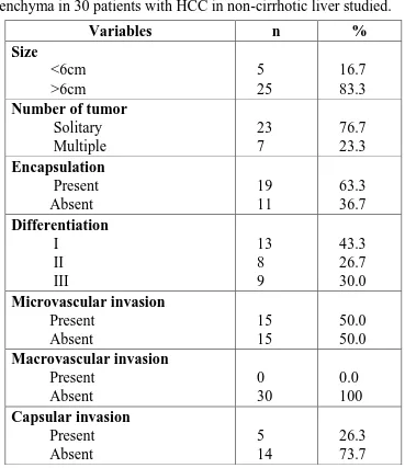
The mean size of tumor on histo-pathological examination was
13.2 ± 5.22 cm (range: 5-25cm). The majority of patients (76.7%) had
[image:58.595.88.495.452.637.2](63.3%) with capsular invasion of tumor observed in 5 patients (26.3%).
Histological differentiation of tumor was grade I in 13 patients (43.3%),
grade II in 8 patients (26.7%) and grade III in 9 patients (30%).
Microvascular invasion was observed in 15 patients (50%) but none of
the patient had macrovascular invasion. The resected margin was found
positive for tumor cells in 8 patients (26.7). The surrounding non-tumor
liver showed steatotic changes in 4 patients (13.3%).
Table 9: Histo-pathologic characteristics of tumor and surrounding parenchyma in 30 patients with HCC in non-cirrhotic liver studied.
Variables n %
Size <6cm >6cm 5 25 16.7 83.3
Number of tumor
[image:59.595.105.474.333.760.2]Margin Positive Negative 8 22 26.7 73.3 Steatosis Present Absent 4 26 13.3 86.7
pTNM-(AJCC-7th edition)
I II III IV 6 14 9 1 21 45 29 5 Okuda stage I II III 9 20 1 30 67 3 pTNM: pathologic tumor-node-metastasis
The follow-up rate was 100% with median follow-up of 405 days
(range: 62-921 days). In the whole study population, tumor recurrence
was identified in 14 patients (46.6%) within the follow-up
period(Table-10). Of these 14 patients, 5 patients (35.7%) had isolated intra-hepatic
recurrence, 3 patients (21.4%) had isolated extra-hepatic recurrence and
6 patients (42.8) had both intra-hepatic and extra-hepatic recurrence. Of
the 5 patients with isolated intra-hepatic recurrence, 2 patients
underwent re-resection, 2 patients received systemic chemotherapy and
1 patient was managed with supportive care. Two patients with isolated
extra-hepatic recurrence and 2 patients with both intra and extra-hepatic
patient with isolated extra-hepatic recurrence and 4 patients with
[image:61.595.95.485.180.633.2]multiple recurrences were managed symptomatically.
Table 10: Pattern of recurrence and its management in 14 of 30 patients with HCC in non-cirrhotic liver studied.
PATTERN OF RECURRENCE (n=14)
n %
Isolated Intra-hepatic recurrence
Unifocal Multifocal 3 2 21.4 14.2
Isolated Extra-hepatic recurrence
Lung Bone 2 1 14.2 7.1
Both Intra-hepatic and Extra-hepatic
Unifocal intra-hepatic + Extrahepatic
Multifocal intra-hepatic + Extrahepatic
2 4
14.2 28.6
MANAGEMENT OF RECURRENCE Intra-hepatic alone
Re-resection
Systemic chemotherapy Best supportive care
2 2 1
Extra-hepatic alone
Systemic chemotherapy Best supportive care
2 1
Both Intra-hepatic and Extra-hepatic
Systemic chemotherapy Best supportive care
2 4
OVERALL SURVIVAL:
At the time of analysis, there had been 11 deaths (36.7%) with 19
study group are 86.7%, 66.7% and 63.3% respectively. At the end of 1,
2, 3 year, there were 26, 20, 19 alive patients respectively.
PROGNOSTIC FACTORS FOR OVERALL SURVIVAL:
Patient who died after operation were excluded from the analysis
of prognostic factors for overall survival after liver resection for HCC in
non-cirrhotic patients. In the univariate analysis, high grade tumor
(p = 0.007), prothrombin time(p = 0.046) and serum bilirubin level
(p = 0.034) were significant predictors of poor long term survival
(Table-11). In the multivariate analysis, only high grade tumor (p =
0.039) was identified as independent prognostic factor (Figure 6) for
overall survival after liver resection for HCC in non-cirrhotic patients
(Table-12).
Table 11: Clinico-pathological and operative prognostic factors for overall and disease-free survival in 30 non-cirrhotic HC patients (Univariate Analysis)
Characteristics
OVERALL SURVIVAL DISEASE-FREE SURVIVAL
n % p-value n % p-value Age (yrs)
<60 >60
11 19
18 ± 2.6 29 ± 2.23
0.458
14 16
22 ± 1.9 33 ± 2.21
0.625 Gender Male Female 12 18
11 ± 2.5 28 ± 2.75
0.223
11 19
19 ± 2.7 26 ± 3.9
0.097 HBs antigen Positive Negative 4 26
25 ± 4.6 33 ± 1.9
0.792 3 27
13 ± 7.1 27 ± 3.8
0.511 Alcohol intake Yes No 4 26
31 ± 3.9 39 ± 4.1
0.091 7 23
16 ± 4.6 21 ± 2.9
[image:62.595.90.498.544.762.2]AFP (ng/ml) <20 >20
9 21
39 ± 2.3 27 ± 1.9
0.191
17 13
26 ± 4.59 30 ± 2.77
0.055 Bilirubin (mg/dl) <1.5 >1.5 3 27
23 ± 7.3 11 ± 1.8
0.034* 3 27
18 ± 6.53 29 ± 2.32
0.012* Prothrombin time <15 sec >15 sec 26 4
25 ± 1.7 13 ± 1.9
0.046* 28 2
27 ± 2.32 11 ± 0.51
0.041* Albumin (g/dl) <3 >3 5 25
16 ± 4.9 46 ± 2.7
0.061 3 27
12 ± 0.82 29 ± 2.31
0.241 Size (cm) <6 >6 11 19
17 ± 2.8 26 ± 1.7
0.576
14 16
18 ± 7.51 31 ± 1.92
0.061 Number Solitary Multiple 26 4
15 ± 6.5 21 ± 2.4
0.061
25 5
18 ± 6.6 29 ± 2.33
0.180 Capsule Present Absent 11 19
25 ± 1.7 33 ± 1.9
0.106
14 16
23 ± 6.1 27 ± 3.21
0.210 Grade High Low 21 9
32 ± 1.8 25 ± 4.4
0.007* 21 9
30 ± 1.99 12 ± 0.71
0.001* Vascular invasion Present Absent 12 18
13 ± 1.7 21 ± 6.4
0.07
15 15
20 ± 5.45 32 ± 1.79
0.0001* Capsular invasion Present Absent 12 18
13 ± 1.7 21 ± 6.4
0.07
3 27
11 ± 0.61 30 ± 7.1
0.266 Steatosis Present Absent 4 26
15 ± 1.9 18 ± 2.6
0.576 3 27
20 ± 2.19 29 ± 2.2
0.082 Major resection Yes No 17 13
29 ± 1.92 21 ± 0.60
0.069
20 10
31 ± 6.7 22 ± 7.1
0.351 Blood transfusion Yes No 2 28
18 ± 0.09 31 ± 2.4
0.199 2 28
11 ± 0.50 27 ± 2.31
0.012*` Post-op stay (days) <15 >15 19 11
36 ± 3.1 23 ± 2.2
0.659
17 13
28 ± 1.71 11 ± 0.07
Wound infection Present Absent
6 24
11 ± 0.50 31 ± 1.91
0.346 8 22
18 ± 2.90 28 ± 2.21
0.021* Sepsis Present Absent 6 24
19 ± 2.96 33 ± 1.71
0.062 5 25
12 ± 2.19 29 ± 2.33
0.001* Post-op liver failure Present Absent 3 27
15 ± 4.6 23 ± 0.79
0.792 4 26
12 ± 0.43 29 ± 2.08
0.023*
*p value <0.05 = statistically significant
Table 12: Significant prognostic factors for overall survival by multivariate analysis in 30 patients with non-cirrhotic HCC
Variables Hazard
ratio 95% CI p-value
Model prediction
High grade tumors 0.820 (0.694-0.698) 0.019*
Bilirubin 1.846 (0.893-3.816) 0.098
Protrombin time 0 .488 (0.117-2.044) 0.327 46.27 95% CI: 95% confidence interval, *p <0.05 = statistically significant
DISEASE FREE SURVIVAL:
Excluding the post-operative death within 30 days, the overall
1-, 2-, 3- year disease-free survival rates after curative liver resection
for HCC in non-cirrhotic patients were 66.6%, 53.3% and 50%
respectively.
PROGNOSTIC FACTORS FOR DISEASE-FREE SURVIVAL:
Post-operative deaths within 90-days were excluded from the
analysis of prognostic factors for disease-free survival after liver
resection for HCC in non-cirrhotic patients. In the univariate analysis,
[image:64.595.86.499.77.230.2]