Copyright
q
1997, American Society for Microbiology
Human T-Cell Leukemia Virus Type 1 Tax Releases Cell
Cycle Arrest Induced by p16
INK4a
KENNETH G. LOW,
1* LYDIA F. DORNER,
1DINALI B. FERNANDO,
1JOSEPH GROSSMAN,
1KUAN-TEH JEANG,
2ANDMICHAEL J. COMB
1Laboratory of Cell Signaling, New England Biolabs, Beverly, Massachusetts 01915,
1and Laboratory of Molecular
Microbiology, National Institute of Allergy and Infectious Diseases, Bethesda, Maryland 20892
2Received 13 August 1996/Accepted 27 November 1996
The human T-cell leukemia virus type 1 (HTLV-1) Tax oncoprotein causes cellular transformation by
de-regulating important cellular processes such as DNA repair, transcription, signal transduction, proliferation,
and growth. Although it is clear that normal cell cycle control is deregulated during HTLV-1-induced cellular
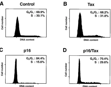
transformation, the effects of Tax on cell cycle control are not well understood. Flow cytometric analyses of
human T cells indicate that cell cycle arrest in late G
1, at or before the G
1/S restriction point, by p16
INK4ais
relieved by Tax. Furthermore, Tax-dependent stimulation of 5-bromo-2
*
-deoxyuridine incorporation and
tran-scriptional activation is inhibited by p16
INK4a. This result suggests that p16
INK4ais able to block Tax-dependent
stimulation of DNA synthesis and cell cycle progression into S phase. In vitro binding assays with recombinant
glutathione
S
-transferase fusion proteins and [
35S]methionine-labeled proteins indicate that Tax binds
spe-cifically with p16
INK4abut not with either p21
cip1or p27
kip1. Furthermore, sequential immunoprecipitation
assays with specific antisera and [
35S]methionine-labeled cell lysates subsequent to coexpression with Tax and
p16
INK4aindicate that the two proteins form complexes in vivo. Immunocomplex kinase assays with
cyclin-dependent kinase 4 antiserum indicate that Tax blocks the inhibition of cdk4 kinase activity by p16
INK4a. This
study identifies p16
INK4aas a novel cellular target for Tax and suggests that the inactivation of p16
INK4afunction is a mechanism of cell cycle deregulation by Tax.
Adult T-cell leukemia/lymphoma is caused by human T-cell
leukemia virus type 1 (HTLV-1) and is characterized by
mul-tistage transformation and immortalization of CD4
1T
lym-phocytes, which includes a long latent period following
infec-tion (6, 13). Oncogenic transformainfec-tion of T lymphocytes by
HTLV-1 is due primarily to the expression of the 40-kDa Tax
retrovirus oncoprotein, which deregulates a variety of
impor-tant cellular processes (6, 13). Tax has previously been shown
to directly interact and interfere with the ability of specific
cellular proteins to perform pivotal and critical roles in
tran-scriptional activation and growth factor signal transduction (6,
13). For example, Tax transactivates the expression of a variety
of cellular genes by increasing the DNA binding of cellular
transcription factors such as NF-
k
B (4, 22, 32), serum response
factor (12), AP-1 proteins, cyclic AMP response element
bind-ing protein (CREB), cyclic AMP response element modulatbind-ing
proteins, and ATF proteins (11, 23, 40, 45). The increased
DNA binding has been shown to be due to enhanced
transcrip-tion factor dimerizatranscrip-tion (2, 5, 44) or increased transcriptranscrip-tion
factor nuclear translocation resulting from the degradation of
cytoplasmic inhibitors (19). Tax can also transactivate gene
expression by interacting directly with components of the basal
transcriptional complex such as the TATA-binding protein (7)
or with transcriptional coactivators such as CREB-binding
pro-tein which mediate interactions between transcription factors
and the basal transcriptional complex (21). Recent studies also
suggest that Tax may interact with and activate specific
com-ponents of growth factor signal transduction pathways such as
the Ras/mitogen-activated protein kinase pathway (31, 43) and
the protein kinase A (PKA) and PKC pathways (18).
How-ever the immortalization of CD4
1T lymphocytes by HTLV-1
Tax requires other deregulatory events since its effects on
transcription or signal transduction alone have not been
dem-onstrated to be sufficient for oncogenic transformation. It is
clear that normal cell cycle control is deregulated during
HTLV-1 leukemogenesis; however, the molecular mechanism
through which this occurs is not well understood.
Normal cell cycle progression is intricately regulated at
dif-ferent checkpoints by a family of cyclin-dependent kinases
(cdks) which are activated by complex formation with cyclins
and phosphorylation (15, 27, 30, 36, 37). Recent studies have
identified a novel family of mammalian cdk inhibitors which
can be divided into two subfamilies based on amino acid
se-quence homology, functionally distinct modes of action, and
range of specificities for cdks (15, 27, 30, 36, 37). Members of
one subfamily of cdk inhibitors, which includes p21
cip1, p27
kip1,
and p57
kip2, have significant amino acid sequence homology
and inhibit a broad range of cdks by binding cdk-cyclin
com-plexes as multimers, whereas members of the other subfamily,
which includes p16
INK4a, p15
INK4b, p18
INK4c, and p19
INK4d,
in-duce cell cycle arrest by specifically inhibiting cdk4 and cdk6
kinase activities through the displacement of cyclin Ds from
the cdk-cyclin complexes and are composed of four ankyrin
motifs (15, 27, 30, 36, 37). Interestingly, Tax has previously
been demonstrated to interact specifically with the ankyrin
motifs of I
k
B
g
(14) and a three-amino-acid residue AAR motif
on CREB (1) which is also present in p16
INK4a(35). Since
p16
INK4aserves a critical role in regulating cell cycle
progres-sion into S phase and can suppress cellular transformation (20,
24, 34), we hypothesized that Tax deregulates normal cell cycle
control by interacting with p16
INK4aand interfering with its
ability to repress cdk4 kinase activity and cellular
transforma-tion.
In this study, we demonstrate that although p16
INK4aover-expression alone induces cell cycle arrest in late G
1, at or
* Corresponding author. Mailing address: New England Biolabs, 32
Tozer Rd., Beverly, MA 01915. Phone: (508) 927-5054, ext. 335. Fax:
(508) 922-7069. E-mail: [email protected].
1956
on November 9, 2019 by guest
http://jvi.asm.org/
before the G
1/S restriction point, it is unable to do so in the
presence of Tax. In addition, the stimulation of DNA synthesis
and cell cycle progression into S phase by Tax alone or with
oncogenic Ras is inhibited by p16
INK4aoverexpression.
Tax-dependent transactivation of proenkephalin gene transcription
is also inhibited by p16
INK4aoverexpression. Furthermore, Tax
blocks the p16
INK4a-dependent inhibition of cdk4 kinase
activ-ity, suggesting that Tax and p16
INK4amight interact directly. In
vitro binding assays indicate that Tax binds p16
INK4abut not
p21
cip1or p27
kip1and forms complexes with p16
INK4ain vivo.
These studies identify a novel cellular target for Tax and
sug-gest a mechanism for cell cycle deregulation by HTLV-1.
MATERIALS AND METHODS
Materials.All reagents, horse serum, and fetal calf serum (FCS) were from Sigma Chemical Company (St. Louis, Mo.) unless otherwise stated. Cell culture media were from GIBCO/BRL (Gaithersburg, Md.). [3H]chloramphenicol (37.9
Ci/mmol), [g-32P]ATP (3,000 Ci/mmol), and [35S]methionine (1,175 Ci/mmol)
were from DuPont New England Nuclear (Boston, Mass.).
Antibodies.Antiserum against p16INK4awas from PharMingen (San Diego,
Calif.), antiserum against Tax was from the AIDS Research and Reference Reagent Program, Division of AIDS, National Institute of Allergy and Infectious Diseases (Bethesda, Md.) (originally contributed by B. R. Cullen), and antisera against cdk4 and cyclin D1 were from Santa Cruz Biotechnology (Santa Cruz, Calif.). Fluorescein-conjugated CD8 antiserum was from Becton Dickinson (San Jose, Calif.), and antiserum against 5-bromo-29-deoxyuridine (BrdU) was from Amersham (Arlington Heights, Ill.).
DNA plasmids.DNA plasmids for the in vitro transcription and translation reactions, the expression of recombinant glutathioneS-transferase (GST) fusion proteins in bacteria, and the expression of recombinant proteins in mammalian cell lines were generous gifts from C. Z. Giam (Case Western University School of Medicine, Cleveland, Ohio) (pET-11d-TaxH6), F. Kashanchi and J. N. Brady
(National Cancer Institute, Bethesda, Md.) (pGEX-2T-Tax), B. R. Cullen (Duke University Medical Center, Durham, N.C.) (pCMV-Tax), M. Serrano and D. Beach (Cold Spring Harbor Laboratory, Cold Spring Harbor, N.Y.) (pBluescript II SK[2]-p16INK4a, pGEX-KG-p16INK4a, and pCMV-cdk4), W. S. El-Deiry and
B. Vogelstein (Johns Hopkins University School of Medicine, Baltimore, Md.) (pGEX-2T-WAF1 and pCEP4-WAF1), H. Toyoshima and T. Hunter (Salk In-stitute, La Jolla, Calif.) (pBluescript II SK[1]-p27kip1and pGEX-KG-p27kip1), P.
Hinds (Harvard Medical School, Boston, Mass.) (pCMV-cyclin D1), M. Meyer-son and E. Harlow (Massachusetts General Hospital Cancer Center, Charles-town) (pGEX-30X-Rb773-928), and R. A. Weinberg (Massachusetts Institute of
Technology, Cambridge) (pEJ 6.6). pBluescript II SK-WAF-1 was con-structed by subcloning a 2.1-kbHindIII/XhoI fragment encoding the p21cip1
cDNA from pCEP4-WAF1 into pBluescript II SK (Stratagene, La Jolla, Calif.). pTM3-p16INK4awas constructed by in-frame subcloning of a 475-bp
PCR-am-plified fragment containing the p16INK4acDNA intoNcoI andXhoI restriction
endonuclease sites of pTM3 (9, 28). This fragment was amplified as previously described (23), using 18 cycles of PCR with 0.25 mM deoxynucleoside triphos-phates and 400 ng each of pBluescript II SK(2)-p16INK4aas the template and
primers 59-GAAACGGATCCATGGAGCCTTCGGCTGACTGG-39and 59-G AATCGGATCCCTCGAGTCAATCGGGGATGTCTGAGGG-39 per 100-ml reaction volume. The DNA sequence of the PCR-amplified fragment was veri-fied by sequencing. pTM3-TaxH6was constructed by in-frame subcloning of an NcoI/BamHI fragment containing the full-length Tax coding sequence with a carboxyl-terminal His6tag from pET-11d-TaxH6into pTM3. pCMVbwas from
Clontech Laboratories (Palo Alto, Calif.).
Culture of cell lines and vaccinia virus strains.All cell lines and vaccinia virus strains were from the American Type Culture Collection (Rockville, Md.) unless otherwise stated. The JPX-9 cell line was a gift from K. Sugamura (Tohoku University School of Medicine, Sendai, Japan) and is a stably transfected deriv-ative of human Jurkat-derived T-lymphocyte cells (H33HJ-JA1) which express Tax under regulation by the murine metallothionein promoter (29). The treat-ment of JPX-9 cells with 30mM CdCl2for 14 h stimulates Tax expression in
greater than 90% of the cells to a level approximately equivalent to that observed in the HTLV-1-transformed human T-cell line MT-2 (reference 29 and unpub-lished observations). JPX-9 and H33HJ-JA1 cells were cultured in RPMI 1640 containing 10% (vol/vol) FCS. JEG-3 human choriocarcinoma, IMR-90 human diploid fetal lung, and BS-C-1 African green monkey kidney cells were cultured in minimal essential medium containing 10% (vol/vol) FCS. Hs27 human fore-skin fibroblast cells were cultured in Dulbecco modified Eagle medium contain-ing 10% (vol/vol) FCS. HeLa S3 human cervical carcinoma epithelial cells were cultured in S-minimal essential medium containing 5% (vol/vol) HS. Vaccinia virus strain WR recombinant vTF7-3 stocks were amplified in HeLa S3 cells, and titers were determined in BS-C-1 cells by plaque assays (8). Cells were infected at a multiplicity of infection of;10 for protein expression as previously described (10).
Transient transfections.JPX-9 cells were transiently transfected by electro-poration (300 V, 960mF, single pulse), and H33HJ-JA1 cells were transiently transfected by the DEAE-dextran protocol as previously described (23). JEG-3, IMR-90, and Hs27 cells were transiently transfected by a modification of the calcium phosphate protocol as previously described (23).
Propidium iodide labeling and flow cytometry.JPX-9 cells (53105) were
synchronized in G0/G1phase by serum starvation for 18 h, incubated with
fluorescein-conjugated CD8 antiserum for 30 min at 48C, washed twice with phosphate-buffered saline (PBS), fixed in 0.25% (wt/vol) paraformaldehyde in PBS for 30 min at 48C, permeabilized with 0.2% (vol/vol) Tween 20 in PBS for 15 min at 378C, labeled with propidium iodide (50mg/ml), and treated with RNase A (50mg/ml) in PBS for 1 h at 378C. CD81cells were then analyzed for DNA content as indicated by propidium iodide labeling.
BrdU labeling and immunohistochemistry.DNA synthesis in JEG-3 cells was determined by BrdU incorporation, using a cell proliferation assay kit (Amer-sham, Arlington Heights, Ill.). Cells were labeled with BrdU–5-fluoro-29 -deoxy-uridine (10:1), and BrdU-positive cells were identified by immunohistochemistry, using a cell proliferation assay kit (Amersham).
CAT andb-galactosidase assays.Following transient transfections, cells were harvested and lysed, and chloramphenicol acetyltransferase (CAT) activity in cell lysates was determined as previously described, with [3H]chloramphenicol as a
substrate and butyryl coenzyme A as an acyl donor (23).b-Galactosidase activity in cell lysates was determined as previously described, usingo-nitrophenyl-b-D
-galactopyranoside as a substrate, and used to normalize for differences in the efficiency of transfection and protein expression between the samples (23). His-tochemical detection ofb-galactosidase activity was determined by fixing cells in 2% (wt/vol) paraformaldehyde and 0.2% (vol/vol) glutaraldehyde for 10 min and using 0.1% (wt/vol) 5-bromo-4-chloro-3-indolyl-b-D-galactopyranoside (X-Gal)
in reaction buffer [5 mM K3Fe(CN)6, 5 mM K4Fe(CN)6, 2 mM MgCl2, 5%
(wt/vol)N,N-dimethylformamide] as a substrate.
In vitro transcription and translation.[35S]methionine-labeled proteins were
synthesized as previously described (23), using the TNT T7-T3 coupled reticu-locyte lysate system (Promega Corporation, Madison, Wis.) with either pBlue-script II SK[2]-p16INK4a, pBluescript II SK-WAF-1, pBluescript II SK[1
]-p27Kip1, or pET-11d-TaxH 6.
Bacterial expression and purification of recombinant GST fusion proteins. Escherichia coliDK-1 was transformed with either pGEX-KG-p16INK4a,
pGEX-2T-WAF1, pGEX-KG-p27Kip1, pGEX-2T-Tax, or pGEX-30X-Rb
773-928.
Recom-binant GST fusion proteins were expressed and immobilized on glutathione-agarose beads, and the amount of protein was estimated as previously described (23, 33, 39).
In vitro binding assays.Five microliters of each [35S]methionine-labeled
pro-tein was added to equal amounts of different agarose-immobilized GST fusion proteins in 100ml of binding buffer (0.25 M NaCl, 50 mM Tris/HCl [pH 7.4], 1% [vol/vol] Nonidet P-40 [NP-40], 10 mM EDTA, 2 mM EGTA, 0.02% [wt/vol] sodium azide, 0.25 mM phenylmethylsulfonyl fluoride [PMSF], 1mg each of aprotinin, leupeptin, and pepstatin A per ml) for 45 min at 48C. The agarose-immobilized complexed proteins were washed five times with binding buffer and analyzed by sodium dodecyl sulfate (SDS)-polyacrylamide gel electrophoresis (PAGE) followed by autoradiography.
[35S]methionine in vivo cell labeling and immunoprecipitations.Cells were
cultured in methionine-free medium for 1 h and then in the same medium containing 0.25 mCi of [35S]methionine/ml for 4 h, harvested, and lysed by
scraping in lysis buffer (0.15 M NaCl, 50 mM Tris-HCl [pH 7.4], 0.5% [vol/vol] NP-40) containing 0.1 mM PMSF and 2.5mg each of aprotinin, leupeptin, and pepstatin A per ml for 10 min at 48C. Cell debris was pelleted at 14,0003gmax
for 10 min at 48C and discarded. Cell extracts were incubated with polyclonal antiserum against either cdk4 (1:100 dilution), cyclin D1 (1:100 dilution), p16INK4a(1:100 to 1:500 dilution), or Tax (1:100 to 1:500 dilution) for 18 h and
then with 25ml of 50% (vol/vol) protein A-Sepharose in lysis buffer for 4 h. Protein A-Sepharose protein complexes were washed three times with 1 ml of lysis buffer and analyzed immediately by SDS-PAGE followed by autoradiogra-phy. In vivo protein complexes were detected by subsequent denaturation of the protein A-Sepharose protein complexes at 958C for 5 min in 0.1 ml of denatur-ation buffer (50 mM Tris-HCl pH 7.5, 0.5% [vol/vol] SDS, 70 mMb -mercapto-ethanol) and incubation with specific polyclonal antiserum against either p16INK4a(1:100 to 1:500 dilution) or Tax (1:100 to 1:500 dilution) in
radioim-munoprecipitation assay (RIPA) buffer (minus SDS) (0.15 M NaCl, 10 mM Tris-HCl [pH 7.5], 1% [vol/vol] NP-40, 1% [wt/vol] deoxycholate, and 0.25 mM PMSF) for 18 h and then with 25ml of 50% (vol/vol) protein A-Sepharose in RIPA buffer (minus SDS) for 4 h. Protein A-Sepharose protein complexes were washed three times with 1 ml of RIPA buffer (minus SDS) and analyzed by SDS-PAGE followed by autoradiography.
Immunocomplex kinase assays.Cells were lysed by sonication at 48C in im-munoprecipitation buffer (0.15 M NaCl, 50 mM HEPES [pH 7.5], 1 mM EDTA, 2.5 mM EGTA, 1 mM dithiothreitol [DTT], 0.1% [vol/vol] Tween 20, 10% [vol/vol] glycerol, 0.1 mM PMSF, 10 mMb-glycerophosphate, 1 mM NaF, 0.1 mM Na3VO4, 10mg each of aprotinin, leupeptin, and pepstatin A per ml). Cell
debris was pelleted at 14,0003gmax for 10 min at 48C and discarded. Cell
extracts were incubated with polyclonal antiserum against cdk4 (1:100 dilution) for 18 h and then with 25ml of 50% (vol/vol) protein A-Sepharose in immuno-precipitation buffer for 4 h. Protein A-Sepharose protein complexes were washed
on November 9, 2019 by guest
http://jvi.asm.org/
four times with 1 ml of immunoprecipitation buffer and twice with 1 ml of 50 mM HEPES (pH 7.5) containing 1 mM DTT. Kinase assays were performed as previously described (26) for 30 min at 308C in a final volume of 20ml (includes protein A-Sepharose bead volume) in kinase buffer (50 mM HEPES [pH 7.5], 10 mM MgCl2, 1 mM DTT, 2.5 mM EGTA, 25mM ATP, 10mCi of [g-32P]ATP)
containing 200 ng of an;45-kDa GST fusion protein containing the carboxyl-terminal portion of the retinoblastoma protein (GST-RbC) as a substrate. Then 200ml of immunoprecipitation buffer at 48C was added to stop the kinase reaction, and supernatants were removed to fresh tubes and incubated with 25ml of 50% (vol/vol) glutathione-agarose in immunoprecipitation buffer for 2 h at 48C. Glutathione-agarose beads were washed three times with 1.0 ml of immu-noprecipitation buffer, and phosphorylated GST-RbC proteins were separated by SDS-PAGE and visualized by autoradiography.
Digitization of autoradiographs and preparation of figures.Autoradiographs were digitized and prepared for the figures as previously described (23). Briefly, autoradiographs were digitized with a Microtek (Torrance, Calif.) Scanmaker III and an Apple (Cupertino, Calif.) Power Macintosh 8100/80, using Photoshop version 3.0 (Adobe Systems, Mountain View, Calif.). Digitized images were saved as tagged image file format files and exported into Pagemaker version 5.0 (Aldus Corp., Seattle, Wash.), where the images were cropped, text was added, and documents were printed at 2,540 dots per inch and a line screen of 150 lines per inch, using a Varityper 5300E (PageWorks, Cambridge, Mass.).
RESULTS
Tax releases JPX-9 cells from cell cycle arrest induced by
p16
INK4a.
The effects of p16
INK4aoverexpression on cell cycle
progression in the absence and presence of Tax were examined
in synchronous JPX-9 human T-cells (Fig. 1). Serum starvation
of JPX-9 cells for 18 h resulted in cell cycle arrest and the
synchronization of
.
95% of the total population of cells in
G
0/G
1phase (data not shown). Subsequent culture of JPX-9
cells in serum-containing medium stimulated reentry into the
cell cycle, resulting in the progression of 30% of the cells into
S phase and the retention of 70% of the cells in G
0/G
1phase
after 14 h (Fig. 1A). The induction of Tax expression in JPX-9
cells with 30
m
M CdCl
2(see Materials and Methods for the
source and a complete description of JPX-9 cells) over the
same time period did not significantly alter the distribution of
cells in G
0/G
1and S phases (68 and 32%, respectively (Fig.
1B). In contrast, the overexpression of p16
INK4ain JPX-9 cells
by transient transfection significantly altered the distribution of
cells, with 84% accumulating in G
0/G
1phase and only 16%
progressing into S phase (Fig. 1C). This result suggests that
p16
INK4aoverexpression arrests cell cycle progression in late
G
1, at or before the G
1/S restriction point. However, in the
presence of Tax, p16
INK4awas unable to arrest cells from
progression into S phase (Fig. 1D), with the distribution of
cells in G
0/G
1and S phases being not significantly different
from that of control cells (Fig. 1A and D). These results
sug-gest that Tax can potentially deregulate the normal restriction
of cell cycle progression in late G
1/S mediated through
p16
INK4aby directly binding and inactivating it.
p16
INK4aoverexpression blocks Tax-dependent stimulation
of cell cycle progression into S phase and transcriptional
ac-tivation.
Since Tax can reverse the effects of p16
INK4aon cell
cycle arrest in vivo, it was hypothesized that p16
INK4amight
also interfere with Tax function. Previous studies have
dem-onstrated that Tax can induce cellular transformation either
alone or synergistically with oncogenic Ras (31, 42). Thus, the
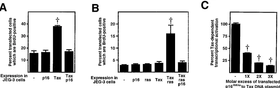
effects of Tax on BrdU incorporation of JEG-3 cells were
examined in the absence and presence of p16
INK4aoverexpres-sion (Fig. 2). The effects of Tax on BrdU incorporation are
most likely due to an increase in DNA synthesis and not an
increase in DNA repair since Tax is known to decrease
expres-sion of the DNA repair enzyme, DNA polymerase
b
(16).
Although the overexpression of p16
INK4aalone did not have a
significant effect on DNA synthesis (Fig. 2A and B), the
ex-pression of Tax alone (Fig. 2A) or with oncogenic Ras (38)
(Fig. 2B) stimulated DNA synthesis of JEG-3 cells to fivefold
compared to control cells in which neither Tax nor oncogenic
Ras was expressed. In contrast to the expression of Tax by
using the vaccinia virus-T7 system, expression using
cytomeg-alovirus vectors did not result in the stimulation of DNA
syn-thesis in JEG-3 cells (Fig. 2A and B). This probably results
from a difference in the levels of Tax expression achieved by
the two different expression systems. Coexpression with
p16
INK4acompletely blocked the ability of Tax alone or with
oncogenic Ras to stimulate DNA synthesis in JEG-3 cells (Fig.
2A and B). Since DNA synthesis occurs only during the S
phase of the cell cycle, these results suggest that the
stimula-tion of cell cycle progression into S phase by Tax alone or with
oncogenic Ras can be completely blocked by p16
INK4aoverex-pression.
To further determine whether p16
INK4amight also regulate
Tax-dependent transcriptional activation of cellular gene
ex-pression, p16
INK4awas overexpressed relative to Tax in human
Jurkat T lymphocytes (Fig. 2C). Previous studies have clearly
demonstrated that Tax is a very potent transcriptional activator
and that it greatly stimulates the expression of a number of
different of cellular and viral genes (6, 13), including the
hu-man proenkephalin gene (17, 23). Expression of Tax alone
stimulated human proenkephalin gene expression fourfold
over basal levels (data not shown) (23). The coexpression of
Tax with increasing amounts of p16
INK4ain human Jurkat T
lymphocytes inhibited Tax-dependent transactivation of the
human proenkephalin gene expression in a dose-dependent
manner (Fig. 2C). Although the mechanism by which p16
INK4aoverexpression inhibits Tax-dependent transactivation of
pro-enkephalin gene transcription is not completely defined by this
experiment, the results are consistent with direct interference
of Tax function by p16
INK4athrough formation of functionally
inactive complexes in vivo.
Tax and p16
INK4ainteract directly in vitro and form
com-plexes in vivo.
The release of human T lymphocytes from
[image:3.612.61.297.68.253.2]p16
INK4a-mediated cell cycle arrest by Tax and the inhibition of
FIG. 1. Tax releases human T cells from cell cycle arrest induced by p16INK4a
overexpression. The DNA content of synchronous JPX-9 cells was analyzed by propidium iodide labeling and flow cytometry in control cells (neither Tax nor p16INK4awas expressed) (A) or in cells subsequent to expression of Tax (B),
p16INK4a(C), and p16INK4a/Tax (D) by electroporation. JPX-9 cells were
tran-siently transfected by electroporation with 20mg of pSV-CD8 in the absence or presence of 10mg of pCMV-p16INK4a(300 V, 960mF, single pulse) and then
cultured for 14 h in the absence or presence of 30mM CdCl2(Tax expression in
JPX-9 cells is maximally induced with 30mM CdCl2by 8 h [data not shown]).
Hatch marks on either axis denote approximately equivalent DNA content or cell number.
on November 9, 2019 by guest
http://jvi.asm.org/
Tax-dependent cell cycle progression and transcriptional
acti-vation by p16
INK4asuggest direct protein-protein interactions
between Tax and p16
INK4a. Therefore, the potential
interac-tions between Tax and p16
INK4aor other cdk inhibitors such as
p21
cip1and p27
kip1were examined by in vitro binding assays
with equal amounts of recombinant GST fusion proteins
im-mobilized on glutathione-agarose beads and in
vitro-tran-scribed and -translated [
35S]methionine-labeled proteins (Fig.
3A to E). Approximately 10% of the total input p16
INK4awas
retained by GST-Tax but not by GST alone (compare Fig. 3A,
lane 2, with Fig. 3E, lane 1; compare lanes 1 and 2 in Fig. 3A).
This result indicates that p16
INK4abinds specifically to the Tax
portion of the recombinant GST-Tax fusion protein. In
con-trast, neither p21
cip1nor p27
kip1was retained by GST-Tax (Fig.
3B and C, lanes 2). Conversely, Tax was retained only by
GST-p16
INK4abut not by either GST alone, GST-p21
cip1, or
GST-p27
kip1(Fig. 3D). Approximately 20% of the total input
Tax was retained by GST-p16
INK4a(compare Fig. 3D, lane 2, to
Fig. 3E, lane 4). These results indicate that Tax specifically
interacts with p16
INK4abut not p21
cip1or p27
kip1in vitro.
To determine whether Tax and p16
INK4aform complexes in
vivo, both proteins were expressed in JEG-3 human
choriocar-cinoma cells by using the vaccinia virus-T7 RNA polymerase
hybrid system, and the cells were subsequently labeled with
[
35S]methionine (Fig. 3F). Protein complexes were
coimmuno-precipitated under native conditions (primary
immunoprecipi-tation) and subsequently disrupted by denaturation prior to
the identification of specific components of the complexes by
immunoprecipitation under denaturing conditions (secondary
immunoprecipitation). Although Tax and p16
INK4aare not
ex-pressed at detectable levels in JEG-3 cells (Fig. 3F, lanes 1 and
2), the expression of either protein by using the vaccinia
vi-rus-T7 RNA polymerase hybrid system resulted in significant
levels of detectable protein (Fig. 3F, lanes 3, 4, and 6). The
coexpression of Tax with p16
INK4aresulted in a significant
amount of p16
INK4acoimmunoprecipitating with Tax in vivo
(Fig. 3F; compare lane 4 or 6 with lane 7). The polyclonal
antiserum against Tax does not cross-react with p16
INK4abecause p16
INK4awas not immunoprecipitated in the absence
of Tax expression (Fig. 3F, lane 5). The results of the converse
experiment to determine whether Tax coimmunoprecipitates
with p16
INK4awere inconclusive because the polyclonal
anti-serum against p16
INK4across-reacts with Tax (data not shown).
These results suggest that Tax and p16
INK4ainteract
specifi-cally in vitro and form stable complexes in vivo. It is also likely
that the formation of these complexes is dependent on ankyrin
or AAR motifs present in p16
INK4awhich are absent in either
p21
cip1or p27
kip1(15, 27, 30, 36, 37).
Tax blocks the inhibition of cdk4 kinase activity by p16
INK4ain vivo.
The formation of stable complexes with p16
INK4ain
vivo and the release of human T lymphocytes from p16
INK4a-induced cell cycle arrest in late G
1phase suggest that Tax
might also interfere with the ability of p16
INK4ato inhibit cdk4
kinase activity. To analyze the functional consequence of the
interactions between Tax and p16
INK4aon cdk4 kinase activity
in vivo, both proteins were either expressed alone or
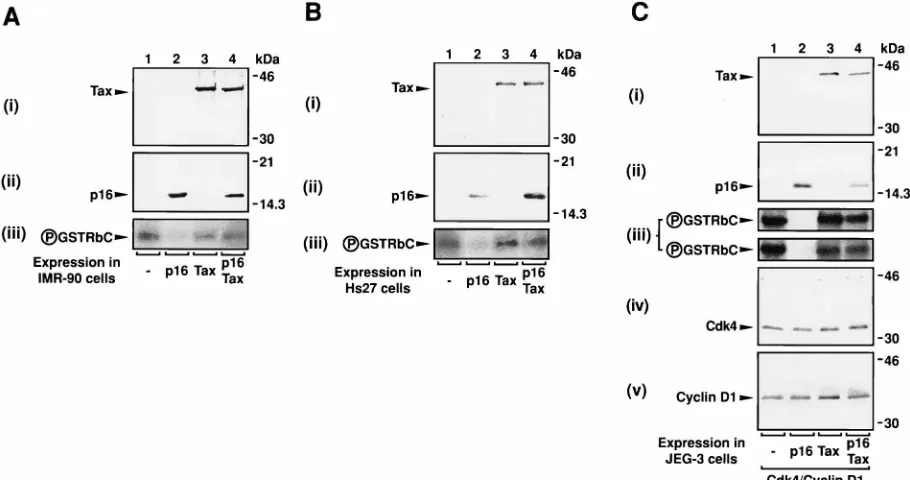
coex-pressed in IMR-90 human diploid fetal lung or Hs27 human
foreskin fibroblast cells (Fig. 4A and B, panels i and ii). Both
cell lines were selected because they do not express detectable
levels of either protein (Fig. 4A and B, panels i and ii, lane 1)
but do express detectable albeit low levels of cdk4 kinase
ac-tivity (Fig. 4A and B, panel iii, lane 1). The expression of Tax
(Fig. 4A and B, panel i, lanes 3 and 4) and p16
INK4a(Fig. 4A
and B, panel ii, lanes 2 and 4) by using the vaccinia virus-T7
RNA polymerase hybrid system resulted in significant levels of
detectable protein. Immunocomplex kinase assays using
cdk4-specific antisera and the recombinant GST-RbC fusion protein
as a substrate revealed that both cell lines possess active
cdk4-cyclin D complexes and low levels cdk4 kinase activity (Fig. 4A
and B, panel iii, lane 1) which are inhibited by p16
INK4aex-pression as expected (Fig. 4A and B, panel iii; compare lanes
1 and 2). Although Tax expression alone did not significantly
FIG. 2. (A) Stimulation of DNA synthesis and cell cycle progression into S phase by Tax is inhibited by p16INK4ain JEG-3 cells. JEG-3 (0.23106) cells were
transiently transfected with 8mg of pCMVbplus pTM3-p16INK4aand/or pTM3-TaxH
6for 20 h. Cells were subsequently infected with 23106PFU of vaccinia virus
strain WR recombinant vTF7-3 for 4 h, washed, cultured for 18 h, and labeled with BrdU for 2 h. BrdU-positive transfected cells were identified by colocalization of immunohistochemical staining for BrdU and histochemical staining forb-galactosidase activity. The histogram depicts the mean6standard error of a representative experiment from three different experiments. †, statistically significant difference from the control (transfected with pTM3 and pCMVbonly) and other treatment groups (P,0.001;n53; unpaired Student’sttest). (B) Stimulation of DNA synthesis and cell cycle progression into S phase by Tax and Ras is inhibited by p16INK4a
in JEG-3 cells. JEG-3 cells (0.23106) were transiently transfected with 6mg of pCMVbplus pCMV-p16INK4a, pCMV-Tax, and/or pEJ6.6 (oncogenic Ras) (38),
cultured for 42 h, and labeled with BrdU for 2 h. BrdU-positive transfected cells were identified as described above. The histogram depicts the mean6standard error of a representative experiment from three different experiments. †, statistically significant difference from the control (transfected with pGEM3 and pCMVbonly) and other treatment groups (P,0.04;n53; unpaired Student’sttest). (C) p16INK4aoverexpression inhibits Tax-dependent transcriptional activation of the human
proenkephalin gene enhancer in a dose-dependent manner in Jurkat human T cells. Jurkat cells (107) were transiently cotransfected with 10mg of pENKAT-12, 10mg
of pCMV-Tax, 5mg of pCMVb, and increasing amounts (10, 20, and 30mg) of pCMV-p16INK4a. Cells were then cultured for 48 h posttransfection before harvesting
and lysis for CAT andb-galactosidase assays. The histogram depicts the mean6standard error of a representative experiment from three different experiments. †, statistically significant difference from the control (transfected with pGEM3 and pCMVbonly) (P,0.0003) and each other (P,0.03) (n53; unpaired Student’st
test).
on November 9, 2019 by guest
http://jvi.asm.org/
[image:4.612.75.542.70.217.2]affect cdk4 kinase activity in either cell line (Fig. 4A and B,
panel iii; compare lanes 1 and 3), coexpression of Tax with
p16
INK4ablocked the inhibition of cdk4 kinase activity by
p16
INK4ain both cell lines in the absence of a significant
dif-ference in the levels of p16
INK4aexpression (Fig. 4A and B,
panels ii and iii; compare lanes 2 and 4).
To better analyze the functional consequence of the
inter-actions between p16
INK4aand Tax in the presence of higher
levels of cdk4 kinase activity, both proteins were either
ex-pressed alone or coexex-pressed with cdk4 and cyclin D1 in JEG-3
cells (Fig. 4C, panels i, ii, iv, and v). The coexpression of cdk4
and cyclin D1 in JEG-3 resulted in consistently equivalent
levels of protein expression (Fig. 4C, panels iv and v; compare
lanes 1 to 4). Although Tax and p16
INK4aare not expressed at
detectable levels in JEG-3 cells (Fig. 4C, panels i and ii, lane
1), the expression of both proteins by using the vaccinia
vi-rus-T7 RNA polymerase hybrid system resulted in significant
and approximately equivalent levels of detectable protein (Fig.
4C, panels i and ii; compare lanes 2 or 3 to lane 4).
Immuno-complex kinase assays using cdk4-specific antiserum and
re-combinant GST-RbC fusion protein as substrates revealed
sig-nificant levels of cdk4 kinase activity which were dramatically
inhibited by p16
INK4aexpression (Fig. 4C, panel iii, compare
lanes 1 and 2). Although Tax expression alone had no
signif-icant effects on cdk4 kinase activity (Fig. 4C, panel iii, compare
lanes 1 and 3), the coexpression of Tax with p16
INK4ablocked
the inhibition of cdk4 kinase activity by p16
INK4a(Fig. 4C,
panel iii; compare lanes 2 and 4). These results suggest that
Tax can directly interact with p16
INK4aand interfere with the
ability of p16
INK4ato inhibit cdk4 kinase activity in vivo.
DISCUSSION
A model describing the functional consequence of
interac-tions between Tax and p16
INK4athat is consistent with
pub-lished data and our results is depicted in Fig. 5. Previous
studies (15, 27, 30, 36, 37) indicate that active complexes of
cdk4 and cyclin D1 are disrupted in the presence of p16
INK4a,
resulting in the preferential formation of inactive complexes of
cdk4 and p16
INK4a(Fig. 5). Our results and other recent
stud-ies (41) clearly demonstrate that Tax interacts and forms in
vivo complexes directly with p16
INK4a(Fig. 3) and that this
interaction blocks the inhibition of cdk4 kinase activity by
p16
INK4a(Fig. 4). This model suggests that in the presence of
Tax, complexes consisting of cdk4 and p16
INK4awould
de-crease concurrently with an inde-crease in the formation of
com-plexes consisting of both Tax and p16
INK4aas well as active
complexes consisting of cdk4 and cyclin D1 (Fig. 5). Complexes
of cdk4 and p16
INK4ahave indeed been observed to be
de-creased in the presence of Tax (41). Furthermore, cdk4 kinase
activity is observed to be increased in the presence of Tax
despite the presence of p16
INK4a(Fig. 4) (41). Both of these
observations provide further support for this model (Fig. 5).
The critical role served by cdk4 activation in regulating cell
cycle progression from G
1phase into S phase is thought to be
dependent on its phosphorylation of the retinoblastoma
pro-tein (pRb) and subsequent release of transcription factors such
as E2F capable of activating the expression of S-phase genes
(37). Thus, the derepression of cdk4 kinase activity by Tax
through interactions with p16
INK4awould result in an increase
in the progression of cells from G
1to S phase. The
observa-tions that Tax releases T lymphocytes from p16
INK4a-mediated
cell cycle arrest in late G
1, at or prior to the G
1/S restriction
point, and that p16
INK4ainhibits a Tax-dependent stimulation
of DNA synthesis are both consistent with a direct role for Tax
[image:5.612.73.285.89.539.2]in deregulating cell cycle progression from G
1phase into S
FIG. 3. Tax interacts specifically with p16INK4abut not p21cip1or p27kip1in
vitro. Interactions between agarose-immobilized recombinant GST or GST-Tax fusion proteins and in vitro-transcribed and -translated35S-p16INK4a(A),35
S-p21cip1(B), or35S-p27kip1(C) were examined by in vitro binding assays. (D)
Interactions between agarose-immobilized recombinant GST, GST-p16INK4a,
GST-p21cip1, or GST-p27kip1and in vitro-transcribed and -translated35S-Tax
were also examined by similar in vitro binding assays. (E) Amounts of input
35S-p16INK4a,35S-p21cip1,35S-p27kip1, and35S-Tax. The amount of [35
S]methi-onine incorporated was different for each protein, although efforts were made to use equal amounts of in vitro-transcribed and -translated [35
S]methionine-la-beled protein. Results are representative of at least three different experiments. (F) Tax comimmunoprecipitates with p16INK4ain vivo. Each lane represents
0.53106JEG-3 cells transiently transfected with 7.5 mg of pTM3-p16INK4a
(lanes 4 to 7) and 7.5mg of pTM3-TaxH6(lanes 3, 6, and 7), infected with 53
106PFU of vaccinia virus strain WR recombinant vTF7-3 for 18 h, and
subse-quently in vivo labeled with [35S]methionine. Protein complexes were identified
by sequential immunoprecipitations under native and then denaturing conditions with polyclonal antiserum against either p16INK4a(ap16) or Tax (aTax). Results
are representative of at least two different experiments. Autoradiographs were digitized and the figures were prepared as described in Materials and Methods.
on November 9, 2019 by guest
http://jvi.asm.org/
phase. Although this study clearly demonstrates that cell cycle
progression from G
1phase to S phase can be deregulated
by Tax in a p16
INK4a-dependent manner, it is unclear
wheth-er Tax can also stimulate progression through the G
1/S
re-striction point in a p16
INK4a-independent manner. For
exam-ple, it is possible that Tax also interacts with and activates
downstream targets of cdk4 such as the pRb tumor
suppres-sor or transactivates S-phase gene expression through
inter-actions with transcription factors such as E2F. However,
pre-liminary studies using in vitro binding assays indicate that
Tax does not interact with pRb directly. Although potential
direct interactions between Tax and other cdks or cyclins
are currently under investigation, it is unclear whether Tax
deregulates progression through other cell cycle checkpoints.
However, there are no detectable in vitro interactions
be-tween Tax and the non-ankyrin motif-containing cdk
inhib-itors such as p21
cip1or p27
kip1which have broader
specific-ities and are involved in regulating cell cycle progression
through other cell cycle checkpoints (Fig. 4) (15, 27, 30, 36,
37).
Recently the adenovirus E1A oncoprotein has been
demon-strated to interfere with transforming growth factor
b
-medi-ated growth arrest in G
1phase of the cell cycle in a p27
kip1-dependent manner (25), suggesting that derepression of cdks
may represent a common mechanism for cell cycle
deregula-tion by viral oncoproteins. The repression of p16
INK4aby Tax is
similar to the interaction between Tax and another ankyrin
motif-containing cytoplasmic inhibitor, I
k
B (14). I
k
B represses
transcriptional activation by NF-
k
B through the binding and
retention of NF-
k
B dimers as inactive cytoplasmic complexes
in unstimulated cells (3). The phosphorylation of I
k
B in
re-sponse to activation by phorbol esters or PKC results in the
release and nuclear translocation of active NF-
k
B dimers
which can stimulate transcription (3). Tax interacts directly
with the ankyrin motifs of I
k
B, resulting in the release and
nuclear translocation of NF-
k
B dimers which are subsequently
[image:6.612.79.534.71.311.2]capable of transcriptional activation (14). Although Tax is
pri-marily known to activate cellular processes directly (6, 13), it is
clear that is can also activate cellular processes indirectly, by
repressing the effects of inhibitory cellular proteins. Although
most likely required, it is unclear whether the deregulation of
cell cycle progression by Tax it is sufficient for complete
cellu-lar transformation of HTLV-1-infected T lymphocytes.
Trans-formation by HTLV-1 probably requires the concurrent
Tax-dependent deregulation of a number of other critical cellular
processes such as transcription and signal transduction in
ad-dition to a deregulation of cell cycle control.
FIG. 4. Overexpression of p16INK4awith Tax in vivo blocks the ability of p16INK4ato inhibit cdk4 kinase activity. p16INK4aand/or Tax were expressed in IMR-90
human diploid fetal lung cells (A), Hs27 human foreskin fibroblast cells (B), and JEG-3 cells (C). Each lane represents 0.53106to 4.03106IMR-90, Hs27, or JEG-3
cells transiently transfected with 7.5mg of pTM3-p16INK4a(lanes 2 and 4) and pTM3-TaxH
6(lanes 3 and 4) and subsequently infected with 53106to 403106PFU
of vaccinia virus strain WR recombinant vTF7-3 for 18 h. JEG-3 cells were also transiently cotransfected with 20mg of pCMV-cdk4 and 20mg of pCMV-cyclin D1 (lanes 1 to 4). Cells were in vivo labeled with [35S]methionine and lysed, and expressed proteins were immunoprecipitated under native conditions with polyclonal antiserum
against either Tax (panel i), p16INK4a(panel ii), cdk4 (panels iii and iv), or cyclin D1 (panel v). Immunocomplex kinase assays were used to determine cdk4-dependent
kinase activities in duplicate sample lysates from a single experiment (panel iii). All results are representative of at least three different experiments. Autoradiographs were digitized and the figures were prepared as described in Materials and Methods.
FIG. 5. Interactions between Tax and p16INK4aresult in a derepression of
cdk4 kinase activity. Previous studies indicate that active complexes of cdk4 and cyclin D1 that form in the absence p16INK4aare disrupted in the presence of
p16INK4a, resulting in the formation of inactive complexes of cdk4 and p16INK4a
(15, 27, 30, 36, 37). Our results indicate that Tax can form complexes with p16INK4aand relieve the inhibition of cdk4 kinase activity p16INK4a.
on November 9, 2019 by guest
http://jvi.asm.org/
[image:6.612.319.558.619.677.2]ACKNOWLEDGMENTS
We thank M. Melner, S. Reeves, and P. Green for helpful
discus-sions and critical reading of the manuscript.
K.G.L. is supported by postdoctoral fellowship PF-3847 from the
American Cancer Society. This work was supported in part by NIH
grants DA00151, DA05706, and DA07745 to M.J.C.
REFERENCES
1.Adya, N., L. J. Zhao, W. Huang, I. Boros, and C. Z. Giam.1994. Expansion of CREB’s DNA recognition specificity by Tax results from interaction with Ala-Ala-Arg at positions 282-284 near the conserved DNA-binding domain of CREB. Proc. Natl. Acad. Sci. USA91:5642–5646.
2.Anderson, M. G., and W. S. Dynan.1994. Quantitative studies of the effect of HTLV-I Tax protein on CREB protein-DNA binding. Nucleic Acids Res.
22:3194–3201.
3.Baeuerle, P. A., and D. Baltimore.1988. IkB: a specific inhibitor of the NF-kB transcription factor. Science242:540–546.
4.Ballard, D. W., E. Bohnlein, J. W. Lowenthal, Y. Wano, B. R. Franza, and W. C. Greene.1988. HTLV-I Tax induces cellular proteins that activate the kB element in the IL-2 receptoragene. Science241:1652–1655. 5.Baranger, A. M., C. R. Palmer, M. K. Hamm, H. A. Giebler, A. Brauweiler,
J. K. Nyborg, and A. Schepartz.1995. Mechanism of DNA-binding enhance-ment by the human T-cell leukaemia virus transactivator Tax. Nature376:
606–608.
6.Cann, A. J., and I. S.-Y. Chen.1990. Human T-cell leukemia virus types I and II, p. 1501–1527.InB. N. Fields, D. M. Knipe, R. M. Chanock, M. S. Hirsch, J. L. Melnick, T. P. Monath, and B. Roizman (ed.), Virology, 2nd ed, vol. 2. Raven Press, New York, N.Y.
7.Caron, C., R. Rousset, C. Be´raud, V. Moncollin, J. M. Egly, and P. Jalinot.
1993. Functional and biochemical interaction of the HTLV-I Tax1 transac-tivator with TBP. EMBO J.12:4269–4278.
8.Earl, P. L., N. Cooper, and B. Moss.1991. Preparation of cell cultures and vaccinia virus stocks, p. 16.16.1–16.16.7.InF. M. Ausubel, R. Brent, R. E. Kingston, D. D. Moore, J. G. Seidman, J. A. Smith, and K. Struhl (ed.), Current protocols in molecular biology, vol. 2. Greene Publishing Associates and Wiley-Interscience, New York, N.Y.
9.Elroy-Stein, O., T. R. Fuerst, and B. Moss.1989. Cap-independent transla-tion of mRNA conferred by encephalomyocarditis virus 59sequence im-proves the performance of the vaccinia virus/bacteriophage T7 hybrid ex-pression system. Proc. Natl. Acad. Sci. USA86:6126–6130.
10. Elroy-Stein, O., and B. Moss.1991. Gene expression using the vaccinia virus/T7 RNA polymerase hybrid system, p. 16.19.1–16.19.9.InF. M. Aus-ubel, R. Brent, R. E. Kingston, D. D. Moore, J. G. Seidman, J. A. Smith, and K. Struhl (ed.), Current protocols in molecular biology, vol. 2. Greene Publishing Associates and Wiley-Interscience, New York, N.Y.
11. Franklin, A. A., M. F. Kubik, M. N. Uittenbogaard, A. Brauweiler, P. Utais-incharoen, M. A. Matthews, W. S. Dynan, J. P. Hoeffler, and J. K. Nyborg.
1993. Transactivation by the human T-cell leukemia virus Tax protein is mediated through enhanced binding of activating transcription factor-2 (ATF-2) response and cAMP element-binding protein (CREB). J. Biol. Chem.268:21225–21231.
12. Fujii, M., H. Tsuchiya, T. Chuhjo, T. Akizawa, and M. Seiki.1992. Interac-tion of HTLV-1 Tax1 with p67SRFcauses the aberrant induction of cellular
immediate early genes through CArG boxes. Genes Dev.6:2066–2076. 13. Gitlin, S. D., J. Dittmer, R. L. Reid, and J. N. Brady.1993. The molecular
biology of human T-cell leukemia viruses, p. 159–192.InB. R. Cullen (ed.), Human retroviruses. Oxford University Press, New York, N.Y.
14. Hirai, H., T. Suzuki, J. I. Fujisawa, J. I. Inoue, and M. Yoshida.1994. Tax protein of human T-cell leukemia virus type I binds to the ankyrin motifs of inhibitory factorkB and induces nuclear translocation of transcription factor NF-kB proteins for transcriptional activation. Proc. Natl. Acad. Sci. USA
91:3584–3588.
15. Hunter, T., and J. Pines.1994. Cyclins and cancer II: cyclin D and cdk inhibitors come of age. Cell79:573–582.
16. Jeang, K. T., S. G. Widen, O. J. Semmes, and S. H. Wilson.1990. HTLV-I trans-activator protein, Tax, is a trans-repressor of the humanb-polymerase gene. Science247:1082–1084.
17. Joshi, J. B., and H. P. G. Dave.1992. Transactivation of the proenkephalin gene promoter by the Tax1protein of human T-cell lymphotropic virus type
I. Proc. Natl. Acad. Sci. USA89:1006–1010.
18. Kadison, P., H. T. Poteat, K. M. Klein, and D. V. Faller.1990. Role of protein kinase A intaxtransactivation of the human T-cell leukemia virus type I long terminal repeat. J. Virol.64:2141–2148.
19. Kanno, T., K. Brown, G. Franzoso, and U. Siebenlist.1994. Kinetic analysis of human T-cell leukemia virus type I Tax-mediated activation of NF-kB. Mol. Cell. Biol.14:6443–6451.
20. Koh, J., G. H. Enders, B. D. Dynlacht, and E. Harlow.1995. Tumour-derived
p16 alleles encoding proteins defective in cell-cycle inhibition. Nature375:
506–510.
21. Kwok, R. P. S., M. E. Laurance, J. R. Lundblad, P. S. Goldman, H. M. Shih, L. M. Connor, S. J. Marriott, and R. H. Goodman.1996. Control of cAMP-regulated enhancers by the viral transactivator Tax through CREB and the co-activator CBP. Nature380:642–646.
22. Leung, K., and G. J. Nabel.1988. HTLV-1 transactivator induces interleu-kin-2 receptor expression through an NF-kB-like factor. Nature333:776– 778.
23. Low, K. G., H. M. Chu, P. S. Schwartz, G. M. Daniels, M. H. Melner, and M. J. Comb.1994. Novel interactions between human T-cell leukemia virus Tax and activating transcription factor 3 at a cyclic AMP-responsive element. Mol. Cell. Biol.14:4958–4974.
24. Lukas, J., D. Parry, L. Aagaard, D. J. Mann, J. Bartkova, M. Strauss, G. Peters, and J. Bartek.1995. Retinoblastoma-protein-dependent cell-cycle inhibition by tumour suppressor p16. Nature375:503–506.
25. Mal, A., R. Y. C. Poon, P. H. Howe, H. Toyoshima, T. Hunter, and M. L. Marter.1996. Inactivation of p27Kip1by the viral E1A oncoprotein in TGFb
-treated cells. Nature380:262–265.
26. Matsushime, H., D. E. Quelle, S. A. Shurtleff, M. Shibuya, C. J. Sherr, and J. Y. Kato.1994. D-type cyclin-dependent kinase activity in mammalian cells. Mol. Cell. Biol.14:2066–2076.
27. Morgan, D. O.1995. Principles of CDK regulation. Nature374:131–134. 28. Moss, B., O. Elroy-Stein, T. Mizukami, W. A. Alexander, and T. R. Fuerst.
1990. New mammalian expression vectors. Nature348:91–92.
29. Nagata, K., K. Ohtani, M. Nakamura, and K. Sugamura.1989. Activation of endogenous c-fosproto-oncogene expression by human T-cell leukemia virus type I-encoded p40taxprotein in the human T-cell line, Jurkat. J. Virol.63:
3220–3226.
30. Peter, M., and I. Herskowitz.1994. Joining the complex: cyclin-dependent kinase inhibitory proteins and the cell cycle. Cell79:181–184.
31. Pozzatti, R., J. Vogel, and G. Jay.1990. The human T-lymphotropic virus type Itaxgene can cooperate with therasoncogene to induce neoplastic transformation of cells. Mol. Cell. Biol.10:413–417.
32. Ruben, S., H. Poteat, T.-H. Tan, K. Kawakami, R. Roeder, W. Haseltine, and C. A. Rosen.1988. Cellular transcription factors and regulation of IL-2 receptor gene expression by HTLV-1taxgene product. Science241:89–92. 33. Sasse, J., and S. R. Gallagher.1991. Detection of proteins, p. 10.6.1–10.6.8.
InF. M. Ausubel, R. Brent, R. E. Kingston, D. D. Moore, J. G. Seidman, J. A. Smith, and K. Struhl (ed.), Current protocols in molecular biology, vol. 2. Greene Publishing Associates and Wiley-Interscience, New York, N.Y. 34. Serrano, M., E. Go´mez-Lahoz, R. A. DePinho, D. Beach, and D. Bar-Sagi.
1995. Inhibition of ras-induced proliferation and cellular transformation by p16INK4a. Science267:249–252.
35. Serrano, M., G. J. Hannon, and D. Beach.1993. A new regulatory motif in cell-cycle control causing specific inhibition of cyclin D/CDK4. Nature366:
704–707.
36. Sherr, C.1994. G1 phase progression: cycling on cue. Cell79:551–555. 37. Sherr, C., and J. M. Roberts.1995. Inhibitors of mammalian G1
cyclin-dependent kinases. Genes Dev.9:1149–1163.
38. Shih, C., and R. A. Weinberg.1982. Isolation of a transforming sequence from a human bladder carcinoma cell line. Cell29:161–169.
39. Smith, J. A.1991. Quantitation of proteins, p. 10.1.1–10.1.3.InF. M. Aus-ubel, R. Brent, R. E. Kingston, D. D. Moore, J. G. Seidman, J. A. Smith, and K. Struhl (ed.), Current protocols in molecular biology, vol. 2. Greene Publishing Associates and Wiley-Interscience, New York.
40. Suzuki, T., J. Fujisawa, M. Toita, and M. Yoshida.1993. The trans-activator Tax of human T-cell leukemia virus type 1 (HTLV-1) interacts with cAMP-responsive element (CRE) binding and CRE modulator proteins that bind to the 21-base-pair enhancer of HTLV-1. Proc. Natl. Acad. Sci. USA90:610– 614.
41. Suzuki, T., S. Kitao, H. Matsushime, and M. Yoshida.1996. HTLV-1 Tax protein interacts with cyclin-dependent kinase inhibitor p16INK4aand
coun-teracts its inhibitory activity towards CDK4. EMBO J.15:1607–1614. 42. Tanaka, A., C. Takahashi, S. Yamaoka, T. Nosaka, M. Maki, and M.
Ha-tanaka.1990. Oncogenic transformation by thetaxgene of human T-cell leukemia virus type I in vitro. Proc. Natl. Acad. Sci. USA87:1071–1075. 43. Uchiumi, F., K. Semba, Y. Yamanashi, J. I. Fujisawa, M. Yoshida, K. Inoue,
K. Toyoshima, and T. Yamamoto.1992. Characterization of the promoter region of thesrcfamily genelynand itstransactivation by the human T-cell leukemia virus type I-encoded p40tax. Mol. Cell. Biol.12:3784–3795.
44. Wagner, S., and M. R. Green.1993. HTLV-I Tax protein stimulation of DNA binding of bZIP proteins by enhancing dimerization. Science262:395– 399.
45. Zhao, L. J., and C. Z. Giam.1992. Human T-cell lymphotropic virus type I (HTLV-I) transcriptional activator, Tax, enhances CREB binding to HTLV-I 21-base-pair repeat by protein-protein interactions. Proc. Natl. Acad. Sci. USA89:7070–7074.