0022-538X/97/$04.0010
Copyrightq1997, American Society for Microbiology
Specific Initiation of Replication at the Right-End Telomere
of the Closed Species of Minute Virus of Mice
Replicative-Form DNA
ANDREAS Q. BALDAUF, KURT WILLWAND,* ELENI MUMTSIDU, JU¨ RG P. F. NU¨ ESCH,
ANDJEAN ROMMELAERE
Department of Applied Tumor Virology, Deutsches Krebsforschungszentrum, and Formation INSERM U375, Heidelberg, Germany
Received 22 August 1996/Accepted 13 October 1996
We have developed an in vitro system that supports the replication of natural DNA templates of the autonomous parvovirus minute virus of mice (MVM). MVM virion DNA, a single-stranded molecule bracketed by short, terminal, self-complementary sequences, is converted into double-stranded replicative-form (RF) DNA when incubated in mouse A9 fibroblast extract. The 3*end of the newly synthesized complementary strand is ligated to the right-end hairpin of the virion strand, resulting in the formation of a covalently closed RF (cRF) molecule as the major conversion product. cRF DNA is not further replicated in A9 cell extract alone. On addition of purified MVM nonstructural protein NS1 expressed from recombinant baculoviruses or vaccinia viruses, cRF DNA is processed into a right-end (5*end of the virion strand) extended form (5*eRF). This is indicative of NS1-dependent nicking of the right-end hairpin at a distinct position, followed by unfolding of the hairpin and copying of the terminal sequence. In contrast, no resolution of the left-end hairpin can be detected in the presence of NS1. In the course of the right-end nicking reaction, NS1 gets covalently attached to the right-end telomere of the DNA product, as shown by immunoprecipitation with NS1-specific antibodies. The 5*eRF product is the target for additional rounds of NS1-induced nicking and displacement synthesis at the right end, arguing against the requirement of the hairpin structure for recognition of the DNA substrate by NS1. Further processing of the 5*eRF template in vitro leads to the formation of dimeric RF (dRF) DNA in a left-to-left-end configuration, presumably as a result of copying of the whole molecule by displace-ment synthesis initiated at the right-end telomere. Formation of dRF DNA is highly stimulated by NS1. The experimental results presented in this report support various assumptions of current models of parvovirus DNA replication and provide new insights into the replication functions of the NS1 protein.
The minute virus of mice (MVM) is a member of the genus
Parvovirus of the Parvoviridae family (48). The MVM genome
is a linear, single-stranded (ss) DNA molecule, 5,149 nucleo-tides (nt) in length, comprising at each end short, self-comple-mentary sequences that fold into stable hairpin structures (2, 4, 5, 7). Given that the great majority of encapsidated MVM genomes have a negative polarity, the 39and 59ends of virion DNA correspond to the left and right ends, respectively. Two nonstructural (NS) proteins are encoded by the MVM ge-nome, the major one of which, NS1, has proved to be essential for viral DNA replication (42, 58), promoter activity (22, 23, 26, 41), and cytotoxicity (8, 9, 31, 32, 59).
According to current models (1, 17, 55), parvovirus DNA replication starts with the self-primed synthesis of a comple-mentary strand at the left-end hairpin of virion DNA, leading to the production of a double-stranded (ds) replicative form (RF). The 39 end of the growing complementary strand is thought to be ligated to the folded-back 59 end of the virion strand, yielding a covalently closed RF (cRF) species. Subse-quent amplification of RF DNA is assumed to involve reopen-ing and copyreopen-ing of the right-end hairpin sequence to generate a right-end (viral 59end) extended duplex molecule designated 59eRF. By folding back into a turnaround structure, the right-end telomere provides a new hairpin primer that can be further
elongated through unidirectional strand displacement synthe-sis. This results in the formation of dimeric RF (dRF) and higher-order concatemeric molecules that are subsequently re-solved into monomeric RF (mRF) DNA.
Evidence in support of this model has been given by a num-ber of in vivo and in vitro studies. In vitro conversion of ss virion DNA into ds RF DNA was achieved with partially pu-rified eukaryotic DNA polymerases alpha (24) and gamma (29), as well as with the large fragment of Escherichia coli DNA polymerase I (7, 45). Unit length open RF (oRF) and cRF DNAs, as well as concatemers of dimer and tetramer length were detected in parvovirus-infected cells (13, 35, 54). Reso-lution of left-to-left-end, as well as right-to-right-end, junction fragments of MVM dimeric and tetrameric RF DNAs was reported to occur in vivo and in vitro and was shown to require the nicking activity of the MVM NS1 protein (14, 15, 20, 21, 34). NS1 was found to get covalently linked to the DNA res-olution products, in keeping with the observed association of NS1 with the 59ends of all newly synthesized viral DNA mol-ecules in MVM-infected cells (18, 25).
The mechanism by which the hairpin termini of MVM RF DNA are replicated is of particular interest. A straightforward process consists of a hairpin transfer reaction involving DNA strand-specific cleavage and displacement synthesis. This type of resolution takes place at both ends of ds DNA intermediates of adeno-associated viruses (AAV, a defective genus of
Parvo-viridae) and is driven by the AAV NS proteins Rep68 and
Rep78, as reproduced under in vitro conditions (28, 40, 50, 61). The terminal sequences of parvovirus DNA constitute imper-* Corresponding author. Mailing address: Deutsches
Krebsfor-schungszentrum, Abt. 0610, Im Neuenheimer Feld 242, Postfach 101949, D-69009 Heidelberg, Germany. Phone: 49-6221-424969. Fax: 49-6221-424962. E-mail: [email protected].
971
on November 9, 2019 by guest
http://jvi.asm.org/
of the folded-back genomic 59end to account for the presence of extra nucleotides at the right-end terminus of extended RF versus virion DNA (1). Given its capacity to resolve the right-to-right-end bridge (14, 20), the NS1 protein can also be as-sumed to drive the processing of the right-end hairpin. How-ever, there is no direct experimental evidence that the right-end hairpin is a target for NS1-specific nicking while the left-end hairpin is not. Another open question concerns the generation of cRF DNA. As mentioned above, it might result from covalent linkage of the folded-back genomic 59end to the newly synthesized complementary strand at the ss-to-ds DNA conversion step, but alternative processes besides conversion may also be considered for its formation (57, 63).
To tackle these questions, we developed an in vitro DNA replication assay, based on extracts of mouse A9 fibroblasts (an MVM-permissive cell line) and supplemented with recombi-nant NS1 protein, to study the processing of defined natural MVM DNA substrates (ss and ds). This system allowed us to substantiate under the experimental conditions tested the above-mentioned predictions of the parvovirus DNA replica-tion model, namely, (i) the formareplica-tion of cRF as a direct major (if not obligatory) product of ss-to-ds DNA conversion, (ii) the capacity of NS1 to induce specific cleavage of the right-end hairpin and formation of an extended duplex right-end telo-mere, in contrast to the inability of the viral protein to trigger resolution of the left-end hairpin, and (iii) the processing of 59eRF into dRF DNA in a left-to-left-end configuration. In-terestingly, efficient formation of dimer length molecules re-quired the presence of the NS1 protein.
MATERIALS AND METHODS
Culture of cells.Mouse A9 fibroblasts (33) and HeLa cells were grown in suspension culture at 378C in spinner or roller flasks containing Joklik’s modified Eagle’s medium (Gibco, Eggenstein, Germany) supplemented with 5% fetal calf serum (PAA, Linz, Austria), 2 mM glutamine, and 100mg of gentamicin per ml. The cell concentration was determined at regular intervals and adjusted to 105/ml to keep the culture in exponential growth.
Sf-9 insect cells were grown in suspension at 278C in TNM-FH medium (Sigma, St. Louis, Mo.) supplemented with 5% fetal calf serum, 2 mM glutamine, and 100mg of gentamicin per ml. Prior to baculovirus infection, cells were plated on tissue culture dishes at about 75% confluence.
Preparation of cell extracts.Cytoplasmic extracts were prepared essentially as described by Stillman and Gluzman (52) and Yamaguchi and DePamphilis (65). Briefly, cells were harvested by centrifugation, washed twice with phosphate-buffered saline and once with hypotonic extraction buffer (20 mM N-2-hydroxy-ethylpiperazine-N9-2-ethanesulfonic acid [HEPES]-KOH [pH 7.5], 5 mM potas-sium acetate, 0.5 mM MgCl2, 0.5 mM dithiothreitol, 1 mM phenylmethylsulfonyl fluoride, 3mg of pepstatin per ml, 3mg of leupeptin per ml, 1.5mg of aprotinin per ml, 3 mg of E64 per ml), and resuspended in 2 cell pellet volumes of hypotonic buffer. After 20 min of incubation on ice, cells were disrupted by 15 strokes in a tight-fitting Dounce homogenizer. The cytoplasmic extract was cleared by centrifugation at 12,0003g for 10 min at 48C and stored at2808C. Production and purification of wild-type and mutant NS1 proteins.Wild-type NS1 was produced in insect Sf-9 cells from a recombinant baculovirus vector (44). Cultures were infected at a multiplicity of 10 PFU/cell, and whole-cell
grown in suspension culture were infected with MVMp at a multiplicity of 1023 PFU/cell and harvested by centrifugation when most of the cells showed a cytopathic effect, usually at around 5 days postinfection. Viral DNA was ex-tracted by the method of Hirt (27), and MVM mRF DNA was purified by centrifugation through 5 to 30% neutral sucrose gradients. Covalently closed and open forms of MVM mRF DNA were further separated on 10 to 30% alkaline sucrose gradients as described by Straus et al. (53).
In vitro replication assay.In vitro replication was performed essentially as described by Stillman and Gluzman (52). The reactions were carried out in a mixture containing 40 mM HEPES-KOH (pH 7.6); 8 mM MgCl2; 500mM dithiothreitol; 100mM each dATP, dGTP, and dTTP; 5mCi of [a-32
P]dCTP (4 Ci/mmol); 100mM each CTP, GTP, and UTP; 3 mM ATP; 40 mM phosphocre-atine; and 20mg of creatine phosphokinase per ml. The reaction mixture (50-ml final volume) contained 10 to 15ml of cytoplasmic A9 cell extract (corresponding to 60mg of proteins) and 10ml of an NS1 preparation (equivalent to about 200 ng of purified protein). The reaction was started by addition of 20 ng of template DNA. After incubation at 378C for 2.5 h, the reaction was terminated by addition of 1% SDS, and proteins were digested for 1.5 h at 378C with 1 mg of proteinase K per ml. Subsequently, unincorporated nucleotides were removed by using the Wizard DNA cleanup kit (Promega, Madison, Wis.).
Immunoprecipitation of NS1-linked DNA.Following in vitro replication, sam-ples were adjusted to the buffer conditions required for cleavage with XbaI endonuclease. After digestion with the restriction enzyme at 378C for 30 min, SDS was added to 1% and noncovalent DNA-protein interactions were dis-rupted by incubation at 658C for 30 min. Thereafter, samples were diluted 10 times with 10 mM Tris (pH 8.0)–1% Nonidet P-40–10 mM EDTA–150 mM NaCl and incubated for 15 h at 48C (21) with a 1:10-diluted antiserum (16) raised against the 91 N-terminal amino acids of the NS1 protein. After additional incubation of the samples with protein A-Sepharose (Pharmacia Biotech, Upp-sala, Sweden) for 1 h at 48C, NS1-linked DNA was pelleted by low-speed cen-trifugation, released from bound protein by proteinase K digestion as described above, and analyzed by agarose gel electrophoresis.
Analysis of replication products.Reaction products were analyzed either directly or following restriction enzyme digestion in accordance with the suppli-er’s instructions. Electrophoresis was performed in 0.8% neutral (Tris-acetate-EDTA-buffered) or 1% alkaline agarose gels, unless indicated differently. Small DNA species were analyzed by neutral or denaturing (in the presence of 7 M urea) polyacrylamide gel electrophoresis (46). Combined one- and two-dimen-sional electrophoresis was conducted in 1% agarose gels, first under neutral conditions and then, after turning the gel 908, under alkaline conditions. For analysis under neutral conditions only, a stripe of the gel containing an identical sample was cut off before alkalization. For one-dimensional analysis under al-kaline conditions, an additional sample was loaded before the second run.
RESULTS
ss virion DNA is converted into cRF in vitro. Genomic ss DNA released from virions is the initial substrate of parvovirus DNA replication. The conversion of virion DNA into a ds RF was studied by using mouse A9 fibroblast extract as a source of cellular DNA replication factors. The32P-labeled products of this reaction were fractionated by combined one- and two-dimensional agarose gel electrophoresis (Fig. 1A). Under neu-tral conditions (Fig. 1A, vertical lane), one major product was detected at the position expected for MVM unit length ds DNA, as determined by analysis of the same sample along with a molecular size marker (Fig. 2A, lane 1). Thus, MVM ss DNA was converted into ds mRF DNA in vitro. Analysis of the same sample under alkaline conditions (Fig. 1A, horizontal lane)
on November 9, 2019 by guest
revealed two major bands, presumably representing subforms of mRF DNA. This was confirmed by two-dimensional elec-trophoresis (central part of the gel), showing that both forms indeed arose from the mRF product. The fact that these spe-cies migrated at a position corresponding to about twice the MVM genomic length under denaturing conditions (Fig. 2B, lane 1) argued for their formation through self-primed elon-gation of the left-end hairpin of MVM virion DNA, resulting in covalently linked parental and complementary strands. It was noted that the apparent molecular size of one mRF species (cRF) varied significantly with the agarose concentration of the alkaline gel, whereas the estimated size of the other (oRF) remained unchanged (data not shown). The former feature is characteristic of ss circular DNA (47), suggesting that the cRF species represented a covalently closed DNA form containing an additional interstrand link due to ligation of the newly synthesized complementary strand to the folded-back genomic 59 end. Therefore, the mRF subforms were designated cRF and oRF, for closed and open RF, respectively, as substanti-ated below.
Several additional bands were detected ahead of the com-plete conversion products under both neutral and alkaline conditions (Fig. 1A). These lower-molecular-weight species were imbedded in a continuous smear that formed a diagonal starting from the oRF species in the two-dimensional gel and may represent intermediates in the filling of the complemen-tary strand. Since this migration pattern was highly reproduc-ible (data not shown), the formation of intermediates may be due to the arrest of DNA synthesis at polymerase pause sites present on the MVM genome.
The structure of the right end of the conversion products was analyzed in more detail. It was determined, in particular, whether extension of the complementary strand stopped when reaching the folded-back genomic 59 end or whether strand displacement synthesis took place and allowed the hairpin se-quence to be copied, giving rise to an extended duplex right-end telomere. This was tested by digesting the conversion products with the PshAI restriction enzyme that cleaves MVM ds DNA at nt 4916, yielding right-end turnaround and ex-tended fragments of 130 and 233 bp, respectively. As
illus-trated in Fig. 1B, the restriction fragment corresponding to the right-end hairpin structure was readily detected by native poly-acrylamide gel electrophoresis, while only a very faint band was seen at the position of the extended right-end terminal frag-ment. It thus appeared that under the in vitro conditions tested, the cellular machinery involved in the conversion pro-cess had very little capacity for unfolding and copying of the right-end hairpin. The heavily labeled upper band migrated at the position expected for the left-end terminal fragment re-sulting from PshAI cleavage of conversion products at nt 670. It was further investigated whether the complementary strand became ligated to the right-end hairpin in a fraction of converted molecules, as suggested by the migration properties of mRF under alkaline conditions. PshAI-digested products were analyzed by denaturing urea-polyacrylamide gel electro-phoresis. As shown in Fig. 1C, a band was visible at the 260-nt position that was expected for the ligated turnaround right-end terminus, confirming the formation of the cRF species. Unli-gated right-end hairpin termini would give rise to a 27-nt-long segment extending from the PshAI site to the folded-back genomic 59end. Such a fragment was not detected, even after long exposure times. This indicates that, at least in the in vitro assay, meeting of the complementary strand 39 end with the parental right-end hairpin 59 end is followed by ligation in most, if not all, RF molecules, making cRF a major, or even obligatory, intermediate of parvovirus DNA replication (Fig. 1D). This result implies that the oRF species detected is likely to consist of randomly nicked cRF molecules rather than of unligated conversion products.
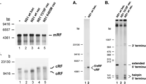
[image:3.612.69.550.67.241.2]Influence of NS1 on the conversion reaction.Given its re-quirement for parvovirus DNA replication in vivo (42, 58), the MVM major NS protein NS1 was tested for its effect on the outcome of the conversion reaction in vitro. NS1 was produced in insect and human cells by means of recombinant baculovi-ruses and vaccinia vibaculovi-ruses, respectively, and purified by nickel-nitrilotriacetic acid-chelate chromatography through a histi-dine tag fused to the recombinant protein. The products of ss DNA conversion were first analyzed by neutral agarose gel electrophoresis. As illustrated in Fig. 2A, similar amounts of mRF molecules were generated in the presence (lanes 2 and 3) FIG. 1. Conversion of MVM virion DNA into ds RF DNA in vitro. (A) MVM ss DNA was incubated in A9 cell extract, and products were analyzed by combined one- and two-dimensional 1.0% agarose gel electrophoresis under neutral (vertical dimension) and alkaline (horizontal dimension) conditions. The marginal lanes show the corresponding one-dimensional fractionation of the same reaction products. (B and C) Analysis of PshAI-digested conversion products (RF) by 5% native (B) or 6% denaturing (C) polyacrylamide gel electrophoresis. 39end-labeled HinfI (B) or HpaII (C) restriction fragments of pGEM5Zf were used as molecular size markers (lane M). The arrow indicates the expected position of the 27-nt fragment that would be generated by PshAI cleavage of unligated product DNA (C). (D) Diagram of the conversion reaction showing the parental viral strand (thin line) with its 39OH end (arrowhead), the newly synthesized complementary strand (thick line), and the positions of PshAI restriction sites. 39, left end; 59, right end.
on November 9, 2019 by guest
http://jvi.asm.org/
and absence (lanes 1, 4, and 5) of functional NS1, indicating that the viral protein had no influence on the efficiency of the conversion reaction.
In contrast, NS1-dependent structural changes in the con-version products were detected by alkaline agarose gel elec-trophoresis (Fig. 2B). The main species obtained in the ab-sence of NS1 (lane 1) or in the preab-sence of the replication-deficient NS1 mutants Y210F (lane 4) and K405M (lane 5) was cRF DNA, as identified above. In the presence of wild-type NS1 expressed from recombinant baculoviruses (lane 2) or vaccinia viruses (lane 3), the relative amount of cRF declined and faster-migrating RF molecules became predominant. At the gel resolution used, these molecules comigrated with the oRF product detected in the absence of NS1, leading us to hypothesize that they result from the NS1-induced resolution of cRF into an open form.
NS1-dependent nicking and extension of the right-end telo-mere of MVM DNA.To test whether cRF is indeed opened in the presence of NS1, this intermediate was used as the tem-plate in an in vitro replication assay. Figure 3A shows the analysis of the reaction products by neutral agarose gel elec-trophoresis. In the absence of NS1 (lane 1), little incorporation of radioactive nucleotides into template cRF DNA was visible and most likely due to repair synthesis. Addition of purified wild-type NS1 (lane 2) resulted in both a striking enhancement of the labeling of input DNA and a slight reduction of the electrophoretic mobility of labeled products, suggesting an in-crease in size. According to the current parvovirus DNA rep-lication model, the turnaround right-end telomere of cRF
DNA is resolved into an extended open structure through site-specific nicking, unfolding, and copying of the original hairpin (1, 17). This prompted us to test whether the labeling and size changes brought about in the cRF substrate by NS1 reflect the ability of the viral protein to induce specific cleavage and extension of the turnaround right-end terminus.
To this end, the cRF replication products were digested with
PshAI and analyzed by native polyacrylamide gel
[image:4.612.313.555.64.341.2]electrophore-sis. As shown in Fig. 3B (lanes 1 and 3), the labeled DNA species formed in the presence of baculo or vaccinia virus-expressed wild-type NS1 gave rise to a PshAI fragment of the size expected for the right-end terminus in the extended con-figuration. In contrast, the replication-deficient NS1 mutant Y210F failed to generate this fragment (lane 2). Thus, the covalently closed right end of MVM cRF DNA was processed into an extended right-end telomere with a defined length in the presence of functional NS1 protein, pointing to the NS1-induced nicking of the right-end hairpin in a site-specific man-ner. A faint band was also visible at the position corresponding to the left-end terminal fragment of PshAI-digested RF DNA. The weak labeling of this fragment did not depend on repli-cation-competent NS1 and was most likely due to nonspecific nick translation. The lack of NS1-induced precursor incorpo-ration into the left end of cRF DNA indicated that the turn-around MVM left-end telomere, in contrast to the right-end telomere, did not serve as an origin for NS1-dependent DNA replication. The band migrating slightly behind the left-end FIG. 2. Effect of the NS1 protein on the in vitro conversion of ss virion DNA
[image:4.612.82.552.68.339.2]into mRF DNA. In vitro replication was performed with an ss virion DNA template in A9 cell extract in the absence of NS1 (lanes 1) or in the presence of purified wild-type (lanes 2 and 3) or mutant (lanes 4 and 5) NS1 produced from recombinant baculoviruses (bac) or vaccinia viruses (vac). Products were ana-lyzed by neutral (A) or alkaline (B) agarose gel electrophoresis. The sizes and positions of DNA fragments used as molecular size markers are shown on the left.
FIG. 3. In vitro processing of cRF DNA. (A) cRF DNA was used as the template for in vitro replication in A9 cell extract in the absence (lane 1) or presence (lane 2) of baculovirus (bac.)-expressed wild-type (wt) NS1. Radiola-beled products were analyzed by neutral agarose gel electrophoresis. (B) Anal-ogous reactions were carried out in the presence of functional (lanes 1 and 3) or mutant (lane 2) NS1 expressed from recombinant baculoviruses (lane 1) or vaccinia viruses (vac) (lanes 2 and 3). Products were digested with the endonu-clease PshAI prior to native polyacrylamide gel electrophoresis. 39and 59are equivalent to the left and right ends, respectively. Molecular size markers are positioned on the left.
on November 9, 2019 by guest
terminal fragment in lane 3 was not observed in other exper-iments.
Interestingly, the restriction-digested cRF replication prod-ucts included an additional DNA species that was specifically formed in the presence of functional NS1 and migrated as a doublet around the position expected for the right-end termi-nal hairpin fragment generated by PshAI digestion (Fig. 3B, lanes 1 and 3). It was very unlikely, however, that either com-ponent of this doublet corresponded to the right-end terminal restriction fragment of input cRF DNA, since the nonspecific labeling of this small piece of DNA should be hardly detectable and independent of NS1. We therefore hypothesized that the doublet resulted from NS1-induced renicking of already ex-tended right-end termini, followed by displacement synthesis. This process would lead to the segregation of a copy of the right-end telomere in the form of an ss molecule that would fold back into a hairpin DNA of about 140 bp, migrating at the position of the observed doublet. This hypothesis implied that the doublet may be generated from a right-end extended sub-strate (59eRF) in an NS1-dependent but PshAI-independent way. These predictions were verified by supplying the in vitro replication system with a 59eRF template and analyzing the products by native polyacrylamide gel electrophoresis without previous restriction enzyme treatment. As illustrated in Fig. 4A, the doublet of right-end hairpin size was indeed produced in the presence of functional NS1 (lane 2), but neither in the absence of NS1 (lane 1) nor in the presence of the NS1 mu-tants Y210F and K405M (lanes 3 and 4). The concomitant labeling of the extended right-end terminus of full-length RF molecules was also expected, accounting for the wild-type NS1-dependent increase in radioactive high-molecular-weight ma-terial retained at the top of the gel (lane 2). These results suggested that not only the turnaround but also the extended right-end telomere was a target for NS1-specific nicking, at least under the in vitro conditions tested. The fact that the displaced right-end telomere was resolved into two distinct
bands may reflect structural variations in the hairpin. This was confirmed by the further analysis of individual gel-purified components of the doublet under denaturing conditions (un-published data).
A scheme summarizing these data is depicted in Fig. 4B. NS1 is shown to induce site-specific cleavage of the right-end (but not the left-end) hairpin of cRF DNA. Subsequent strand displacement synthesis gives rise to the extended 59eRF inter-mediate, which can serve as a substrate for an additional round(s) of NS1-driven nicking and extension. The covalent attachment of NS1 to the 59end created at the nick is substan-tiated below.
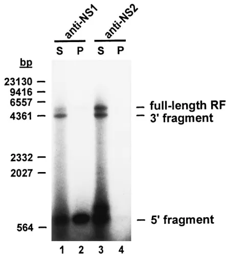
NS1 becomes covalently attached to the in vitro-extended right-end telomere.The NS1 protein has been reported to be covalently linked to the 59 ends of viral and complementary strands of replicated parvovirus DNA isolated from infected cells (18) and of in vivo and in vitro resolution products of cloned bridge fragments from multimeric RF intermediates (14, 15, 20, 21). This attachment is thought to be a conse-quence of the site-specific nicking of parvovirus DNA by NS1. It was therefore of interest to determine whether this viral protein was also bound to the extended right-end telomere that was generated from the cRF right-end hairpin in in vitro rep-lication reactions supplemented with purified NS1. Immuno-precipitation experiments using NS1-specific antibodies were performed to this end.
Replication reactions were carried out with a cRF DNA template in the presence of NS1, and the products were cleaved with XbaI at nt 4342, creating an 807-bp-long right-end extended fragment. Samples were heated at 658C in the pres-ence of 1% SDS to disrupt noncovalent protein-DNA associ-FIG. 4. NS1-induced nicking and strand displacement synthesis at the
right-end telomere. (A) In vitro reactions using a 59eRF template were carried out with A9 cell extract in the absence of NS1 (lane 1) or in the presence of purified wild-type (wt) (lane 2) or mutant (lanes 3 and 4) NS1 expressed from recombi-nant vaccinia viruses (vac). Products were analyzed by neutral polyacrylamide gel electrophoresis. 59is equivalent to the right end. Size markers consisted of TaqI (M1) and HindIII (M2) restriction fragments of pBR322 and bacteriophagel
[image:5.612.324.545.68.317.2]DNA, respectively. (B) Schematic illustration of the NS1-dependent resolution of turnaround (cRF) and extended (59eRF) right-end telomeres. Heavy lines represent newly synthesized DNA, and 39 OH primers are shown by small arrowheads. Filled circles symbolize NS1 proteins that cleave RF DNA in a site-specific way and become covalently bound to the resultant 59ends of the DNA strands. The positions of PshAI restriction sites are indicated. See the text for details.
FIG. 5. Covalent attachment of NS1 to in vitro-processed right-end telo-meres. cRF template DNA was incubated with A9 cell extract in the presence of vaccinia virus-expressed wild-type NS1. Products were partially digested with
XbaI and subsequently immunoprecipitated by using antibodies specific for NS1
(lanes 1 and 2) or NS2 (lanes 3 and 4). Pellets containing the immunoprecipi-tated material (P) and corresponding supernatants (S) were analyzed by 1.2% neutral agarose gel electrophoresis. 39and 59are equivalent to the left and right ends, respectively. The sizes and positions of HindIII restriction fragments of bacteriophagelDNA used as molecular size markers are shown on the left.
on November 9, 2019 by guest
http://jvi.asm.org/
vitro-labeled RF population. The specificity of the immune reaction for NS1 was ascertained by incubating samples with an antiserum directed against an NS2 protein portion unre-lated to NS1 and showing that the immunoprecipitate was free of both right-end XbaI fragments and full-length RF DNA (lanes 3 and 4). The origin of the additional band given by the supernatant of the anti-NS2-treated sample slightly above the right-end fragment position (lane 3) is unclear. Finally, it should be mentioned that NS1 was also found to become covalently attached to the free right-end hairpin DNA stretches that were segregated as a result of 59eRF nicking and extension (data not shown).
It was concluded from these experiments that the in vitro assay reproduced a typical feature of in vivo parvovirus DNA replication, i.e., the covalent binding of NS1 to extended right-end telomeres. This attachment is associated with the resolu-tion of right-end hairpin termini, confirming the involvement of NS1 rather than a copurified cellular protein in the right-end nicking and extension processes.
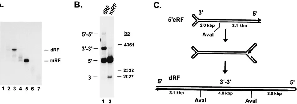
5*eRF DNA is processed into dRF molecules in the presence of NS1.To test the capacity of the in vitro system for further replication of 59eRF DNA, reactions were conducted with an mRF template that was covalently closed at one end. This template was purified by neutral and subsequent alkaline su-crose gradient centrifugations and characterized by restriction analysis, showing that most of the left-end termini were in the closed configuration and most of the right-end termini were in the extended configuration, while the converse structure was found in only a small minority of telomeres (data not shown). As illustrated in Fig. 6A, little incorporation of radioactive nucleotides took place when the 59eRF template was incu-bated with A9 cell extract in the absence of NS1 (lane 2) or in the presence of wild-type baculovirus-infected insect cell ex-tract treated as for NS1 purification (lane 1). Addition of purified baculovirus-produced NS1 (lane 3) resulted in a strong enhancement of mRF labeling, as expected from the above-mentioned NS1-induced nicking and strand displace-ment synthesis at the right-end telomere. In addition, the for-mation of a distinct DNA species was observed at a position corresponding to an apparent molecular size of about 10 kbp, i.e., the size of dRF intermediates. Incubation of the same template with HeLa cell extract in conjunction with wild-type NS1 expressed from recombinant vaccinia viruses led to the formation of the 10-kbp species as well (Fig. 6B, lane 2). In contrast, no high-molecular-weight product was detected in the presence of the replication-deficient NS1 mutants K405M (lane 1) and Y210F (lane 3).
To ascertain that dRF DNA was indeed formed, the prod-ucts of 59eRF replication in A9 cell extract supplemented with wild-type NS1 were fractionated by neutral sucrose gradient centrifugation (Fig. 7A). This allowed individual 10-kbp and
mRF species to be recovered and further characterized by digestion with the AvaI endonuclease and by neutral agarose gel electrophoresis. The restriction pattern of mRF consisted of the left-end (2-kbp) and right-end (3.1-kbp) fragments (Fig. 7B, lane 2) that were expected from the asymmetric cleavage of MVM DNA at nt 2070 (Fig. 7C). NS1-induced displacement synthesis at the right-end telomere accounted for the much higher level of radioactive precursor incorporation into the right-end fragment, compared with the nonspecifically labeled left-end fragment. Analysis of the higher-molecular-weight species revealed two major products, one of which comigrated with the right-end AvaI fragment of mRF, while the other had about twice the length of the corresponding left-end fragment (Fig. 7B, lane 1). The latter size was expected for the central
AvaI fragment of a dimeric intermediate in which the
individ-ual subunits are arranged in a left-to-left-end orientation (Fig. 7C). Therefore, these data supported the identity of the 10-kbp species with a dRF molecule generated through replication initiation at the right end of mRF DNA. Since one strand of dRF DNA is a complete copy of the original 59eRF template, incorporated radiolabeled nucleotides should be equally dis-tributed over the molecule. This is compatible with the stron-ger signal given by the right-end fragment compared with the left-to-left-end bridge (Fig. 7B, lane 1), despite the smaller size of the former, since these fragments are present in a 2:1 stoi-chiometry, with two right-end termini flanking each left-to-left-end bridge. It should also be mentioned that two additional minor DNA species were detected after restriction of dRF DNA. One of these comigrated with the left-end mRF frag-ment, while the other had the length expected for the central
AvaI dRF fragment in the right-to-right-end configuration
(Fig. 7B, lane 1). Therefore, these species appeared to repre-FIG. 6. In vitro replication of right-end-extended RF DNA. The DNA prod-ucts generated in vitro from a 59eRF template were analyzed by electrophoresis through 0.8% neutral agarose gels. (A) Reactions were carried out with A9 cell extract in the absence (lane 2) or presence (lane 3) of NS1 purified from recombinant baculovirus (bac)-infected Sf-9 cells or in the presence of identically processed wild-type (wt) baculovirus-infected Sf-9 cells (lane 1). (B) In vitro reactions were performed with HeLa cell extract in the presence of vaccinia virus (vac)-expressed wild-type (lane 2) or mutant (lanes 1 and 3) NS1. The positions and sizes of molecular size markers (HindIII restriction fragments oflDNA) are given on the right.
on November 9, 2019 by guest
sent the restriction products of dRF molecules generated from the minor fraction of the 39eRF template.
These results showed that the in vitro replication assay ful-filled the requirements for dRF formation. As proposed in current parvovirus DNA replication models (1, 17, 55), the extended right-end telomere probably folds back into the so-called “rabbit-eared” structure, providing a hairpin primer for strand displacement synthesis and subsequent copying of the displaced strand (Fig. 7C). It is worth noting that some dRF formation could be detected in the absence of NS1 when the autoradiographs were exposed for very long times (data not shown). A novel finding of this study therefore lies in the capacity of NS1 to strongly stimulate the multimerization re-action.
DISCUSSION
This report describes an in vitro system that mimics distinct events in MVM DNA replication. The system makes use of A9 or HeLa cell extract as a source of cellular factors for the replication of natural viral DNA templates (ss virion DNA and ds replicative intermediates isolated from infected cells). This allowed the role of the viral protein NS1 to be studied by providing the assay with purified NS1 expressed from recom-binant baculoviruses or vaccinia viruses. Analysis of the pro-cessing of defined MVM DNA substrates in this in vitro system revealed a number of interesting features, including (i) the frequent, if not necessary, passing through a covalently closed conversion product (cRF) in which the newly synthesized com-plementary strand is ligated to the folded-back genomic right-end hairpin; (ii) the capacity of NS1 to drive cleavage and extension of the closed right-end hairpin telomere, contrasting with the inability of the viral protein to resolve the left-end hairpin telomere; and (iii) the strong stimulation of dimer length intermediate formation from mRF in the presence of NS1.
Formation of a covalently closed conversion product. Con-version of ss virion DNA into ds RF DNA is thought to result
from elongation of the folded-back genomic 39 end. This is supported by the turnaround structure of the left-end telomere of a fraction of RF molecules extracted from parvovirus-in-fected cells (54) and the ability of parvovirus virion DNA to provide several DNA polymerases with both a template and a primer for leading-strand synthesis in vitro (7, 24, 29, 45). Our in vitro assay proved able to sustain MVM ss-to-ds DNA con-version in the absence of NS1. The major concon-version product was found to be cRF DNA terminating in turnaround struc-tures on both sides. Thus, the cellular extract appears to have little capacity, by itself, to unfold and copy the right-end genomic hairpin which becomes ligated to the growing com-plementary strand. The cRF species was recently detected in cells infected with the autonomous parvoviruses MVM (13) and Aleutian disease virus (35) and was postulated to be an early intermediate in parvovirus DNA replication (17, 56). Our data argue for this possibility by showing that cRF DNA is the direct product of the conversion reaction taking place under in vitro conditions.
NS1-dependent resolution of the right-end telomere.While having no detectable effect on the efficiency of the overall conversion reaction in A9 cell extract, the MVM protein NS1 was found to be essential for the further replication of ds DNA intermediates. No specific processing of cRF DNA took place in vitro unless replication-competent NS1 was supplied in
trans. When used as a substrate, cRF DNA was cleaved and
[image:7.612.70.551.71.240.2]extended at its right end in an NS1-dependent fashion. Con-sistently, most of the mRF molecules generated from virion ss DNA in the presence of NS1 were not in the covalently closed configuration, probably due to the NS1-induced resolution of right-end hairpin termini. This may also account for the de-layed detection of cRF within the RF pool in MVM-infected cells (57). Furthermore, our data indicate that the NS1-medi-ated cleavage of the right-end turnaround telomere is site specific, as is apparent from the distinct size of the extended right-end termini generated from open right-end hairpins by strand displacement synthesis. Although it can be inferred from the present work that nicking takes place in the vicinity of FIG. 7. Purification and characterization of RF species generated from a 59eRF template in vitro. An in vitro replication reaction was conducted with a 59eRF substrate, A9 cell extract, and baculovirus-expressed wild-type NS1. (A) Separation of dRF and mRF products on a 5 to 30% neutral sucrose gradient. Fractions were collected from the bottom of the tube and analyzed by 0.8% neutral agarose gel electrophoresis. (B) dRF (lane 1) and mRF (lane 2) products were recovered from gradient fractions 3 and 5, respectively (see panel A), digested with the restriction endonuclease AvaI, and analyzed by 0.8% neutral agarose gel electrophoresis. The smear above the mRF right-end fragment (lane 2) presumably consists of 59eRF molecules in which hairpin extension at the right-end telomere had already started. 39and 59, left- and right-end fragments; 39-39and 59-59, left-to-left-end and right-to-right-end bridge fragments of concatemeric DNA. The sizes and positions oflDNA
HindIII fragments used as molecular size markers are shown on the right. (C) Schematic representation of dRF formation from 59eRF DNA through folding back of a rabbit-eared structure (middle) and copying of the whole molecule by right-end hairpin-primed strand displacement synthesis. The small arrowheads indicate DNA strand 39ends. The thick lines represent newly synthesized DNA. The positions of the AvaI restriction sites and the approximate sizes of the corresponding fragments are given. See the text for details.
on November 9, 2019 by guest
http://jvi.asm.org/
ensuing displacement synthesis were detected not only with a right-end turnaround (cRF) substrate but also with a right-end extended (59eRF) substrate, indicating that the hairpin config-uration is not required for recognition, cleavage, and process-ing of the right-end telomere. This result is consistent with other studies showing that the right-end sequence present in the cloned right-to-right-end bridge of concatemeric RF DNA in a duplex configuration is cleaved and resolved in the pres-ence of NS1 both in vivo (20) and in vitro (14). While mean-ingful for the resolution of right-to-right-end junctions of mul-timeric RF intermediates, the processing of extended right-end termini is of questionable significance for the mRF substrate used in our assay. Indeed, displacement synthesis at the nicked right-end terminus of 59eRF merely regenerates the original template and leads to the segregation of a copy of the right-end telomere. An integral part of current parvovirus DNA replica-tion models consists of melting of the extended right end of RF DNA, followed by the refolding of individual strands into hair-pins to form the so-called rabbit-eared structure (55). The formation of rabbit ears may be more efficient in vivo than under our in vitro conditions, minimizing the initiation of dis-placement synthesis from extended right-end telomeres.
Insensitivity of the left-end hairpin to NS1.Another inter-esting finding of this study is that the MVM left-end hairpin can be distinguished from its right-end counterpart by its in-ability to be cleaved and resolved in the presence of NS1. This result gives direct proof of a central postulate of autonomous parvovirus DNA replication models; i.e., the left-end inverted repeat does not constitute a replication origin in the hairpin configuration and needs to be copied in the form of a left-to-left-end bridge to be subsequently resolved at the multimeric RF DNA stage (1, 6, 17). The stem of the left-end hairpin contains a so-called bubble region exhibiting a mismatch of three (GAA) opposite two (GA) noncomplementary nucleo-tides. When extended and copied into a bridge structure, the left-end inverted repeat is cleaved by NS1 specifically on the GA side of the bridge that functions as a replication origin (21). The lack of specific replication initiation from the GAA arm (21) is extended by the present work to the left-end hair-pin mismatch configuration that does not constitute an active RF origin either. This contrasts with the case of AAV, in which Rep-dependent hairpin resolution and RF replication initia-tion take place at both ends of the viral DNA (50), in keeping with the presence of the same telomere on either side of the AAV genome, while distinct left-end and right-end telomeres are characteristic of the parvovirus genus (6). Altogether, these data account for the above-mentioned flip and flop alternate configurations detected at both the left and right ends of AAV DNA but only at the right end of MVM DNA (1, 36), given that this sequence heterogeneity is a reflection of the hairpin transfer process.
persistence of the nick. Our data argue for this possibility by showing that a mutation affecting tyrosine residue Y-210, which is necessary for NS1 covalent attachment to DNA (39), prevents the viral protein from resolving the cRF species in vitro. Interestingly, covalent attachment to DNA is an intrinsic feature of other site-specific nickases, like the gene A protein of bacteriophage FX174 (60). It should also be stated that covalently bound NS1 may take part in additional steps of the parvovirus life cycle besides DNA replication, such as progeny strand packaging (19).
NS1-stimulated formation of dimer length RF DNA.59eRF DNA was found to be processed in vitro into dRF DNA in the presence of NS1. The two subunits of most in vitro-synthesized dRF molecules were connected through a left-to-left-end bridge, while a small minority (about 5%) of the dimers con-tained a right-to-right-end junction. A similar bias towards left-to-left-end bridges applies to dRF DNA isolated from infected cells (57). The initiation of dRF formation thus ap-pears to take place mainly at the right-end terminus under both in vitro and in vivo conditions. This is not unexpected, since most in vivo RF products containing a single terminal cross-link are closed on the left end and have an open, extended right-end terminus (data not shown). As stated above, this open terminus is assumed to fold back into a rabbit-eared structure, providing a hairpin primer for further copying of the whole molecule, leading to the generation of dRF DNA (55). It should be mentioned that the efficiency of in vitro reactions was low, as is apparent from our failure to reveal replication products by ethidium bromide staining. Therefore, to obtain significant production of dRF DNA, its immediate precursor, 59eRF, had to be used as the template. The cRF substrate, whose processing into dRF DNA was presumably rate limited by the additional right-end telomere extension step, generated dRF DNA in amounts that could be visualized in only a few experiments.
Our in vitro system sustained some formation of dRF DNA from a 59eRF substrate in the absence of NS1, but at a very low level that was hardly detectable. This NS1-independent reac-tion is consistent with a recent report of Cossons et al. (12) showing that the excised MVM DNA constituent of an infec-tious molecular clone can apparently undergo re-formation and elongation of a right-end hairpin terminus in the absence of NS1. A new finding of the present investigation is that dRF production from monomeric right-end extended molecules is strikingly enhanced by the addition of replication-competent NS1. A possible explanation for this stimulatory effect of NS1 lies in the known helicase activity of the viral product (64). This NS1 function may promote the initiation of dRF formation by allowing the extended right-end telomere to fold back into a rabbit-eared structure, and/or it may facilitate the elongation of the newly synthesized strand by enabling the replication
on November 9, 2019 by guest
complex to displace the parental viral strand. The involvement of the NS1 helicase activity in dRF production is supported by the failure of the NS1 mutant K405M to drive dimeric DNA synthesis in the in vitro assay. This mutation affects lysine 405, located in the nucleoside triphosphate-binding–ATPase do-main of NS1 (38), and severely reduces the helicase activity of the protein (39). It is worth noting that the enhancement of dRF formation shows specificity for NS1. Indeed, no stimula-tion was observed when the simian virus 40 (SV40) large T antigen was substituted for NS1 in the in vitro reaction (data not shown). The SV40 large T antigen exhibits structural and functional homology with NS1, including a potent helicase activity (3, 51) that was confirmed for the partially purified protein used in our experiments (11). The role of the NS1 helicase function in enhanced dRF production is questioned, however, by the fact that the Y210F mutant, which is deficient in DNA nicking and covalent attachment but proficient in helicase activity (39), is also unable to induce dRF formation in vitro. Further studies are required to elucidate the possible contribution of an NS1 activity (or activities) besides helicase to the facilitation of dRF synthesis.
In conclusion, the assay presented in this report proved able to reproduce defined steps of MVM DNA replication up to the dRF stage under in vitro conditions. The analysis of these reactions provided experimental evidence for various assump-tions made in current models of parvovirus DNA replication, in particular, with regard to the role of the regulatory NS1 protein in the processing of the right-end telomere.
ACKNOWLEDGMENTS
We are greatly indebted to D. Pintel (University of Missouri, Co-lumbia) and N. Salome´ (DKFZ, Heidelberg, Germany) and to P. Tattersall (Yale University, New Haven, Conn.) for providing recom-binant baculoviruses and vaccinia viruses, respectively, and for helpful discussions. We also thank J. Christensen (The Royal and Agricultural University of Copenhagen, Frederiksberg, Denmark) for his generous gift of SV40 large T protein and for useful suggestions. A.Q.B. and K.W. contributed equally to this work.
This work was supported by the Commission of the European Com-munities (Biomedicine and Health Research Program).
REFERENCES
1. Astell, C. R., M. B. Chow, and D. C. Ward. 1985. Sequence analysis of the termini of virion and replicative forms of minute virus of mice DNA suggests a modified rolling hairpin model for autonomous parvovirus DNA replica-tion. J. Virol. 54:171–177.
2. Astell, C. R., E. M. Gardiner, and P. Tattersall. 1986. DNA sequence of the lymphotropic variant of minute virus of mice, MVM(i), and comparison with the DNA sequence of the fibrotropic prototype strain. J. Virol. 57:656–669. 3. Astell, C. R., C. D. Mol, and W. F. Anderson. 1987. Structural and functional homology of parvovirus and papovavirus polypeptides. J. Gen. Virol. 68:885– 893.
4. Astell, C. R., M. Smith, M. B. Chow, and D. C. Ward. 1979. Structure of the 39hairpin termini of four rodent parvovirus genomes: nucleotide sequence homology at origins of DNA replication. Cell 17:691–703.
5. Astell, C. R., M. Thomson, M. Merchlinsky, and D. C. Ward. 1983. The complete DNA sequence of minute virus of mice, an autonomous parvovi-rus. Nucleic Acids Res. 11:999–1018.
6. Berns, K. I. 1990. Parvovirus replication. Microbiol. Rev. 54:316–329. 7. Bourguignon, G. J., P. J. Tattersall, and D. C. Ward. 1976. DNA of minute
virus of mice: self-priming, nonpermuted, single-stranded genome with a 59-terminal hairpin duplex. J. Virol. 20:290–306.
8. Brandenburger, A., D. Legendre, B. Avalosse, and J. Rommelaere. 1990. NS-1 and NS-2 proteins may act synergistically in the cytopathogenicity of parvovirus MVMp. Virology 174:576–584.
9. Caillet Fauquet, P., M. Perros, A. Brandenburger, P. Spegelaere, and J. Rommelaere.1990. Programmed killing of human cells by means of an inducible clone of parvoviral genes encoding non-structural proteins. EMBO J. 9:2989–2995.
10. Christensen, J., S. F. Cotmore, and P. Tattersall. 1995. Minute virus of mice transcriptional activator protein NS1 binds directly to the transactivation region of the viral P38 promoter in a strictly ATP-dependent manner. J. Vi-rol. 69:5422–5430.
11. Christensen, J., M. Pedersen, B. Aasted, and S. Alexandersen. 1995. Purifi-cation and characterization of the major nonstructural protein (NS-1) of Aleutian mink disease parvovirus. J. Virol. 69:1802–1809.
12. Cossons, N., E. A. Faust, and M. Zannis-Hadjopoulos. 1996. DNA polymer-ase delta-dependent formation of a hairpin structure at the 59terminal palindrome of the minute virus of mice genome. Virology 216:258–264. 13. Cotmore, S. F., M. Gunther, and P. Tattersall. 1989. Evidence for a ligation
step in the DNA replication of the autonomous parvovirus minute virus of mice. J. Virol. 63:1002–1006.
14. Cotmore, S. F., J. P. Nu¨esch, and P. Tattersall.1992. In vitro excision and replication of 59telomeres of minute virus of mice DNA from cloned pal-indromic concatemer junctions. Virology 190:365–377.
15. Cotmore, S. F., J. P. Nu¨esch, and P. Tattersall.1993. Asymmetric resolution of a parvovirus palindrome in vitro. J. Virol. 67:1579–1589.
16. Cotmore, S. F., and P. Tattersall. 1986. Organization of nonstructural genes of the autonomous parvovirus minute virus of mice. J. Virol. 58:724–732. 17. Cotmore, S. F., and P. Tattersall. 1987. The autonomously replicating
par-voviruses of vertebrates. Adv. Virus Res. 33:91–174.
18. Cotmore, S. F., and P. Tattersall. 1988. The NS-1 polypeptide of minute virus of mice is covalently attached to the 59termini of duplex replicative-form DNA and progeny single strands. J. Virol. 62:851–860.
19. Cotmore, S. F., and P. Tattersall. 1989. A genome-linked copy of the NS-1 polypeptide is located on the outside of infectious parvovirus particles. J. Vi-rol. 63:3902–3911.
20. Cotmore, S. F., and P. Tattersall. 1992. In vivo resolution of circular plas-mids containing concatemer junction fragments from minute virus of mice DNA and their subsequent replication as linear molecules. J. Virol. 66:420– 431.
21. Cotmore, S. F., and P. Tattersall. 1994. An asymmetric nucleotide in the parvoviral 39hairpin directs segregation of a single active origin of DNA replication. EMBO J. 13:4145–4152.
22. Doerig, C., B. Hirt, J. P. Antonietti, and P. Beard. 1990. Nonstructural protein of parvoviruses B19 and minute virus of mice controls transcription. J. Virol. 64:387–396.
23. Doerig, C., B. Hirt, P. Beard, and J. P. Antonietti. 1988. Minute virus of mice non-structural protein NS-1 is necessary and sufficient for trans-activation of the viral P39 promoter. J. Gen. Virol. 69:2563–2573.
24. Faust, E. A., and C. D. Rankin. 1982. In vitro conversion of MVM parvovirus single-stranded DNA to the replicative form by DNA polymerase alpha from Ehrlich ascites tumour cells. Nucleic Acids Res. 10:4181–4201.
25. Gunther, M., and P. Tattersall. 1988. The terminal protein of minute virus of mice is an 83 kilodalton polypeptide linked to specific forms of double-stranded and single-double-stranded viral DNA. FEBS Lett. 242:22–26.
26. Hanson, N. D., and S. L. Rhode. 1991. Parvovirus NS1 stimulates P4 expres-sion by interaction with the terminal repeats and through DNA amplifica-tion. J. Virol. 65:4325–4333.
27. Hirt, B. 1967. Selective extraction of polyoma DNA from infected mouse cell cultures. J. Mol. Biol. 26:365–369.
28. Im, D. S., and N. Muzyczka. 1990. The AAV origin binding protein Rep68 is an ATP-dependent site-specific endonuclease with DNA helicase activity. Cell 61:447–457.
29. Kollek, R., and M. Goulian. 1981. Synthesis of parvovirus H-1 replicative form from viral DNA by DNA polymerase gamma. Proc. Natl. Acad. Sci. USA 78:6206–6210.
30. Laemmli, U. K. 1970. Cleavage of structural proteins during the assembly of the head of bacteriophage T4. Nature 227:680–685.
31. Legendre, D., and J. Rommelaere. 1992. Terminal regions of the NS-1 protein of the parvovirus minute virus of mice are involved in cytotoxicity and promoter trans inhibition. J. Virol. 66:5705–5713.
32. Li, X., and S. L. Rhode III. 1990. Mutation of lysine 405 to serine in the parvovirus H-1 NS1 abolishes its functions for viral DNA replication, late promoter trans activation, and cytotoxicity. J. Virol. 64:4654–4660. 33. Littlefield, J. W. 1964. Three degrees of guanylic acid-inosinic acid
pyro-phosphorylase deficiency in mouse fibroblasts. Nature 203:1142–1144. 34. Liu, Q., C. B. Yong, and C. R. Astell. 1994. In vitro resolution of the dimer
bridge of the minute virus of mice (MVM) genome supports the modified rolling hairpin model for MVM replication. Virology 201:251–262. 35. Lo¨chelt, M., H. Delius, and O. R. Kaaden.1989. A novel replicative form
DNA of Aleutian disease virus: the covalently closed linear DNA of the parvoviruses. J. Gen. Virol. 70:1105–1116.
36. Lusby, E., K. H. Fife, and K. I. Berns. 1980. Nucleotide sequence of the inverted terminal repetition in adeno-associated virus DNA. J. Virol. 34: 402–409.
37. Muzyczka, N. 1991. In vitro replication of adeno-associated virus DNA. Semin. Virol. 2:281–290.
38. Nu¨esch, J. P., S. F. Cotmore, and P. Tattersall.1992. Expression of func-tional parvoviral NS1 from recombinant vaccinia virus: effects of mutations in the nucleotide-binding motif. Virology 191:406–416.
39. Nu¨esch, J. P., S. F. Cotmore, and P. Tattersall.1995. Sequence motifs in the replicator protein of parvovirus MVM essential for nicking and covalent attachment to the viral origin: identification of the linking tyrosine. Virology 209:122–135.
on November 9, 2019 by guest
http://jvi.asm.org/
Spring Harbor, N.Y.
47. Shin, S., and L. A. Day. 1995. Separation and size determination of circular and linear single-stranded DNAs by alkaline agarose gel electrophoresis. Anal. Biochem. 226:202–206.
48. Siegl, G., R. C. Bates, K. I. Berns, B. J. Carter, D. C. Kelly, E. Kurstak, and P. Tattersall.1985. Characteristics and taxonomy of Parvoviridae. Intervi-rology 23:61–73.
49. Snyder, R. O., D. Im, and N. Muzyczka. 1990. Evidence for covalent attach-ment of the adeno-associated virus (AAV) Rep protein to the ends of the AAV genome. J. Virol. 64:6204–6213.
50. Snyder, R. O., R. J. Samulsky, and N. Muzyczka. 1990. In vitro resolution of covalently joined AAV chromosome ends. Cell 60:105–113.
51. Stahl, H., P. Droge, and R. Knippers. 1986. DNA helicase activity of SV40 large tumor antigen. EMBO J. 5:1939–1944.
52. Stillman, B. W., and Y. Gluzman. 1985. Replication and supercoiling of simian virus 40 DNA in cell extracts from human cells. Mol. Cell. Biol. 5:2051–2060.
53. Straus, S. E., E. D. Sebring, and J. A. Rose. 1976. Concatemers of alternating plus and minus strands are intermediates in adenovirus-associated virus
parvovirus cytotoxic functions. J. Virol. 67:7668–7672.
60. van Mansfeld, A. D., H. A. van Teeffelen, P. D. Baas, and H. S. Jansz. 1986. Two juxtaposed tyrosyl-OH groups participate in phi X174 gene A protein catalysed cleavage and ligation of DNA. Nucleic Acids Res. 14:4229–4238. 61. Ward, P., E. Urcelay, R. Kotin, B. Safer, and K. I. Berns. 1994. Adeno-associated virus DNA replication in vitro: activation by a maltose binding protein/Rep68 fusion protein. J. Virol. 68:6029–6037.
62. Willwand, K., and O. R. Kaaden. 1988. Capsid protein VP1 (p85) of Aleutian disease virus is a major DNA-binding protein. Virology 166:52–57. 63. Willwand, K., and O. R. Kaaden. 1990. Proteins of viral and cellular origin
bind to the Aleutian disease virus (ADV) DNA 39-terminal hairpin: presen-tation of a scheme for encapsidation of ADV DNA. J. Virol. 64:1598–1605. 64. Wilson, G. M., H. K. Jindal, D. E. Yeung, W. Chen, and C. R. Astell. 1991. Expression of minute virus of mice major nonstructural protein in insect cells: purification and identification of ATPase and helicase activities. Virol-ogy 185:90–98.
65. Yamaguchi, M., and M. L. DePamphilis. 1986. DNA binding site for a factor(s) required to initiate simian virus 40 DNA replication. Proc. Natl. Acad. Sci. USA 83:1646–1650.