0022-538X/96/$04.0010
Copyrightq1996, American Society for Microbiology
Specific Binding of Human Immunodeficiency Virus Type 1
(HIV-1) Gag-Derived Proteins to a 5
9
HIV-1
Genomic RNA Sequence
UTE GEIGENMU¨ LLER†ANDMAXINE L. LINIAL*
Division of Basic Sciences, Fred Hutchinson Cancer Research Center, Seattle, Washington 98104
Received 18 July 1995/Accepted 26 September 1995
We developed an in vitro binding assay to study the specific interaction between human immunodeficiency virus type 1 (HIV-1) RNA and the Gag polyprotein. Binding of the in expressed protein to in vitro-transcribed RNA was determined by altered migration of the protein in polyacrylamide gels. We found that a Gag precursor lacking the matrix domain bound specifically to HIV-1 RNA, while deletion of both matrix and capsid domains diminished the specificity of binding. Among several regions of HIV-1 RNA tested, strongest
binding was seen with the 5*-most 261 nucleotides, while antisense RNA from the same region did not bind.
The precise packaging of the viral genomic RNA into ret-roviral particles requires specific interactions between a cis-acting sequence in the viral RNA (calledC, or E) and the viral Gag polyprotein (reviewed in reference 20). For Rous sarcoma virus, murine leukemia virus, and spleen necrosis virus (13, 28–30, 32), Csequences have been located at the 59end of the genome. For human immunodeficiency virus type 1 (HIV-1), sequences be-tween the splice donor and start of the HIV-1 gag gene have been implicated in packaging (2, 6, 18). Deletion of these sequences, however, decreases genome packaging by only about fivefold (6, 16a, 22). Recently, another region of the HIV-1 genome at the 59 end of the coding region for the Gag polyprotein has been shown to be important for Gag recognition and packaging (5, 21, 22). Thus, for HIV-1, there may be several regions contributing to specific genomic encapsidation.
The retroviral Gag protein is synthesized as a polyprotein which is cleaved by a virally encoded protease shortly after, or concomitant with, viral budding (12, 25). In HIV-1, cleavage results in four major mature proteins, matrix (MA), capsid (CA), nucleocapsid (NC), and p6. NC, which has been shown to specifically bind to several regions of the HIV-1 RNA (4, 5, 7, 21), contains two cysteine-histidine (Cys-His) boxes sepa-rated by a region of basic amino acids. Deletion of one Cys-His box allows RNA binding and packaging, but deletion of both boxes abrogates both functions (2, 4, 5, 7, 10, 23). The basic region is also required for binding in vitro (7, 31). However, it is not clear that cleaved mature NC is present at the time that RNA is selected for encapsidation. It is therefore more likely that the uncleaved Gag polyprotein is involved in RNA selec-tion and that the NC Cys-His boxes and basic amino acids within the context of the uncleaved Gag precursor are involved in specific association with genomic RNA.
In this study, we have looked at the in vitro binding of various truncated forms of the HIV-1 Gag polyprotein to RNAs derived from different regions of the HIV-1 genome.
We used an in vitro binding assay, in which in
vitro-tran-scribed3H-labeled RNA was combined with in vitro-synthe-sized,35S-labeled, deleted forms of the Gag protein. RNA-protein complexes were detected on a nondenaturing polyacrylamide gel, on which their migration is different from that of free protein. This type of gel-shift assay differs from more conventional ones in that the labeled protein is detected rather than the nucleic acid. An assay of this type had previously been used to study binding of Sindbis virus capsid protein to RNA (9).
For in vitro transcription of HIV-1-derived RNA, various regions of the HIV-1 genomic cDNA (pBru3 [26]; GenBank accession no. K02013) were cloned in one or both orientations under the control of a viral T7 DNA-dependent RNA poly-merase promoter in the pSP73 vector (Promega, Madison, Wis.) such that after linearization of the DNA with appropri-ate restriction endonucleases the sense or antisense strand could be transcribed (Fig. 1A). Nonspecific RNAs were tran-scribed from control plasmids as detran-scribed in Table 1. In vitro transcription from the SP6 or T3 DNA-dependent RNA poly-merase promoter was performed under standard conditions (15). For transcriptions with T7 DNA-dependent RNA poly-merase, RNA yields were maximized by using the conditions described previously (24). Transcripts were labeled by incor-poration of3H-UTP and the yields were quantified by trichlo-roacetic acid precipitation. All transcripts were routinely ana-lyzed for size and intactness on denaturing 7 M urea–6% polyacrylamide gels.
HIV-1 Gag deletion proteins were expressed in a rabbit reticulocyte lysate-based coupled in vitro transcription-trans-lation system (Promega), following the manufacturer’s proto-col. Uncut plasmid DNA, containing the HIV-1 coding se-quence under the control of an SP6 promoter and upstream of a (dA)30 stretch, was used as the template. The details of plasmid construction are given in Fig. 1B. Initiation of trans-lation depended on the selection of the first AUG on the mRNA as the initiation codon, which in all cases was in frame with the Pr55Gagopen reading frame. For the N-terminal de-letion mutants of Gag, the AUG utilized is an internal methi-onine codon in the wild-type provirus context. Protein products were labeled by incorporation of [35S]methionine. Incorpora-tion was determined by trichloroacetic acid precipitaIncorpora-tion and typically ranged between 5 and 25% of the input, depending on the actual Gag fragment expressed. When analyzed on a 15%
* Corresponding author. Mailing address: Division of Basic Sci-ences, Fred Hutchinson Cancer Research Center, 1124 Columbia St., Seattle, WA 98104. Phone: (206) 667-4429. Fax: (206) 667-5939. Elec-tronic mail address: [email protected].
† Present address: Division of Gastroenterology, Stanford Univer-sity School of Medicine, Stanford, CA 94305.
667
on November 9, 2019 by guest
http://jvi.asm.org/
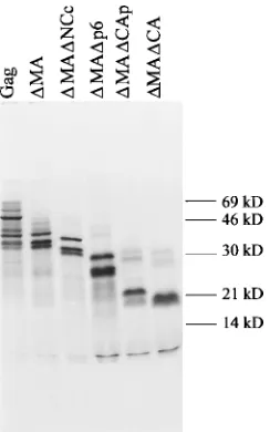
polyacrylamide–sodium dodecyl sulfate (SDS) gel (16), all translation products showed multiple bands (Fig. 2). The larg-est major band in each case migrates at a position consistent with the molecular mass of the expressed protein. DMA, DMADNCc, and DMADp6, all of which share the same N-terminal sequence, display an identical pattern of three bands, which is shifted according to the sizes of the proteins. The same pattern of three bands is also visible in the Pr55Gaglane. Therefore, a likely explanation for the multiple bands is that they represent proteins initiated at successive AUG codons on the same mRNA.
For RNA-protein binding reactions, optimized amounts of in vitro-transcribed RNA (3 pmol) and the unpurified in vitro transcription-translation reaction mixture (1 to 2ml) contain-ing the specific HIV-1 protein were incubated in a volume of 10ml. The actual amount of HIV-1-specific protein present in the binding reaction mixture is not known, but from the
[image:2.612.62.299.79.325.2]amount of trichloroacetic acid-precipitable [35S]methionine, it is estimated to be at least 100-fold-lower than the amount of RNA. The binding reaction mixture contained 10 mM HEPES (N-hydroxyethylpiperazine-N9-2-ethanesulfonic ac-id)-KOH (pH 7.2), 5 mM MgCl2, 2 mM dithiothreitol, and 1 mg of heparin per ml. The addition of up to 200 mM KCl or 10mM ZnCl2did not alter the detectable binding. After a 20-min incubation at room temperature, 1ml of 50% glycerol was added, and the entire binding reaction mixture was loaded
FIG. 1. In vitro-synthesized HIV-1-derived RNAs and Gag protein frag-ments. (A) Location of in vitro-transcribed RNAs on HIV-1 genome. Open boxes shown below the diagram of the 59end of the genome represent HIV-1-derived sequences, thin lines represent vector-HIV-1-derived stretches, and the arrow-heads indicate the orientation [sense (1) or antisense (2)] of RNA.p, before transcription, DNAs were linearized at the following restriction enzyme sites:
BssHII (EPHIV), EcoRI (BEHIV), AccI (BPHIV, ANHIV, and PPHIV), AlwNI
(PEHIV), and AvrII (HBgHIV). (B) Diagram of Gag deletion proteins ex-pressed in vitro. The Gag domains exex-pressed are shown as boxes: NC is indicated by the diagonally striped boxes, and the position of the Cys-His boxes within the NC domain is highlighted by solid black areas. pPA6gag was constructed by inserting the entire Pr55Gag
-coding region followed by the first 178 nucleotides of the overlapping protease-coding region and preceded by 6 nucleotides of the 59 untranslated sequence into the pSP64poly(A) vector (Promega). The 178 nucle-otides derived from the protease-coding region are not sufficient to allow ex-pression of an active protease (14). For construction of pDMA, pDMADCAp, and pDMADCA, HIV-1-derived sequences upstream of the PvuII site (position 694 in the HIV-1 sequence), the HindIII site (position 1259), and the PpuMI site (position 1399), respectively, were deleted. Deletion of the HIV-1-derived se-quences downstream of the BglII site (position 1642 in the HIV-1 sequence) and insertion of a 14-bp XbaI linker containing TAG stop codons in all three reading frames gave rise to pDMADp6. pDMADNCc was constructed by introducing an in-frame deletion from the blunt-ended ApaI to the filled-in BglII site (position 1553 to 1642 in the HIV-1 sequence). All deletion junctions were verified by dideoxy nucleotide sequencing by using the Sequenase version 2.0 kit from U.S. Biochemical Corp. (Cleveland, Ohio). aa, amino acids.
[image:2.612.316.555.93.195.2]FIG. 2. SDS-PAGE analysis of in vitro translation products. A total of 2.5ml of each 50ml of the in vitro transcription-translation reaction mixture pro-grammed with the plasmids indicated at the top of the lanes was loaded per lane; 2.5ml of the nonradioactive, prestained molecular marker (Rainbow marker; Bethesda Research Laboratories, Bethesda, Md.) was run in parallel. After gel electrophoresis, the dried gel was lined up with the autoradiogram, and the positions of the colored marker protein bands were transferred to the autora-diogram. Sample preparation and gel electrophoresis were conducted according to the method of Laemmli (16).
TABLE 1. Summary of binding ofDMA to in vitro-transcribed non-HIV-1-derived RNAs
RNA (length)a Plasmid usedb Bindingc
59261 nt of HIV-1 (282) pEPHIV (BssHII) 111
RSVC(271) pASY159 (HindIII) 111
b-Globin promoter (276) pSX (HgaI) 2 b-Galactosidase coding region
(222)
pEC1206 (MluI) 2 Neomycin resistance coding
region (318)
pBSneo (PvuII) 2 pSP64 vector sequence (266) pSP64poly(A) (PvuII) 1 pSP73 vector sequence (186) pSP73 (NdeI) 2
a
Total length in nucleotides, including vector-derived sequences. RSV, Rous sarcoma virus.
b
Restriction enzyme sites used to linearize plasmids for in vitro transcription are given in parentheses. pASY159 (provided by A. Yeo, Fred Hutchinson Cancer Research Center, Seattle, Wash.) contains nucleotides 357 to 628 of the Rous sarcoma virus sequence (GenBank accession no. J02342) inserted into pBSII (Stratagene, La Jolla, Calif.). pSX, which contains the SphI-XbaI fragment of the sickle allele of the humanb-globin gene inserted into pBS, was obtained from E. Epner and M. Groudine, Fred Hutchinson Cancer Research Center. pECL206, which carries the 339-bp TaqI fragment spanning the SstI site of the
lacZ gene inserted into pBS, was kindly provided by A. Geballe, Fred
Hutchin-son Cancer Research Center. pBSneo (provided by A. Yeo) has the neomycin gene inserted into pBSII.
cIn vitro binding reactions and gel electrophoresis were carried out as
de-scribed in the text.111, strong binding;1, weak binding;2, no binding.
on November 9, 2019 by guest
http://jvi.asm.org/
[image:2.612.376.498.456.651.2]onto a 5% polyacrylamide–5% glycerol gel in 45 mM Tris-borate–1 mM EDTA (pH 8.5). The gel was run for 3 h at 170 to 200 V and fixed, and the radioactivity was visualized by fluorography. All binding experiments were repeated four or five times, and the results were reproducible.
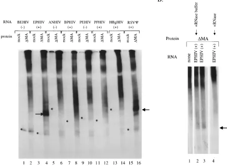
Initial binding reactions were carried out with a35S-labeled matrix-domain-deleted form of the Gag polyprotein,DMA. It has been previously shown that the MA domain is not required for RNA encapsidation into virions (17). Several RNAs de-rived from different regions within the 59proximal part of the HIV-1 genomic RNA were tested. As controls, the antisense RNAs corresponding to two of these regions were also in-cluded. All RNAs are named according to the plasmid DNAs from which they were transcribed, and their orientation is indicated by1(sense) or2(antisense) (Fig. 1A)3H-labeled RNA and 35S-labeled protein were combined in solution as described above, and binding was analyzed on a nondenaturing polyacrylamide gel (Fig. 3A). In the presence of a mock tran-scription-translation reaction mixture (no template DNA add-ed; odd-numbered lanes), all RNAs migrate as single bands (the positions are indicated by asterisks; the3H-labeled RNA bands cannot be visualized at this exposure of the gel). On the
[image:3.612.85.534.82.411.2]addition of a transcription-translation reaction mixture con-tainingDMA, a smear appears along the top of the lanes. This diffuse signal originates from the 35S-labeled protein, which does not run as a discretely sized band on a nondenaturing gel. The diffuse region of label may also represent binding to un-specific RNA or protein-protein complexes of various sizes. On the addition ofDMA to EPHIV1RNA (lane 4), a strong and discrete new band appears. In order to show that this band represents a complex between DMA and EPHIV1 RNA, RNase was added in a control experiment (Fig. 3B). While DMA by itself shows only a smear (Fig. 3B, lane 1), the addi-tion of EPHIV1RNA results in a new band (Fig. 3B, lanes 2 and 3), which disappears after treatment with RNase (Fig. 3B, lane 4). In contrast, no new bands are visible when DMA is added to BEHIV2, ANHIV1, PEHIV2, or PPHIV1RNA (Fig. 3A, lanes 2, 6, 10, and 12, respectively). The addition of DMA to BPHIV1 and HBgHIV1 RNAs (lanes 8 and 14, respectively), and in some experiments to PPHIV1 and PEHIV2RNAs (data not shown), also results in new bands. These signals, however, are significantly weaker and less de-fined than the one seen with EPHIV1RNA (lane 4). Thus, in our in vitro binding assay, an MA-domain-deleted form of
FIG. 3. In vitroDMA Gag protein-HIV-1 RNA binding assay. (A) Each transcription-translation reaction mixture (2ml) containing no HIV-1 protein (mock; odd-numbered lanes) or35S-labeledDMA Gag protein (even-numbered lanes) was added to 3 pmol of3H-labeled RNA in binding buffer (see the text). The RNAs
used, as indicated above the lanes, are described in the legend to Fig. 1. Lanes 15 and 16, ASY159 (HindIII) containing the Rous sarcoma virusCsite. Asterisks indicate the locations of the RNA band, in the lanes labeled mock, determined from a longer exposure of this autoradiograph. The arrows point to the major shifted protein-RNA complexes. All lanes illustrated are from the same gel. (B) A transcription-translation reaction mixture (2ml) containing35S-labeledDMA protein was
added to binding buffer (see the text) containing no HIV-1-specific RNA (lane 1) or 3 pmol of3H-labeled EPHIV1RNA (lanes 2 to 4). After incubation for 20 min
at room temperature, 1ml of either 10 mM Tris (pH 7.6)–15 mM NaCl containing 1mg of RNase A (lane 4) or RNase buffer alone (lane 2) was added, and the incubation continued for 30 min at room temperature before the gel was loaded. The arrow points to the major labeled complex. All lanes illustrated are from the same gel.
on November 9, 2019 by guest
http://jvi.asm.org/
Pr55Gag,DMA, shows the strongest binding to EPHIV1RNA, which contains the 59 261 nucleotides of the HIV-1 genomic RNA. No binding to the corresponding negative-strand RNA, BEHIV2, or a number of RNAs from other regions of the HIV-1 genome is seen. However, in addition to the 59 261 nucleotides, several other regions of the HIV-1 RNA also bound to the Gag fragment, although not as consistently, and the complexes formed were more diffuse. One of these RNAs (BPHIV1) covers the region around the start codon of the Gag protein, which contains sequences previously shown to be important for binding to Gag protein and for packaging (2, 6, 18, 21, 22). A survey of the literature suggests that there may be more than one region of the HIV-1 genome which is in-volved in RNA packaging. There is not as yet any evidence for a single region of HIV-1 RNA which is sufficient to allow encapsidation of heterologous RNA, as is the case for oncoret-roviruses (1, 3). It may be that several regions of the HIV-1 RNA must combine to form a complex C structure or that several independentCelements may have to be bound by one or more Gag proteins before assembly of the viral capsid and concomitant packaging of the genomic RNA are triggered.
To further explore the specificity of the in vitro binding of DMA to RNA, we tested a number of unrelated RNAs (sum-marized in Table 1). No binding to RNAs transcribed from the promoter region of the humanb-globin gene, from the coding region of the b-galactosidase gene or the neomycin gene, or from the pSP73 plasmid used for cloning the HIV-1 RNAs was detected. However, there was strong binding to an RNA con-taining the packaging region of Rous sarcoma virus (3) (also shown in Fig. 3A, lane 16). Berkowitz et al. (5) also found binding of a bacterially expressed Gag protein to packaging regions from bovine leukemia virus and Mason-Pfizer monkey virus. These findings indicate that some general feature of the Cstructure is conserved among retroviruses and that this fea-ture is recognized in in vitro binding assays. We also detected weaker but significant binding to RNA transcribed from the pSP64poly(A) plasmid. It is not possible to say whether this RNA forms a structure similar to that of HIV-1Cor whether this result reflects some lack of specificity in our assay.
It was difficult to examine the binding properties of the full-length Gag protein in our assay system since the addition of EPHIV1RNA did not change the pattern of bands already seen with the protein alone (data not shown). It is likely that the full-length Gag protein binds to its own mRNA in the in vitro transcription-translation reaction, which at its 59end con-tains sequences previously implicated in specific binding to Pr55Gag(21, 22).
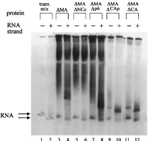
In order to determine the domains of theDMA Gag protein which contribute to binding specificity, additional constructs encoding deleted forms of the protein were generated. These derivatives of HIV-1 Gag lack the MA and p6 proteins (DMADp6), MA and part of the CA proteins (DMADCAp), MA and the entire CA domains (DMADCA), and MA protein and the region of the NC protein encompassing the second Cys-His box and most of the basic region in between the two Cys-His boxes (DMADNCc; see Fig. 1B).DMA, DMADNCc, DMADp6,DMADCAp, andDMADCA were incubated in the presence of either BEHIV2 or EPHIV1 RNA under stan-dard binding conditions and analyzed on a nondenaturing gel (Fig. 4). DMADCAp and DMADCA do not show the back-ground smear seen with the other Gag deletion proteins, pos-sibly because in these two smallest proteins tested, the basic nature of the NC domain becomes more apparent and pre-vents migration of the proteins into the gel.
In the presence of sense EPHIV1 RNA, the addition of DMA (Fig. 4, lane 4),DMADp6 (lane 8),DMADCAp (lane 10),
orDMADCA (lane 12) gives rise to a fairly discrete, shifted band. The size of the shift decreases fromDMA toDMADp6 to DMADCAp andDMADCA, consistent with the decreasing size of the protein binding to the RNA. No binding is detected with DMADNCc (Fig. 4, lane 6) or with a deletion construct lacking the first Cys-His box as well as part of the internal basic region of the NC protein (data not shown). For both DMA and DMADp6, a new 35S-containing band appears only on the addition of EPHIV1RNA but not BEHIV2RNA. However, in the case ofDMADCAp andDMADCA, a weaker but distinct band is also visible with BEHIV2 RNA. DMADCAp and DMADCA thus display less-specific binding to HIV-1 RNA than doDMA andDMADp6, and the CA domain must there-fore contribute to the specificity of RNA binding. Since no independent RNA binding could be demonstrated for the CA domain (see below), CA probably acts by increasing the spec-ificity of binding of the NC domain. A contribution of the CA domain to the specificity of RNA binding would meet the requirements of virus assembly in vivo. During infection, the HIV-1 RNA must first be specifically selected from the pool of intracellular RNAs by the Gag precursor, while after protease cleavage in the virion, the entire viral RNA must be sequence independently coated by the NC protein.
[image:4.612.315.556.70.301.2]Several studies regarding the specific RNA binding activity of the NC domain in vitro have been published (7, 19). In only one study, however, was RNA binding of the NC compared with that of a Gag protein also containing the CA domain (5). Using bacterially expressed glutathione S-transferase (GST) fusion proteins, these investigators found comparable specific RNA binding activities for the full-length Gag and the NC proteins. However, only the GST fusion proteins and not the
FIG. 4. In vitro binding of various Gag deletion proteins to 59end of HIV-1 RNA. BEHIV2(lane 1) or EPHIV1RNA (lane 2) in binding buffer (see the text) was incubated with 2ml of transcription-translation reaction mixture con-taining no HIV-1 protein (trans. mix). To test the binding specificity of the Gag deletion proteins, 2ml of the translation mix containingDMA (lanes 3 and 4), DMADNCc (lanes 5 and 6),DMADp6 (lanes 7 and 8), orDMADCAp (lanes 9 and 10) or 1ml of the translation mixture containingDMADCA together with 1 ml of the mock reaction mixture (lanes 11 and 12) was added to EPHIV1RNA (even-numbered lanes) or BEHIV2RNA (odd-numbered lanes). The two ar-rows indicate the locations of the RNA only bands (top arrow shows BEHIV2 and the bottom arrow EPHIV1). All lanes illustrated were from the same gel.
on November 9, 2019 by guest
http://jvi.asm.org/
cleaved fragments were tested for their RNA binding activity. It is conceivable that the GST domain acts much like the CA domain in altering the conformation of the NC domain to confer specificity on RNA binding. It should also be noted that all of these investigators used different regions of the HIV-1 RNA as specific binding substrates.
A Gag fragment with deletions of the entire MA domain and either the first or the second Cys-His box as well as part of the basic region of the NC domain showed no RNA binding ac-tivity in our assay. This result confirms the importance of the Cys-His boxes as well as the basic region between those two boxes within NC, which has been well documented in the literature (2, 5, 8, 10, 11, 27). It also allows the conclusion that neither the CA nor the p6 domain, in the context of the partially deleted NC domain, has independent RNA binding activity strong enough to be detected in our assay system. With a different in vitro binding system, it was reported that there is residual specific RNA binding activity of a GST-Gag fusion protein deleted in either the first or the second Cys-His box of the NC domain (5). However, these investigators also reported a sharp, nonlinear drop in RNA-protein complex formation, when the protein concentration was lowered. In our assay, the Gag protein concentration is probably very low due to its expression in an in vitro translation system. Therefore, it is not surprising that in our assay, a Gag fragment carrying a deletion in part of the NC domain shows no RNA binding activity at all. In summary, our in vitro binding assay confirms that the MA and p6 regions of Gag are not required for binding to RNA and that the Cys-His box and basic region of NC are essential for binding to RNA. In addition, we show that deletion of the CA domain decreases the specificity of binding. The results presented here suggest that the 59end of the HIV-1 genome contains information which is important for the association with the viral Gag protein and therefore may be involved in genomic encapsidation. This region could act in concert with other regions of the genome to ensure efficient encapsidation of genomic RNA.
We thank Michael Emerman, Andrei Tikhonenko, and Mark Roth for comments on the manuscript and members of our laboratory for critical assessment of the data. We are particularly indebted to Peggy P. Lee for input in design of the RNA probes. T7 RNA polymerase was a gift from B. Lewicki, Max-Planck-Institut fu¨r Molekulare Genetik, Berlin, Germany.
This work was supported by grants AI 27291 and CA 18282 from the NIH to M.L.L.
REFERENCES
1. Adam, M., and A. D. Miller. 1988. Identification of a signal in a murine retrovirus that is sufficient for packaging of nonretroviral RNA into virions. J. Virol. 62:3802–3806.
2. Aldovini, A., and R. A. Young. 1990. Mutations of RNA and protein se-quences involved in human immunodeficiency virus type 1 packaging results in production of noninfectious virus. J. Virol. 64:1920–1926.
3. Aronoff, R., A. M. Hajjar, and M. L. Linial. 1993. Avian retroviral RNA encapsidation: reexamination of functional 59RNA sequences and the role of nucleocapsid Cys-His motifs. J. Virol. 67:178–188.
4. Berkowitz, R. D., and S. P. Goff. 1994. Analysis of binding elements in the human immunodeficiency virus type 1 genomic RNA and nucleocapsid pro-tein. Virology 202:233–246.
5. Berkowitz, R. D., J. Luban, and S. P. Goff. 1993. Specific binding of human immunodeficiency virus type 1 Gag polyprotein and nucleocapsid protein to viral RNAs detected by RNA mobility shift assays. J. Virol. 67:7190–7200. 6. Clavel, F., and J. M. Orenstein. 1990. A mutant of human immunodeficiency
virus with reduced RNA packaging and abnormal particle morphology. J. Virol. 64:5230–5234.
7. Dannull, J., A. Surovoy, G. Jung, and K. Moelling. 1994. Specific binding of HIV-1 nucleocapsid protein to PSI RNA in vitro requires N-terminal zinc finger and flanking basic amino acid residues. EMBO J. 13:1525–1533.
8. Dorfman, T., J. Luban, S. P. Goff, W. A. Haseltine, and H. G. Gottlinger. 1993. Mapping of functionally important residues of a cysteine-histidine box in the human immunodeficiency virus type 1 nucleocapsid protein. J. Virol.
67:6159–6169.
9. Geigenmu¨ller-Gnirke, U., H. Nitschko, and S. Schlesinger.1993. Deletion analysis of the capsid protein of Sindbis virus: identification of the RNA binding region. J. Virol. 67:1620–1626.
10. Gorelick, R. J., D. J. Chabot, A. Rein, L. E. Henderson, and L. O. Arthur. 1993. The two zinc fingers in the human immunodeficiency virus type 1 nucleocapsid protein are not functionally equivalent. J. Virol. 67:4027–4036. 11. Gorelick, R. J., S. M. Nigida, Jr., J. W. Bess, Jr., L. O. Arthur, L. E.
Henderson, and A. Rein.1990. Noninfectious human immunodeficiency vi-rus type 1 mutants deficient in genomic RNA. J. Virol. 64:3207–3211. 12. Kaplan, A. H., M. Manchester, and R. Swanstrom. 1994. The activity of the
protease of human immunodeficiency virus type 1 is initiated at the mem-brane of infected cells before the release of viral proteins and is required for release to occur with maximal efficiency. J. Virol. 68:6782–6786.
13. Katz, R. A., R. W. Terry, and A. M. Skalka. 1986. A conserved cis-acting sequence in the 59 leader of avian sarcoma virus RNA is required for packaging. J. Virol. 59:163–167.
14. Krausslich, H.-G., H. Schneider, G. Zybarth, C. A. Carter, and E. Wimmer. 1988. Processing of in vitro-synthesized gag precursor proteins of human immunodeficiency virus (HIV) type 1 by HIV proteinase generated in
Esch-erichia coli. J. Virol. 62:4393–4397.
15. Krieg, P. A., and D. A. Melton. 1984. Functional messenger RNAs are produced by SP6 in vitro transcription of cloned cDNAs. Nucleic Acids Res.
12:7057–7070.
16. Laemmli, U. K. 1970. Cleavage of structural proteins during the assembly of the head of bacteriophage T4. Nature (London) 227:680–685.
16a.Lee, P. P., and M. Linial. Unpublished data.
17. Lee, P. P., and M. L. Linial. 1994. Efficient particle formation can occur if the matrix domain of human immunodeficiency virus type 1 Gag is substituted by a myristylation signal. J. Virol. 68:6644–6654.
18. Lever, A., H. Gottlinger, W. Haseltine, and J. Sodroski. 1989. Identification of a sequence required for efficient packaging of human immunodeficiency virus type 1 RNA into virions. J. Virol. 63:4085–4087.
19. Li, X., Z. Gu, R. Geleziunas, L. Kleiman, M. A. Wainberg, and M. A.
Parniak.1993. Expression, purification, and RNA-binding properties of HIV-1 p15gag nucleocapsid protein. Protein Expr. Purif. 4:304–311. 20. Linial, M. L., and A. D. Miller. 1990. Retroviral RNA packaging: sequence
requirements and implications. Curr. Top. Microbiol. Immunol. 157:125– 152.
21. Luban, J., and S. P. Goff. 1991. Binding of human immunodeficiency virus type 1 (HIV-1) RNA to recombinant HIV-1 Gag polyprotein. J. Virol.
65:3203–3212.
22. Luban, J., and S. P. Goff. 1994. Mutational analysis of cis-acting packaging signals in human immunodeficiency virus type 1 RNA. J. Virol. 68:3784– 3793.
23. Meric, C., E. Gouilloud, and P.-F. Spahr. 1988. Mutations in Rous sarcoma virus nucleocapsid protein p12 (NC): deletions of Cys-His boxes. J. Virol.
62:3328–3333.
24. Milligan, J. F., D. R. Groebe, G. W. Witherell, and O. C. Uhlenbeck. 1987. Oligoribonucleotide synthesis using T7 RNA polymerase and synthetic DNA templates. Nucleic Acids Res. 15:8783–8798.
25. Oroszlan, S., and R. B. Luftig. 1990. Retroviral proteinases, p. 153–186. In R. Swanstrom and P. K. Vogt (ed.), Retroviruses: strategies of replication. Springer-Verlag, Berlin.
26. Peden, K., M. Emerman, and L. Montagnier. 1991. Changes in growth properties on passage in tissue culture of viruses derived from infectious molecular clones of HIV-1LAI, HIV-1MAL, and HIV-1ELI. Virology 185: 661–672.
27. Sakaguchi, K., N. Zambrano, E. T. Baldwin, B. A. Shapiro, J. W. Erickson,
J. G. Omichinski, G. M. Clore, A. M. Gronenborn, and E. Appella.1993. Identification of a binding site for the human immunodeficiency virus type 1 nucleocapsid protein. Proc. Natl. Acad. Sci. USA 90:5219–5223.
28. Shank, P. R., and M. Linial. 1980. Avian oncovirus mutant (SE21Q1b) deficient in genomic RNA: characterization of a deletion in the provirus. J. Virol. 36:450–456.
29. Sorge, J., D. Wright, V. D. Erdman, and A. E. Cutting. 1984. Amphotropic retrovirus vector system for human cell gene transfer. Mol. Cell. Biol.
4:1730–1737.
30. Stoker, A. W., and M. J. Bissell. 1988. Development of avian sarcoma and leukosis virus-based vector-packaging cell lines. J. Virol. 62:1008–1015. 31. Surovoy, A., J. Dannull, K. Moelling, and G. Jung. 1993. Conformational
and nucleic acid binding studies on the synthetic nucleocapsid protein of HIV-1. J. Mol. Biol. 229:94–104.
32. Watanabe, S., and H. M. Temin. 1982. Encapsidation sequences for spleen necrosis virus, an avian retrovirus, are between the 59long terminal repeat and the start of the gag gene. Proc. Natl. Acad. Sci. USA 79:5986–5990.