Copyright © 1999, American Society for Microbiology. All Rights Reserved.
Pathogenicity of Different Rabies Virus Variants Inversely
Correlates with Apoptosis and Rabies Virus Glycoprotein
Expression in Infected Primary Neuron Cultures
KINJIRO MORIMOTO,
1D. CRAIG HOOPER,
1SERGEI SPITSIN,
2HILARY KOPROWSKI,
1AND
BERNHARD DIETZSCHOLD
1*
Center for Neurovirology, Department of Microbiology and Immunology,
1and Biotechnology Foundation
Laboratories,
2Thomas Jefferson University, Philadelphia, Pennsylvania 19107-6799
Received 30 July 1998/Accepted 23 September 1998
The mouse-adapted rabies virus strain CVS-24 has stable variants, CVS-B2c and CVS-N2c, which differ
greatly in their pathogenicity for normal adult mice and in their ability to infect nonneuronal cells. The
glycoprotein (G protein), which has previously been implicated in rabies virus pathogenicity, shows substantial
structural differences between these variants. Although prior studies have identified antigenic site III of the G
protein as the major pathogenicity determinant, CVS-B2c and CVS-N2c do not vary at this site. The possibility
that pathogenicity is inversely related to G protein expression levels is suggested by the finding that CVS-B2c,
the less pathogenic variant, expresses at least fourfold-higher levels of G protein than CVS-N2c in infected
neurons. Although there is some difference between CVS-B2c- and CVS-N2c-infected neurons in G protein
mRNA expression levels, the differential expression of G protein appears to be largely determined by
post-translational mechanisms that affect G protein stability. Pulse-chase experiments indicated that the G protein
of CVS-B2c is degraded more slowly than that of CVS-N2c. The accumulation of G protein correlated with the
induction of programmed cell death in CVS-B2c-infected neurons. The extent of apoptosis was considerably
lower in CVS-N2c-infected neurons, where G protein expression was minimal. While nucleoprotein (N protein)
expression levels were similar in neurons infected with either variant, the transport of N protein into neuronal
processes was strongly inhibited in CVS-B2c-infected cells. Thus, downregulation of G protein expression in
neuronal cells evidently contributes to rabies virus pathogenesis by preventing apoptosis and the apparently
associated failure of the axonal transport of N protein.
Accumulating evidence indicates that the rabies virus
glyco-protein (G glyco-protein) plays an essential role in the pathogenicity
of the virus. For example, the pathogenicity of the tissue
cul-ture-adapted rabies virus strains ERA, HEP, and CVS has
been shown to correlate with the presence of a determinant
located in antigenic site III of the G protein (5, 18). Variants
with an Arg
3
Glu mutation at position 333 in site III of the G
protein are less pathogenic for immunocompetent adult mice
than the wild-type parental viruses (5, 18). The greatly reduced
spread of these antigenic site III mutants within the nervous
system (6) indicates that the appropriate G protein structure is
absolutely essential for the rapid axonal/transsynaptic spread
of rabies virus that leads to a lethal infection in adult animals.
Based on these findings, one might expect that site III
muta-tions are the single most important indicator of decreased
pathogenicity. However, by comparison with street rabies virus
strains, rabies virus strains adapted to nonneuronal cells in
vitro, such as ERA, are already highly attenuated with respect
to pathogenicity for immunocompetent mammals (15), despite
having an unchanged antigenic site III. Thus, the pathogenicity
phenotype of a particular rabies virus strain is determined not
only by the determinants located in antigenic site III of the G
protein but also by other mechanisms which may or may not be
related to the G protein. We recently found that the mouse
brain-adapted CVS-24 strain of rabies virus consists of two
phenotypically and genotypically distinct variants, CVS-B2c
and CVS-N2c (16). The amino acid sequences of the
nucleo-proteins (N nucleo-proteins) of both variants are identical, whereas
the G proteins of CVS-B2c and CVS-N2c differ in 10 amino
acids outside antigenic site III. While CVS-N2c is the
domi-nant variant when CVS-24 is passaged in mouse brain or
neu-roblastoma cells, passage in baby hamster kidney (BHK-21)
cells results in rapid selection of the B2c variant.
N2c replicates more readily in neuronal cells, whereas
CVS-B2c replicates better in nonneuronal cells. For normal adult
mice, the pathogenicity index (defined as the 50% lethal dose
of a particular virus stock preparation divided by the virus
titer) of N2c is up to 50 times higher than that of
CVS-B2c, depending on the route of inoculation (16). The disparity
in the pathogenicities of these two variants must therefore
depend either on differences outside site III in the G protein or
on other factors that determine the capacity of the virus to
invade, replicate, spread in neuronal tissue, and produce
neu-rological disease.
In the present study, we used variants N2c and
CVS-B2c as probes to examine the mechanisms involved in rabies
virus neuropathogenicity. We demonstrated that G protein
expression in infected primary neuron cultures is different
be-tween the two variants and that the downregulation of G
pro-tein expression in neurons is associated with increased
patho-genicity.
MATERIALS AND METHODS
Cells and viruses.NA neuroblastoma cells of A/J mouse origin were grown at 37°C in Eagle’s minimum essential medium supplemented with 10% heat-inac-tivated fetal bovine serum (FBS). Primary neuron cultures were prepared from the hippocampi of prenatal Swiss Webster mice (day 17 of gestation) as
de-* Corresponding author. Mailing address: Center for Neurovirology,
Department of Microbiology and Immunology, Thomas Jefferson
Uni-versity, 1020 Locust St., Philadelphia, PA 19107-6799. Phone: (215)
503-4692. Fax: (215) 923-7145. E-mail: bdietzschold@reddi1.uns.tju
.edu.
510
on November 9, 2019 by guest
http://jvi.asm.org/
scribed previously (2). Briefly, the isolated hippocampi were treated for 15 min at 37°C with 4 ml of 0.04% trypsin. After the addition of 5 ml of Dulbecco’s minimal essential medium (GIBCO BRL, Grand Island, N.Y.) and 1 ml of FBS, cells were dispersed with a pipette with a heat-polished tip and strained through a Falcon cell strainer. Cells were layered on a 2-ml FBS cushion and centrifuged for 5 min at 1,000 rpm, and the pelleted cells were resuspended in Neurobasal medium (GIBCO BRL) at a concentration of 53105cells/ml. Cells (250ml) were then plated on 18-mm round polylysine-coated coverslips and incubated in a 5% CO2atmosphere for 1 h at 37°C, and the coverslips were placed on a layer of astrocytes isolated from the brains of 2-day-old Swiss Webster mice and cultured in 10-cm-diameter petri dishes as described previously (4). The primary neurons grown on coverslips were more than 95% pure, as judged by size, morphology, and immunostaining for the panneuronal marker protein PGP9.5. After 2 days of culturing in Neurobasal medium, neurons were infected with the CVS-24 variants CVS-N2c and CVS-B2c at a multiplicity of infection (MOI) of 1, which resulted in infection of more than 90% of the neurons within 24 h postinfection (p.i.), as determined by staining with N protein-specific antibodies. Isolation and characterization of these variants have been described elsewhere (16). Because CVS-B2c replicates better in nonneuronal cells than CVS-N2c and the latter does not cause plaques in nonneuronal cells, the titers of both viruses were determined with mouse neuroblastoma cells and the fluorescent-focus assay.
Immunofluorescence analysis.Neurons were fixed in 80% acetone at 24, 48, and 72 h p.i. To detect N protein, fixed neuron cultures were subjected to a direct fluorescence staining technique with fluorescein isothiocyanate (FITC)-labeled anti-rabies virus N protein-specific mouse monoclonal antibody (MAb) (Cento-cor, Malvern, Pa.) as described previously (7). To detect G protein, fixed neuron cultures were incubated with a monospecific rabbit antibody that recognizes rabies virus G protein, and reactions were visualized with FITC-labeled goat anti-rabbit immunoglobulin G (Sigma, St. Louis, Mo.). Evans blue was used as a counterstain and appeared red under green fluorescence.
Immunoprecipitation.Primary mouse neuron cultures were infected with CVS-B2c or CVS-N2c at an MOI of 1. At 2, 24, or 48 h p.i., the culture medium was replaced with methionine-free Neurobasal medium containing 10 mCi of [35S]methionine/ml, and incubation was continued for 24 h at 37°C. Cells were then lysed with lysis buffer (10 mM Tris-HCl [pH 7.4], 150 mM NaCl, 1% Triton X-100, 0.5% sodium deoxycholate), protein content was determined, and lysates were calibrated to the same protein concentration (400mg/ml). Two micrograms of G protein-specific MAb 6-15c (kindly provided by A. Osterhaus, Institute of Virology, Erasmus University Hospital, Rotterdam, The Netherlands), 2mg of N protein-specific MAb 377-7 (7), or 2mg of actin-specific antibody was added to 400ml of lysate, and the mixtures were incubated overnight at 4°C. The resulting immune complexes were adsorbed to protein A-Sepharose beads. After the beads were washed with lysis buffer containing 0.45% sodium dodecyl sulfate (SDS), the precipitated proteins were solubilized in a small volume of SDS sample buffer and subjected to electrophoresis on an SDS–10% polyacrylamide gel. The gel was dried and exposed to X-ray film, and protein bands were quantitated by densitometry with the NIH 1.58 IMAGE program.
Pulse-chase analysis.Primary neuron cultures were infected with CVS-B2c or CVS-N2c at an MOI of 1. At 24 h p.i., the cells were pulse-labeled for 1 h with 50mCi of [35S]methionine/ml. The medium was then replaced with Neurobasal medium containing 2 mM unlabeled methionine. After 2, 6, and 12 h of chase with unlabeled methionine, neurons were lysed, and the lysates were subjected to immunoprecipitation with G or N protein-specific MAbs. Proteins were analyzed by SDS-polyacrylamide gel electrophoresis as described above.
RNA extraction and Northern blot analysis.Total RNA was isolated from infected neuron cultures by the RNAzol B method (Biotex Laboratories, Inc., Houston, Tex.). For Northern blot analysis, aliquots containing 7.5mg of RNA were electrophoresed on a 1% agarose gel containing 2.2 M formaldehyde–0.1 M morpholinepropanesulfonic acid (MOPS) buffer (pH 7.0), blotted on Nytran nylon membranes, and hybridized with nick-translated [a-32P]dCTP-labeled cloned N or G protein cDNA as described previously (16). Membranes were exposed to X-ray film, and RNA bands were quantitated by densitometry with the NIH 1.58 IMAGE program.
Quantitative PCR.Absolute and relative amounts of G and N protein mRNAs produced in CVS-N2c- and CVS-B2c-infected neurons were also assessed by the real-time quantitative PCR method (10). Reverse transcription (RT) reactions were carried out with 2.5mg of total RNA and 1mM reverse primer as described previously (16). Serial dilutions of the RT products were then subjected to PCR amplification with forward and reverse primers and a dual-fluorescence dye-labeled probe (FAM-TAMRA). The nucleotide sequences of the probes and primers were as follows: N-reverse primer, 59-TCATCAGAGTTGACGGTTCC G-39; G-reverse primer, 59-TTGATTCATGTCGAGTCCGCT-39; G3PDH-re-verse primer: 59-AGATGGTGATGGGCTTCCC-39; N-forward primer, 59-CA AGAATATGAGGCGGCTGAA-39; G-forward primer, 59-GATGACTCTGTG CTTGGGCA-39; G3PDH-forward primer, 59-GGCAAATTCAACGGCACAG T-39; FAM-TAMRA dual-labeled N-probe, 59-AAAGTCCGACGTGGCACTG GCAGA-39; FAM-TAMRA dual-labeled G-probe, 59-AAAGAGGTCGTAGT GTGCCCCCCGA-39; and FAM-TAMRA dual-labeled G3PDH-probe, 59-AG GCCGAGAATGGGAAGCTTGTCATC-39.
The sequences of the N and G mRNA-specific probes and primers are con-served between CVS-N2c and CVS-B2c. Amplification of
glyceraldehyde-3-phosphate dehydrogenase (G3PDH) mRNA served as an internal standard. Amplification was carried out for 40 cycles of denaturation at 95°C for 15 s, annealing, and polymerization at 60°C for 1 min with a TaqMan EZ RT-PCR kit (The Perkin-Elmer Corp.). PCR products were detected with the ABI Prism 7700 Sequence Detection System. Standard curves were constructed by plotting CTvalues (points at which the amplification blot crosses the threshold) against log nanogram amounts of total input RNA. Based on the slope of the standard curves, the total amount of a particular mRNA species was calculated as x5(b2 y)/m, where x is the total amount of mRNA, b is the intercept at log 0 ng of RNA, and m is the slope of the standard curves. For y, a CTvalue of 20 was arbitrarily chosen. To determine the normalized amounts, the relative quantities (recipro-cals of the total amounts) of G and N protein mRNAs were divided by the relative quantity of G3PDH mRNA.
In situ terminal end labeling.The terminal deoxynucleotidyltransferase-me-diated dUTP nick end-labeling (TUNEL) method was used to detect DNA strand breaks indicative of apoptotic cell death. At different times after infection, neurons grown on round coverslips were treated with 4% paraformaldehyde in phosphate-buffered saline (PBS) (pH 7.4) for 30 min at room temperature and washed with PBS. A TUNEL assay with propidium iodide counterstain (red-orange) was performed with an Apoptosis Detection System, Fluorescein, in accordance with the manufacturer’s protocol (Promega, Madison, Wis.). Cover-slips were mounted with the antifading mounting medium Vectashield (Vector Laboratories) on microslides and examined on a Leitz Microlab microscope under visible and UV illumination.
Annexin V assay.Primary neuron cultures grown on round glass coverslips were infected with the rabies virus variants and, at 24, 48, and 72 h p.i., washed with 0.1 M HEPES (pH 7.2) and incubated for 10 min with FITC-labeled Annexin V and for 2 min with propidium iodide (APOPTEST-FITC kit; NeXins Research, B. V., Hoeve, The Netherlands) in accordance with the manufactur-er’s protocol. Cells were washed again with HEPES buffer and fixed with 4% paraformaldehyde in PBS (pH 7.4) for 30 min at room temperature. Coverslips were mounted with Vectashield on microslides and examined on a fluorescence microscope with the standard setting for FITC fluorescence detection.
RESULTS
Expression of the rabies virus N and G proteins in primary
neuron cultures.
Neurons were cultured for 2 days in
Neuro-basal medium and infected with variant CVS-N2c or CVS-B2c
virus at an MOI of 1, which results in optimal infection (more
than 90% of the neurons at 24 h p.i.). Immunofluorescence
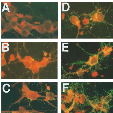
analysis of G protein expression in neurons at 24, 48, and 72 h
p.i. revealed intense staining in cell bodies and processes of
neurons infected with the CVS-B2c variant (Fig. 1D to F).
Much less staining was detected in neurons infected with
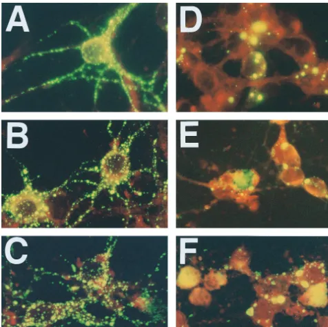
CVS-N2c (Fig. 1A to C). A similar analysis of N protein expression
revealed intense staining in all infected neurons, regardless of
the virus variant used for infection (Fig. 2). However,
CVS-N2c-infected neurons (Fig. 2A to C) showed a constant fine
granular staining pattern which appeared to be more
concen-trated near the cell surface and which extended into the
neu-ronal processes, whereas CVS-B2-infected neurons (Fig. 2D to
F) showed large, discrete N protein-positive inclusion bodies in
the cell body cytoplasm and almost no N protein-specific
stain-ing in neuronal processes for the first 48 h of culturstain-ing. At 72 h
p.i., when CVS-B2c-infected neurons had begun to degenerate,
large amorphous masses stained positively for the N protein.
These data suggest that the N protein is transported into the
processes of neurons infected with CVS-N2c but is retained in
the cell bodies of CVS-B2c-infected neurons, with minimal
peripheral translocation into neuronal processes.
Immunoprecipitation analysis of lysates from primary mouse
neuron cultures infected with CVS-N2c or CVS-B2c (MOI, 1)
indicated that, while similar amounts of the N protein were
produced in both cultures between 2 and 24 h, 24 and 48 h, and
48 and 72 h p.i., significantly more G protein was produced in
CVS-B2c-infected neurons than in CVS-N2c-infected neurons
throughout the period studied (Fig. 3A). The G/N protein ratio
was approximately fourfold higher in CVS-B2c-infected
neu-rons than in their CVS-N2c-infected counterparts (Fig. 3B).
Similar results were obtained with monoclonal and polyclonal
antibodies against the N or G protein (data not shown). These
V
OL. 73, 1999
NEUROPATHOGENESIS OF RABIES VIRUS
511
on November 9, 2019 by guest
http://jvi.asm.org/
data suggest that G protein but not N protein expression is
differentially regulated in primary neuron cultures infected
with CVS-N2c or CVS-B2c. The differences in the levels of
expression of the G protein relative to those of the N protein
between CVS-B2c and CVS-N2c appeared to be dependent
upon the nature of the cells infected. In the experiments
pre-sented here, G/N protein ratios in neuroblastoma cells infected
with CVS-B2c or CVS-N2c did not differ significantly (Fig. 4).
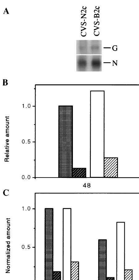
Analysis of rabies virus mRNA expression in neuronal cells.
Northern blot analysis to determine the relative mRNA levels
of the G and N proteins in primary neuron cultures infected
with CVS-N2c or CVS-B2c indicated that slightly more N
mRNA and G mRNA was produced in CVS-B2c-infected
neu-rons than in CVS-N2c-infected neuneu-rons at 48 h p.i. (Fig. 5A),
but little difference between the two variants in their G/N
mRNA ratios (0.13 for CVS-N2c and 0.22 for CVS-B2c) (Fig.
5B) was seen. Real-time quantitative PCR (10), also used to
determine the G and N mRNA levels in infected primary
neuron cultures, revealed G/N mRNA ratios of 0.19 for
CVS-N2c and 0.26 for CVS-B2c at 48 h p.i., as calculated from the
relative quantities of G and N mRNAs normalized to the
relative quantity of G3PDH mRNA (Fig. 5C). Essentially
iden-FIG. 1. Immunofluorescence analysis of G protein expression in primary neuron cultures infected with CVS-N2c (A, B, and C) or CVS-B2c (D, E, and F) virus and examined at 24 (A and D), 48 (B and E), and 72 (C and F) h p.i. Hippocampal neurons isolated from prenatal mouse brains (day 17 of gestation) were seeded on polylysine-coated glass coverslips, which were then placed on top of a cell layer of mouse astrocytes. After 2 days of culturing in Neurobasal medium, neurons were infected at an MOI of 1 and subjected to immunofluorescence analysis as described in Materials and Methods.on November 9, 2019 by guest
http://jvi.asm.org/
[image:3.612.63.545.68.549.2]tical ratios were obtained from mRNA amounts normalized to
the amount of 18S rRNA (data not shown). Thus, both
quan-titative PCR analysis and Northern blot analysis revealed that
G/N mRNA ratios in CVS-B2c-infected neurons were only
slightly higher than those in CVS-N2c-infected neurons. This
finding contrasts with the protein expression data, which
dem-onstrated at least fourfold-higher G protein expression levels
in CVS-B2c-infected neurons than in CVS-N2c-infected
neu-rons. Together, these results suggest that differences in
trans-lational regulation or posttranstrans-lational processing account, in
large part, for the differences in G protein expression levels.
Stability of rabies virus G and N proteins in cultured
neu-rons infected with CVS-B2c and CVS-N2c.
Pulse-chase analysis
to determine whether differences in G protein stability might
underlie differences in G protein expression in
CVS-B2c-in-fected versus CVS-N2c-inCVS-B2c-in-fected neurons indicated rapid
deg-radation of the G protein from both variants (Fig. 6A) but a
faster decline of the G proteins from CVS-N2c than from
CVS-B2c. Interestingly, the rates of decay of both G proteins
slowed at 2 h after synthesis, with no further decay seen after
6 h (Fig. 6B). However, the difference in the percentages of G
protein that decayed in CVS-B2c-infected versus
CVS-N2c-infected neurons was sufficient to result in higher levels of
stable G protein in CVS-B2c-infected neurons, consistent with
the immunofluorescence and immunoprecipitation data. The
N protein was relatively stable after synthesis in both infected
cultures (Fig. 6A).
G protein expression, N protein transport, and the
induc-tion of apoptosis in cultured neurons infected with rabies
[image:4.612.67.543.71.545.2]viruses CVS-B2c and CVS-N2c.
Because the distributions of
FIG. 2. Immunofluorescence analysis of N protein expression in primary neuron cultures infected with CVS-N2c (A, B, and C) or CVS-B2c (D, E, and F) virus. Cells were stained at 24 (A and D), 48 (B and E), and 72 (C and F) h p.i.
V
OL. 73, 1999
NEUROPATHOGENESIS OF RABIES VIRUS
513
on November 9, 2019 by guest
http://jvi.asm.org/
the N protein in neurons infected with CVS-B2c and CVS-N2c
differed (Fig. 2) despite the identical amino acid sequences of
this protein in the variants (16), the differences in the
distri-bution patterns were probably not due to any effect of the N
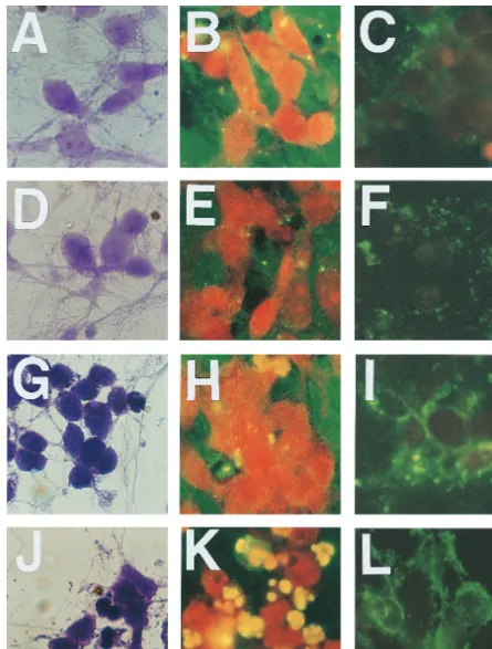
protein itself. Analysis of cells stained with crystal violet
re-vealed no major morphological changes in CVS-N2c-infected
neurons at 24 and 48 h p.i. (Fig. 7A and D, respectively),
whereas neurons infected with CVS-B2c showed shrinking of
cell bodies as well as thinning and degeneration of neuronal
processes by 24 h p.i. (Fig. 7G) and even more extensive
mor-phological changes by 48 h p.i. (Fig. 7J). A TUNEL assay of
infected neuron cultures revealed the presence of only a few
TUNEL signal-positive nuclei in CVS-N2c-infected neurons at
24 and 48 h p.i. (Fig. 7B and E, respectively) but a large
number of TUNEL signal-positive nuclei in CVS-B2c-infected
neurons at 48 h p.i. (Fig. 7K). Furthermore, high-level Annexin
V binding in CVS-B2c-infected neurons was detected as early
as 24 h p.i. (Fig. 7I), whereas no significant Annexin V binding
in CVS-N2c-infected neurons was observed at either 24 or 48 h
p.i. Because DNA cleavage is likely to be a late event in the
apoptotic pathway, it is not surprising that TUNEL-positive
cells appear late in infection. However, Annexin V binding,
which detects the exposure of phosphatidylserine at the plasma
membrane, is an early event in apoptosis. Together, these
results indicate that CVS-B2c causes programmed cell death in
parallel with high levels of G protein expression within the first
24 h of infection, whereas CVS-N2c-infected neurons express
low levels of G protein in cultures and evidently do not
un-dergo extensive apoptosis during the first 48 h of infection.
DISCUSSION
[image:5.612.324.527.66.402.2]The induction of apoptosis in rabies virus-infected neurons
has been proposed as a potential pathogenic mechanism for
FIG. 3. Immunoprecipitation analysis of the N and G proteins in primary [image:5.612.60.287.67.501.2]neurons infected with CVS-N2c or CVS-B2c virus (MOI, 1). At 2, 24, or 48 h p.i., the culture medium was replaced with methionine-free Neurobasal medium containing 10mCi of [35S]methionine per ml, and incubation was continued for 24 h at 37°C. At 24 (lane 1), 48 (lane 2), or 72 (lane 3) h p.i., cells were lysed and subjected to immunoprecipitation with G protein-specific MAb (row 1), N pro-tein-specific MAb (row 2), or actin-specific antibody (row 3). Immune complexes were analyzed by SDS–10% PAGE as described in Materials and Methods. The gel was dried and exposed to X-ray film (A), and protein bands were quantitated by densitometry with the NIH 1.58 IMAGE program (B). Cross-hatched bar, CVS-N2c; hatched bar, CVS-B2c.
FIG. 4. Immunoprecipitation analysis of the N and G proteins in NA neu-roblastoma cells infected with CVS-N2c or CVS-B2c (MOI, 1). At 48 h p.i., the culture medium was replaced with methionine-free RPMI 1640 medium con-taining 10mCi of [35S]methionine per ml, and incubation was continued for 24 h at 37°C. Cells were lysed with lysis buffer and immunoprecipitated with G protein- or N protein-specific MAb. The resulting immune complexes were analyzed by SDS–10% PAGE as described in Materials and Methods. The gel was dried and exposed to X-ray film (A), and protein bands were quantitated by densitometry with the NIH 1.58 IMAGE program (B). Cross-hatched bar, CVS-N2c; hatched bar, CVS-B2c.
on November 9, 2019 by guest
http://jvi.asm.org/
rabies (13). If apoptosis is an important pathogenic mechanism
in rabies, the CVS-B2c variant, which is clearly more cytotoxic
than CVS-N2c for neurons in vitro, should be the more
patho-genic of the variants. However, our in vivo studies with normal
adult mice have shown that the pathogenicity index (50%
le-thal dose/virus titer) of CVS-N2c is up to 50-fold higher than
that of CVS-B2c, depending on the route of inoculation (16).
This observation is not completely unexpected, because
apo-ptosis is an integral part of the host defense response and
should help limit the spread of infection. It is possible that a
rabies virus strain well adapted to its host can avoid processes,
such as apoptosis, that interfere with its survival. There is
evidence that apoptosis plays a role in the attenuation of
pathogenicity of other viruses, such as Sendai virus (12). The
fact that many pathogenic viruses contain antiapoptosis genes
(reviewed in reference 19) attests to the likelihood that
apo-ptosis contributes more to defense from infection than
patho-genicity.
[image:6.612.62.287.66.517.2]The availability of two closely related rabies virus variants,
one that rapidly induces apoptosis in neurons and one that
does not, has allowed us to delineate mechanisms which may
be relevant to neuronal cell death and the pathogenesis of
FIG. 5. Northern blot (A and B) and quantitative PCR (C) analyses of N andG protein mRNAs in primary neuron cells infected with CVS-N2c or CVS-B2c virus (MOI, 1). At 24 h p.i., total RNA was extracted, electrophoresed in a 1% agarose gel containing formaldehyde, transferred to nylon membranes, and hy-bridized with specific32P-labeled probes as described in Materials and Methods. Membranes were exposed to X-ray film (A), and mRNA bands were quantitated by densitometry with the NIH 1.58 IMAGE program (B). For quantitative PCR analysis, RT reactions were carried out with 2.5mg of total RNA and 1mM primer, and serial dilutions of the RT products were subjected to PCR amplifi-cation as described in Material and Methods. To obtain normalized amounts, the relative quantities of G and N protein mRNAs were divided by the relative quantity of G3PDH mRNA (C). From left to right, bars represent CVS-N2c N protein mRNA, CVS-N2c G protein mRNA, CVS-B2c N protein mRNA, and CVS-B2c G protein mRNA.
FIG. 6. Pulse-chase analysis of the N and G proteins in primary neurons infected with CVS-N2c or CVS-B2c virus (MOI, 1). At 24 h p.i., neuron cultures were pulsed with 50mCi of [35S]methionine per ml for 1 h and then chased with unlabeled methionine for 2, 6, and 12 h at 37°C. After the chase, cells were lysed and immunoprecipitated with G protein- or N protein-specific MAb. Immune complexes were analyzed by SDS–10% PAGE as described in Materials and Methods. The gel was dried and exposed to X-ray film (A), and G protein bands were quantitated by densitometry with the NIH 1.58 IMAGE program (B).E,
CVS-N2c protein;F, CVS-B2c protein.
V
OL. 73, 1999
NEUROPATHOGENESIS OF RABIES VIRUS
515
on November 9, 2019 by guest
http://jvi.asm.org/
[image:6.612.318.542.84.478.2]FIG. 7. Morphological changes and induction of apoptosis in primary cultured neurons infected with CVS-N2c (A to F) or CVS-B2c (G to L) virus. Neurons were stained with crystal violet (A, D, G, and J) or stained for determination of apoptosis with the TUNEL assay (B, E, H, and K) or FITC-labeled Annexin V (Ca21-dependent phospholipid binding protein) (C, F, I, and L) at 24 h p.i. (A to C and G to I) and 48 h p.i. (D to F and J to L).
on November 9, 2019 by guest
http://jvi.asm.org/
rabies. Our data suggest a direct correlation between G protein
expression level and induction of apoptosis in cultured
neu-rons. Similar results have been obtained with the relatively
apathogenic ERA rabies virus strain and a more virulent CVS
rabies virus strain for G protein expression and the induction
of apoptosis in Jurkat T cells (20). Presumably, apoptosis
re-sults in a limited infection in vivo, particularly if infection is in
the periphery, where the released G protein can stimulate a
protective immune response. The fact that stereotactic
intcerebral injection or intranasal administration of avirulent
ra-bies virus strains can cause either lethal or transient disease
while intramuscular infection does not (5, 11, 24) tends to
support the concept that apoptosis of peripheral neurons
in-fected with rabies virus may prevent the spread of the virus to
the brain and, perhaps, may promote more rapid induction of
immunity. CVS-N2c-infected neurons, despite producing N
protein in amounts comparable to those produced by
CVS-B2c-infected neurons, produce at least fourfold less G protein.
This differential regulation of G protein expression appears to
be only partially the result of variations in rates of transcription
or stability of the G protein mRNAs, since Northern blot and
quantitative PCR analyses indicated only slight differences
be-tween CVS-N2c and CVS-B2c in the levels of expression of
their N and G protein mRNAs in primary neuron cultures. It
is noteworthy in this regard that the 3
9
and 5
9
untranslated
regions of the G protein mRNA are identical in CVS-N2c and
CVS-B2c (data not shown) and that no AU-rich elements,
which are the most common RNA-destabilizing elements (23),
were identified in these regions. Analysis of G protein mRNA
decay in transiently transfected cells might provide more
con-clusive evidence for possible differences in G protein mRNA
turnover between these two variants, but DNA transfection
experiments with cultured primary neurons, the only relevant
cells for such studies, are not possible, probably because of the
extreme vulnerability of these cells to in vitro manipulations.
Pulse-chase experiments showed that the higher G protein
levels in CVS-B2c-infected neurons were largely the result of
the lower rate of degradation of the CVS-B2c G protein than
of the CVS-N2c G protein. Structural motifs, such as
destabi-lizing N-terminal residues or PEST domains found in proteins
that are subject to rapid degradation (1, 22), are not present in
the G protein of CVS-N2c or CVS-B2c. However, since the
differences between these rabies virus variants in G protein
expression levels are largely restricted to neuronal cells, it is
likely that a neuron-specific proteolytic pathway is utilized to
regulate the expression of rabies virus G proteins. In this
con-text, it has been shown that proteasomal proteolysis plays an
important role in neural regulation (8). For example,
degra-dation of the regulatory subunits of cyclic AMP-dependent
protein kinases has been identified as a molecular mechanism
underlying long-term synaptic plasticity (9). It is conceivable
that rabies virus G proteins differ in their affinity for
chaper-ones that target degradation, translocation, or
posttransla-tional modification pathways that lead to differences in G
pro-tein longevity.
Our data suggest an association among three events that
may be related to the pathogenesis of rabies. In neurons
in-fected with the highly cytopathic CVS-B2c variant, high levels
of G protein are expressed on the cell surface, transport of N
protein into cell processes is inhibited, and cells rapidly
un-dergo apoptosis. On the other hand, neurons infected with the
CVS-N2c variant do not express high levels of G protein, allow
efficient transport of N protein into neuronal processes, and do
not exhibit signs of apoptosis, at least in the first 48 h after
infection. Although further studies are required to determine
the relationship between the induction of apoptosis and G
protein expression, we speculate that the failure of N protein
transport is linked to the apoptotic pathway, perhaps through
the depolymerization of actin filaments (14), which are
essen-tial for the intracellular transport of the N proteins of several
RNA viruses, including rabies virus (3, 17).
A number of studies have concluded that the rabies virus G
protein is a major determinant of rabies virus pathogenicity (5,
18). Mutations in antigenic site III of the G protein reduce the
spread of the virus (6), reduce receptor binding activity (21),
and abolish its pathogenicity for adult mice (5, 18). In the
present study, we identified a mechanism, evidently involving
apoptosis, that regulates the pathogenicity of rabies virus
in-dependently of receptor binding. The fact that the induction of
apoptosis in neurons appears to correlate with the expression
of high levels of the G protein may indicate that there is a
cause and effect relationship between these two events.
How-ever, this interpretation must be viewed with caution. We
found that the CVS variants studied here have identical N
proteins, 1 amino acid difference in the NS protein, 4 amino
acid differences in the M protein, and 10 in the G protein (16;
data not shown). Therefore, we cannot exclude a possible
con-tribution of the NS, M, or L protein to the induction of
apo-ptosis and the related pathogenic phenotype. The involvement
of these proteins in rabies pathogenesis can only be resolved
through the use of reverse genetics technology. Nevertheless,
preliminary results indicate that street rabies virus strains
(dog-and silver-haired bat-derived rabies virus strains), which are
considerably more pathogenic than tissue culture-adapted
strains, express very limited levels of the G protein in neuronal
cells and do not induce apoptosis.
ACKNOWLEDGMENTS
We thank Heather Carbaugh for excellent help in the preparation of
neuron cultures. We also thank R. Pereira for invaluable assistance in
quantitative PCR analysis.
This work was supported by Public Health Service grant AI 09706.
REFERENCES
1. Bergold, P., S. A. Beushausen, T. C. Sacktor, S. Cheley, H. Bayley, and J. H. Schwartz.1992. A regulatory subunit of the cAMP-dependent protein kinase down-regulated in aplysia sensory neurons during long-term sensitization. Neuron 8:387–392.
2. Brewer, G. J., J. R. Torricelli, E. K. Evege, and P. J. Price. 1993. Optimized survival of hippocampal neurons in B27-supplemented Neurobasal, a new serum-free medium combination. J. Neurosci. Res. 35:567–576.
3. Ceccaldi, P.-E., F. Valtorta, S. Braud, R. Hellio, and H. Tsiang. 1997. Al-teration of the actin-based cytoskeleton by rabies virus. J. Gen. Virol. 78: 2831–2835.
4. Cole, R., and J. de Vellis. 1997. Astrocyte and oligodendrocyte cultures, p. 117–130. In S. Fedoroff and A. Richardson (ed.), Protocols for neural cell cultures. Humana Press, Totowa, N.J.
5. Dietzschold, B., W. H. Wunner, T. J. Wiktor, A. D. Lopes, M. Lafon, C. L. Smith, and H. Koprowski.1983. Characterization of an antigenic determi-nant of the glycoprotein that correlates with pathogenicity of rabies virus. Proc. Natl. Acad. Sci. USA 80:70–74.
6. Dietzschold, B., T. J. Wiktor, J. Q. Trojanowski, R. I. Macfarlan, W. H. Wunner, M. J. Torres-Anjel, and H. Koprowski.1985. Differences in cell-to-cell spread of pathogenic and apathogenic rabies virus in vivo and in vitro. J. Virol. 56:12–18.
7. Dietzschold, B., C. E. Rupprecht, M. Tollis, J. Mattei, T. J. Wiktor, and H. Koprowski.1988. Antigenic diversity of the glycoprotein and nucleocapsid proteins of rabies and rabies-related viruses: implications for epidemiology and control of rabies. Rev. Infect. Dis. 10:5785–5798.
8. Gastel, J., P. H. Roseboom, P. A. Rinaldi, J. L. Weller, and D. C. Klein. 1998. Melatonin production: proteasomal proteolysis in serotonin N-acetyltrans-ferase regulation. Science 279:358–360.
9. Hegde, A. N., K. Inokuchi, W. Pei, A. Casadio, M. Ghirardi, D. G. Chain, K. C. Martin, E. R. Kandel, and J. H. Schwartz.1997. Ubiquitin C-terminal hydrolase is an immediate-early gene essential for long-term facilitation in aplysia. Cell 89:115–126.
10. Heid, C. A., J. Stevens, K. J. Livak, and P. M. Williams. 1996. Real time quantitative PCR. Genome Res. 6:986–994.
11. Hooper, D. C., K. Morimoto, M. Bette, M. Weihe, H. Koprowski, and B.
V
OL. 73, 1999
NEUROPATHOGENESIS OF RABIES VIRUS
517
on November 9, 2019 by guest
http://jvi.asm.org/
Dietzschold.1998. Collaboration of antibody and inflammation in the clear-ance of rabies virus from the central nervous system. J. Virol. 72:3711–3719. 12. Itoh, M., H. Hotta, and M. Homma. 1998. Increased induction of apoptosis by a Sendai virus mutant is associated with attenuation of mouse pathoge-nicity. J. Virol. 72:2927–2934.
13. Jackson, A. C., and J. P. Rossiter. 1997. Apoptosis plays an important role in experimental rabies virus infection. J. Virol. 71:5603–5607.
14. Kothakota, S., T. Azuma, C. Reinhard, A. Klippel, J. Tang, K. Chu, T. J. McGarry, M. W. Kirschner, K. Koths, D. J. Kwiatkowski, and L. T. Wil-liams.1997. Caspase-3-generated fragment of gelsolin: effector of morpho-logical change in apoptosis. Science 278:294–297.
15. Lawson, K. F., R. Hertler, K. M. Charlton, J. B. Campbell, and A. J. Rhodes. 1989. Safety and immunogenicity of ERA strain of rabies virus propagated in a BHK-21 cell line. Can. J. Vet. Res. 53:438–444.
16. Morimoto, K., D. C. Hooper, H. Carbaugh, Z. F. Fu, H. Koprowski, and B. Dietzschold.1998. Rabies virus quasispecies: implications for pathogenesis. Proc. Natl. Acad. Sci. USA 95:3152–3156.
17. Ravkov, E. V., S. T. Nichol, C. J. Peters, and R. W. Compans. 1998. Role of actin microfilaments in Black Creek Canal virus morphogenesis. J. Virol. 72:2865–2870.
18. Seif, I., P. Coulon, P. E. Rollin, and A. Flamand. 1985. Rabies virulence: effect on pathogenicity and sequence characterization of rabies virus muta-tions affecting antigenic site III of the glycoprotein. J. Virol. 53:926–934. 19. Teodoro, J. G., and P. E. Branton. 1997. Regulation of apoptosis by viral
gene products. J. Virol. 71:1739–1746.
20. Thoulouze, M.-I., M. Lafage, J. A. Montano-Hirose, and M. Lafon. 1997. Rabies virus infects mouse and human lymphocytes and induces apoptosis. J. Virol. 71:7372–7380.
21. Tuffereau, C., J. Benejean, A.-M. R. Alfonso, A. Flammand, and M. C. Fishman.1998. Neuronal cell surface molecules mediate specific binding to rabies virus glycoprotein expressed by a recombinant baculovirus on the surfaces of lepidopteran cells. J. Virol. 72:1085–1091.
22. Varshavsky, A. 1992. The N-end rule. Cell 69:725–735.
23. Xu, N., C.-Y. Chen, and A.-B. Shyu. 1997. Modulation of the fate of cyto-plasmic mRNA by AU-rich elements: key sequence features controlling mRNA deadenylation and decay. Mol. Cell. Biol. 17:4611–4621. 24. Yang, C., and A. C. Jackson. 1992. Basis of neurovirulence of avirulent rabies
virus variant Av01 with stereotaxic brain inoculation in mice. J. Gen. Virol. 73:895–900.
on November 9, 2019 by guest
http://jvi.asm.org/