0022-538X/95/$04.0010
Copyrightq1995, American Society for Microbiology
Both Viral E2 Protein and the Cellular Factor PEBP2 Regulate
Transcription via E2 Consensus Sites within the Bovine
Papillomavirus Type 4 Long Control Region
MARIA E. JACKSON*ANDM. SAVERIA CAMPOThe Beatson Institute for Cancer Research, Cancer Research Campaign, Beatson Laboratories, Bearsden, Glasgow G61 1BD, Scotland
Received 12 April 1995/Accepted 5 July 1995
The bovine papillomavirus type 4 (BPV4) long control region (LCR) contains three consensus binding sites,
E2(1), E2(2), and E2(3) (ACCN6GGT), for the viral E2 transcription factor and a fourth degenerate site, dE2
(ATCN6GGT), which lies 3 bp upstream of E2(3). The E2(2) site was found to bind the cellular transcription
factor PEBP2, and mutations at this site reduced basal promoter activity by as much as 60%, indicating an important role for PEBP2 in LCR function. Mutation of the E2(3) or dE2 site slightly decreased basal promoter activity, but the cellular proteins binding these sites have not yet been characterized. E2 protein was found to have considerable influence upon LCR promoter activity in primary bovine palate keratinocytes. Thus, when high levels of BPV1 E2 were present, almost complete repression of the BPV4 LCR was observed, whereas smaller amounts of BPV1 or BPV4 E2 led to transactivation. Mutational analysis indicated that E2(1) and dE2 mediated transactivation by E2, whereas E2(2) and E2(3) were responsible for repression by E2. In vitro complexes of binding sites E2(1) and E2(2) with E2 protein demonstrated much greater stability than complexes formed by the E2(3) and dE2 sites. These data suggest that the four E2 sites in the BPV4 LCR each perform different functions in the control of transcription and that competition between cellular transcription factors and viral E2 proteins is essential in regulating the level of viral gene expression during papilloma development.
Bovine papillomavirus type 4 (BPV4) induces papillomas of the upper alimentary tract which may progress to carcinomas in cattle feeding on bracken fern, which contains both carcin-ogens and immunosuppressants (6). The major oncoproteins of BPV4 have been identified as the products of the E7 and E8 open reading frames, and in cooperation with activated ras, BPV4 can transform primary bovine fibroblasts (25, 40). If the cells are additionally treated with quercetin, a component of bracken, they become fully transformed to the malignant phe-notype and are tumorigenic in nude mice (39). The precise cellular changes which induce the transition from benign pap-illoma to carcinoma are not yet understood, but the possibility that a change in viral gene expression is one of the factors which influence progression is currently under investigation.
Papillomavirus gene expression is regulated by both virally encoded and cellular transcription factors. The papillomavirus E2 open reading frame encodes a full-length protein which can act as a transactivator (45) in addition to truncated proteins which lack the N-terminal transactivation domain and act to repress transcription (8, 9, 11, 29, 30). Both forms of E2 bind the consensus sequence ACCN6GGT, which is usually reiter-ated in the viral long control region (LCR) (1, 21). Under some circumstances the full-length transactivator form of E2 has been demonstrated to act as a repressor of transcription. Thus, for the genital human papillomaviruses (HPVs), E2 binding to promoter-proximal E2 sites represses transcription (4, 11, 42, 50, 51) by preventing formation of the initiation complex (12) and competing for the binding of Sp1 to an adjacent site (11,
48, 49). E2 proteins have also been shown to repress promoter activity by competition at a site within the BPV1 LCR which can bind either E2 or a cellular factor which is essential for promoter activity (47, 52). In addition, HPV type 8 (HPV8) E2 protein has recently been demonstrated to transactivate the HPV8 LCR via an element which acts as a transcriptional silencer in the absence of E2 (33). Thus, evidence that com-petition between cellular factors and E2 is important in regu-lation of papillomavirus transcription is accumulating.
The BPV4 LCR contains three consensus E2 binding sites, E2(1), E2(2), and E2(3), in addition to a degenerate site, dE2 (ATCN6GGT), which lies 3 bp upstream of the promoter-proximal E2(3) site (23). Cellular nuclear factors have been demonstrated to produce DNase I footprints over E2(2) and dE2, suggesting the possibility of competition between E2 and cellular transcription factors at these sites (23). The dE2 and E2(3) sites are adjacent to a TATA box, and this arrangement is interesting in view of the presence of paired consensus E2 sites immediately upstream of TATA boxes in many genital HPVs, each of which appears to contribute to E2-mediated repression of promoter activity (8, 11, 42, 48, 50, 51). In view of these observations regarding HPVs it seemed that alteration of one of the two promoter-proximal E2 sites of BPV4 to a degenerate version might have important consequences for transcriptional regulation.
The present study indicated that the E2(2) site of the BPV4 LCR bound the cellular transcription factor PEBP2 and that the degenerate dE2 site possessed E2 binding activity. The effect of E2 binding varied between the sites, with transactiva-tion via E2(1) and dE2 and repression via E2(2) and E2(3). The results suggest the potential for competition between E2 and cellular factors and for differential effects depending upon the concentration of E2.
* Corresponding author. Mailing address: The Beatson Institute for Cancer Research, CRC Beatson Laboratories, Garscube Estate, Switchback Rd., Bearsden, Glasgow G61, 1BD, Scotland. Phone: 141 942 9361. Fax: 141 942 6521.
6038
on November 9, 2019 by guest
http://jvi.asm.org/
MATERIALS AND METHODS
Plasmids and in vitro mutagenesis.For promoter assay LCR constructs (nucleotides 6710 to 331) were cloned into the BamHI site of p0luc (5) upstream of the luciferase coding sequence. For expression of full-length BPV4 E2 a fragment of the BPV4 genome (nucleotides 2738 to 4330) encoding the entire E2 open reading frame was cloned into the BamHI site of pBG331 (J. Barsoum, Biogen, Cambridge, Mass.) downstream of the adenovirus major late promoter (pBG331-BV4E2). pBG331-BV1E2 (J. Barsoum) was used for expression of BPV1 E2. Oligonucleotide-directed mutagenesis was performed with the Altered Sites system (Promega), and all mutant LCR derivatives were confirmed by DNA sequencing.
Cell culture.PalK cells (primary fetal bovine palate keratinocytes) were de-rived from fetal palate biopsies by the method described by Cuthill et al. (10) for human cervical keratinocytes. These cells were cultured on irradiated 3T3 feeder layers in SLM (Gibco BRL) supplemented with 10% fetal calf serum (Gibco BRL), 0.1 nM cholera enterotoxin, 0.5mg of hydrocortisone per ml, 5mg of insulin per ml, and 180mM adenine (all from Sigma). Epidermal growth factor (10 ng/ml; Sigma) was added to cultures after the cells had adhered to the flask.
Transfection.PalK cells (plated without feeders) were transfected by Poly-brene-dimethyl sulfoxide treatment essentially as described by Jiang et al. (27), using 10mg of LCR-luciferase test plasmid, 5mg of control plasmid pCH110 (18), which carries b-galactosidase downstream of the simian virus 40 early promoter, and various amounts of E2-expressing plasmid or vector control as described below. Cells were plated at 106per 25-cm2flask, and the following
morning the medium was replaced with 2 ml of medium containing plasmid DNA and 10mg of Polybrene (Aldrich) per ml; the flasks were then returned to the incubator for 6 h with occasional shaking. After 6 h the medium was replaced with 5 ml of medium containing 35% dimethyl sulfoxide (BDH) for 3 min, and then the cells were washed twice with phosphate-buffered saline and refed. The cells were harvested 30 h later in Reporter Lysis Buffer (Promega). Luciferase assays were performed by using the Luciferase Assay System (Promega) with a BioOrbit 1251 luminometer. Luminescence values (millivolts per second) were typically as follows: background, 30; p0luc vector, 100 to 200; and pLCRluc, 2,500 to 10,000, all integrated over a 1-min period.b-Galactosidase assays (17) were used to standardize luciferase activities.
Oligonucleotides.Oligonucleotides were synthesized on an Applied Biosys-tems (Foster City, Calif.) 381A DNA synthesizer, purified on denaturing poly-acrylamide gels, annealed, and labelled with32
P by using Klenow fragment or T4 polynucleotide kinase and standard protocols (43). The oligonucleotide se-quences used were as follows with nuclear factor binding sites in bold (the mutations described in sections below were introduced into these oligonucleo-tides when appropriate):
E2(1), TCGAGACGCTCTGCACCGAAAACGGTCACATTTGAC CTGCGAGACGTGGCTTTTGCCAGTGTAAACTGAGCT E2(2), TCGAGACAAAGTGTACCGATTGCGGTCGAAACTCTC
CTGTTTCACATGGCTAACGCCAGCTTTGAGAGAGCT E2(3), TCGAGGTCGTACCGAATCGGGTGCATATATAAC
CCAGCATGGCTTAGCCCACGTATATATTGAGCT E2(3)S, TCGAGGTCGTACCGAATCGGGTGCATC
CCAGCATGGCTTAGCCCACGTAGAGCT dE2, TCGAGTATCAGTTGCATCCCATTCGGTCGTACC
CATAGTCAACGTAGGGTAAGCCAGCATGGAGCT AP-2, GATCCAAAGTGTAGCCTGCAGGCCCAACCTC
TTTCACATCGGACGTCCGGGTTGGAGTCTAG NF-D, TCGAGTCAGAAGATGGCGGAGGGCCTCCAACACAGC
CAGTCTTCTACCGCCTCCCGGAGGTTGTGTCGAGCT PEA, TCGAGGAAGTGACTAACTGACCGCAGCTGGCCGTGC
CCTTCACTGATTGACTGGCGTCGACCGGCACGAGCT AP1, CCGAAGTTCAGATGACTAACTCAGGG
GGCTTCAAGTCTACTGATTGAGTCCC
Nuclear extract preparation and electrophoretic mobility shift assays (EMSA).
Nuclear extracts were prepared exactly as described by McCaffery and Jackson (34). DNA binding reactions were performed with 2.5ml of nuclear extract and 1mg of poly(dI-dC) in a solution containing 10 mM Tris-Cl (pH 7.0), 100 mM KCl, 1 mM MgCl2, 1 mM dithiothreitol, 1 mM EDTA, and 5% glycerol. After 10
min of preincubation on ice, 0.005 pmol of labelled oligonucleotide and 0.5 pmol of cold competitor oligonucleotides when appropriate were added and incuba-tion was continued at room temperature for 15 min in a final volume of 50ml. Samples were electrophoresed on 6% polyacrylamide gels at 9 V/cm in 300 mM glycine–50 mM Tris–1 mM EDTA (pH 8.7).
Bacterially synthesized PEBP2 subunits. A truncated histidine-tagged PEBP2aA1 polypeptide (amino acids 94 to 226) (38) containing the DNA bind-ing and dimerization domains of the protein was produced by isopropyl-b-D -thiogalactoside (IPTG) induction of JM109/pQEaN94C226 followed by purifi-cation on nickel chelate affinity resin (Qiagen) according to the manufacturer’s protocol. The full-size PEBP2b2 subunit (37) was produced by IPTG induction of BL21(DE3)/pET3ab2 and partially purified as described by Ogawa et al. (37). The PEBP2 extracts were stored in aliquots at2708C and used in EMSA reactions under the conditions described above.
In vitro transcription and translation of BPV4 E2.A fragment of the BPV4
genome (nucleotides 2738 to 4330) encoding the entire E2 open reading frame was cloned into the BamHI site of Bluescript pSK1(Stratagene) with E2 under the transcriptional control of the T3 promoter. Transcription from linearized template using bacteriophage T3 RNA polymerase (Promega) and translation using nuclease-treated rabbit reticulocyte lysate (Promega) were carried out according to protocols supplied by Promega. The translation mix was stored at
2708C, and 6ml was used per 50-ml EMSA reaction mixture with buffers as described above.
Dissociation rates for E2 complexes.Dissociation rates for E2 complexes were determined as described by Li et al. (31). A 171.5-ml binding reaction mixture with labelled oligonucleotide was set up as described above. After 15 min of incubation, 24.5ml was loaded onto a running polyacrylamide gel and 3ml of unlabelled oligonucleotide (to give a 200-fold excess) was added to the remaining 147ml. Samples (25ml) taken at various times following addition of competitor were loaded onto the running gel. The gel was autoradiographed without screens, and free and bound oligonucleotides were quantified with a Molecular Dynamics laser scanning densitometer.
Reverse transcriptase PCR (RT-PCR) of RNA from transiently transfected cells.RNA was isolated from transiently transfected cells by using RNAzolB (Biogenesis Ltd). A 15-mg portion of RNA was treated with 50 U of DNase I (Amersham) for 60 min at 378C in PCR buffer (Roche) with 40 U of rRNasin (Promega), and the DNase I was then inactivated by incubation for 20 min at 908C. A total of 2.5 pmol of reverse primer (RP; TCCAGCGGTTCCATCC TCTAG) was added to the RNA, which was incubated for 5 min at 858C before being cooled to room temperature. A further 40 U of rRNasin was added, together with reverse transcription buffer (Life Sciences Inc.) and diethyl pyro-cabonate-treated water, to a final volume of 60ml. A 30-ml portion of each sample was added to 40 U of reverse transcriptase (Life Sciences Inc.). Both plus and minus reverse transcriptase samples were incubated for 2 h at 428C. The PCR primers used were FP1 (GTCGTACCGAATCGGGTGCA), FP1mt (GTCGTAAAGAATCGAATGCA), FP3 (TTGGCATTCCGGTACTGTTGG), and RP, which map to the positions shown in Fig. 8C. For PCRs a master mix of all components (Roche) including 12.5 pmol of each primer (FP1 [or FP1mt], FP3, and RP) was first prepared, and 90ml of this mix was added to 10ml of sample. PCRs were performed in a Perkin-Elmer Cetus Geneamp PCR 9600 machine programmed as follows: 2 min at 948C; 30 cycles of 30 s at 948C, 30 s at 558C, and 30 s at 728C; 4 min at 728C; and 48C hold. PCR products were analyzed by electrophoresis at 100 V on 6% acrylamide gels in 13Tris-borate-EDTA (TBE) buffer and stained with ethidium bromide.
RESULTS
E2 site mutations affect BPV4 LCR promoter activity even
in the absence of E2.The LCR of BPV4 contains three
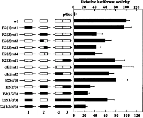
con-sensus E2 binding sites (ACCN6GGT) at nucleotides 7050 [E2(1)], 175 [E2(2)], and 267 [E2(3)] (Fig. 1A and B). In addition a degenerate E2 site (dE2; ATCN6GGT) is present at nucleotide 252, 3 bp upstream of E2(3); it has been demon-strated by direct sequencing of DNA amplified from papillo-mas by PCR that this degenerate E2 site is not a cloning artifact (23). Each of the four sites E2(1), E2(2), E2(3), and dE2 was individually mutated to AAAN6AAT, and a further construct, in which the T at position 2 of the degenerate dE2 site was mutated to C, yielding a consensus E2 site, was made (Fig. 1B). The wild-type and mutant LCRs were then cloned upstream of a promoterless luciferase gene in the vector p0luc. The BPV4 LCR exhibited promoter activity in primary bovine fetal palate keratinocytes (PalK) in the absence of any E2 protein (Fig. 2). Mutation of E2(1) and conversion of dE2 to a consensus E2 site (dE2mt1) had no significant effect on this basal LCR promoter activity; however, LCR-E2(2)mt1 pos-sessed only 43% of the wild-type LCR activity, and LCR-E2(3)mt1 and LCR-dE2mt2 also demonstrated significant de-creases in basal promoter activity. The E2(1)mt1, E2(2)mt1, E2(3)mt1, and dE2mt2 mutants were also tested in combina-tions, with the finding that E2(2)mt1 always led to a consider-able decrease in promoter activity but mutation of the remain-ing E2 sites led to further decreases in promoter activity such that an LCR derivative in which all four E2 sites were mutated [LCR-E2(1/2/d/3)] possessed only 22% of the wild-type activ-ity. These results indicated that the E2 sites, and particularly E2(2), were important for E2-independent basal promoter ac-tivity, and indeed, DNase I footprints formed by cellular pro-teins have been observed over the E2(2) site (Fig. 1A) (23).
on November 9, 2019 by guest
http://jvi.asm.org/
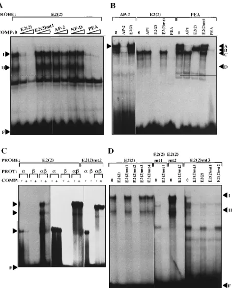
E2(2) binds the cellular transcription factor PEBP2.The considerable drop in LCR promoter activity following muta-tion of E2(2) suggested that a cellular factor important for transcriptional activity of the LCR bound at this site. An E2(2) site oligonucleotide incubated with PalK nuclear extract formed two specific complexes in EMSA which were inhibited by competition with excess unlabelled E2(2) oligonucleotide (Fig. 3A). An E2(2)mt1 oligonucleotide had lost the ability to
form specific complexes in EMSA and to compete for complex formation by the wild-type oligonucleotide (Fig. 3A, B, and D). Comparison of the E2(2) sequence with known nuclear factor binding sites identified three factors, AP-2 (22, 35), NF-D (7), and PEBP2 (also termed PEA2) (41), for which the potential binding site would be disrupted by the E2(2)mt1 mutation (Fig. 1C). An oligonucleotide carrying the NF-D binding site was unable to compete for complex formation by E2(2), and while the AP-2 oligonucleotide competed weakly (Fig. 3A), this oligonucleotide itself formed complexes which could not be inhibited competition with E2(2) (Fig. 3B). The E2(2) com-plexes were efficiently inhibited by competition with an oligo-nucleotide derived from polyomavirus enhancer A (PEA), which contained binding sites for both PEBP2 and AP1 (41) (Fig. 3A and B). PEA oligonucleotides form four specific com-plexes in EMSA: complex A, formed by binding of both AP1 and PEBP2; complex B, containing AP1 only; complex C, con-taining PEBP2 only; and complex D, which is related to PEBP2 (44). The E2(2) oligonucleotide was able to compete for the formation of complexes A, C, and D, which contain PEBP2, but not complex B, which contains AP1 only (Fig. 3B; note that complex D is not visible in this exposure). An AP1-specific oligonucleotide could not compete for complex formation by E2(2), and competition with the E2(2)mt1 oligonucleotide did not inhibit the formation of PEA complexes (Fig. 3B). Com-plexes I and II formed by E2(2) (Fig. 3A) appear to be equiv-alent to complexes C and D formed by PEA, although com-plexes II and D were present at very low levels in this EMSA (Fig. 3B).
The analysis described above indicated that the cellular fac-tor binding to the E2(2) site was PEBP2. Further evidence to support this conclusion was obtained by the binding of bacte-rially synthesized PEBP2 polypeptides to the E2(2) oligonu-cleotide. PEBP2 is a heterodimer composed of two subunit families (28), the PEBP2a family, possessing DNA binding activity (3, 38), and the PEBP2bfamily, the members of which enhance the binding of the a subunits (37). A truncated PEBP2aA1 polypeptide (amino acids 94 to 226) possessing the
FIG. 1. (A) Schematic representation of the BPV4 LCR subclone (nucleotides 6710 to 331) used in this study, showing the relative positions of the four E2 sites and DNase I footprints identified previously (23). T, TATA box. (B) DNA sequences of the BPV4 E2 sites and their mutant derivatives. LCR-E2(d/3), LCR-E2(2/3), LCR-E2(1/2/3), LCR-E2(1/d/3), and LCR-E2(1/2/d/3) combine the mutations of E2(1)mt1, E2(2)mt1, dE2mt2, and E2(3)mt1 as appropriate, i.e., each site is mutated to AAAN6AAT. The PEBP2 binding site at E2(2) is indicated. (C) Homologies between the E2(2) site and binding sites for the nuclear factors AP-2 (22, 35), NF-D
[image:3.612.118.498.74.272.2](7), and PEBP2 (38). The E2 consensus is boxed, and mismatched positions are indicated by solid squares.
FIG. 2. Promoter activities of the wild-type LCR and E2 site mutants in PalK cells. The LCR constructs are represented diagrammatically with mutated sites shown as solid boxes. The activity of the wild-type LCR is designated as 100%, and the mutant LCR activities are expressed as percentages of the wild-type activity. The results represent average values and standard deviations for 10 sets of duplicates. The individual sets of transfections contained various amounts of pBG331 vector from 1 to 10mg as controls for the E2 transactivation measure-ments shown in Fig. 6; the data sets were combined, since pBG331 had no apparent effect upon luciferase activities.
on November 9, 2019 by guest
http://jvi.asm.org/
[image:3.612.60.298.455.652.2]PEBP2aA1 DNA binding and heterodimerization domains and the entire PEBP2b2 protein were produced in Escherichia
coli and tested for their ability to bind E2(2). The PEBP2aA1 polypeptide alone was able to form a retarded complex in EMSA with the E2(2) oligonucleotide, whereas the PEBP2b2 protein alone demonstrated no DNA binding ability, as ex-pected (Fig. 3C). However, when PEBP2aA1 and PEBP2b2 polypeptides were present together, novel complexes were formed and DNA binding was enhanced relative to that of PEBP2aA1 alone. No complexes were formed when the E2(2)mt1 oligonucleotide was incubated with the PEBP2 polypeptides (data not shown). The E2(2) site therefore pos-sesses the ability to bind the cellular transcription factor PEBP2, and the loss of PEBP2 binding activity in E2(2)mt1 correlates with a 57% decrease in transcription from the LCR.
Thus, PEBP2 appears to be an important transcription factor for basal LCR promoter activity.
Mutagenesis of the E2(2) site.The nucleotide substitutions
in E2(2)mt1 altered both halves of the E2 binding site, whereas the PEBP2 consensus overlapped only the 39 half of the E2 sequence. Additional mutations (Fig. 1B) were therefore in-troduced into E2(2) in order to confirm the observations made with E2(2)mt1. E2(2)mt2, with substitutions only in the 59half of the E2 site, was still able to bind PEBP2 in EMSA (Fig. 3C and D). For E2(2)mt3 (Fig. 3D) and E2(2)mt4 (data not shown) the PEBP2 consensus was disrupted, and neither of these oligonucleotides was able to bind PEBP2 in EMSA, as expected. E2(2)mt2, E2(2)mt3, and E2(2)mt4 were individu-ally introduced into the BPV4 LCR, and promoter activity was assayed as described above. The two mutants in which the PEBP2 site was abolished, LCR-E2(2)mt3 and LCR-E2(2) mt4, had promoter activities equivalent to that of the original mutant, LCR-E2(2)mt1 (Fig. 2). However, LCR-E2(2)mt2, in which only the 59half of the E2 site was mutated, leaving the PEBP2 consensus intact, had higher promoter activity than LCR-E2(2)mt1 but still significantly less than the wild-type level. This result suggested that the 59 half of the E2(2) site also bound a cellular factor important for basal LCR promoter activity. However, oligonucleotides E2(2) and E2(2)mt2 form identical complexes with PalK nuclear extract, i.e., E2(2)mt2 does not appear to have lost any binding activity (Fig. 3D). Furthermore, the E2(2)mt3 oligonucleotide, which is mutated only in the 39half and is unable to bind PEBP2, forms com-plexes which can be efficiently inhibited by competition with the E2(2)mt3 and E2(2)mt1 mutant oligonucleotides but not by competition with the wild-type E2(2) oligonucleotide (Fig. 3D), suggesting that these complexes are artifacts specific to the mutant oligonucleotide. Because of the symmetrical nature of E2(2), the 59half of this site shows a partial match with the PEBP2 consensus, making it possible for a second PEBP2 complex to bind to E2(2) in cooperation with PEBP2 already bound to the consensus site. However, increasing the ratio of nuclear extract to oligonucleotide did not reveal any additional complexes (data not shown), and at the saturating PEBP2 concentrations obtainable by using bacterially synthesized polypeptides, E2(2) did not form any complexes which were not also formed by E2(2)mt2 (Fig. 3C). Thus, although the reduction in promoter activity resulting from the E2(2)mt2 substitutions suggests that this mutation affects a nuclear factor binding site, attempts to identify such a factor by EMSA have not been successful.
E2 binding to BPV4 E2 sites and mutant derivatives.The
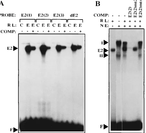
binding of E2 to oligonucleotides carrying each of the four BPV4 E2 sites was tested by using in vitro-synthesized BPV4 E2. All the sites, including the dE2 site, were found to bind E2, and thus the replacement of C by T at position 2 of dE2 does not prevent E2 binding (Fig. 4A). However, the stability of E2 binding appeared to differ considerably among the sites. Thus, E2(1) and E2(2) complexes with E2 were relatively stable over a 30-min incubation, whereas E2(3) and dE2 complexes with E2 dissociated much more rapidly (Fig. 5). The E2(2) mutants behaved as expected in EMSA, i.e., mutants 1, 2, and 3 were unable to bind E2, whereas E2(2)mt4, in which the PEBP2 consensus only was mutated, retained the ability to bind E2 (data not shown) and compete for E2 binding to E2(2) (Fig. 4B). The considerable overlap between the E2 and PEBP2 consensus sites at E2(2) makes it unlikely that these two pro-teins would bind simultaneously. Indeed, when both propro-teins were present in EMSA, no complexes were formed other than those observed when these proteins were present separately (Fig. 4B).
FIG. 3. (A) Complexes formed in EMSA by labelled E2(2) oligonucleotide probe in the absence of competitor (0) and in the presence of 10- and 100-fold excess of unlabelled competitor oligonucleotide (COMP) as indicated. The po-sitions of the two specific complexes (I and II) formed by E2(2) and free oligonucleotide (F) are shown on the left. (B) Complexes formed in EMSA by labelled AP-2, E2(2), and PEA oligonucleotide probes in the absence of com-petitor (0) and in the presence of 100-fold excess of cold comcom-petitor oligonucle-otide as indicated. The positions of the AP-2 complex and the four complexes (A, B, C, and D) formed by the PEA oligonucleotide are shown by arrows. (C) Complexes formed in EMSA by E2(2) and E2(2)mt2 oligonucleotides with bacterially synthesized PEBP2a andbsubunits. Reactions were performed either in the absence (2) or in the presence (1) of a 100-fold excess of unla-belled E2(2) competitor oligonucleotide. The 6 tracks on the left contain 50-fold less protein than the 10 tracks on the right, in which the EMSA was carried out at saturating PEBP2 concentrations sufficient to bind both probe and competitor oligonucleotides. PEBP2 complexes and free oligonucleotide (F) are indicated by arrows. PROT, protein. (D) Complexes formed in EMSA using PalK nuclear extract by the wild-type E2(2) oligonucleotide and its mutant derivatives either in the absence (0) or in the presence of 100-fold excesses of unlabelled competitor oligonucleotides (COMP) as indicated. The different intensities of complexes formed by E2(2) and E2(2)mt2 relate to the specific activity of the probe and the autoradiograph exposure time.
on November 9, 2019 by guest
http://jvi.asm.org/
[image:4.612.62.298.71.364.2]The BPV4 LCR E2 binding sites can mediate
transactiva-tion or repression by full-length E2. The effect of BPV1 E2
and BPV4 E2 upon transcription from the BPV4 LCR was assayed by cotransfecting the LCR luciferase constructs with either pBG331-BV1E2 or pBG331-BV4E2, which express BPV1 E2 and BPV4 E2, respectively, from the adenovirus major late promoter. For each construct the activity in the absence of E2 protein was set at 1 and any change in activity in the presence of E2 was expressed in relation to this value (Fig. 6). Cotransfection of 5 mg of pBG331-BV1E2 was found to cause 85% repression of the LCR, which could be abrogated by mutation of E2(3). A marginal alleviation of the repression was also seen for the E2(2) mutations, and when the mutations of E2(2)mt1 and E2(3)mt1 were combined to produce LCR-E2(2/3), four- to sixfold transactivation by BPV1 E2 was ob-served. Thus, repression by BPV1 E2 appeared to be mediated mainly by E2(3), but it was also influenced by E2(2). However, the E2(1) and dE2 sites appeared to mediate transactivation by E2, since, as demonstrated by comparing E2(1/2/3) with E2(2/ 3), E2(1/d/3) with E2(d/3), and E2(d/3) with E2(3)mt1, trans-activation by BPV1 E2 was consistently decreased when E2(1) or dE2 was mutated. When all four E2 sites were mutated, no effect of BPV1 E2 was observed, i.e., LCR-E2(1/2/d/3) had the same activity in the presence and in the absence of BPV1 E2. To test whether LCR repression occurred at all E2 concen-trations or was dependent on the source of E2, various amounts of either pBG331-BV1E2 or pBG331-BV4E2 were cotransfected with the wild-type LCR luciferase plasmid. Some transactivation was observed with 0.01mg of pBG331-BV1E2, but increasing repression was observed with higher concentra-tions of this plasmid (Fig. 7). Cotransfection of
pBG331-BV4E2 produced increasing transactivation of the LCR with maximal transactivation at 1.0mg of plasmid; however, at 5mg of pBG331-BV4E2 the level of transactivation was sharply reduced. Repression of the BPV4 LCR by E2 therefore ap-pears to be dependent on the origin and cellular concentration of E2 protein, with transactivation occurring at low protein concentrations and repression occurring only as the E2 con-centration rises.
The effect of E2 site mutations on transactivation by BPV4 E2 was assayed by using 1.0 mg of pBG331-BV4E2, which produced an approximately 3.5-fold increase in wild-type LCR activity (Fig. 6B). The levels of transactivation obtained in individual experiments varied somewhat, but the trend was always the same. Thus, although the standard deviations in Fig. 6B are large, the average values give an accurate representa-tion of the differences in transactivarepresenta-tion seen among the mu-tants in four independent sets of transfections. LCR-E2(1)mt1 and LCR-dE2mt2 were invariably transactivated to a lower degree than the wild type, but although the dE2 site clearly mediated transactivation by E2, the conversion of this site to a consensus E2 binding sequence in dE2mt1 did not increase transactivation by E2. The E2(2) and E2(3) site mutations produced levels of transactivation similar to or higher than the wild-type level. The E2(2/3) double mutant demonstrated the greatest level of transactivation by E2, while the E2(1/2/3) triple mutant showed a greatly reduced ability to be vated by BPV4 E2 and, similarly, LCR-E2(d/3) was transacti-vated to a much lesser extent than LCR-E2(3)mt1. Interest-ingly, some transactivation was seen even when all four E2 sites were mutated.
[image:5.612.61.296.74.288.2]When the amount of pBG331-BV4E2 was increased to 10.0 mg (Fig. 6C), transactivation of the wild-type LCR was mark-edly reduced but the differences between the mutants became more defined. LCR-E2(1)mt1 and LCR-dE2mt2 again dem-onstrated lower activity than the wild type. Of the LCR-E2(2) mutants, mt1, mt2, and in particular mt3 showed greater trans-activation than the wild type, whereas mt4, which is the only E2(2) mutant retaining the ability to bind E2, demonstrated an E2 response identical to that of the wild type. LCR-E2(3)mt1 showed approximately fourfold transactivation, and the double mutant, LCR-E2(2/3), showed sevenfold transactivation; how-ever, the additional mutation of dE2 [LCR-E2(d/3)] or E2(1)
FIG. 5. Dissociation rates of E2-oligonucleotide complexes formed by E2(1), E2(2), E2(3), and dE2 oligonucleotides. Unlabelled self oligonucleotide com-petitor (200-fold excess) was added at time zero to labelled oligonucleotides preincubated for 15 min with in vitro-translated BPV4 E2 protein. Samples were loaded onto a running acrylamide gel at 0, 2, 5, 10, 20, and 30 min (A). Auto-radiographs were scanned by laser densitometry, and the amount of E2-oligo-nucleotide complex was calculated as the bound oligoE2-oligo-nucleotide/total oligonu-cleotide ratio with the initial amount of complex set at 100% in each case (B).
f——f, E2(1);å----å, E2(2);v——v, E2(3);r----r, dE2. FIG. 4. (A) Complexes formed by E2(1), E2(2), E2(3), and dE2
oligonucle-otides in EMSA using in vitro-translated BPV4 E2 protein. The indicated probe oligonucleotides were incubated with unprogrammed reticulocyte lysate (C) or E2-programmed reticulocyte lysate (E) in either the absence (2) or the presence (1) of a 100-fold excess of cold self-competitor oligonucleotide. Free oligonu-cleotide (F) and the E2-oligonuoligonu-cleotide complexes (E2) are indicated by arrows. RL, reticulocyte lysate; COMP, competitor. (B) Complexes formed by E2(2) oligonucleotide in the presence of both E2 and PEBP2 proteins. PalK nuclear extract (NE) and E2-programmed reticulocyte lysate (RL) were added as shown above the lanes; 100-fold excesses of the indicated cold competitor oligonucle-otides were added when appropriate. The positions of free oligonucleotide, E2 complex, and PEBP2 complexes I and II are indicated by arrows.
on November 9, 2019 by guest
http://jvi.asm.org/
[image:5.612.326.546.512.651.2][LCR-E2(1/2/3)] again resulted in a loss of transactivation. In conclusion, E2(1)mt1 and dE2mt2 reduced the ability of the LCR to be transactivated by E2, whereas mutation of E2(2) and E2(3) abrogated E2-mediated repression, allowing higher levels of transactivation.
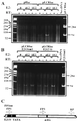
In order to ascertain whether transcription was being initi-ated, as expected, from the TATA box immediately down-stream of E2(3), RT-PCR was used. RNA was prepared from transiently transfected cells and cDNA was produced as de-scribed in Materials and Methods, by using a reverse primer (RP) homologous to part of the luciferase coding sequence. For PCR amplification RP was used in conjunction with for-ward primers FP1/FP1mt, annealing to the E2(3) site, and FP3, annealing immediately upstream of the luciferase initiation codon (Fig. 8C). FP1 was used for amplifications in which the transfected plasmids had a wild-type E2(3), and FP1mt was used to analyze transfections of E2(3)mt1 plasmids. The 94-bp amplification product of FP3 plus RP represents all luciferase transcripts, and the 204-bp amplification product of FP1/ FP1mt plus RP would represent any luciferase transcripts ini-tiated upstream of the BPV4 TATA box. For each sample a
[image:6.612.152.464.71.240.2]control in which reverse transcriptase had been omitted from the cDNA synthesis step in order to detect any amplification arising from DNA contamination of the RNA preparation was included; in no case was any amplification product visible in
FIG. 6. Relative promoter activities of the wild-type and mutant LCR constructs in the presence of 5mg of BPV1 E2 plasmid (A), 1mg of BPV4 E2 plasmid (B), and 10mg of BPV4 E2 plasmid (C). For each construct the activity in the absence of E2 protein (with an equivalent quantity of vector pBG331) has been set at 1 and the activities in the presence of E2 are expressed relative to this value. Results represent the means and standard deviations for three (A), four (B), and two (C) duplicate sets of data.
[image:6.612.354.506.366.603.2]FIG. 7. Promoter activities of the wild-type BPV4 LCR (solid bars) and p0luc vector (open bars) in the presence of increasing concentrations of either BPV1 or BPV4 E2. pBG331 vector plasmid was added to the indicated amounts of pBG331-BV1E2 and pBG331-BV4E2 to produce a total of 5mg of pBG331-derived plasmid per transfection. LCR activity in the absence of E2 has been set at 1, and results for both p0luc and the LCR are expressed relative to this value.
FIG. 8. RT-PCR of RNA from transient transfections. (A) RT-PCR analysis using primers FP1, FP3, and RP of RNA from PalK cells transfected with p0luc and pLCRluc in the absence of E2 (0), in the presence of 1mg of BPV4 E2 (4E2), or in the presence of 5mg of BPV1 E2 (1E2). Samples to which reverse tran-scriptase (RT) enzyme was added are indicated by plus signs, and control sam-ples lacking RT are indicated by minus signs. The first lane from the left contains
fX174 HaeIII standards, and the last two lanes represent a control PCR without added template and a control reaction with 1 ng of pLCRluc as a template. (B) RT-PCR analysis using primers FP1mt, FP3, and RP of RNA from PalK cells transfected with pLCR-E2(3)mt1 and pLCR-E2(2/3). Other details are as for panel A. The last lane represents a control reaction with 1 ng of pLCR-E2(3)mt1 as a template. (C) Representation of part of pLCRluc showing the positions of the primers used for RT-PCR. The E2(3) site, the TATA box, and the ATG codon are shown as solid boxes; the shaded area represents sequences from the p0luc vector.
on November 9, 2019 by guest
http://jvi.asm.org/
[image:6.612.113.247.533.676.2]these samples (Fig. 8). For cells transfected with p0luc, no luciferase transcripts were detected by RT-PCR. For cells transfected with pLCRluc, pLCR-E2(3)mt1, and pLCR-E2 (2/3) only a 94-bp RT-PCR product was observed, indicating that the majority of the transcripts must be initiated down-stream of the BPV4 TATA box. Similarly, in the presence of BPV1 or BPV4 E2 protein, no transcripts initiating upstream of the TATA box were observed, indicating that changes in luciferase activity were not due to E2 protein acting on cryptic promoters within p0luc or the BPV4 LCR. LCR derivatives carrying different permutations of the E2 site mutations all gave the same result (data not shown). Thus, as for all the mucosal papillomaviruses studied to date, the BPV4 TATA box immediately downstream of the paired E2 sites is the target for transcriptional modulation by E2 proteins.
DISCUSSION
The results described above indicated that mutation of the four E2 sites in the BPV4 LCR caused a reduction in LCR activity even in the absence of E2. Mutation of E2(2) in par-ticular led to a 57 to 60% drop in LCR promoter activity in PalK cells. Thus, the cellular factor(s) binding E2(2) makes a considerable contribution to basal LCR promoter activity. An earlier study similarly demonstrated that deletion of 37 bp encompassing E2(2) resulted in an 85% decrease in LCR en-hancer activity (23). The E2(2) site overlaps a consensus bind-ing site for the cellular factor PEBP2 (PEA2), and this site was indeed found to bind PEBP2 in EMSA. PEBP2, which was initially identified as a cellular transcription factor binding the polyomavirus enhancer (28, 41), is induced during differentia-tion (14) and undergoes striking alteradifferentia-tions in response to cell transformation by ras (44). Potential binding sites for PEBP2 are present in the enhancer elements of murine and feline leukemia viruses (13, 16) and in the enhancer regions of sev-eral T-lymphocyte-specific genes (37, 38). PEBP2 is a member of a family of transcription factors which includes the
Drosoph-ila melanogaster segmentation gene runt and the human AML1
gene (2, 38), and it is a heterodimer composed of two groups of subunits,aandb(28). Expression of theasubunits, which possess the DNA binding activity of PEBP2, is cell type spe-cific, while the b subunits appear to be ubiquitous (37, 38). Thus, the PEBP2 family of transcription factors appears to be involved in developmental regulation and differentiation, and the genes for both aand bsubunits have been found at the translocation breakpoints in human leukemia (2, 32). The ex-pression pattern of PEBP2 in differentiating keratinocytes is not known, but PEBP2 may be one of the cellular factors which couple the BPV4 life cycle to keratinocyte differentiation.
The overlap between E2 and PEBP2 binding sites at E2(2) makes it appear unlikely that these proteins would bind to-gether, and indeed, when both PEBP2 and E2 were present in EMSA, complexes representing each protein alone were ob-served but no additional complexes which might represent simultaneous binding of the two proteins were apparent. Thus, E2 and PEBP2 would be expected to compete for binding to E2(2) and the relative concentrations of these proteins within the cell may have considerable influence on promoter activity. The finding that E2(2)mt2, which was able to bind PEBP2, possessed only 60% of the wild-type promoter activity in the absence of E2 protein suggested the possibility that another cellular transcription factor can bind the 59 half of E2(2). Attempts to identify such a factor were unsuccessful, as were attempts to demonstrate binding of a second molecule of PEBP2 to the 59side in cooperation with a molecule already bound at the 39side. However, it remains a possibility that such
putative cooperative binding between PEBP2 molecules re-quires a subunit sequences absent from the truncated form used in this study.
The affinity of BPV1 E2 protein for its binding sites has been demonstrated to vary dramatically depending on the nucle-otide sequence of the site, with the optimal binding site being ACCGN4CGGT (20, 31, 36), although the internal N4 se-quence and the nucleotides immediately adjacent to the E2 site also have a considerable influence on binding affinity (31). The results presented here demonstrate that the BPV4 E2 sites also vary considerably with respect to E2 binding, such that E2(1) and E2(2) form more stable oligonucleotide-E2 complexes than E2(3) and dE2. E2(1) and E2(2) have the optimal ACC GN4CGGT sequence and were expected to have the highest affinity for E2. The finding that the dE2 site exhibited E2 binding equivalent to that exhibited by E2(3) was unexpected in view of the presence of T rather than C at position 2 of dE2. It may be that T is tolerated better than other position 2 base substitutions which have been found to considerably decrease binding of BPV1 E2 to its sites (31). Another possibility is that BPV4 E2 has evolved to permit this substitution. Sequencing of the LCR regions of BPV3 and BPV6, which are closely related to BPV4 (24, 26), revealed that while BPV3 possessed paired E2 consensus sites adjacent to the TATA box, BPV6 exhibited an arrangement identical to that of BPV4, i.e., dE2/ E2/TATA (34a); furthermore, the substitution in the degener-ate site was identical in BPV4 and BPV6, i.e., ATCN6GGT, indicating that the degenerate site may have an important function which is conserved between BPV4 and BPV6. Since dE2 is able to bind E2 protein in vitro and is functional in in vivo studies, the arrangement of the E2 sites of the BPV4 LCR resembles that of the E2 sites of the LCRs of the other mucosal papillomaviruses, with a pair of E2 binding sites adjacent to the TATA box in addition to the upstream sites (21).
The effect of E2 on LCR promoter activity was dependent on the E2 protein concentration. Cotransfection of very small amounts of BPV1 E2 plasmid produced a moderate transacti-vation of the BPV4 LCR, but higher concentrations of BPV1 E2 resulted in increasing repression. With BPV4 E2 increasing transactivation was seen up to 1mg of BPV4 E2 plasmid with decreased transactivation at higher concentrations of plasmid. Thus, almost complete repression could be obtained with BPV1 E2, whereas even at the highest concentrations of BPV4 E2 plasmid tested LCR activity was still equivalent to that observed in the absence of E2. Similar results demonstrating strong repression by BPV1 E2 but not the homologous E2 protein have been obtained for the HPV18 and HPV11 LCRs (8, 11, 51). These results may well reflect a functional differ-ence between BPV1 E2 and the other papillomavirus E2 teins, although differences in levels of expression of E2 pro-teins from the plasmid vectors cannot be excluded. The action of E2 differed among the four BPV4 E2 sites, since mutation of E2(1) and dE2 led to a consistent decrease in transactivation by BPV1 or BPV4 E2 and mutation of E2(2) and E2(3) led to increased transactivation by BPV1 or BPV4 E2. Thus, E2(1) and dE2 mediate transactivation while E2(2) and E2(3) medi-ate repression. The HPV11 and HPV18 E2 binding sites equiv-alent in position to both E2(2) and E2(3) of BPV4 have also been demonstrated to mediate repression by the homologous E2 proteins (11, 50). For HPV11 and HPV16 repression via the promoter-proximal pair of E2 sites is at least in part due to competition between E2 binding and Sp1 binding to an up-stream nonconsensus Sp1 site (11, 48, 49). No equivalent Sp1 motif is found immediately upstream of dE2/E2(3) in BPV4, although an AT-rich region similar to that described by Gloss and Bernard (15) is present (Fig. 1B). The absence of an Sp1
on November 9, 2019 by guest
http://jvi.asm.org/
motif may explain why the dE2 site of BPV4 does not mediate repression by E2 as do the equivalent sites of HPV11, HPV16, and HPV18 (11, 42, 50). The LCR construct in which all four E2 binding sites were mutated showed no response to BPV1 E2 protein, but it could still be transactivated approximately twofold by BPV4 E2. BPV1 E2 has been shown to be capable of transactivation independent of DNA binding (19), and BPV4 E2 appears to possess a similar activity.
Many examples of competition between cellular factors and viral E2 proteins for binding to papillomavirus E2 sites have been reported (11, 33, 47–49, 52), suggesting that such inter-actions play a vital role in regulating papillomavirus transcrip-tion. Displacement of the cellular factor Sp1 by E2 protein has been demonstrated to occur in vitro when the binding sites overlap, and this appears to be one mechanism of E2 repres-sion in HPV11 and HPV16 (11, 48, 49). The results described here suggest that in BPV4 the cellular factor PEBP2 is simi-larly displaced by E2, leading to repression, and PEBP2 can be added to the increasing list of cellular factors which interact at papillomavirus E2 sites.
The observations regarding PEBP2 binding at E2(2) and the differing interactions of E2 at the four BPV4 E2 sites suggest a model for autoregulation of levels of viral gene expression by E2. Following initial viral infection there would be no viral E2 protein present, and PEBP2 binding to E2(2) would transac-tivate transcription of the early genes, including E2, from the LCR. At the resultant low E2 concentrations the E2(1) site would become preferentially occupied because of greater E2 complex stability at this site, mediating further transactivation. Although the E2(2) site also forms stable complexes with E2, cellular PEBP2 may be able to compete effectively for binding to this site at low E2 concentrations, thus allowing activation. As the cellular concentration of E2 protein rose, the E2(2) and E2(3) sites would be expected to become increasingly occupied by E2, thus displacing PEBP2 and any other cellular transcrip-tion factors binding these sites, leading to a reductranscrip-tion in viral gene expression. The role of the dE2 site seems less clear. By analogy with HPV11, HPV16, and HPV18 it was expected that this site would contribute to repression, but the BPV4 dE2 site was found to mediate transactivation via both BPV1 and BPV4 E2 proteins. However, the repression at the more distal of the pair of sites adjacent to the promoter in HPV11 and HPV16 appears to be due to competition for binding of Sp1 to a neighboring site (11, 48, 49). Such an Sp1 site does not appear to be present in BPV4, and this may explain the functional difference in E2 activity at this site.
For BPV1 it has been demonstrated that in addition to the full-length E2 transcription factor, a shorter form (sE2) which lacks the N-terminal transactivation domain and acts as a re-pressor is also produced (30). Although the existence of sE2 protein has not been demonstrated for BPV4, minor tran-scripts which have the potential to encode sE2 proteins have been detected in BPV4-induced papillomas (46); thus, sE2 may add a further level of complexity to regulation of BPV4 pro-moter activity.
ACKNOWLEDGMENTS
We thank John Wyke for critical reading of the manuscript and the following for their generous gifts of plasmids: James Barsoum for pBG331 and pBG331-BV1E2; Yoshiaki Ito for pQEaN94C226 and pET3ab2; and Allan Brasier for p0luc.
This work was supported by the Cancer Research Campaign.
REFERENCES
1. Androphy, E. J., D. R. Lowy, and J. T. Schiller. 1987. Bovine papillomavirus E2 trans-activating gene product binds to specific sites in papillomavirus DNA. Nature (London) 325:70–73.
2. Bae, S. C., E. Ogawa, M. Maruyama, H. Oka, M. Satake, K. Shigesada, N. A.
Jenkins, D. J. Gilbert, N. G. Copeland, and Y. Ito.1994. PEBP2aB/mouse AML1 consists of multiple isoforms that possess differential transactivation potentials. Mol. Cell. Biol. 14:3242–3252.
3. Bae, S. C., Y. Yamaguchi-Iwai, E. Ogawa, M. Maruyama, M. Inuzuka, H.
Kagoshima, K. Shigesada, M. Satake, and Y. Ito. 1993. Isolation of PEBP2aB cDNA representing the mouse homolog of human acute myeloid leukemia gene, AML1. Oncogene 8:809–814.
4. Bernard, B. A., C. Bailly, M. C. Lenoir, M. Darmon, F. Thierry, and M.
Yaniv.1989. The human papillomavirus type 18 (HPV18) E2 gene product is a repressor of the HPV18 regulatory region in human keratinocytes. J. Virol. 63:4317–4324.
5. Brasier, A. R., J. E. Tate, and J. F. Habener. 1989. Optimized use of the firefly luciferase assay as a reporter gene in mammalian cell lines. BioTech-niques 7:1116–1122.
6. Campo, M. S., B. W. O’Neil, R. J. Barron, and W. F. H. Jarrett. 1994. Experimental reproduction of the papilloma-carcinoma complex of the ali-mentary canal in cattle. Carcinogenesis 15:1597–1601.
7. Caruso, M., C. Iacobini, C. Passananti, A. Felsani, and P. Amati. 1990. Protein recognition sites in polyomavirus enhancer: formation of a novel site for NF-1 factor in an enhancer mutant and characterisation of a site in the enhancer D domain. EMBO J. 9:947–955.
8. Chin, M. T., R. Hirochika, H. Hirochika, T. R. Broker, and L. T. Chow. 1988. Regulation of human papillomavirus type 11 enhancer and E6 promoter by activating and repressing proteins from the E2 open reading frame: func-tional and biochemical studies. J. Virol. 62:2994–3002.
9. Cripe, T. P., T. H. Haugen, J. P. Turk, F. Tabatabai, P. G. Schmid, M. Durst,
L. Gissman, A. Roman, and L. P. Turek.1987. Transcriptional regulation of the human papillomavirus 16 E6-E7 promoter by a keratinocyte dependent enhancer, and by viral E2 transactivator and repressor gene products: im-plications for cervical carcinogenesis. EMBO J. 6:3745–3753.
10. Cuthill, S., G. J. Sibbet, and M. S. Campo. 1993. Characterisation of a nuclear factor, papillomavirus enhancer binding factor-1, that binds to the long control region of human papillomavirus type 16 and contributes to enhancer activity. Mol. Carcinog. 8:96–104.
11. Dong, G., T. R. Broker, and L. T. Chow. 1994. Human papillomavirus type 11 E2 proteins repress the homologous E6 promoter by interfering with the binding of host transcription factors to adjacent elements. J. Virol. 68:1115– 1127.
12. Dostatni, N., P. F. Lambert, R. Sousa, J. Ham, P. M. Howley, and M. Yaniv. 1991. The functional BPV1 E2 transactivating protein can act as a repressor by preventing formation of the initiation complex. Genes Dev. 5:1657–1671. 13. Fulton, R., M. Plumb, L. Shield, and J. C. Neil. 1990. Structural diversity and nuclear protein binding sites in the long terminal repeats of feline leukemia virus. J. Virol. 64:1675–1682.
14. Furukawa, K., Y. Yamaguchi, E. Ogawa, K. Shigesada, M. Satake, and Y.
Ito.1990. A ubiquitous repressor interacting with an F9 cell-specific silencer and its functional suppression by differentiated cell-specific positive factors. Cell Growth Differ. 1:135–147.
15. Gloss, B., and H.-U. Bernard. 1990. The E6/E7 promoter of human papil-lomavirus type 16 is activated in the absence of E2 proteins by a sequence-aberrant Sp1 distal element. J. Virol. 64:5577–5584.
16. Golemis, E. A., N. A. Speck, and N. Hopkins. 1990. Alignment of U3 region sequences of mammalian type C viruses: identification of highly conserved motifs and implications for enhancer design. J. Virol. 64:534–542. 17. Gorman, C. 1985. High efficiency gene transfer into mammalian cells, p.
143–190. In D. M. Glover (ed.), DNA cloning: a practical approach, vol II. IRL Press, Oxford.
18. Hall, C. V., P. E. Jacob, G. M. Ringold, and F. Lee. 1983. Expression and regulation of Escherichia coli lacZ gene fusions in mammalian cells. J. Mol. Appl. Genet. 2:101–109.
19. Haugen, T. H., L. P. Turek, F. M. Mercurio, T. P. Cripe, B. J. Olson, R. D.
Anderson, D. Seidl, M. Karin, and J. Schiller.1988. Sequence specific and general transcriptional activation by the bovine papillomavirus-1 E2 trans-activator require an N-terminal amphipathic helix-containing E2 domain. EMBO J. 7:4245–4253.
20. Hawley-Nelson, P., E. J. Androphy, D. R. Lowy, and J. T. Schiller. 1988. The specific DNA recognition sequence of the bovine papillomavirus E2 protein is an E2 dependent enhancer. EMBO J. 7:525–531.
21. Iftner, T. 1990. Papillomavirus genomes: sequence analysis related to func-tional aspects, p. 181–202. In H. Pfister (ed.), Papillomaviruses and human cancer. CRC Press, Inc., Boca Raton, Fla.
22. Imagawa, M., R. Chiu, and M. Karin. 1987. Transcription factor AP-2 mediates induction by two different signal-transduction pathways: protein kinase C and cAMP. Cell 51:251–260.
23. Jackson, M. E., and M. S. Campo. 1991. Positive and negative E2 indepen-dent regulatory elements in the long control region of bovine papillomavirus type 4. J. Gen. Virol. 72:877–883.
24. Jackson, M. E., and M. S. Campo. 1992. Bovine papillomaviruses. Papillo-mavirus Rep. 3:91–94.
25. Jaggar, R. T., W. D. Pennie, K. T. Smith, M. E. Jackson, and M. S. Campo. 1990. Cooperation between bovine papillomavirus type 4 and ras in the
on November 9, 2019 by guest
http://jvi.asm.org/
morphological transformation of primary bovine fibroblasts. J. Gen. Virol.
71:3041–3046.
26. Jarrett, W. F. H., M. S. Campo, B. W. O’Neil, H. M. Laird, and L. W.
Coggins.1984. A novel bovine papillomavirus (BPV6) causing true epithelial papillomas of the mammary gland skin: a member of a proposed new BPV subgroup. Virology 136:255–264.
27. Jiang, C.-K., D. Connolly, and M. Blumenberg. 1991. Comparison of meth-ods for transfection of human epidermal keratinocytes. J. Invest. Dermatol.
97:969–973.
28. Kamachi, Y., E. Ogawa, M. Asano, S. Ishida, Y. Murakami, M. Satake, Y.
Ito, and K. Shigesada.1990. Purification of a mouse nuclear factor that binds to both the A and B cores of the polyomavirus enhancer. J. Virol. 64:4808– 4819.
29. Lambert, P. F., N. L. Hubbert, P. M. Howley, and J. T. Schiller. 1989. Genetic assignment of multiple E2 gene products in bovine papillomavirus-transformed cells. J. Virol. 63:3151–3154.
30. Lambert, P. F., B. A. Spalholz, and P. M. Howley. 1987. A transcriptional repressor encoded by BPV1 shares a common carboxy-terminal domain with the E2 transactivator. Cell 50:69–78.
31. Li, R., J. Knight, G. Bream, A. Stenlund, and M. Botchan. 1989. Specific recognition nucleotides and their DNA context determine the affinity of E2 protein for 17 binding sites in the BPV-1 genome. Genes Dev. 3:510–526. 32. Liu, P., S. A. Tarle, A. Hajra, D. F. Claxton, P. Marlton, M. Freedman, M. J.
Siciliano, and F. S. Collins.1993. Fusion between transcription factor CBFb/ PEBP2b and a myosin heavy chain in acute myeloid leukemia. Science
261:1041–1044.
33. May, M., K. Grassman, H. Pfister, and P. G. Fuchs. 1994. Transcriptional silencer of the human papillomavirus type 8 late promoter interacts alter-natively with the viral trans activator E2 or with a cellular factor. J. Virol.
68:3612–3619.
34. McCaffery, R. E., and M. E. Jackson. 1994. An element binding a C/EBP related transcription factor contributes to negative regulation of the bovine papillomavirus type 4 long control region. J. Gen. Virol. 75:3047–3056. 34a.McCaffery, R. E., and W. D. Pennie. Unpublished data.
35. Mitchell, P. J., C. Wang, and R. Tjian. 1987. Positive and negative regulation of transcription in vitro: enhancer-binding protein AP-2 is inhibited by SV40 T antigen. Cell 50:847–861.
36. Moskaluk, C. A., and D. Bastia. 1988. Interaction of the bovine papilloma-virus type 1 E2 transcriptional control protein with the viral enhancer: purification of the DNA-binding domain and analysis of its contact points with DNA. J. Virol. 62:1925–1931.
37. Ogawa, E., M. Inuzuka, M. Maruyama, M. Satake, M. Naito-Fujimoto, Y.
Ito, and K. Shigesada.1993. Molecular cloning and characterisation of PEBP2b, the heterodimeric partner of a novel Drosophila runt-related DNA binding protein PEBP2a. Virology 194:314–331.
38. Ogawa, E., M. Maruyama, H. Kagoshima, M. Inuzuka, J. Lu, M. Satake, K.
Shigesada, and Y. Ito.1993. PEBP2/PEA2 represents a family of transcrip-tion factors homologous to the products of the Drosophila runt gene and the human AML1 gene. Proc. Natl. Acad. Sci. USA 90:6859–6863.
39. Pennie, W. D., and M. S. Campo. 1992. Synergism between bovine papillo-mavirus type 4 and the flavonoid quercetin in cell transformation in vitro. Virology 190:861–865.
40. Pennie, W. D., G. J. Grindlay, M. Cairney, and M. S. Campo. 1993. Analysis of the transforming functions of bovine papillomavirus type 4. Virology
193:614–620.
41. Piette, J., and M. Yaniv. 1987. Two different factors bind to thea-domain of the polyomavirus enhancer, one of which also interacts with the SV40 and c-fos enhancers. EMBO J. 6:1331–1337.
42. Romanczuk, H., F. Thierry, and P. M. Howley. 1990. Mutational analysis of cis elements involved in E2 modulation of human papillomavirus type 16 P97
and type 18 P105promoters. J. Virol. 64:2849–2859.
43. Sambrook, J., E. F. Fritsch, and T. Maniatis. 1989. Molecular cloning: a laboratory manual, 2nd ed. Cold Spring Harbor Laboratory Press, Cold Spring Harbor, N.Y.
44. Satake, M., T. Ibaraki, and Y. Ito. 1988. Modulation of polyomavirus en-hancer binding proteins by Ha-ras oncogene. Oncogene 3:69–78. 45. Spalholz, B. A., Y. C. Yang, and P. M. Howley. 1985. Transactivation of a
bovine papillomavirus transcriptional regulatory element by the E2 gene product. Cell 42:183–191.
46. Stamps, A. C., and M. S. Campo. 1988. Mapping of two novel transcripts of bovine papillomavirus type 4. J. Gen. Virol. 69:3033–3045.
47. Stenlund, A., and M. R. Botchan. 1990. The E2 transactivator can act as a repressor by interfering with a cellular transcription factor. Genes Dev.
4:123–136.
48. Tan, S.-H., B. Gloss, and H.-U. Bernard. 1992. During negative regulation of the human papillomavirus-16 E6 promoter, the viral E2 protein can displace Sp1 from a proximal promoter element. Nucleic Acids Res. 20:251–256. 49. Tan, S.-H., L. E.-C. Leong, P. A. Walker, and H.-U. Bernard. 1994. The
human papillomavirus type 16 E2 transcription factor binds with low coop-erativity to two flanking sites and represses the E6 promoter through dis-placement of Sp1 and TFIID. J. Virol. 68:6411–6420.
50. Thierry, F., and P. M. Howley. 1991. Functional analysis of E2-mediated repression of the HPV18 P105promoter. New Biol. 3:90–100.
51. Thierry, F., and M. Yaniv. 1987. The BPV1-E2 transacting protein can be either an activator or a repressor of the HPV18 regulatory region. EMBO J.
6:3391–3397.
52. Vande Pol, S., and P. M. Howley. 1990. A bovine papillomavirus constitutive enhancer is negatively regulated by the E2 repressor through competitive binding for a cellular factor. J. Virol. 64:5420–5429.