JOURNAL OFVIROLOGY, June 1995, p. 3247–3257 Vol. 69, No. 6 0022-538X/95/$04.0010
Copyrightq1995, American Society for Microbiology
The vif Gene Is Essential for Efficient Replication of Caprine
Arthritis Encephalitis Virus in Goat Synovial Membrane Cells
and Affects the Late Steps of the Virus Replication Cycle
ABDALLAH HARMACHE,1MICHE` LE BOUYAC,1GILLES AUDOLY,2CORINNE HIEBLOT,1
PAOLA PEVERI,3† ROBERT VIGNE,1ANDMARIE SUZAN1*
Institut National de la Sante´ et de la Recherche Me´dicale (INSERM) U372, 13276 Marseille Cedex 09,1
and INSERM U271, 69424 Lyon Cedex 03,2France, and Institute of Veterinary Virology,
CH-3001 Berne, Switzerland3
Received 10 November 1994/Accepted 21 February 1995
Complex retrovirus genomes contain a variable number of accessory genes, among which is thevifgene. We investigated in vitro the role of the vifgene of caprine arthritis encephalitis virus (CAEV) by studying the phenotype of fivevifmutants after infection of primary goat synovial membrane (GSM) cells and blood-derived monocytes/macrophages. Any deletion introduced into thevif gene resulted in slow and low viral replication and production of virions with an infectious titer lower than that of wild-type viral particles. The wild-type phenotype could be restored by thetransexpression of thevifgene in a complementation assay. Quantitative PCR and reverse transcription-PCR analyses were performed in order to determine which stage of the replicative cycle was impaired by thevifdeletion. Our results demonstrated that CAEV Vif did not act at the level of reverse transcription or transcription but rather at the late stage of virus formation and/or release, as lower amounts of virus were produced after a single replicative cycle. Thevif-deleted CAEV produced after 24 h of infection was still able to infect GSM cells, indicating that thevifgene is not essential for virus infectivity but is required for efficient virus production.
Caprine arthritis encephalitis virus (CAEV) is a retrovirus that naturally infects goats and induces chronic inflammatory and degenerative disease of the joints, mammary glands, and central nervous system (3, 20). CAEV is closely related to visna virus, which infects sheep, and exhibits more distant homology with other animal and human lentiviruses. However, the ge-netic organization and the gene regulation of CAEV expres-sion are characteristics of the complex retroviruses of which human immunodeficiency virus (HIV) is the prototype (4). Both the CAEV and visna virus genomes contain three genes coding for the structural proteins Gag, Pol, and Env, two genes encoding the regulatory proteins Tat and Rev, and an addi-tional gene previously named Q (24, 28, 32), which will here be referred to as vif because of the increasing evidence of homol-ogy with the vif gene of other lentiviruses (23). The transcrip-tion pattern of CAEV is complex and temporally regulated (14), as it is for other complex lentiviruses such as visna virus (35) and HIV (17). All of these vif genes are transcribed late during the viral cycle and are expressed as a singly spliced large mRNA (30, 35). Other similarities are noticeable at the protein level. The Vif proteins of HIV and visna virus are expressed and are immunogenic during in vivo infection, as revealed by the detection of anti-Vif antibodies in the sera of HIV-infected patients or visna virus-infected sheep (1, 15, 18). Vif proteins are present in the cytoplasm of infected cells and are not associated with cell-free virions (1, 11, 19).
The function of the Vif protein has been mostly studied with
vif-deleted (vif2) HIV-1 viruses and remained to be clearly
defined. Vif has been described as necessary for virus
infectiv-ity and cell-free or cell-to-cell transmission (6, 34). This
phe-notype is dependent on host cells from which vif2viruses are
derived (6, 27, 31, 36), and Vif is essential for infection of peripheral blood mononuclear cells (PBMC) (5, 7). The stage
at which the defect in the replication cycle of vif2 viruses
occurs is now under investigation. Some reports have shown
that in nonpermissive cells, the replication of vif2 HIV-1 is
blocked after virus entry, at the DNA synthesis level (33, 36), whereas other authors suggest that the defect occurs at a later stage of replication/maturation (2). Sakai et al. (27) demon-strated that Vif modulates the gp120 content of newly synthe-sized virions, although not at the intracellular level, suggesting a role for Vif at a posttranslational level. Other reports pro-posed that Vif could compensate for cellular factors required for the production of infectious virus particles, i.e., Vif en-hances viral infectivity during virus production (7, 36).
This is the first report describing the phenotypic analysis of
a vif2 CAEV infection of primary goat synovial membrane
(GSM) cells and blood-derived monocytes/macrophages as well as a quantitative study of the different stages of the rep-licative cycle. We show that CAEV Vif is essential for rapid and efficient virus replication. Quantitative PCR and reverse transcription (RT)-PCR analyses demonstrate that the defect
in vif2CAEV replication during a single cell cycle must occur
at the late stage of virus formation and/or production, since there is no defect in either provirus synthesis or RNA
tran-scription. The vif2CAEV produced after one replicative cycle
could infect GSM cells, but vif2 virus production after this
second replication cycle was even more decreased than that of the wild-type (wt) virus. These results suggest a crucial role for CAEV Vif in virus formation and/or release.
MATERIALS AND METHODS
Molecular clone and construction of mutants.The infectious molecular clone was obtained after ligation of the 9-kb and 0.5-kb HindIII clones of CAEV (24, * Corresponding author. Mailing address: INSERM U372, BP 178,
13276 Marseille cedex 09, France. Phone: (33) 91 82 75 82. Fax: (33) 91 82 60 61.
† Present address: Institute of Animal Breeding, CH-3012 Berne, Switzerland.
3247
on November 9, 2019 by guest
http://jvi.asm.org/
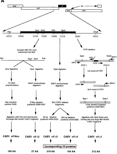
28). The overall strategy for mutant production is depicted in Fig. 1A. The 9-kb
HindIII clone was digested with PstI (positions 4353 to 5408) and cloned into a
Bluescript vector (BsK). The vif Nco2mutant was obtained by digestion at the
NcoI site (position 5285) and filling in with T4 DNA polymerase, resulting in a
4-bp insertion which removed the NcoI site and introduced a frameshift, creating a stop codon at position 5426. The vifD2 mutant was obtained by cleavage at the
SauI site (position 5240) followed by Bal 31 digestion. A 276-bp out-of-frame
deletion was obtained (positions 5068 to 5343), which introduced a stop codon in position 5361. Cleavage of the 9-kb HindIII clone with SpeI (position 5524) and digestion with Bal 31 resulted in two mutants: vifD3, with a 33-bp in-frame deletion (positions 4499 to 5533), and vifD4, with a 225-bp in-frame deletion (positions 5434 to 5660). The vifD1 mutant was created by PCR to delete a 51-bp FIG. 1. (A) vif deletions introduced into the 9-kb HindIII clone of CAEV and (B) structure of the corresponding predicted proteins. (A) The vif gene spans nucleotides 5006 to 5692 (black box). The different strategies used to construct the mutants are described in Materials and Methods. vif Nco2resulted from a 4-bp insertion at the NcoI site (position 5285); vifD2,D3, andD4 were obtained by Bal 31 digestion from the SauI (position 5240) or the SpeI (position 5524) site, respectively;
vifD1 was a PCR deletion mutant. (B) The CAEV wt Vif protein sequence (CAEV Co [28]) is compared with the Vif sequences from visna virus isolates EV1 (29), SA OMVV (25), and K1514 (32). Below the sequences, an asterisk indicates AA identity and a dot indicates homologous AA residues. Underlined is the sequence deleted in the vifD2 mutant (AA 28 to 229). The shaded box (AA 95 to 140) indicates the sequence modified in the vif Nco2mutant, which ends at AA 140. The sequence in brackets (AA 83 to 94) indicates the region where reversions from vif Nco2infected cells were detected. Bars above the sequences indicate the deletions introduced by vifD1 (AA 189 to 205),D3 (AA 165 to 176), andD4 (AA 144 to 217). The open box (AA 170 to 175) indicates the motif conserved among all lentivirus isolates (23).
on November 9, 2019 by guest
http://jvi.asm.org/
[image:2.612.115.497.77.582.2]fragment (positions 5569 to 5621). In the first reaction, two independent PCRs were run with primers VIF4 and VIFDand primers VIF5 and ENV1, with VIFD and VIF5 having overlapping ends. In the second PCR, the amplified products were mixed and run with primers VIF4 and ENV1. The final amplified product was digested with SpeI and SmaI and replaced into the 9-kb HindIII clone.
Cells, transfection, and infection.Primary GSM cells were derived from carpal joints as previously described (21) and maintained in culture in Eagle’s minimal essential medium (MEM) supplemented with 1% glutamine, penicillin, strepto-mycin, and 10% fetal calf serum. Monocytes/macrophages were derived from PBMC freshly isolated on Ficoll-Paque gradients (Pharmacia) as described be-fore (22) and cultured for 10 to 12 days in Teflon bags in RPMI 1640 medium supplemented with 1% glutamine, penicillin, streptomycin, 10 mM HEPES (N-2-hydroxyethylpiperazine-N9-2-ethanesulfonic acid), 1025
Mb-mercaptoethanol, and 10% sheep serum.
Transfections of GSM cells were performed with 5mg of proviral DNA per 50 3104
cells with the Lipofectin reagent (Gibco BRL) as previously described (16). The medium was changed every 3 to 4 days, and the supernatants were harvested to measure the reverse transcriptase (RT) activity and virus titers.
Infections were carried out at a multiplicity of infection (MOI) of 0.01 to 0.04 for 2 h at 378C on subconfluent GSM cells or on six-well plates (Falcon) con-taining PBMC-derived mononuclear cells or macrophages after 10 to 12 days of culture in Teflon bags. Infection of GSM cells was monitored by RT activity. The 50% tissue culture infectious dose (TCID50) per milliliter was determined on
supernatants from infected mononuclear cells or macrophages at days 3 and 7. For PCR and RT-PCR analyses of the replicative cycle, GSM cells were infected under the same conditions except that viral supernatants were pre-treated with 20mg of DNase I (Boehringer) per ml and 10 mM MgCl2for 30 min
at 308C before being added to the cells. Heat-inactivated (30 min at 808C) wt CAEV was used as a negative control. After 2 h at 378C, the cells were washed twice with phosphate-buffered saline (PBS), incubated with 15mg of trypsin type XIII (Sigma) per ml for 15 min at 48C, washed again twice with PBS, and then cultured in MEM–10% fetal calf serum. At different times postinfection, cells were trypsinized and lysed for DNA or RNA extraction.
Transcomplementation assay.The vif gene of CAEV was cloned into the eucaryotic expression vector pRC-CMV (InvitroGen), which contains a neomy-cin resistance gene. Briefly, the vif gene was amplified with the VIF1 and VIF7 primers described below and subcloned into the SmaI site of the Bluescript vector. After sequence verification, a HindIII-XbaI fragment was excised and ligated to HindIII- and XbaI-digested pRC-CMV. GSM cells were transfected by
lipofection with 10mg of the pRC-CMV vector as a negative control (pRC) or 10 mg of this plasmid containing the CAEV vif gene (pRC vif). After selection with geneticin (Gibco BRL; 1 mg/ml) for neomycin resistance, the transfected cells were grown to confluency and then infected with wt or vif2CAEV supernatants containing the same RT activity (10,000 cpm per flask). Complementation was monitored by RT activity measurements in the cell-free supernatants.
RT activity and virus titration.Culture supernatants were clarified by centrif-ugation for 10 min at 5003g and then stored at2808C. The virion particles contained within 1 ml were pelleted by centrifugation for 5 min at 100,0003g
and lysed in 10ml of 10 mM Tris-HCl (pH 7.8)–100 mM NaCl–1 mM EDTA– 0.1% Triton X-100. This solution was mixed with 40ml of 50 mM Tris-HCl (pH 7.8)–20 mM MgCl2–20 mM KCl–2 mM dithiothreitol–0.5mg of
poly(A)-oli-go(dT) (Pharmacia)–2.5mCi of [3
H]dTTP (Amersham). The reaction mix was incubated for 1 h at 378C, spotted onto DE81 paper, air dried, and washed three times for 5 min each in 5% anhydrous Na2HPO4, twice in distilled water, and
once in 70% ethanol. Incorporated radioactivity was measured in a liquid scin-tillation counter (Packard Instruments).
Virus titers were determined on confluent monolayers of GSM cells in 24-well plates (Falcon). One milliliter of undiluted supernatant or 10-fold dilutions, up to 105
, were inoculated in quadruplicate. The plates were incubated for 10 to 12 days; the cells were then fixed with 0.2% glutaraldehyde–2% formaldehyde and stained with 1% crystal violet (Sigma). Wells were scored for the presence of multinucleated syncytia, and the TCID50was determined as the highest dilution
causing syncytium formation in two of the four wells.
DNA and RNA extraction for PCR and RT-PCR analyses.DNA extraction was performed on GSM cells at 2, 6, and 24 h postinfection with the nucleic acid extraction kit Isoquick (Microprobe) according to the manufacturer’s protocol. Extracted DNA was dissolved in distilled water and quantified by optical density. RNA was extracted from GSM cells at 24 h postinfection or from cell super-natants. Ten milliliters of supernatant was ultracentrifuged in a Beckman SW41 rotor for 45 min at 40,000 rpm. The pellet was dried and resuspended in 300ml of RNAzol (Bioprobe), whereas pelleted cells were lysed in 500ml of RNAzol. In both cases, RNA was double extracted according to the manufacturer’s in-structions and then dissolved in 10ml of RNase-free distilled water (for virion-associated RNA) or quantified by optical density (for total cellular RNA).
[image:3.612.136.471.69.399.2]PCR and RT-PCR analyses.For the RT reaction, total cellular RNA was reverse transcribed in a final volume of 20ml with 10 U of avian myeloblastosis virus reverse transcriptase (Promega), 250 ng of random hexamer primers (Phar-macia), and 1 mM each of the four deoxynucleoside triphosphates (dNTPs). The FIG. 1—Continued.
VOL. 69, 1995 ROLE OF vif DURING CAEV REPLICATION IN VITRO 3249
on November 9, 2019 by guest
http://jvi.asm.org/
reaction was run on a Hybaid thermocycler as follows: denaturation for 3 min at 658C, 30 s at 608C, a ramp for 21 min to 398C, followed by 25 min of extension at 398C. Control RT reactions were performed under similar conditions, but the reverse transcriptase was omitted and in this case, no RT-PCR product was detected. Virion RNA was reverse transcribed as described above except that only 40 ng of the random hexamer primers was used.
PCR was carried out in a final volume of 25ml of 13PCR buffer (Promega), 200mM each of the dNTPs, 1.5 mM MgCl2, 6 ng of each primer perml, 1.5 U of Taq polymerase (Promega), and various amounts of cellular DNA or RT
reac-tion product, using the following condireac-tions: a first denaturareac-tion step for 3 min at 948C, followed by 35 amplification cycles of a 1-min denaturation step at 948C, 1 min of annealing at a temperature determined for each primer pair, followed by 1 to 2 min of extension at 728C (depending on the size of the expected PCR product), with a final 10 min of extension. For each temperature change, a scale of 1.5 s/8C was imposed to minimize the effect of the heterogeneity of the apparatus.
After amplification, each sample was run on a 1.5% agarose gel, transferred to a nylon membrane (Hybond N1; Amersham), and hybridized with a specific
32P-labeled oligonucleotide probe. Hybridization was performed at 528C
over-night in 1 M NaCl–10 mM PIPES [piperazine-N,N9-bis(2-ethanesulfonic acid), pH 7.0]–0.1% sodium dodecyl sulfate (SDS)–5% dextran sulfate–0.2% Den-hardt’s reagent. Filters were washed once or twice in 23SSC–0.1% SDS and once in 0.83SSC–0.1% SDS at 528C before being exposed to X-ray film (Kodak) at2708C (13SSC is 0.15 M NaCl plus 0.015 M sodium citrate).
Quantitative competitive RT-PCR.The competitor plasmids were kindly pro-vided by A. P. Ravazolo, P. Turelli, and G. Que´rat. The CAEVDenv mutant was
obtained by deletion of an AatII fragment (positions 8398 to 8581) from the env gene. The CAEVDpol mutant was constructed by deleting 55 bp (positions 3824
to 3879) from the pol gene of the 9-kb HindIII CAEV clone. Amplification of these plasmids with the ENV2/ENV3 and POL S/POL A primers, respectively, led to shorter products than the RT-PCR product obtained from the RNA samples. The amount of viral cDNA in the RT reaction was first approximated by titration against a fivefold dilution series of the mutant env or pol fragment. This was followed by a more precise quantitation by titrating 2ml of cDNA against a threefold dilution series spanning the first determination (26), using the conditions described above.
Primers and oligonucleotide probes.The exact sequences and positions of the primers used have been chosen according to the nucleotide sequence of CAEV (28) and are as follows: R (sense primer), 59-GAGTTCTAGGAGAGTCCCT CC-39(1 to 21); U5 H (hybridization oligonucleotide), 59-GTATTGCACAGAT TAAGGGAC-39(145 to 125); U5 (antisense primer), 59-CTGCGAGAGCCG CTCTGGTA-39(162 to 143); GAG (antisense primer), 59-CAGGATTCTCGA CCACCAAG-39(432 to 413); POL S (sense primer), 59-GATAGGATAGGAG TGCATTG-39(3721 to 3740); POL H (hybridization oligonucleotide), 59-TATT TCCGAAATATATTTGTC-39(3801 to 3781); POL A (antisense primer), 59 -TGAGTCTATGATTCCTCCT-39(4020 to 4002); POL B (sense primer), 59 -TCAGGAGAGTGGAAAGGACC-39(4902 to 4921); VIF1 (sense primer), 59 -CAGGATCCATGCAAAATTCATCCCGCC-39(BamHI site [underlined] and 5006 to 5024); VIF2 (antisense primer), 59-GGTAATTCTGGTCCAGGT-39 (5067 to 5050); VIF3 (sense primer), 59-TGCACCTAAGGAGAGTGA-39(5235 to 5252); VIF4 (sense primer), 59-GTCTGGAAAGACTAGTAC-39(5514 to 5531); VIFD(antisense primer), 59-CCCGTTGTGTGCAATACTTGAAACA CATGC-39(5631 to 5621 [underlined], complementary to the underlined se-quence in VIF5, and 5569 to 5551); VIF5 (sense primer), 59-GACACAAC GGGATACACGCA-39(5621 to 5640); VIF6 (antisense primer), 59-ATGCGT GTATCCCGTTGTGT-39 (5641 to 5622); VIF7 (antisense primer), 59-CAG GATCCCTTTGAGGCAGTTCTTCAC-39(BamHI site [underlined] and 5710 to 5692); REV (sense primer), 59-GATACATGCGCTTAACTGGG-39(6028 to 6047); ENV1 (antisense primer), 59-CCCAGTTAAGCGCATGTATC-39(6047 to 6028); ENV2 (sense primer), 59-CATCAATACTGTATAACCTC-39(8184 to 8203); ENV H (hybridization oligonucleotide), 59-GGACGAGCAGCCTCAG CAGG-39(8745 to 8726); ENV3 (antisense primer), 59-AGATTCCCATCGT CAGCG-39(8766 to 8749); and U3 (sense primer), 59-CCGCAAGTGCTGA CAGATGT-39(8998 to 9017).
RESULTS
Molecular clones and mutants.Figure 1A summarizes the overall strategy developed to construct the different CAEV vif
mutants used in this study. The vif Nco2mutant coded for a
140-amino-acid (AA) protein truncated at its COOH-terminal sequence (Fig. 1B). Moreover, the deduced amino acid se-quence of the truncated protein was divergent after AA 95 (Fig. 2C, panel 2). All the other mutants obtained either by Bal
31 digestion (vifD3 and vifD4) or by PCR (vifD1) were deleted
in domains located in the most conserved regions of the
dif-ferent ungulate lentiviral Vif proteins (Fig. 1B). Vif D1 was
deleted from AA 189 to 205, VifD3 was deleted from AA 165
to 176, and both deletions are contained within the Vif D4
mutant, deleted from AA 144 to 217. The deletion introduced
into the Vif D3 mutant concerned the amino acid motif
SLQXL (positions 170 to 175), described as being conserved
among almost all the retroviral Vif proteins (23). The vif D2
mutant coded for a protein expressing only the first 27 NH2
-terminal amino acids, among which the last 6 were the result of the frameshift introduced by the deletion (Fig. 1B).
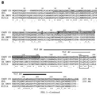
vifgene of CAEV is necessary for efficient viral replication.
The first mutant studied was vif Nco2, with a 4-bp insertion at
the NcoI site (position 5285), which codes for a 140-AA pro-tein instead of the 229 AA of the wt. Viral supernatants
ob-tained after transfection of GSM cells with the wt or vif Nco2
CAEV molecular clone were used to infect GSM monolayers, and infection was monitored by RT activity in the cell super-natants every 3 to 4 days. As shown in Fig. 2A, no obvious difference was observed between the replication of wt and vif
Nco2 CAEV. Total cellular DNA was extracted, and PCR
amplification of the vif gene was performed with the VIF1/ VIF7 primer pair, which produced a 720-bp fragment for both
vif wt and vif Nco2(Fig. 2B, lanes 1 and 2). Digestion of the
amplified products, which contain another NcoI site at position 5609, with NcoI generated fragments of the expected sizes and revealed that only one NcoI site (position 5609) was present in
vif Nco2instead of the two observed in vif wt (Fig. 2B, lanes
3 and 4). This result demonstrated that the initial mutation introduced into the vif gene was still conserved but did not exclude any modification in other parts of the gene.
Amplified products from two independent transfection/in-fection experiments were sequenced, and the nucleotide se-quences are shown in Fig. 2C (panel 1). In the first case, vif R1, a 4-bp deletion occurred (positions 5251 to 5254) upstream from the 4-bp insertion at the NcoI site (underlined). The deduced amino acid sequence (Fig. 2C, panel 2) revealed that an entire Vif protein identical to the wt one except for a stretch of 12 AA at positions 83 to 94 could be expressed. In the second case, vif R2, one base was deleted (position 5249) in the same region as for vif R1 and resulted in a 230-AA protein (one additional asparagine residue) with the same character-istics as Vif R1. It should be noted that the reversion took place in a region corresponding to a variable domain of the ungulate Vif proteins (Fig. 1B). In both cases, the same results were obtained by sequencing DNA prepared from GSM cells infected with supernatants taken at two different times after transfection. Moreover, we were able to detect the reverted sequences in DNA extracted from the transfected cells (not shown). These experiments suggested that the revertant muta-tions occurred during transfection and were contained within the viral inoculum used for subsequent infections, explaining why the initial virus production during infection was not af-fected by the initial vif mutation. These data imply that CAEV needs a functional vif gene for efficient replication. To test this hypothesis, we went on studying the effects of larger vif dele-tions on virus replication and phenotype.
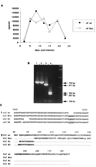
vif2CAEV have slow and low replication rates.The pheno-types of the four vif deletion mutants were established by infecting GSM cells with the same amount of RT activity from transfection supernatants (20,000 cpm per flask), correspond-ing to an MOI of 0.01 to 0.04. The RT activity curves obtained
for vifD1,D2,D3, andD4 were superimposable and revealed
slower and lower growth kinetics compared with that of the wt virus (Fig. 3A). Total cellular DNA was extracted on day 20 postinfection and subjected to PCR amplification with the VIF1/VIF7 primer pair. As shown in Fig. 3B, Southern blot
hybridization of the amplified products with the 32P-labeled
VIF2 oligonucleotide revealed the expected band size in each
case: 720 bp for the wt virus (lane 2), 670 bp for vifD1 (lane 4),
on November 9, 2019 by guest
http://jvi.asm.org/
FIG. 2. Analysis of the vif Nco2mutant. (A) RT activity measurements in the supernatants from GSM cells infected with wt or vif Nco2CAEV. The medium was changed every 3 to 4 days, and RT activity was measured in 1 ml of supernatant concentrated 100-fold. (B) PCR analysis of DNA extracted from cells infected wt (lanes 2 and 4) or vif Nco2(lanes 1 and 3) virus. The amplified products (VIF1/VIF7 primers) were analyzed on an agarose gel, either undigested (lanes 1 and 2) or after
NcoI digestion (lanes 3 and 4). The vif wt has two NcoI sites (positions 5285 and 5609) and generated three fragments of 324, 287, and 109 bp (lane 4). vif Nco2harbors only one NcoI site (position 5609) and generated two fragments, 611 and 109 bp (lane 3). (C) Panel 1. Nucleotide sequences from two revertants (vif R1 and vif R2) isolated from independent experiments, compared with the wt and vif Nco2sequences. The VIF1/VIF7 primers were used to amplify the vif gene to be sequenced, and the region (5243 to 5301) where mutational events resulted in a frameshift is shown. Underlined is the initial 4-bp insertion at the NcoI site. ACAA is the upstream sequence deleted in vif R1, while one A was deleted in vif R2. Panel 2. Deduced amino acid sequences of the Vif proteins encoded by the different vif mutants. Only the region (AA 80 to 185) where the different mutations introduced into the vif gene resulted in sequence modification is shown. Dots indicate identity with the wt sequence.
VOL. 69, 1995 ROLE OF vif DURING CAEV REPLICATION IN VITRO 3251
on November 9, 2019 by guest
http://jvi.asm.org/
445 bp for vifD2 (lane 5), 688 bp for vifD3 (lane 6), and 496
bp for vif D4 (lane 7). In lane 3 of Fig. 3A is shown the
amplified product from vif Nco2 infected cells in a parallel
experiment, whereas no product could be amplified from mock-infected cells (lane 1). These experiments revealed that
deletions introduced into any part of the 39half of the vif gene
of CAEV, as in vif D1, D3, and D4, resulted in the same
attenuated phenotype as in the largest deletion mutant, vifD2.
This supports the importance of the 39 moiety of the protein
for function and confirms the need for a complete and func-tional vif gene in CAEV replication.
Virus titration was carried out on day 27 postinfection
su-pernatants, and the TCID50 appeared to be 10- to 100-fold
lower for all the vif2viruses than for the wt virus (data not
shown). Infection experiments were performed on primary un-differentiated mononuclear cells or on un-differentiated
macro-phages to determine the phenotype of vif2 viruses on cells
which are the natural targets of infection (8, 9, 22). For the
remainder of this study, we tested only vifD2, as being
repre-sentative of all the other vif2 viruses, and the results are
summarized in Table 1. It can be seen that the vif2virus did
not grow on undifferentiated mononuclear cells and grew
poorly on differentiated macrophages (0 and 102TCID
50/ml in
day 7 supernatants, respectively), compared with the wt virus,
which grew at a low level on mononuclear cells and at a high
level on macrophages (102 and 106.75 TCID
50/ml in day 7
supernatants, respectively). These results demonstrated that the slow-and-low phenotype observed on GSM cells can be reproduced on primary blood-derived macrophages, which can
both be considered semipermissive cells for vif2CAEV
rep-lication.
Transcomplementation of vif2 CAEV by vif. In order to
assess whether the modified phenotype of the vif2CAEV was
due to the deletion introduced into the vif gene, a transcomple-mentation assay was carried out by transfection of GSM cells with the pRC vif CAEV construct. Cells expressing the neo-mycin resistance gene contained in this vector were selected in
the presence of geneticin and then infected with wt or vif2
CAEV. As shown in Fig. 4A, the wt phenotype could be re-stored in cells transfected with pRC vif CAEV before infection
with vif2virus, whereas cells transfected with the expression
vector pRC alone before vif2virus infection produced a small
amount of virions, characteristic of the vif2phenotype. The
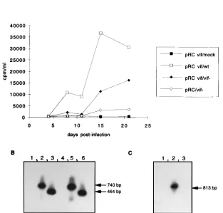
lower RT activity produced in Vif-expressing cells could be explained by the fact that the cells were not cloned and so had variable levels of expression of the vif gene. Virion-associated RNA was analyzed on day 21 postinfection, and RT-PCR was carried with the POL B/VIF6 primer pair, allowing only viral genomic DNA amplification.
The amplified products were analyzed by Southern blot
hy-bridization with the 32P-labeled VIF2 oligonucleotide probe;
the pattern obtained is shown in Fig. 4B. Virions produced by cells infected with the wt virus contained the wt vif gene (740 bp) whether transfected with the vector alone (lane 2) or one expressing the vif gene (lane 5). RNA associated with the
virions produced by the vif2virus-infected GSM cells revealed
the same 464-bp amplified product, in the absence (lane 3) or in the presence (lane 6) of the vif gene product. No signal could be amplified from mock-infected cells whether transfected with pRC (lane 1) or pRC vif (lane 4).
In Fig. 4C are shown the results obtained by RT-PCR anal-ysis on virion-associated RNA. The primer pair used, VIF3/ ENV1, allowed amplification only from wt RNA, since the VIF3 primer is located within the vif deletion. As can be seen, among GSM cells transfected with the pRC vif construct, only those infected with wt virus could produce an amplified signal of 813 bp (lane 2), while no amplification could be obtained
from virions produced by vif2virus-infected cells (lane 3) or
from mock-infected-cell supernatant (lane 1). These results indicated that restoration of the wt phenotype was due neither to contamination nor to recombination and demonstrated that the Vif protein synthesized from an expression vector could act
in trans to complement a vif2 CAEV. We then went on to
FIG. 3. (A) RT activity production by the vifD1,D2,D3, andD4 deletion mutants in infected GSM cells compared with that by wt CAEV. The medium was changed every 3 to 4 days, and RT activity was measured in 1 ml of supernatant concentrated 100-fold. (B) PCR analysis with the VIF1/VIF7 prim-ers on cellular DNA extracted at day 20 postinfection from GSM cells either mock infected (lane 1) or infected with wt (lane 2), vif Nco (lane 3), vifD1 (lane 4), vifD2 (lane 5), vifD3 (lane 6), or vifD4 (lane 7). The Southern blot of the amplified products was hybridized with the32P-labeled VIF2 oligonucleotide
[image:6.612.62.296.65.387.2]probe.
TABLE 1. Titers of wt and vif2CAEV produced by infected-blood-derived mononuclear cells or macrophagesa
Cells
TCID50/ml
wt CAEV vif2CAEV
Day 3 Day 7 Day 3 Day 7
Mononuclear 0 102 0 0
Macrophages 102.5 106.75 0 102
a
Infections were done at an MOI of 0.01, and supernatants were taken on days 3 and 7 postinfection to determine the TCID50on GSM cells. Mononuclear cells
were derived from heparin-treated blood and infected while undifferentiated at day 0. Macrophages were obtained and infected on day 12 after culture of mononuclear cells in Teflon bags.
on November 9, 2019 by guest
http://jvi.asm.org/
[image:6.612.314.556.93.161.2]analyze a single replication cycle of the virus to determine which stage was modified by the vif deletion.
Reverse transcription is as efficient in wt CAEV- as invif2
CAEV-infected cells.GSM cells were infected with superna-tants containing the same amount of RT activity (25,000 cpm
per flask) of wt or vif2CAEV and of heat-inactivated wt virus
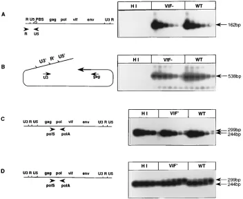
as a negative control. The cells were lysed at 6 h postinfection, and DNA was extracted and analyzed for the levels of strong-stop DNA, which is synthesized early during reverse transcrip-tion. PCR amplification was performed with the R/U5 primers, and the amplified products were revealed by Southern blot
hybridization with the32P-labeled U5H oligonucleotide probe
(Fig. 5A). Serial threefold dilutions of input total DNA indi-cated similar levels of strong-stop DNA, as the penultimate
dilution (1.23 ng) produced the same signal for the wt and vif2
viruses. The complete double-stranded proviral DNA was then analyzed with the U3/GAG primers, and in each case, the last dilution of input DNA (1.03 ng) generated signals of the same intensity (Fig. 5B), indicating that the second jump of the RT
occurred with the same efficiency in wt and vif2CAEVs.
To compare the relative amounts of proviral DNA in wt and
vif2CAEV-infected cells, we used a quantitative competitive
PCR assay, in which a constant amount of cellular DNA was coamplified with serial threefold dilutions of a pol-mutated internal standard containing a 55-bp deletion in the amplified fragment, allowing determination of the DNA amount in the
extract. The analyses were carried out on DNA extracted at 2 and 6 h postinfection. Figure 5C shows that detection of pro-viral DNA was possible at 2 h postinfection, with the same amount of competitor DNA (0.04 pg) being the 50% compet-ing dose. We could not determine whether this signal was due to DNA synthesis following infection or to DNA contained within the viral inoculum. Thus, the analysis was carried out at 6 h postinfection (Fig. 5D), and we determined that the 50%
competing dose was 0.37 pg for the wt as well as the vif2
genomic DNA. This result demonstrated a ninefold increase in DNA synthesis between 2 and 6 h postinfection, indicating active reverse transcription of viral RNA, but did not allow us
to demonstrate any differences between the wt and vif2viruses
at the reverse transcription stage.
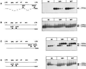
vif2 CAEV produces quantitatively less virion-associated RNA during a single replicative cycle.Transcription was
stud-ied on cellular RNA extracted from 24-h wt or vif2
CAEV-infected GSM cells. After a nonspecific RT reaction allowing the amplification of all the RNA species, specific primer pairs were chosen to amplify the rev, env, and genomic viral RNAs (Fig. 6). As shown in Fig. 6A, the REV/ENV3 primers ampli-fied the spliced rev mRNA from serial threefold dilutions of the RT reaction. We obtained a signal of the same intensity for
the fifth dilution of either wt or vif2rev RNA, suggesting no
[image:7.612.157.470.72.371.2]difference in the transcription of early multispliced mRNAs. An example of a late unspliced or singly spliced mRNA was the
FIG. 4. Transcomplementation of vif2CAEV by the vif gene. (A) GSM cells were transfected with 10mg of the expression vector alone (pRC) or one expressing the vif gene of CAEV (pRC vif), selected in the presence of 1 mg of geneticin per ml, and then infected with wt or vif2viral supernatants (10,000 cpm per flask). (B) PCR analysis with the POL B/VIF6 primers on cellular DNA extracted at day 21 postinfection from cells transfected with pRC (lanes 1 to 3) or pRC vif (lanes 4 to 6) and either mock infected (lanes 1 and 4) or infected with wt (lanes 2 and 5) or vif2(lanes 3 and 6) CAEV. The Southern blot of the amplified products was hybridized with the32
P-labeled VIF2 oligonucleotide probe. (C) RT-PCR analysis of virion-associated RNA extracted from day 21 supernatant. Lane 1, pRC vif-transfected, mock-infected GSM cell supernatant; lane 2, pRC vif-transfected, wt CAEV-infected cell supernatant; lane 3, pRC vif-transfected, vif2CAEV-infected cell supernatant. Amplification was done with the VIF3/ENV1 primers, VIF3 being inside the vif deletion. The Southern blot of the amplified products was hybridized with the32
P-labeled VIF6 oligonucleotide probe.
VOL. 69, 1995 ROLE OF vif DURING CAEV REPLICATION IN VITRO 3253
on November 9, 2019 by guest
http://jvi.asm.org/
env mRNA (Fig. 6C). The relative amount of env mRNA was estimated by using a quantitative competitive PCR assay with the ENV2/ENV3 primers. Constant volumes of the RT reac-tion mix were coamplified with serial threefold dilureac-tions of a competitor DNA containing a 183-bp deletion in the env gene. The 50% competing dose was 1 pg in each case, and this allowed us to conclude that the same amount of env mRNA
was produced at 24 h postinfection in the wt and vif2
CAEV-infected cells. The same approach was used to quantitate the genomic mRNA using the POL S/POL A primer pair (Fig. 6B); the 50% competing dose was 0.37 pg for the wt as well as
for the vif2 virus. All these results suggested that, at the
intracellular level, viral mRNAs were transcribed with the
same efficiency during one replicative cycle of wt or vif2
CAEV, which appeared to be complete within 24 h instead of 72 h, as previously described for visna virus (35) and CAEV (14).
Viral supernatants were collected at 24 h postinfection, and virion-associated RNA was extracted and subjected to quanti-tative competitive RT-PCR with the POL S/POL A primers to quantify virus production. As shown in Fig. 6D, the 50%
com-peting dose was 0.1 pg for the vif2virus, compared with 0.3 pg
for the wt virus. Since these experiments demonstrated a sig-nificant quantitative difference in the relative amount of virion-associated RNA, it could be assumed that the defect intro-duced by the vif deletion was located in the very late stages of the replication cycle, most probably at the moment of virus formation and/or release.
Quantitative defect invif2CAEV production is amplified after a second replicative cycle.The wt and vif2viral particles
produced in the supernatant after the first replication cycle (24 h) were used to infect GSM cells, to analyze the consequences of the vif deletion on a second replicative cycle. Cellular DNA was extracted at 6 h postinfection and subjected to PCR am-plification to detect the strong-stop cDNA and completion of the double-stranded DNA. Semiquantitative analysis of the amplified products revealed that for the early reverse-tran-scribed product (Fig. 7A), the lowest amounts of DNA pro-ducing a detectable signal were 12.3 ng of wt DNA and 37 ng
of vif2DNA. The same threefold difference was also observed
when analyzing the late reverse-transcribed product (Fig. 7B), in which the last dilution giving rise to an amplified signal corresponded to 30.8 ng for the wt DNA and 92.5 ng for the
vif2DNA. Since this difference reflected the input
quantita-tive difference, these results indicated that the vif2 viruses
produced after one replication cycle were not modified in their infectious capacity and that reverse transcription continued normally.
Cellular DNA was prepared at 24 h postinfection and sub-jected to quantitative competitive PCR amplification of the viral genomic DNA with the POL S/POL A primers as de-scribed above. Figure 7C shows that 50% competition was obtained with 0.1 pg of competitor DNA in the case of the wt
virus (third dilution), compared with 0.03 pg for the vif2virus
(fourth dilution), suggesting that transcription during the sec-ond cycle was equally efficient in both cases. Finally, virions produced at 24 h postinfection were concentrated, and associ-ated RNAs were quantified in the same way (Fig. 7D). This
experiment revealed a ninefold decrease in vif2virus
[image:8.612.131.483.71.359.2]produc-tion; the 50% competing dose was 11 fg for the wt virus (third
FIG. 5. PCR analysis of the reverse transcription steps during one replicative cycle of heat-inactivated wt (HI), wt, or vif2CAEV. Cellular DNA was extracted at 2 h (C) or 6 h (A, B, and D) postinfection. (A) Amplification with the R/U5 primers and semiquantitative analysis of serial threefold dilutions of DNA, starting with 100 ng. (B) Amplification with the U3/GAG primers of serial threefold dilution of DNA, starting with 250 ng. In both cases, Southern blots of the amplified products were hybridized with the32P-labeled U5H oligonucleotide probe. Cellular DNAs (100 ng) extracted at 2 h (C) or 6 h (D) postinfection were coamplified with the POL
S/POL A primers in the presence of threefold dilutions of theDpol competitor plasmid, starting with 3.3 pg (C) or 10 pg (D). Both Southern blots were hybridized with
the32P-labeled POL H oligonucleotide probe.
on November 9, 2019 by guest
http://jvi.asm.org/
dilution) and 1.2 fg for the vif2virus (fifth dilution). There-fore, analysis of a second replication cycle gave the same find-ing as the first, namely, no difference in the reverse transcrip-tion or transcriptranscrip-tion events but impaired virus productranscrip-tion, which is more pronounced in the second replication cycle, confirming the role of the vif gene product in the very late steps of viral replication. An increase in this defect during the time course of an infection could account for the slow and low
replication of vif2mutants that we observed in GSM cells (Fig.
3) and primary macrophages (Table 1).
DISCUSSION
This paper describes the phenotype of several vif deletion mutants of CAEV which appeared to have a slow and low replication profile on GSM cells compared with the wt virus. The four vif mutants were obtained by deletion of different
conserved domains among the Vif proteins of ungulate (vifD1
and D4) or all (vif D3) lentiviruses (Fig. 1B), and they all
exhibited the same phenotype as the largest deletion mutant,
vifD2, suggesting the importance of these domains in protein
structure and function. This conclusion was also reached with
the vif Nco2mutant, in which the initial mutation, while being
conserved, was compensated for by a second mutational event, resulting in restoration of the wt vif phenotype. These events took place in a region upstream from the mutation site, which is one of the most variable regions in the ungulate Vif proteins (Fig. 1B). Furthermore, a complementation experiment dem-onstrated that the wt vif phenotype could be restored by
ex-pression in trans of the vif gene (Fig. 4). This suggested the necessity for CAEV to have a functional vif gene in order to replicate efficiently.
We previously reported (1) that the vif gene of visna virus was expressed late in the replication cycle and that the Vif protein was localized in the cytoplasm of infected cells but was not associated with the viral particles. On the basis of the close relationship between visna virus and CAEV, one could assume that these features are also valid for Vif of CAEV. The Vif protein of HIV-1 also shares these characteristics (11, 18, 19), and one report described the existence of HIV-1 Vif as a soluble cytosolic form and as a membrane-associated form in infected cells (11). These authors demonstrated that the C-terminal part of the Vif protein was important in both mem-brane association and function and that the hydrophobic re-gion (residues 142 to 154) containing the highly conserved motif SLQXL was required for complementation of Vif func-tion in trans. Our results demonstrated the importance of this
domain in CAEV Vif function, as the vifD3 mutant, coding for
a protein which is deleted in this domain, has the same
im-paired phenotype as the largest deletion mutant, vifD2. This
[image:9.612.129.481.75.355.2]domain is conserved in 34 of 38 lentivirus Vif proteins, and it is also found in nonviral proteins associated with cell mem-branes (23). It is most likely that at least part of the function of the Vif proteins is conserved among all the lentiviruses as a result of the conservation of this short domain in spite of little or no global sequence similarity. However, the precise function of Vif remains to be definitively established in primate and nonprimate lentiviruses.
FIG. 6. RT-PCR analysis of the transcription steps during one replicative cycle of heat-inactivated wt (HI), wt, or vif2CAEV. Cellular RNAs (A, B, and C) or RNA associated with released virions (D) was extracted at 24 h postinfection. LTR, long terminal repeat. (A) Amplification of rev mRNA with the REV/ENV3 primers from threefold dilutions of the RT reaction mix, starting with 5ml. (B and D) PCRs were performed with the POL S/POL A primers on mixtures containing a constant amount of 5ml of the RT reaction mix and serial threefold dilutions of theDpol competitor plasmid, starting with 10 pg (B) or 1 pg (D). Both Southern blots were
hybridized with the32P-labeled POL H oligonucleotide probe. (C) PCR was performed with the ENV2/ENV3 primers on a mixture of 5ml of the RT reaction mix and
threefold dilution of theDenv competitor plasmid, starting with 3 pg. The Southern blot was hybridized with the32P-labeled ENV H oligonucleotide probe.
VOL. 69, 1995 ROLE OF vif DURING CAEV REPLICATION IN VITRO 3255
on November 9, 2019 by guest
http://jvi.asm.org/
We investigated which stage of the replicative cycle was affected by the vif deletion during replication in primary GSM cells. Quantitative analyses of different steps of the reverse transcription and transcription process through two consecu-tive replication cycles revealed no detectable differences
be-tween the wt and vif2 viruses, indicating that the vif gene
product might act at a posttranscriptional level. However, the
extracellular production of vif2virus showed a threefold
de-crease compared with that of the wt virus; this difference was more marked after the second replication cycle. This quanti-tative difference argues for a role of Vif in particle formation and/or release. Our work did not address the question of the protein content and morphological structure of the released
vif2virions, as recently reported for vif2HIV-1 (13).
How-ever, our experiments demonstrated that the vif2 viral
parti-cles produced at 24 h postinfection were still able to enter GSM cells, suggesting no major effect of Vif on the virion structure and therefore on the infectivity of the virus particles. These results are in agreement with the conclusions drawn by
Blanc et al. (2) and Fan and Peden (5) from studies of vif2
mutants of several HIV-1 isolates grown on permissive cell
lines. These authors and others (7, 10, 19) showed that vif2
HIV-1 and HIV-2 are unable to productively infect activated fresh PBMC. Our results established that primary GSM cells or primary macrophages could be considered semipermissive
for vif2CAEV replication. It thus appeared that, in contrast to
studies on vif2 primate lentiviruses, we did not have fully
restrictive cell lines or primary cells with which to study the
function of the vif gene of CAEV. This could be a potential reason to explain the difference between our results and those concerning the HIV vif gene, but we cannot exclude the pos-sibility that the vif genes of primate and nonprimate lentivi-ruses might have diverged in their function(s).
The ungulate lentiviruses are known to latently infect blood monocytic cells (8, 9, 22) and dendritic cells (12). Our
exper-iments showed that the vif2CAEV did not grow on
undiffer-entiated monocytes and grew poorly on differundiffer-entiated macro-phages compared with the wt virus. This would suggest an important role for Vif during CAEV infection in vivo. Infec-tion of goats with the infectious molecular clone of CAEV resulted in the establishment of persistent infection and patho-genesis characterized by the induction of arthritic lesions in the joints a few weeks after inoculation (34a). Experiments in vivo are in progress to study the effect of the vif deletion on virus replication, persistence, and disease progression in this animal model.
ACKNOWLEDGMENTS
We are grateful to Ana Paula Ravazzolo, Priscilla Turelli, and Gilles Que´rat for providing the env and pol mutant CAEV clones and to our colleagues from INSERM U372 for helpful support and discussions. We thank Pierre Filippi, Bruno Spire, Gilles Que´rat, and Marian Major for critically reading the manuscript.
[image:10.612.125.486.74.360.2]A. Harmache is a doctoral fellow from the French agency against AIDS (ANRS). This work was supported by INSERM and ANRS. FIG. 7. PCR and RT-PCR analyses of a second replication cycle of heat-inactivated wt (HI), wt, or vif2CAEV produced at 24 h postinfection. Cellular DNA was extracted at 6 h (A and B) or 24 h (C) postinfection. Virion-associated RNA was prepared from cell supernatant at 24 h postinfection (D). Primers and32P-labeled
oligonucleotide probes were as described in the legend to Fig. 5. For semiquantitative PCR analyses, the highest inputs of DNA were 1mg (A) and 2.5mg (B) which were then threefold diluted. For quantitative competitive RT-PCR analyses, 5ml of the RT reaction mix was coamplified with a threefold dilution of theDpol competitor
plasmid, starting with 0.1 pg (C and D).
on November 9, 2019 by guest
http://jvi.asm.org/
REFERENCES
1. Audoly, G., N. Sauze, G. Harkiss, C. Vitu, P. Russo, G. Que´rat, M. Suzan,
and R. Vigne.1992. Identification and subcellular localization of the Q gene product of visna virus. Virology 189:734–739.
2. Blanc, D., C. Patience, T. Schulz, R. Weiss, and B. Spire. 1993. Transcomple-mentation of Vif2HIV-1 mutants in CEM cells suggests that Vif affects late steps of the viral life cycle. Virology 193:186–192.
3. Cheevers, W. P., and T. C. McGuire. 1988. The lentiviruses: maedi/visna, caprine arthritis encephalitis and equine infectious anemia. Adv. Virus Res.
34:189–215.
4. Cullen, B. R. 1991. Human immunodeficiency virus as a prototypic complex retrovirus. J. Virol. 65:1053–1056.
5. Fan, L., and K. Peden. 1992. Cell-free transmission of Vif mutants of HIV-1. Virology 190:19–29.
6. Fisher, A. G., B. Ensoli, L. Ivanoff, M. Chamberlain, S. Petteway, L. Ratner,
R. C. Gallo, and F. Wong-Staal.1987. The sor gene of HIV-1 is required for efficient virus transmission in vitro. Science 237:888–893.
7. Gabuzda, D. H., K. Lawrence, E. Langhoff, E. Terwilliger, T. Dorfman, W. A.
Haseltine, and J. Sodroski.1992. Role of vif in replication of human immu-nodeficiency virus type 1 in CD41T lymphocytes. J. Virol. 66:6489–6495. 8. Gendelman, H. E., O. Narayan, S. Molineaux, J. E. Clements, and Z. Ghotbi.
1985. Slow, persistent replication of lentiviruses: role of tissue macrophages and macrophage precursors in bone marrow. Proc. Natl. Acad. Sci. USA
82:7086–7090.
9. Gendelman, H. E., O. Narayan, S. Kennedy-Stoskopf, P. G. E. Kennedy, Z.
Ghotbi, J. E. Clements, J. Stanley, and G. Pezeshkpour.1986. Tropism of sheep lentiviruses for monocytes: susceptibility to infection and virus gene expression increases during maturation of monocytes to macrophages. J. Virol. 58:67–74.
10. Gibbs, J. S., D. A. Regier, and R. C. Desrosiers. 1994. Construction and in vitro properties of HIV-1 mutants with deletions in ‘‘nonessential’’ genes. AIDS Res. Hum. Retroviruses 10:343–350.
11. Goncalves, J., P. Jallepalli, and D. H. Gabuzda. 1994. Subcellular localiza-tion of the Vif protein of human immunodeficiency virus type 1. J. Virol.
68:704–712.
12. Gorrell, M. D., M. R. Brandon, D. Sheffer, R. J. Adams, and O. Narayan. 1992. Ovine lentivirus is macrophagetropic and does not replicate produc-tively in T lymphocytes. J. Virol. 66:2679–2688.
13. Ho¨glund, S., A. O¨ hagen, K. Lawrence, and D. H. Gabuzda. 1994. Role of vif
during packing of the core of HIV-1. Virology 201:349–355.
14. Kalinski, H., A. Yaniv, P. Mashiah, T. Miki, S. Tronick, and A. Gazit. 1991. Rev-like transcripts of caprine arthritis encephalitis virus. Virology 183:786– 792.
15. Kan, N. C., G. Franchini, F. Wong-Staal, G. C. DuBois, W. G. Robey, J. A.
Lautenberger, and T. S. Papas.1986. Identification of HTLV-III/LAV sor gene product and detection of antibodies in human sera. Science 231:1553– 1555.
16. Karger, B. D., and C. Komro. 1990. Evaluation of procedures for transfec-tion using Lipofectinyreagent. Focus 12:25–27.
17. Kim, S., R. Byrn, J. Groopman, and D. Baltimore. 1989. Temporal aspects of DNA and RNA synthesis during human immunodeficiency virus infection: evidence for differential expression. J. Virol. 63:3708–3713.
18. Lee, T.-H., J. E. Coligan, J. S. Allan, M. F. McLane, J. E. Groopman, and M.
Essex.1986. A new HTLV-III/LAV protein encoded by a gene found in cytopathic retroviruses. Science 231:1546–1549.
19. Michaels, F. H., N. Hattori, R. C. Gallo, and G. Franchini. 1993. The human
immunodeficiency virus type 1 (HIV-1) Vif protein is located in the cyto-plasm of infected cells and its effect on viral replication is equivalent in HIV-2. AIDS Res. Hum. Retroviruses 9:1025–1030.
20. Narayan, O. 1990. Immunopathology of lentiviral infections in ungulate animals. Curr. Opin. Immunol. 2:399–402.
21. Narayan, O., J. E. Clements, J. D. Strandberg, L. C. Cork, and D. E. Griffin. 1980. Biological characterization of the virus causing leukoencephalitis and arthritis in goats. J. Gen. Virol. 50:69–79.
22. Narayan, O., S. Kennedy-Stoskopf, D. Sheffer, D. E. Griffin, and J. E.
Clements.1983. Activation of caprine arthritis encephalitis virus expression during maturation of monocytes to macrophages. Infect. Immun. 41:67–73. 23. Oberste, M. S., and M. A. Gonda. 1992. Conservation of amino acid motifs
in lentivirus Vif proteins. Virus Genes 6:95–102.
24. Pyper, J. M., J. E. Clements, M. A. Gonda, and O. Narayan. 1986. Sequence homology between cloned caprine arthritis encephalitis virus and visna virus, two neurotropic lentiviruses. J. Virol. 58:665–670.
25. Que´rat, G., G. Audoly, P. Sonigo, and R. Vigne. 1990. Nucleotide sequence analysis of SA-OMVV, a visna-related ovine lentivirus: phylogenetic history of lentiviruses. Virology 175:434–447.
26. Ramakrishnan, R., D. J. Fink, G. Jiang, P. Desai, J. C. Glorioso, and M.
Levine.1994. Competitive quantitative PCR analysis of herpes simplex virus type 1 DNA and latency-associated transcript RNA in latently infected cells of the rat brain. J. Virol. 68:1864–1873.
27. Sakai, H., R. Shibata, J.-I. Sakuragi, S. Sakuragi, M. Kawamura, and A.
Adachi.1993. Cell-dependent requirement of human immunodeficiency vi-rus type 1 Vif protein for maturation of vivi-rus particles. J. Virol. 67:1663– 1666.
28. Saltarelli, M., G. Que´rat, D. A. Konings, R. Vigne, and J. E. Clements. 1990. Nucleotide sequence and transcriptional analysis of molecular clones of CAEV which generate infectious virus. Virology 179:347–364.
29. Sargan, D. R., I. D. Bennet, C. Cousens, D. J. Roy, B. A. Blacklaws, R. G.
Dalziel, N. J. Watt, and I. McConnell.1991. Nucleotide sequence of EV1, a British isolate of maedi-visna virus. J. Gen. Virol. 72:1893–1903. 30. Schwartz, S., B. Felber, and G. N. Pavlakis. 1991. Expression of human
immunodeficiency virus type 1 vif and vpr mRNAs is Rev-dependent and regulated by splicing. Virology 183:677–686.
31. Sodroski, J., W. C. Goh, C. Rosen, A. Tartar, D. Portetelle, A. Burny, and W.
Haseltine.1986. Replicative and cytopathic potential of HTLV-III/LAV with
sor gene deletions. Science 231:1549–1553.
32. Sonigo, P., M. Alizon, K. Staskus, D. Klatzmann, S. Cole, O. Danos, E.
Retzel, P. Tiollais, A. Haase, and S. Wain-Hobson.1985. Nucleotide se-quence of the visna lentivirus: relationship to the AIDS virus. Cell 42:369– 382.
33. Sova, P., and D. J. Volsky. 1993. Efficiency of viral DNA synthesis during infection of permissive and nonpermissive cells with vif-negative human immunodeficiency virus type 1. J. Virol. 67:6322–6326.
34. Strebel, K., D. Daugherty, K. Clouse, D. Cohen, T. Folks, and M. A. Martin. 1987. The HIV ‘A’ (sor) gene product is essential for virus infectivity. Nature (London) 328:728–730.
34a.Suzan, M., et al. Unpublished data.
35. Vigne, R., V. Barban, G. Que´rat, V. Mazarin, I. Gourdou, and N. Sauze. 1987. Transcription of visna virus during its lytic cycle: evidence for a se-quential early and late gene expression. Virology 161:218–227.
36. Von Schwedler, U., J. Song, C. Aiken, and D. Trono. 1993. vif is crucial for human immunodeficiency virus type 1 proviral DNA synthesis in infected cells. J. Virol. 67:4945–4955.
VOL. 69, 1995 ROLE OF vif DURING CAEV REPLICATION IN VITRO 3257