58
A DISSERTATION ON
A STUDY OF ANTERIOR DECOMPRESSION
AND INSTRUMENTATION IN
TUBERCULOSIS OF SPINE
Dissertation submitted for
M.S. DEGREE EXAMINATIONS
BRANCH - II - ORTHOPAEDIC SURGERY
DEPARTMENT OF ORTHOPAEDIC SURGERY
MADRAS MEDICAL COLLEGE,
CHENNAI – 600 003.
THE TAMIL NADU DR. M.G.R. MEDICAL UNIVERSITY
CHENNAI – TAMIL NADU
59
Certificate
This is to certify that this dissertation entitled “A STUDY
OF ANTERIOR DECOMPRESSION AND
INSTRUMENTATION IN TUBERCULOSIS OF SPINE”
submitted by
Dr. R. PRABHAKAR appearing for Part-II M.S. Branch-II
Orthopaedic Surgery Degree examination in September 2006 is
a bonafide record of work done by him under my direct
guidance and supervision in partial fulfillment of regulations of
the Tamil Nadu Dr. M.G.R. Medical University, Chennai.
PROF.
MAYILVAHANAN
NATARAJAN
M.S Ortho., M.Ch. Ortho (Liverpool), Ph.D., (Ortho. Oncology) D.Sc (Ortho)
Professor & Head of the Department
Department of Orthopaedic Surgery,
Madras Medical College,
Chennai – 600 003.
Dr. KALAVATHY PONNIRAIVAN B.Sc., M.D. (Bio)
DEAN,
60
CONTENTS
1.
INTRODUCTION 1
2.
AIM OF THE STUDY
4
3.
REVIEW OF LITERATURE
5
a.
Historical Background
b.
Baceteriology
c.
Aetio Pathogenesis of tuberculosis
d.
Clinical Aspects
e.
Treatment
f.
Biomechanics of spine
g.
Biomechanics of instrumentation
4.
MATERIALS AND METHODS
29
5.
RESULTS 37
6.
CASE ILLUSTRATIONS
42
7.
DISCUSSION 43
8.
CONCLUSION 50
9.
BIBLIOGRAPHY 51
61
ACKNOWLEDGEMENT
My sincere thanks and gratitude to
Dr. KALAVATHY PONNERIVAN B.Sc., M.D.,(Bio),Dean, Madras Medical College, for permitting me to
utilize the clinical materials of this hospital.
I have great pleasure in thanking my teacher and guide
Prof. MAYILVAHANAN NATARAJAN M.S. Orth (M’as), M.Ch Trauma (L Pool),Ph.D (Orth. Onco)., DSc., Professor & Head of the Department, Department of
Orthopaedic Surgery, Madras Medical College for permitting me to use the
clinical materials and for his valuable advice and encouragement in
preparing this dissertation.
I am very much grateful to Prof. K. ANBAZHAGAN
M.S.Ortho., D. Ortho.,for his valuable support and guidance that he has provided me throughout
this study.
I have great pleasure in acknowledging the help rendered by
Prof. R.H. GOVARDHAN M.S.Ortho., D. Ortho.,for his valuable advice and
guidance.
62
I am very much grateful to Rtd.,
Prof. R. DHANAPAL,
M.S.Ortho., D.Ortho.,for his valuable support and guidance that he has provided me
throughout this study.
I have to make my special thanks to Dr. Nalli R. UVARAJ,
Ms. (Ortho)., D.Ortho who had been my guide for this study and hisconstant guidance and efforts made this study possible.
My sincere thanks to
Dr.R.SELVARAJ, Dr.S.KARUNAKARANfor
their suggestion and help during my study.
I also like to thank Dr. NAGARAJ, Cardiothoracic Surgeon for
helping us in the Thoracotomies.
I am also thankful to all my colleagues and staff members of the
Department of Orthopaedics and Traumatology who helped me in all
possible ways.
60
CONTENTS
1.
INTRODUCTION 1
2.
AIM OF THE STUDY
4
3.
REVIEW OF LITERATURE
5
a.
Historical Background
b.
Baceteriology
c.
Aetio Pathogenesis of tuberculosis
d.
Clinical Aspects
e.
Treatment
f.
Biomechanics of spine
g.
Biomechanics of instrumentation
4.
MATERIALS AND METHODS
29
5.
RESULTS 37
6.
CASE ILLUSTRATIONS
42
7.
DISCUSSION 43
8.
CONCLUSION 50
9.
BIBLIOGRAPHY 51
1
INTRODUCTION
In April 1993, World Health Organization (WHO) has declared
Tuberculosis as a global emergency because it was out of control in
many parts of the world. More than 3.8 million new cases of all forms
of tuberculosis, 90% of them from developing countries, were reported
to the WHO in 2001. However, because of low level of case detection
and incomplete notification, reported cases represent only the tip of the
iceberg and it is estimated that 8.5 million new cases of tuberculosis
occurred Worldwide in 2001, 95% of them in developing countries. It is
also estimated that 1.8 million deaths from tuberculosis occurred in
2000, 98% of them in developing countries.
In India approximately 10 million cases of tuberculosis exists.
1-3% of the 10 million have involvement of bone and joints. The
commonest skeletal lesion is the vertebral lesion which is responsible
for 50% of all bone and joint tuberculosis. The estimated number of
spinal tuberculosis cases in India is between 30,000 and 90,000 cases.
Skeletal tuberculosis constitutes 1-3% of tuberculosis affections
of the human body and spinal tuberculosis accounts for 50% of skeletal
tuberculosis. It is a preventable disease to some extent and totally
curable if treatment is started in early stage. For successful
2
understand the importance of early recognition of the disease and
adherence to the presented regimen of antitubercular treatment.
The advent of modern day multi drug chemotherapy for
tuberculosis has drastically changed the management and the results of
the disease today. But spinal tuberculosis is a unique entity in that the
late complications can have devastating effects because of proximity to
the cord. Ambulant chemotherapy could eradicate the disease
completely, but could not prevent the development of kyphotic
deformity or the neurological deficit precipitated by cord compression.
Earliest method of treatment was to keep the patient in bed with cast or
ambulation with braces. But this form of treatment could only prevent
the deformity to a very small extent.
The commonly used method of placing anterior strut grafts in the
defect created by excision of the infected vertebral bodies corrects the
kyphotic deformity, but the grafts are prone to failure or resorption
especially when more than two vertebrae have been excised.
Instrumentation for tuberculosis began as a correction of
deformities following eradication of disease and was later used even
during active disease to prevent deformity. Oga et al8 study confirms
that the biofilm formation of tuberculous bacilli is not very prominent,
so that penetration of Antituberculosis drug is effective in skeletal
3
protect the graft, stabilize the segments, and prevent progression of
deformity. The idea behind the posterior instrumentation was that it
was away from the infective focus.
Anterior instrumentation, which are very close to the infective
focus has been used in case of moderate to severe kyphotic deformity in
skeletal tuberculosis of dorsal and lumbar region and found to be very
4
AIM OF THE STUDY
To assess the results of anterior decompression and
anterior instrumentation in patients with dorsal and lumbar
5
HISTORICAL BACKGROUND
The tubercle bacillus has co-existed with Homo sapiens since
time immemorial. The Rig Veda, Atharva Veda (3000-1800 BC) and
Samhita of Charaka & Sushruta (1000 & 600BC) recognized the disease
as “Yakshme” in humans, which by its symptoms and signs could only
be tuberculosis of the lungs. Tuberculous lesions have been found in
Egyptian mummies and the Greco Roman civilization recognized
phthisis or consumption as a problem of the lungs.
Laennec (1781-1826), described in the beginning of nineteenth
century, the basic microscopic lesion, the ‘tubercle’ the name by which
the disease is universally known at present.
Tuberculosis of the spinal column was first described by Percival
Pott in 1779. The classical destructive lesion of the disc space and the
adjacent vertebral bodies, collapse of the spinal elements and severe
and progressive kyphosis subsequently became known as Pott’s disease.
BACTERIOLOGY
Tubercle bacilli are mainly of two types; human & bone.
According to Western reports, bovine tubercle bacilli are responsible
for 80% of osteo-articular lesions below the age of 10 years.
The human bacillus is responsible for almost all cases of
6
identification of the bacillus in cold abscess aspirate or biopsy taken
from the site of the lesion or culture of bacilli in Lowenstein Jenson
medium would be necessary in certain cases. In the Indian Scenario,
various studies have shown varying rates of confirmation. 40-80% by
Dahl, 70.8% by Tuli, and 87% by Lakhanpal after culture and guinea pig
inoculation11.
Dobson et al have confirmed Dahl’s findings of 1951 and 197311.
Atypical myobacthsia, other than M. Tuberculosis tumanis or bones
have been reported in lesions of the syrovial sheath. The transmission
of atypical mycobacteria can not be by contact. The following factors
would have to be considered in this regard: (1) Trauma (2) Local steroid
injection (3) Surgical trauma (4) Diabetic status (5) use of chemical
immuno suppressive drugs like cydosporin in organ transplantation
and (6) Acquired immunodeficiency syndrome.
CLINICAL ASPECTS OF SPINAL TUBERCULOSIS
The commonest skeletal lesion in the vertebral lesion which is
responsible for 50% of all bone and joint tuberculosis.
The commonest age of occurrence is the first three decades of life
but it can occur at any age and has been reported from the first year of
life to among those 80 years old. The disease occurs most frequently in
both the sexes. In most cases, the lesion is insidious in onset and only
7
symptoms are weight loss, lassitude and evening rise of temperature.
Locally, there is stiffness, painful restricted joint movements in all the
planes and severe spasm of the surrounding muscles. If the lesion has
been present for a sufficiently long time, a cold abscess occurs in the
soft tissues, tracking its way through the inter muscular planes. A
deformity, in the spine can be present as kyphosis along with local
tenderness and proximal lymphadenopathy.
Tuberculosis of the spine can occur
1. Usually secondary to tuberculosis elsewhere by
haemotogenus or lymphatic spread, most commonly
through Batson’s prevertebral venous plexus.
2. By contiguous extension from a pulmonary abscess,
commonly leading to thoracic spondylitis.
3. As primary infection. This is being increasingly reported,
possibly by ingested bacteria reaching here by
haematogenous route from gastro-intestinal tract.
Tuberculosis of the spine has the following distribution: thoracic
- 42%, thoraco-lumbar - 12%, Lumbar - 26%, Cervical - 12%, Cervico
dorsal - 5%, and Lumbo - Sacral - 3%.
Lesions in the spine is often classified into the following
8
1. Paradiscal: This is the commonest variety and involves the
adjacent margins of two consecutive vertebrae. The
intervening disc space is reduced and the vertebral
margins appear fuzzy. The infection is believed to be via
the arterial blood supply, which is segmental and follows
embryonic pattern that supplies inferior half of superior
vertebra and superior half of inferior vertebra.
2. Central: This involves the central portion of a single
vertebra, keeping the proximal and distal disc spaces
intact. The possible lodgment of the infection comes via
the venous route in this variety.
3. Anterior marginal; The lesion begins as a destructive lesion
in one of the body of a vertebra, minimally involving the
disc space but not involving past of the vertebra on either
sides.
4. Posterior: The disease localizes itself in the posterior
element i.e., the lamina, pedicle or the spinous process.
The infection is said to be coming via the arterial supply to
these structures. There is no involvement of the body of the
vertebra. This variety many a times may present primarily
as neural deficit without any lesion in the body. The
9
posterior midline spinal tenderness over the involved
vertebra much more than vertebral body lesion.
5. Synovial: The disease involves synovium of atlanto-axial
and atlanto-occipital joints.
The clinical presentation of spinal tuberculosis is extremely
protean. The type and severity of symptoms vary depending on the level
of involvement, the severity of the disease and the duration of the
infection. Patient usually present with combination of constitutional
manifestation such as weight loss, fever, fatigue and malaise, a well as
focal pain. Most of the patients present with relatively moderate and
chronic symptoms despite severe vertebral destruction. The pain varies
from mild and constant to severe and activity related. Pain is typically
localized to the site of involvement and is most common in the thoracic
spine. It can be constant and indolent, reflecting the progressive
destruction of the involved disc space and vertebral elements, or it can
be intake and directly linked to spinal motion, coughing and weight
bearing, which is caused by more advanced disc disruption and spinal
instability, nerve root compression, or pathological fracture.
Patients may present with cold abscess tracking out. Since the
cold abscess is the most common and important finding for establishing
10
cold abscess is of great importance. In any region, prevertebral
accumulation of pus is a very noticeable feature.
In the cervical spine, it present as a retropharyngeal a preverbal
shadow, but could also be anatomically located in the following sites: 1)
Behind the prevertebral fasica (2) along the posterior border of
stemomastoid muscle (3) in the supraclavicular area and rarely (4)
down the mediastinum to become an upper mediastinum mass visible
on x-ray (5) in the back of the neck lateral to the posterior spinal
muscles and (6) tracking down the brachial plexus to present in the
axilla or even the elbow joint, along one of the main nervous of the
upper extremity.
A thoracic cold abscess is quite frequently prevertebral or
posterior mediastinal in location. It could, however, track along the
intercostal nerves to present at the following sites:
a) Anterior end of intercostal space.
b) Abdominal wall behind the rectus sheath.
c) Midaxillary line and
d) Along the posterior division of the intercostals nerve
lateral to the sacro-spinalis muscle mass.
In lower thoracic lesions, below D10, the cold abscess might take
11
a) Behind the lateral lumbo-costal arch of the origin of the
diaphragm and present in the per nephritic space or in the
layers of the anterior abdominal wall.
b) Behind the medial lumbo-costal arch of the origin of the
diaphragm and enter the psoas sheath and present as a
psoas cold abscess, palpable above the inguinal ligament or
on the medial aspect of the thigh, if it traverses below the
inguinal ligament.
c) It can go behind the median arcuate ligament of the origin
of the diaphragm along the aorta and its branches and can,
thus have wider sites of presentation, as the lumbar cold
abscess does.
A lumbar cold abscess can spread along the aorta and its
branches to present at the (1) Ischiorectal fossa (2) in the buttock,
under the gluteus maximus (3) along the psoas sheath or (4) in the
lumbo-dorsal (Petit’s) triangle. It can also track down along the femoral
or obtrurator artery and present on the medial side of the thigh;
femoral triangle; popliteal fossa or on the medial side of tendo achilles.
Neurological symptoms of spinal tuberculosis may be subtle, but
will progress on time. Compressive myelopathy is the most common
12
The vertebral regions commonly involved in paraplegia of
hematogenous origin are thoracic, thoraco-lumbar, lumbar and cauda
equina in that order. The level of spinal cord involvement determines
the level of impairment.
Griffith, Seddon & Roaf classified tuberculosis paraplegia in two
grades.
Grade A and Grade B.
Grade A with early onset, within 2 years after onset of symptoms
of tuberculosis and.
Group B: with late onset i.e., after more than 2 years. Group B
paraplegia might be due to recrudescence of disease, mechanical
pressure result of severe kyphosis, inadequate blood supply to the
spinal cord as a result of slow exsanguinations resulting in a fibrous
cord and pathy meningitis. Grade B, in general, has a poor prognosis.
Grade A paraplegia (Pott’s paraplegia) have also been described
as (Goel, 1967, Tuli 1985 Kumar, 1988)
Grade 1: The patient is not aware of the problem. On clinical
examination, there are signs of compression, usually exhibited by long
tract involvement signs or segmental paresis. The patient is able to
13
Grade 2: There is evident spasticity but the patient is able to
walk, often with jumpiness in the gait. Long term involvement signs are
significantly present.
Grade 3: The patient is bedridden and has spastic paraplegia in
extension with demonstrable neurological deficits, both sensory and
motor.
Grade 4: Paraplegia occurs with flexor spasm. There is bladder
and bowel involvement and total sensory and motor loss. The prognosis
is poor.
DIAGNOSIS:
The investigations to establish the diagnosis are primarily x-ray
examination and imaging technique such as computerized axial
tomography (CAT) and MRI.
RADIOGRAPHIC APPEARANCE:
The paradiscal lesions shows a reduction in disc space before
osseous destruction occurs, but focal osteoporosis is seen easier than
disc space reduction. One of the most important diagnostic radiological
criteria is the delineation and study of paravertebral shadows. In the
cervical region, the normal retropharyngeal space is 1.5 cms below the
cricoids cartilage any increase beyond this should make one respect the
14
region, below the 4th dorsal vertebra typical fusiform ‘Bird Nest’
abscess is commonly seen.
Specific radiological appearance in the spine may include, an
aneurysmal type scalloping along the anterior margin of the vertebral
body, mostly as a route of cold abscess under the anterior longitudinal
ligament.
Rarely, lateral curvature of the spine (scoliosis) may be seen but
the most common is kyphotic deformity i.e., increase in the
antero-posterior curvature.
CT scan in spinal tuberculosis has its value in detecting the lesion
in areas otherwise difficult to be seen on x-rays like posterior arch,
cranio vertebral, cervicodorsal, lumbosacral and sacrococcygeal
junction and sacrum.
MRI demonstrates relative sparing of the disc space and at the
same time, involvement of the vertebral bodies on either ride of the
disc. Dissection of the anterior soft tissues, with abscess formation and
collection and expansion of granulation tissue adjacent to the vertebral
body, is highly suggestive of tuberculosis. MRI studies are more able to
reveal epidural abscesses, compression of nerve root or compression of
spinal cord.
Tubercle bacilli grow slowly in culture and confirmation may not
15
liquid media with radiometric growth detection (BACTEC 460) and the
identification of isolates by nucleic acid probes a high pressure liquid
chrompatophy of mycolic acid, help make reports available in 2-3
weeks.
Polymerase chain reaction (PCR) testing is highly specific for
tuberculosis bacillus and provides rapid confirmation of a positive
culture.
DIFFERENTIAL DIAGNOSIS:
Following conditions have to be considered for differential
diagnosis.
1. Traumatic compression fracture:
Normally, the anterior wedging of vertebra and no involvement
of the disc space help. These fractures can be of multiple vertebrae and
no abscess shadow or paravertebral mass is visualized on x-ray
examination.
2. Pyogenic Osteomyelitis:
Clinical presentation is acute with high rise of temperature. The
ESR is well above 100mm/m. There is possibility of septicemia.
Radiological destruction is limited to one or two vertebrae and the
16
3. Salmonella Osteomyelitis:
This can be easily missed. Sickle cell disease individuals are more
prone to get it. Drainage of abscess and culture of organism helps to
establish a diagnosis.
4. Mycetoma Actinomycosis:
It is a rare condition and difficult to differentiate. The diagnosis
needs a radical laminectomy and biopsy examination.
5. Brucellosis:
Drainage and culture of bacilli are needed.
6. Luetic:
A rare condition confirmed by blood examination for syphilis.
7. Echinococcus:
The type of destruction in vertebral body is as punched out with
concomitant destruction of the disc space. Soft tissue extension and
hepatic involvement are present.
8. Chronic Infection:
Rheumatoid (Seronegative) involvement and ankylosing
spondylitis can be differentiated with haematological investigation, no
17
9. Metabolic skeletal osteoporosis:
Senile, postmenopausal, the density of bones is decreased, loss of
osseous trabeculae is noted. No paravertebral mass or abscess shadow
is noted. It is important to exclude hyperthyroidism and
hyperparathyroidism (primary or secondary) as cause of compression
fractures of vertebrae. In all these problems, the interverteberal disc
space is well preserved. The same is true of cortisone induced
osteoporosis.
10. Tumours of the vertebral column:
Either benign or malignant may have to be differentiated from
skeletal tuberculosis. Malignant lesion may either be primary or
secondary.
All malignant tumours have a characteristic bone destruction
pattern but the intervertebral disc space is well preserved.
The other possible pathological lesions that might simulate
tuberculosis of spine are osteochondritis of the scheurmam type and
hemivertibrae. These are rare lesion and have specific radiological
18
TREATMENT
Antituberculosis drug regimens:
The Medical Research Council of the United Kingdom carried out
a series of trials in the late 60’s and early 70’s to establish the
antituberculosis regimen necessary for treatment of tuberculosis lesion
of bones and joints. Though the emphasis in the trials was primarily on
tuberculosis spine, the recommendations are for all types of
musculo-skeletal lesions. Briefly, there is a four drug regimen for the first three
months with dosages of the drugs based on age and body weight of the
patient. The drugs of choice are Rifampicin, Isoniazid, ethambutol and
pyrazinamide, followed by three drugs i.e., Rifampicin, isoniazid and
ethambutol for 16 to 24 months if toxicity develops, the offending drug
is changed. Allergic reaction can occur to any drug; careful attention,
must be paid to toxicity. Thus streptomycin can effect the VIII nerve
resulting in deafness or vestibular functional derangement. Rifampicin
can produce hepatotoxicity and hence SGOT & SGPT levels must be
monitored. Ethambutol can produce depressed thyroid function.
The major aim of treatment is to prevent paraplegia. Most
authors have adopted the use of 4 anti-tuberculosis drugs for a period
of three months initially followed by three drugs for 18 to 24 months.
The drugs used are streptomycin, rifampicin, Isoniazid, ethambutol and
pyrazinamide in children below the age of 12 years. Both streptomycin
19
treatment modality is the middle path. i.e., bed rest, drugs, periodic
review of progress by x-ray and ESR done every 4 weeks. A careful
detailed neurological examination every 3 or 4 days is mandatory. If
there is an increase in neurological deficit, surgical intervention is
desirable.
AUXILLARY TREATMENT:
Steroids are not recommended to be given routinely. Short term
steroid therapy can be given in patients who are in a moribund state,
till anti-tuberculosis treatment starts acting or when patchy meningitis
is present. The addition of steroid might prove crucial. Short term
therapy with anabolic steroids in debilitated malnourished patients
enhances the protein intake but it should avoided in women and
children.
Indications for surgery being:
1. Neurological complications which fail to respond to
conservative care.
2. Paraplegia of the flexor spasm type, with bladder and
bowel involvement and sensory deficit.
3. Neurological status remaining static or
20
5. Mechanical instability after healing.
6. Recurrence of the disease
7. Multiple vertebral involvement in children with severe
kyphosis.
The various techniques of surgical treatment are
1) Costo-tranversectomy where there is a large abscess in the
thoracic regions.
2) Antero-lateral decompression for the paravertebral mass,
either an abscess or granulation tissue. The technique is
primarily an extra pleural exposure of the
abscess/granulation tissue and the vertebral lesion, and
the partial excision of the vertebral body. So that the
pressure on the cord is relieved. Normally 2 or 3 ribs are
removed for about 2 to 3 inches at their vertebral end. The
intercostal artery and nerve are identified and ligated. The
cord with its covering membranes exposed anteriorly and
laterally so that the pulsation of the cord commences after
the decompression. The vertebra are then fused with the
resected ribs. This is the commonest procedure used in this
country.
21
The anterior approaches initially described by Hodgson
(1915-1993) has created lot of interest among ortho surgeons today.
For lesions in the cervical spine extending from the fourth to the
sixth cervical segment, the lateral approach described by Hodgson and
associates to preferred.
To reach thoracic lesions between the seventh cervical and the
fourth thoracic vertebra, a periscapular approach similar to that used
for a first - stage thoracoplasty is employed. The side of the approach is
usually right ride for lesion above D5.
For lesions below the level of the fourth thoracic vertebra, the
approach to usually on the left side. Usually approach is thro’ the rib
bed 2 levels above the apex of the lesion.
For thoraco-lumbar region between the eight thoracic and third
lumbar segments. The ninth rib is removed and as a rule, the
diaphragm is divided along its posterior attachment.
Another satisfactory approach is described by Fey. The eleventh
rib is removed, providing satisfactory access to lesions from the
eleventh thoracic down to second lumbar vertebra. In this procedure
for adequate exposure of the vertebral bodies the psoas muscle must be
detached at its upper end and turned downward. In this region, it must
22
level of the centre of their corresponding vertebral bodies and must
therefore be controlled as the bodies are approached.
For disease involving the second to the fourth or fifth lumbar
vertebra, the twelfth -rib incision of Digby is also used.
Another way to approach the middle lumbar vertebra is to use the
conventional renal incision. Through it one can expose adequately the
first to the fifth lumbar vertebrae.
Anterior cage fixation and posterior instrumentation have been
done and described in various literatures. Anterior instrumentation
which have all along been used in deformity correction has been used in
caries spine in our study. To understand the basis of instrumentation,
we should first know about biomechanics of spine and biomechanics of
23
BIOMECHANICS OF SPINE
4Intervertebral load transmission through the thoracolumbar
spine is a complex condition of spinal balance between the anterior,
middle and posterior spinal column. In the neutral position,
approximately 80% of the spinal load is transmitted through the
posterior facets. However, postural charges, conditions of facet or disc
degeneration or spinal destabilization and reconstruction may alter
these load-bearing properties significantly. The thoracolumbar spinal
loading paths are coupled intricately with the kinematics of the motion
segments, as the anterior and posterior musculoligamentous
constraints serve to actively and passively control three-dimensional
multidirectional flexibility of the functional spinal units. The axis of
intervertebral rotation during flexion and extension in the frontal plane
has been shown to range from the anterior 1/3 to the middle
24
BIOCHEMICS OF SPINE INSTRUMENTATION
2Spinal implants are used to apply corrective forces, to maintain
the correction achieved, and to provide the necessary rigidity to
optimize rates of arthrodesis. All spinal implants serve as temporary
internal splints. The failure to achieve union will result in prolonged
cyclical loading which ultimately results in fatique failure of the
implant. These devices share loads with the spine in a dynamic
relationship in which the implant initially bears most of the load.
Implant loading gradually diminishes as healing progresses and should
be minimal after consolidation of the fusion mass.
Via their attachment sites to the spine both anterior and
posterior implants may apply corrective forces including distraction,
compression and translation.
Posterior implants gain purchase through placement of hooks on
the pedicles, laminae, or transverse process or by threading of wires
around the lamina or through the bases of the spinous process and by
placement of transpedicular screws.
Posterior fixation, works at an increasing distance from the axis
of intervertebral rotation, affords greater leverage and resistance to
25
column support however, leads to a potentially unstable mechanism
and an instrumentation load - bearing configuration.
Anterior devices generally rely on single vertebral body screws.
As the bone-implant interface is within the cancellous vertebral body,
these systems may be inherently less stable than posterior constructs.
Mechanical studies assessing anterior vertebral screws have revealed
maximal stability is achieved with bicortical purchase but with the
addition of a staple preparation of far cortex is not necessary.
Evolution of anterior system2:
The first system was developed by Dwyer and included vertebral
body screws and a braided titanium cable, used in scoliosis correction.
Significant loss of correction occurred in upto 40% of patients. The
flexibility of the system resulted in nonunion in upto 33% of cases.
Second system is the Zielke system5, which used a semi-rigid, 3.2
mm threaded rod. This method relies on compression.
The need for further refinement led to the solid rod systems. The
first was the Texas Scottish Rite Hospital (TSRH) system. A solid rod
(4.8mm or 6.4mm) is precontoured and placed within the screws and
tightened.
Most recently the dual rod constructs have been developed to
26
Kaneda multi- segmental system used two semi-rigid, 4mm rods that
are attached to triangulated vertebral body screws. The screws are
placed through a vertebral plate at each level, improving the pull out
strength by 50%.
For improving the strength of anterior single rod constructs,
strategy involves the provision of structural anterior inter-body
support, which may maintain anterior column length and improve
stability.
Structural interbody devices include femoral ring allografts,
titanium cages and traditionally used iliac crest and rib grafts.
Rib grafts and Iliac crest when used alone were found to collapse,
in the long run.
Femoral ring allografts despite some early subsidence,
maintained anterior column height. An in vivo study in sheep suggested
that distraction could be maintained with titanium cages despite some
loss of height in the early post operative period.
Yilmiz et al13, Govender et al3, have previously adopted this
system of anterior instrumentation, which were used in spinal
deformity correction in scoliosis for deformity correction in caries
27
Oga et al8, have conducted a study using six stainless steel discs
and proved that biofilm formation by tubercule bacilli is less, so that
antibacterial drug penetration on the tuberculosis bacillus is more and
that implant can be used in a tuberculous focus, without an increase in
the risk of infection.
Based on this study, posterior instrumentation, with anterior
strut graft were done by several authors.
Anterior instrumentation used traditionally in scoliosis to
maintain correction, was used in tuberculosis, by Yilmiz et al13 and by
Govender et al3. Based on their observation we have conducted this
28
MATERIALS AND METHODS
This study was conducted in Government General Hospital,
Chennai from May 2002 to April 2006. 20 patients with tuberculosis
spondylitis with neurological deficit for whom anterior decompression
and stabilization was done; were assessed, out of which 13 cases where
taken up for study. Females were marginally high in our study. Male to
female ratio was 6:7.
The age group of the patient ranged from 15-60 years, mean age
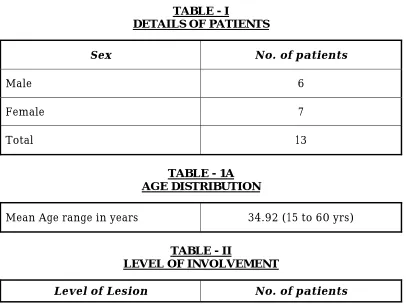
being 34.92. Table-I Dorsal Involvement was more in study Dorsal 8,
[image:34.612.105.511.397.704.2]Dorsolumbar - 3 and Lumbar - 2 (Table -II).
TABLE - I
DETAILS OF PATIENTS
Sex No. of patients
Male 6
Female 7
Total 13
TABLE - 1A AGE DISTRIBUTION
Mean Age range in years 34.92 (15 to 60 yrs)
TABLE - II
LEVEL OF INVOLVEMENT
29
D3 - D4 1
D5 - D6 2
D8 - D9 1
D9 - D10 3
D10 - D11 1
D11 - D12 3
L1 - L2 2
Total 13
INCLUSION CRITERIA:
We included cases of tuberculosis spondylitis, affecting dorsal,
dorso-lumbar and lumbar spine which had been treated by anterior
radical debridement and anterior fusion combined with anterior
instrumentation. All patients had completed their course of
antituberculous chemotherapy and had been followed up for a
minimum period of 5 months.
The indications for surgery were a neurological deficit
(progressive, complete or partial), a cold abscess detected clinically or
radiologically, vertebral destruction with significant angulation and
30
EXCLUSION CRITERIA:
All cases of cervical spine tuberculosis and those cases of dorsal
& lumbar tuberculosis treated by conservative management were not
included in the study.
Those patients with less than 5 months follow up were also
excluded from the study.
Neurological assessment were made according to Frenkel’s
grading. Kyphus angles were recorded as Konstam’s angle on plain
lateral radiographs.
KONSTAM’S & BLEOVSKY METHOD:
The Kyphus angle is determined by drawing lines along the
superior end plate of the normal vertebra above and the inferior end
plate of the normal vertebra below. Usually these lines fail to intersect
on the x-rays. So a construction method is adopted and perpendiculars
are drawn to these lines and the angle made at their intersection is
determined.
This method of determining Kyphus angle was used in few cases,
whereas in cases where the x-rays were stored as images in computer,
31
Auto CAD Program:
This is a computer design software used for structural drawings
and for lot of other purposes. X-rays images are brought into the
program, lines are drawn using the tools and the angles obtained.
Anterior fusion, graft height, graft related problems (fracture,
absorption, subsidence and slippage) and implant related problems
(loosening and breakage) were recorded and assessed.
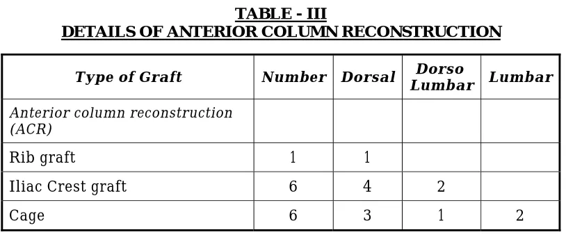
All our patients had an involvement of 1 disc space level. So
fusion 1 space above and 1 disc below was done. Totally 3 motion
segments were immobilised in all our cases.
12 cases had an prevertebral abscess with an average about 70ml
of pus (40-100ml) 1 case had only granulation tissue with no pus.
All patients were managed with anterior debridements
decompression, interbody arthodesis with rib graft in 1 case, Iliac crest
strut graft with zeta instrumentation in 6 cases and titanium cage with
zeta instrumentation in 6 cases. Anterior instrumentation is done in the
form of zeta (moss-miami system). The instrumentation consisted of
rods and screws placed in the vertebral body and extended one level
cephalad and one level caudad to the affected vertebrae. Staple washers
32
TABLE - III
DETAILS OF ANTERIOR COLUMN RECONSTRUCTION
Type of Graft Number Dorsal Lumbar Dorso Lumbar
Anterior column reconstruction (ACR)
Rib graft 1 1
Iliac Crest graft 6 4 2
Cage 6 3 1 2
OPERATIVE TECHNIQUE:
Right Sided Thoracotomy:
This approach was used in cases with lesions upto D5 Vertbera. It
is a periscapular approach with incision through the third rib bed.
Tributaries of the azygos veins needed to be ligated and the vertebra
approached and debridement done.
Left thoracotomy:
For D5 to D11 region, thoracotomy is performed on the left side,
since the pulsating aorta, forms an excellent land mark for the vertebra.
Usually vertebra is approached by resecting a rib two levels above the
level of the lesion.
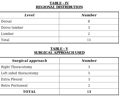
Extrapleural approach:
This approach was used between D11 and L1 after excision of the
[image:38.612.104.511.103.280.2]33
Retroperitoneal approach:
The lumbar region was approached retriperiotoneally, and psoas
muscles erased from the transverse process. Vertebral intersegmental
arteries which run across the middle of each vertebral body needs to be
[image:39.612.107.507.240.566.2]dealt with for exposure.
TABLE - IV
REGIONAL DISTRIBUTION
Level Number
Dorsal 8
Dorso lumbar 3
Lumbar 2 Total 13
TABLE - V
SURGICAL APPROACH USED
Surgical approach Number
Right Thoracotomy 3 Left sided thoracotomy 5 Extra Pleural 3 Retro Peritoneal 2
TOTAL 13
OBSERVATION
The average operating time was 3.7 hours (2.5 hours - 5 hours)
and the average blood loss was 525ml (range 250-300ml for
34
Post operatively, histopathological examination of the caseous
material and the involved vertebra was done, and the diagnosis was
confirmed.
All patients were started on 4 drug course of chemotherapy. INH
- 300mg, Rifampicin - 450mg, Ethambutol-800mg, Pyrazinamide-1g
preoperatively and continued post operatively till suture removal. Then
the patients were registered under RNTCP programme and treatment
continued at home.
The patients were nursed in bed in the post operative period, and
were made to sit up with braces after suture removal on the 12th post
operative day. The brace was advised to be worn for a minimum period
of three months.
Lateral and antero posterior radiographs were made in the
immediate post operative periods, at six weeks and at three months;
after which they were made to come every six months until the time of
the latest follow-up evaluation.
The presence of fusion was determined by the absence of
localized pain and tenderness over the site of arthrodesis, the
maintenance of correction of deformity and evidence of fusion on
radiographs. The erythrocyte sedimentation rate was monitored for the
35
Neurological examination was performed at each follow up visit
with the use of the classification system of Frankel et al. According to
this sytem
Type A - Indicated a complete spinal cord injury.
Type B - A spinal cord injury with only sensation preent.
Type C - An injury with motor function present but not useful.
Type D - An injury wish useful motor function
Type E - An injury with intact neurological function.
Length of the graft varies from 2.5cm to maximum 4 cm. in all
36
RESULTS
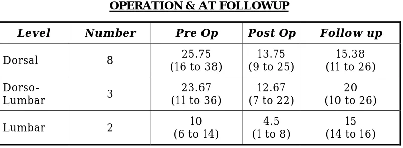
Of the 13 patients who had involvement had an pre-operative
kyphosis that ranged from 60 to 380 with a mean of 19.81. Post
operatively the kyphosis ranged from 10 to 250 with a mean of 10.31. At
follow up the kyphosis ranged from 40 to 260 with a range of 16.79.
All cases showed a progression of the kyphus angle, i.e. there was
loss of correction in all cases. The loss of correction varied from 10 to
170.
The correction achieved was calculated by subtracting pre
operative Kyphus angle from post operative angle.
The final correction was calculated by subtracting the loss of
correction of the kyphosis from the degree of correction achieved at
operation. The mean final correction was 6.380 (–100 to 240).
There was negative final correction in two cases (i.e. loss of
correction more than correction achieved) in 2 cases and in one case
the final correction was zero (i.e. loss of correction equal to the
correction achieved).
Of 2 cases in the lumbar region, one case had an deterioration of
correction to –100 over 19 months period and the other case had O0
37
One case in the Dorso-lumbar region had an negative correction
of –20, in which there was slippage of cage.
Correction was well maintained in the dorsal region, than in
Dorso Lumbar and lumbar region.
Rib graft was found to be inferior, subsidence of graft occurred is
one case. This leads to increase in Kyphus angle.
Maximum correction occurred in the dorsal region and the
correction was also well maintained in the dorsal region.
Dorso lumbar region also had a good correction, but one case had
a slippage of cage, which increased the kyphus angle.
Lumbar region had an negative correction, in the two cases which
we have done one case lost the correction it had achieved and the other
case had more loss of correction.
Maximum correction obtained in our series was 260, which was
achieved in D10- D11 region.
Neurological recovery:
Preoperatively 2 cases were in grade A, 7 in Grade B, 3 in Grade C
and 1 in Grade E. 10 out of 13 cases showed complete recovery. 7 cases
achieved a Frenkel grade E and returned back to their normal activities.
38
regained his normal power which was Grade D and returned back to his
daily activities.
Frenkel Pre Op Follow up
A 2 1 B 7 1 C 3 - D 2 E 1 9
One child regained upto Grade D power at three months follow
up and is still under active physiotherapy at home.
One case recovered completely regained normal power grade E.
But progressively deteriorated and returned to Grade B. This was the
case in which rib graft was used.
One patient had preoperatively and post operatively normal
power.
Two cases which preoperative neurological Grade A, one case did
not recover at all. Both of them developed sacral pressure sores and
were treated wit flap cover. One patient died of pressure sores at 6
months following surgery.
The other patient had co-existent Tuberculosis meningitis also
39
recovery alone, and was lost for further follow up after the last flap
cover.
There was one case of superficial wound infection; which healed
with antibiotics and dressing alone.
Consolidation of the graft occurred at an average of 3 to 5 months
following surgery. Subsidence of the rib graft was found in the only
case, in which we used it.
Three patients died during follow up. One patient died as a result
of pressure sore and its complications. One patient died of myocardial
infarction 1 year following surgery, and the other patient died of fungal
[image:45.612.108.509.449.597.2]granuloma of the eye which had infected the cavernous sinus and died.
TABLE - VI
MEAN (RANGE) KYPHUS ANGLE (DEGREES) BEFORE AND AFTER OPERATION & AT FOLLOWUP
Level Number Pre Op Post Op Follow up
Dorsal 8 (16 to 38) 25.75 (9 to 25) 13.75 (11 to 26) 15.38
Dorso-Lumbar 3
23.67 (11 to 36)
12.67 (7 to 22)
20 (10 to 26)
Lumbar 2 (6 to 14) 10 (1 to 8) 4.5 (14 to 16) 15
TABLE - VII
40
Level Number Correction LOC correction Final
Dorsal 8 (3 to 26) 12 (0 to 3) 1.75 (1 to 24) 10.25
Dorso-Lumbar 3
11 (2 to 17)
7.33 (1 to 19)
3.67 (-2 to 12)
41
ILLUSTRATION
CASE – I :
60 years old lady presented with D9 - D10 caries spine, anterior
decompression and stabilization with Zeta and Iliac crest was done.
Patient had Good recovery. This case has the longest follow up - 42
months.
CASE - II:
38 years old female presented with D10 - D11 caries spine.
Anterior decompression and stabilization with Zeta and Iliac crest was
done. Good recovery was seen in this case. This patient died of fungal
granuloma of the eye.
CASE - III:
40 years old male, a known case of post polio-residual paralysis
presented to us with paraparesis with involvement of D9 - D10 level.
Anterior decompression and stabilization with Titanium cage and Zeta
instrumentation was done. Good recovery.
CASE - IV:
13 years old boy presented with paraparesis with flexor spasm of
both lower limb with intact sensory with involvement of D5 - D6 level.
Titanium cage and Zeta rode instrumentation was done. Patient showed
42
DISCUSSION
The prevalence of spinal tuberculosis has been increasing every
year. Despite the good results of medical treatment with regard to
eradicating the micro organism, kyphosis remains an unresolved
problem. The insertion of strut grafts in the space created after
debridement of the affected vertebral bodies provides some support
anteriorly, but this is usually some what insufficient.
This has led to the use of posterior instrumentation for
additional support either at the time of debridement or at a later stage.
However, posterior instrumentation is associated with increased
operating time, leading to greater blood loss, prolonged anaesthesia
and increased post operative morbidity.
The use of anterior instrumentation can decrease the operating
time, blood loss and post operative morbidity, at the same time give
results comparable to posterior instrumentation.
Our study, though a small study consisting of 13 patients
followed over a period of 5 months to 42 months. We have compared
over study with Sundararaj et al10, Yilmiz et al13 study, Govender et al3
43
Adults were affected in 92.0% of cases which is comparable with
97.4% in Sundararaj et al10, Chen et al study, but differs from the
paediatric profile of Rajasekaran and Soundarapandian9; Oga et al8.
The highest incidence of disease was seen in the dorsal spine
(62%), comparable with Sundararaj et al10 (57.15%), rather than the
dorso lumbar spine reported by Tuli14.
The mean preoperative kyphosis in the dorsal spine (25.750) was
similar to that in Sudararaj et al10 (26.90), MRC trial7, Chen et al12
reported a greater preoperative kyphotic angle of 33.10. Yilmaz et al13
also reported a greater kyphotic angle of 59 degrees (range 340 to 770).
Govendar et al also had mean preoperative kyphotic angle of 590.
The mean kyphosis in the lumbar spine 100 compared with
Sudararaj et al10 study (15.360) was less.
The mean surgical correction in the entire study was9.50, which
is less than compared to 13.70 of Sudararaj et al10, 230 Rajasekaran and
Soundarapandian9. The amount of correction achieved has not been clearly
44
Immediate post operative correction obtained in dorsal region in
our study is 13.750 comparable to Sudararaj et al1015.860, Chen et al12
had an average correction angle of 17.30, 150correction was obtained by
Govender et al3.
In Dorso-lumbar region post operative correction obtained in our
study is 12.670, comparable to 14.60 in Sudararaj et al10 study, and in
lumbar region, our correction was 4.50 compared to – 1.280 in
Sudararaj et al10 study. Other studies have not clearly defined their post
operative correction in dorso-lumbar and lumbar region.
At follow up, our correction in dorsal region has been well
maintained at 15.380 compared to 20.250 in Sudararaj et al10.
In Dorso-lumbar and lumbar regions our mean follow up values
are 200 & 150 respectively, which showed more progression and are
significantly higher values compared to 17.40 and 2.240 in Sudararaj et
al10 study.
The maximum correction achieved in our case was in the Dorsal
region 260, whereas in the Sudararaj et al10 study the correction was
maximum 17.80 in the dorso lumbar spine.
A considerable loss of correction with anterior fusion has been
45
combined with anterior instrumentation and fusion, it has been less
and comparable to posterior instrumentation and fusion.
The mean loss of correction in the dorsal spine in our series was
1.750, comparable to Yilmaz et al13 30, Chen et al12 30, and Sudararaj et
al10 4.80.
In Dorso-lumbar region, we had a loss of correction 7.330, in one
case there was negative correction due to cage slippage, which distorted
our figures, and produced a higher loss of correction, compared to 2.80
correction in Sudararaj et al10, Govender et al3 study showed a loss of
correction from 50 to 250 as they progressed down the lower dorsal
spine.
This could probably be attributed to the increased load
transmission across the dorso lumbar region, where an rethinking
about anterior instrumentation and use of global fusion is very valid.
We had only 2 cases in the lumbar region, of which one had
negative correction (–100) and the other loss of correction O0,
comparing this Sudararaj et al10 study of posterior instrumentation loss
of correction of more than 100 (120, 120 & 250) has occurred.
This again brings to the fore, the doubt about the case of anterior
or posterior instrumentation alone the in lumbar region. Probably
46
Bhojraj et al’s6 observation that “Pedicle instrumentation have
poor purchase in the osteoporotic bone about the infective focus” may
still be valid. Still more cases needs to be done to find out the result.
There was slippage of cage in one case, compared to loosening of
47
ANTERIOR COLUMN RECONSTRUCTION
(ACR & GRAFT RELATED PROBLEMS)
The tricortical iliac crest was used in 6 patients, titanium cage
with morsellized rib graft in 6 patients and rib graft in 1 case.
Despite an intact cortical shell, the weakness of rib grafts can be
attributed to their unfavourable length width ratio, their curvature and
the small surface area of contact witht eh adjacent normal vertebral end
plates. The rib graft is subjected to excess loads with enormous forces
transmitted across the graft and if it spanned more than two levels,
graft fractures have been reported resulting in progressive kyphosis,
Govender et al3. We had one case of graft failure following rib grafting.
Our incidence of infection is very less only one case had
superficial wound infection which settled with antibiotics and dressing.
This implies that there is no additional risk of persistent infection after
adjuvant anterior instrumentation confirming the conclusion by Oga et
al8. Further more Mycobacterium tuberculosis seems to have less
48
NEUROLOGY:
Neurological lesions were observed in 12 patients (92%) of which
8 (62%) recovered Frankel Grade E power, 1 patient who was a case of
post polio residual paralysis regained back his normal power of Grade
D. Two graded as Frenkel A, one of them unchanged after surgery, one
had sensory recovery alone. One child recovered upto Frankel Grade D
power at 5 months follow up.
Our results regarding neurological recovery are comparable with
78% of Yilmiz et al13 study; 92% Sudararaj et al10 study, and 91% of
Chen et al study.
The average operating time in our study was 3.7 hours (3-4.5
hours 3 hours for Dorsal & 4.5 hours for DL & lumbar regions)
compared 5 hours in Sudararaj et al10 study for combined anterior and
posterior surgery and 4.7 hours for Yilmaz et al13 study. In Govender et
al3 study, the operating time was 3.5 hours which was comparable with
our study.
The average blood loss during procedure was 525 ml (400-650ml,
400ml for dorsal region and 650ml for lumbar region) compared to 862
ml (347-1583ml) of Govender et al3, 1000ml (500 to 1400ml) in
Sudararaj et al10 study. In Yilmaz et al13., the loss was at a higher level
49
Our patients were mobilized usually on the 12th post operative
50
CONCLUSION
Based on the results of our study, we like to conclude that
adjuvant anterior stabilization results in early mobilization and
rehabilitation, thereby helping in reducing the morbidity of the
patients.
Radical anterior debridement clears the diseased focus allowing
reconstruction and restoration of the anterior column, as well as stabilization
in the same sitting. Healing of the disease and fusion of the graft across the
affected vertebra are hastened while neurological recovery is unaffected.
The incidence of graft - related problems and the progression of
the kyphosis is significantly less when compared with anterior
debridement and grafting alone.
The post operative loss of correction during follow up is
insignificant for anterior stabilization in the Dorsal spine. For
Dorso-lumbar and Dorso-lumbar regions, global fusion involving both anterior and
posterior stabilization will be the method of choice.
Intra or postoperative complications related to surgery were not seen.
These was no additional risk of infection with the use of implant
anteriorly, even in the presence of large quantities of pus.
51
BIBLIOGRAPHY
1. Bryan W. Cunningham & LTC David W. Polly Jr. The use of
interbody cage devices for spinal deformity. A Bio
mechanical prespective. Clin. Orthop 2002; 394: 73-83.
2. David A. Spiegel, John M. Flym & Dennis. S. Drummond.
Anterior Instrumentation in the treatment of scoliosis.
UPOJ Volume II Spring 1998: 19-26.
3. Govender S. The outcome of allografts and anterior
instrumentation in spinal tuberculosis. Clin. Orthop 2002;
398: 60-66.
4. James A Askhin, Miller and Albert B. Schultz.
Biomechanics of human spine. Basic Orthopaedic
Biomechanics 2nd Ed. 1997; 353-393. Lippincott - Raven.
5. Kostuik JP, Casl A, Ferron S. Anterior Zielke
instrumentation for spinal deformity in adults. JBJS (Am)
1989; 71: 898-912.
6. Mehta JS, Bhojraj SY. Tuberculosis of thoracic spine. A
classification based on the selection of surgical strategies.
52
7. MRC A ten year assessment of a controlled trial comparing
debridgement and anterior spinal fusion in the
management and tuberculosis of the spine in patients on
standard chemotherapy in Hong Kong. JBJS (Br) 1982;
64-B: 393-398.
8. Oga M, Arizono T, Takashita M, Sugoika Y. Evaluation of
the risk of instrumentation as a foreign body in spinal
tuberculosis; Clinical and biological study. Spine 1993; 18:
1890-4.
9. Rajesekaran S, Soundarapandian S. Progression of
kyphosis in tuberculous spine treated by anterior
arthrodesis. J. Bone Jont Surg (Am) 1989;71-A: 1314-23.
10. Sundararaj G.D. Behara S. Ravi V, Venkatesh K., Cherian
V.M, Lee V.Role of posterior stabilization in the
management of tuberculosis of the dorsal and lumbar spine
JBJS (Br) 2003; 85-B: 100-6.
11. Sankaran B. Tuberculosis of Bones & Joints. Ind. Journal
of Tuberculosis 1993, 40-109.
12. Chen WJ, Wu CC, Jung CH, Chen LH, Nui CC, Lai PL.
Combined anterior and posterior surgeries in the
treatment of spinal tuberculosis spondylitis. Clin. Orthop
53
13. Yilmaz C, Sclele HY, Gurnam I, Erdemli B, Korkuruz Z:
Anterior instrumentation for the treatment of spinal
tuberculosis. JBJS 8/A: 1261-1267, 1999.
54
PROFORMA
CS:
STUDY OF ANTERIOR DECOMPRESSION AND INSTRUMENTATION IN TUBERCULOSIS OF SPINE
Name: Age: Sex: IP No:
Unit:
Ward:
Address: Occupation: DOA: DOS:
DOD:
Regional Distribution of spinal TB:
Neurological Status: Frenkel’s Grade:
Vertebral Loss:
Kyphus angle: Pre-OP: Post-OP Loss of correction at follow up
Surgery performed:
Approach to spine:
Operative findings: Pus quantity/graduation tissue:
Duration of surgery: Blood loss:
55
Ant. Column reconstruction (Cage/Graft used):
Length of graft/cage: Instrumentation used:
Length of hospital stay: Time of mobilization:
Complications: Pulmonary/Abdominal/Infection/Graft/Implant
Follow up:
Other remarks:
Neurological assessment: MOTOR
Right Left Right Left Right Left Right Left Shoulder-Abd
Elbow- Flex. - Ext.
Wrist- Flex. - Ext.
Hand Grip Hip - Abd.
Add.
- Flex - Ext.
Knee - Flex. - Ext.
Ankle - DF - PF EHL FHL
Sensory system Reflexes
56
Triceps Supinator Knee
Ankle Plantar
Creamasteric Abdominal
Bladder Bowel
57
MASTER CHART Neurological
Grading Kyphotic Angle S.No Name Age Sex I.P. No. Unit Ward Period Level of
Lesion Procedure Pre
OP Follow up Pre OP Post OP Follow up
Correction
achieved correction Loss of Period of follow
up Complications
1. Pitchammal 60 F 553661 I 19 22.7.02 - 6.9.02 D9-D10 Zeta with
Iliac crest B E 180 150 150,160,160 30 20 42 mths Nil 2. Muthulakshmi 35 F 570090 I 19 4.11.02 - 4.12.02 D8-D9 Zeta with
Rib graft B E,B 290 230 260 60 30 40 mths Recovery Loss of 3. Anbuchezian 40 M 614733 I 2A 19.8.03 - 10.9.03 D11-D12 Zeta with
Iliac crest A A 110 90 100 20 10 6 mths pressure scores Died of 4. Murugan 18 M 674733 I 2A 22.12.03 - 4.2.04 D11-D12 Zeta with
Iliac crest B E 360 220 240 140 20 17 mths Nil
5. Shyamala 38 F 6339733 I 19 11.12.03 - 12.1.04 D10-D11 Zeta with
Iliac crest B E 350 90 110 260 20 17 mths
Died of fungal granuloma of
eye
6. Rani 51 F 664921 I 19 14.6.04- 2.9.04 L1-L2 Cage with
Zeta C E 60 10 20,40,90,160 50 150 19 mths correction Loss of 7. Farzana 26 F 682324 I 19 17.9.04 - 12.10.04 D9-D10 Cage with
Zeta A B 150 30 40 120 10 7 mths Pressure sores
8. Chinnadurai 49 M 675508 I 2A 13.7.04 - 24.8.04 D3-D4 Zeta with
Iliac crest C E 180 30 40 160 10 12 mths Died of MI
9. Valli 24 F 679843 I 19 3.9.04 - 5.10.04 D11-D12 Cage with
Zeta B E 240 70 110,160,210,260 170 190 18 mths Slippage of cage
10. Anandevel 40 M 689236 I 2A 3.10.04 -
8.12.04 D9-D10
Cage with
Zeta C D 160 100 110 60 10 16 mths
11. Vetrivel 18 M 698369 I 2A 14.12.04 -
8.2.05 D5-D6
Zeta with
iliac crest B E 380 230 260 150 30 14 mths
12. Rajiv Gandhi 15 M 788287 I 168 3.9.05 - 16.11.05 D5-D6 Zeta with cage B D 370 250 250 120 – 5 mths
65
CASE - 1
PRE OP
66
FOLLOW UP67
68
CASE II
PRE OP
70
CASE III
PRE OP
71
POST OP
72
ANGLES DRAWN USING AUTOCAD
73
CASE IV
CHILD WITH FLEXOR SPASM
PRE OP
74
POST OP
FOLLOWUP
75
COMPLICATIONS
SLIPPAGE OF CAGE
76
COMPLICATIONS
COLLAPSE OF RIB GRAFT
8 3 2 12 11 5.5 1.75 7.33 10.5 10.25 3.67 -5 -6 -4 -2 0 2 4 6 8 10 12
Number Pre Op Post Op Follow up
MEAN (RANGE) CORRECTION, LOSS OF CORRECTION (LOC) AND FINAL CORRECTION (DEGREES) OF THE KYPHUS ANGLE
Level
Number
Pre Op
Post Op
Follow
up
Dorsal 8
25.75
13.75
15.38
Dorso-Lumbar
3
23.67
12.67
20
Lumbar
2
10
4.5
15
8 3 2 25.75 23.67 10 13.75 12.67 4.5 15.38 20 15 0 5 10 15 20 25 30
Number Pre Op Post Op Follow up