A STUDY OF POST OPERATIVE
COMPLICATIONS AFTER THYROID SURGERY
Dissertation Submitted
for M.S. Degree in General Surgery
Branch I
The Tamil Nadu
Dr. M.G.R. Medical
University
Chennai
CERTIFICATE
This is to certify that this dissertation entitled "A STUDY OF POST
OPERATIVE COMPLICATIONS AFTER THYROID SURGERY"
submitted by Dr. S. MARY PUNITHA to the faculty of General Surgery, The
Tamil Nadu Dr. M.G.R. Medical University, Chennai, in partial fulfillment of
the requirement for the award of M.S. Degree Branch I (General surgery) is a
bonafide research work carried out by her under our direct supervision and
guidance.
Prof. Dr. S.K. PREM THAMARAI SELVI, M.S., Dr. K.P. ARUNKUMAR, M.S.,
Unit Chief Professor and Head of the Department
Dr. T.P. KALANITHI, M.D., Dean
DECLARATION
I, Dr. S. MARY PUNITHA solemnly declare that the dissertation titled
"A STUDY OF POST OPERATIVE COMPLICATIONS AFTER THYROID SURGERY" has been prepared by me.
This is submitted to The Tamil Nadu Dr. M.G.R. Medical University,
Chennai, in partial fulfillment of the requirement for the award of M.S., degree
Examination (General Surgery) to be held in SEPTEMBER 2006.
Place : Coimbatore.
ACKNOWLEDGEMENT
I am extremely thankful to our Dean Dr. T.P. KALANITHI, M.D., for having allowed me to conduct this study in this hospital.
I am extremely grateful to Prof. Dr. K.P. ARUNKUMAR, M.S., Professor and Head of the Department of Surgery for his expert help and friendly advice.
I express my sincere gratitude and extreme thankfulness to Prof. Dr. S.K. PREM
THAMARAI SELVI, M.S., my Unit Chief and for her valuable guidance at all stages of this study.
My greateful thanks to other Unit Chiefs Prof. Dr. R. PERUMAL RAJAN, M.S., Prof. Dr. EASWARAN, M.S., Prof. Dr. A. RAMAMURTHY, M.S., and
Dr. G.S. RAMACHANDRAN M.S., and all E.N.T. Unit Chiefs for their kind Co-operation and for permitting me to study their patients.
I thank all the assistant professors in the surgical units without whom the study
would not have succeeded.
I thank my Co-PGs and last but not the least, I thank all the Patients for their kind
INDEX
S. NO.
CONTENTS
PAGE NO
1. INTRODUCTION 1
2. AIM OF THE STUDY 2
3. REVIEW OF LITERATURE 3
(i) HISTORICAL PERSPECTIVE 3
(ii) SURGICAL ANATOMY 6
(iii) PHYSIOLOGY 12
(iv) PATHOLOGY 15
(v) INVESTIGATIONS 16
(vi) SURGICAL PROCEDURES 17
(vii) COMPLICATIONS AFTER SURGERY 21
(viii) MANAGEMENT AND PREVENTION OF
COMPLICATIONS 22
4. MATERIALS AND METHODS 43
5. OBSERVATION 46
6. DISCUSSION 51
7. DO'S AND DONT'S FOLLOWED IN THIS
INSTITUTION 56
8. CONCLUSION 57
(i) PROFORMA
(ii) BIBLIOGRAPHY
" Surgeons who take unnecessary risks and operate by the clock are
exciting from the onlookers standpoint but they are not necessarily those
in whose hand you would by preference choose to place yourself ".
INTRODUCTION
Surgical procedures on the thyroid gland are generally safe and well tolerated.
Nonetheless, the occasional complications that may follow such surgery may be life
threatening or atleast permanently disabling. The complications derive from
1)anatomical variations of many vital structures associated with the gland in the
neck
2) proximity of the vital structures like RLN and Parathyroid glands to the gland.
Since these complications occur so infrequently despite the high volume of surgery
on the thyroid no surgeon is likely to encounter a large experience with a particular
complication.
A thorough knowledge of the complications, their prevention, and the ability
to recognize early and accordingly manage them has become more important because
of the increased frequency of surgery on this gland.
The exemplary work of Theodor Kocher during the last century in developing
a safe technique for thyroidectomy resulted in a "noble prize" and this allowed
continuing progress in surgery by his successors.
The importance of safe operative technique can hardly be ever emphasized for
it is far better to "prevent" a complication than to "treat" it.
This study is a review of the morbidity attributed to surgical procedures on
thyroid gland.
“The extirpation of thyroid gland for goitre typifies,
Perhaps better than any operation the supreme
Triumph of surgeons art”
AIM OF THE STUDY
(i) To understand the anatomy, pathophysiology and surgical procedures of
thyroid gland.
(ii) To asses varies reasons for complications that arise during the surgery of
thyroid gland.
(iii) To analyse the long and short-term complications of surgery done for various
goiters and malignancies of thyroid gland.
(iv) To identify basic ideas for safety during thyroid surgeries.
(v) Enlist various methods to rectify the attendant complications following thyroid
REVIEW OF LITERATURE
A HISTORICAL PERSPECTIVE:
It was probably Galen who first gave a descriptive account though very brief,
about the thyroid gland in the second century A.D. the time when the art of Medicine
was slavish following the doctrines laid out by him. It was then well accepted that
the diseases were caused by the humors-bile, phlegm and blood. Of course, no
surgery was possible on tumours.
An improved description of the gland is seen in “De Humani Corporis
Fabrica” of Vasalius in 1543. In 1656, Wharton who was interested in the peculiar
shape of the gland, found the resemblance of it to an oblong shield and thus came the
thyroid (GK Thyros-Shield). But his concept of the role of the gland in the human
body was not of a physician but more of a beautician. To him it was an organ to fill
spaces in front of neck, so to have a delicate smooth and elegant neck. This concept
virtually explained and probably convinced many that why females did have a larger
thyroid gland.
Funnier explanations of its existence in the body came later. Many were of the
opinion that it was a lymphatic gland and it received worms! For some it provided
the lubricants for larynx. Probably till towards the middle of the nineteenth century
The first ever thyroidectomy which was in the year of AD 952, is credited to
the bold and venturesome operator of Moorish Origin Albucasis who did it in the
Spanish Arab City of Zahra. He knew very well how to control haemorrhage by
ligature and hot iron, it is quite natural for one to wonder about the indication of
thyroidectomy then. ‘Was it for a female who thought a few crevices, curves and a
scar in front of the neck would improve her looks?
In fact, no basic techniques of thyroidectomy were known and nothing
progressed for hundreds of years. Surgical literature of nineteenth century is deplete
with details of fatalities from thyroid surgery. The hemorrhage which then surgeons
encountered was massive and simply uncontrollable. Desperation and despotism that
was growing among the surgeons were rather tremendous, statement from Leipzig in
1848 said: “If we review all we know concerning operations upon hard goitres we
can only regard with tremendous aversion, these foolhardy performances”. Samuel
D.Gross of Philadelphia went too far to write in 1866. ‘Can the thyroid in the state of
enlargement be removed? Emphatically, experience answers ‘No’. Should a surgeon
be so foolhardy to undertake it ....every stroke of the knife will be followed by a
torrent of blood and lucky it would be for him if his victim lived long enough for him
to finish his blatantly horrid butchery. No honest and sensible surgeon would ever
engage in it.
In this technical and professional void, the dedicated genius of Emile Theodor
Kocher1 (1841-1901), Prof. of Surgery in Berne, Switzerland, was the primary force
that moved thyroid surgery forward. Switzerland being a severe goitre endemic area,
combined antiseptic techniques, thorough knowledge of anatomy and meticulous and
gentle handling and dissection of tissues in his operative methods. He advocated a
thyroidectomy that observed then yet to be discovered parathyroids and anatomical
appreciation of recurrent laryngeal nerve (RLN). Complications of tetany and RLN
injury were 'rarely' encountered and he eventually achieved an astonishingly low
mortality rate of 0.5% for total thyroidectomy, which was more than 90% prior to
Kocher era.
These enviable results were contradistinction to those of Billroth, Kocher’s
only contemporary with similarly large experience in thyroid surgery, who had
complication rates orders of magnitude higher despite being a great surgeon.
Halsted1, who was personally acquainted with both these men, offered the following
explanation.
"I have pondered the question for many years and that the explanation
probably lies in the operative method of the two illustrious surgeons. Kocher, Neat
and precise, operating in a relatively bloodless manner, scrupulously removed the
entire thyroid gland doing little damage outside its capsule. Billroth operating more
rapidly, as I recall, with less regard for the tissues and less concern for haemorrhage,
might easily have removed the parathyroids or atleast have interfered with blood
supply and have left fragments of the thyroid".
Thus it is evident that Kocher’s technique captivated Halsted and evoked his
interest in thyroid surgery Halsted later evolved his own method of thyroidectomy
and described it in a monograph ‘the operative surgery of goitre’ which to this day
SURGICAL ANATOMY
The thyroid gland is placed in front of the neck. It has an average weight of
20 gms It is composed of two lobes joined together by an isthumus. The lobes are 2 to
2.5 cms thick and wide and 4 cm long. Each lobe occupies the thyroid cartilage in its
middle and extends below to the 6th tracheal ring. The thyroid is related to the larynx
and trachea in its posterior aspect and pharynx and oesophagus in its medial aspect
posterolaterally it is related to the carotid sheath2.
Anteriorly it is covered by the ribbon muscles namely the sternohyoid and
sternothyroid, medially it is related to the inferior constrictor muscle and the cricoid
cartilage.
Sometimes a pyramidal lobe ascending from the upper border of the isthmus to
one side of midline is seen. It may be attached to the thyroid cartilage by a fibro
muscular slip called levator glandulae thyroideae2.
The thyroid is invested by the pre tracheal fascia a fibrous sheath derived
from deep cervical fascia. It is thick on the posteromedial aspect and these
thickenings are called the lateral ligaments of Berry2.
The posterior lamina fuses with the pre-vertebral fascia and participates in the
formation of a well-defined posterior wall of retro pharyngeal and retro oesophageal
space. The gland can be easily separated from the trachea except at the posterior
aspect of the isthmus were it is intimately connected. The gland is also provided by a
true capsule, which envelops it closely and sends in numerous fibrous septae to
DEVELOPMENT
The thyroid gland develops as a median downward growth of column of cells
from the pharyngeal floor between the 1st and 2nd pharyngeal pouches, subsequently
marked by the foramen caecum of the tongue. The diverticulum named as median
thyroid diverticulum1 grows down in the midline into the neck, its tip bifurcates and
proliferation of the cells of this bifid end gives rise to the two lobes of the thyroid
gland. A portion of the duct forms the pyramidal lobe of thyroid. The proximal part
of the duct usually disappears and persistence of a part of it gives rise to thyroglossal
cyst3.
Paired lateral anlages originate from the fourth branchial pouch and fuse with
the median anlage at approximately the fifth week of gestation. The lateral anlages
are neuroectodermal in origin (ultimobranchial bodies) and provide the calcitonin
producing parafollicular or C cells, which come to lie in the superoposterior region of
the gland. Thyroid follicles are initially apparent by 8 weeks and colloid formation
begins by the 11th week of gestation1.
HISTOLOGY
Microscopically, the thyroid is divided into lobules that contain 20 to 40
follicles. There are roughly 3x10⁶ follicles in the adult male thyroid gland. The
follicles are spherical and average 30 μm in diameter. Each follicle is lined by
cuboidal epithelial cells and contains a central store of colloid secreted from the
epithelial cells under the influence of the pituitary hormone, TSH. The second group
of thyroid secretory cells is the "C cells" or "parafollicular cells", which contain and
small groups in the interfollicular stroma and located in the upper poles of the thyroid
lobes4.
BLOOD SUPPLY
The thyroid gland is supplied by the superior and inferior thyroid arteries.
Superior thyroid artery arises as the first branch of external carotid artery. At
its origin it is closely related to the external laryngeal branch of superior laryngeal
nerve. As it nears the superior pole of the thyroid gland it divides into an anterior and
a posterior branch. A part of the anterior branch runs close to the medial side of the
thyroid lobe to anastamose above the isthmus with its fellow from the opposite side5.
Inferior thyroid artery arises from the thyrocervical trunk, which is a branch of
the first part of the subclavian artery. Typically it ascends along the medial side of
the anterior scalene muscle behind the prevertebral fascia and loops down medially
on the anterior surface of longus colli. It penetrates the prevertebral fascia and at
about the point of its branching, crosses the more vertically directed inferior laryngeal
nerve, then it turns medially behind the carotid sheath to reach the middle of the back
of the thyroid and divides into upper and lower branches5.
Occasionally the Arteria Thyroidea Ima branch may supply the gland arising
from either the innominate artery or the aortic arch3.
Normally anastamosis of considerable size exists between the arteries of the
thyroid lobes of the same and opposite side. These vessels also anastamose with the
vessels of the trachea and the oesophagus.
VENOUS DRAINAGE
The superior thyroid vein emerges from the upper pole of the gland, runs
The inferior thyroid vein emerges from the isthmus and the medial aspect of the
lower part of the gland. It descends in front of the trachea to end in the inominate
vein6.
The middle thyroid vein emerges from the lower part of the lateral border of
the gland. It runs across the common carotid artery to join the internal jugular vein4.
LYMPHATIC DRAINAGE
There is an extensive lymphatic network within the thyroid gland the superior
lymphatic channel drains the cranial border of the isthmus, much of the medial
surface of the lobes and ventral and dorsal surface of the upper part of the lobes. The
inferior channel drains the major portion of the isthmus and the lower portion of the
lobes.
The upper channel empties into the upper deep cervical nodes. The lower ones
drain into the lower deep cervical nodes including the supraclavicular, pre tracheal
and pre laryngeal nodes. There is an additional drainage from the middle of each
lobe, which passes directly laterally to enter the deep cervical nodes. The deep
cervical nodes communicate with the mediastinal group of lymph nodes6.
NERVE SUPPLY
The thyroid gland in relation to the recurrent laryngeal nerve and to the
external laryngeal branch of the superior laryngeal nerve is of major surgical
significance since damage to these structures leads to a disability in phonation.
The recurrent laryngeal nerve innervates the intrinsic muscles of the larynx
except for the cricothyroid muscle, damage to this nerve leads to vocal cord paralysis
on the sameside. Reddell indicated that among cases in which surgeons avoid rather
damage. It is very important for the surgeon to carefully identify this nerve at the
time of operation.
The right recurrent laryngeal nerve resides in the tracheoesophageal groove in
64% of cases whereas the left recurrent laryngeal nerve was similarly located in 77%
of cases7. The nerve was lateral to the trachea in 28% of cases on the right side and
17% on the left side. In some cases it was found antero lateral to the trachea and in
danger of division during subtotal thyroidectomy if it were not carefully exposed and
its course visualised. The inferior thyroid artery is often used as a landmark for
demonstration of the recurrent laryngeal nerve.
The recurrent laryngeal nerve is thought by many surgeons to run posteriorly
to the inferior thyroid artery. However it passed anterior to the inferior thyroid artery
in 37% of cases on the right side and 27% of cases on the left. In almost 10% of
cases it runs in between the branches of the artery. In 50% of cases the nerve may be
embedded in the ligament of Berry which is of importance because traction of the
gland would put the nerve on stretch and make it subject to section6,10.
A non-recurrent laryngeal nerve can also occur more often on the right side
than on the left6. On the right side it occurs in the presence of a vascular anomaly of
the right subclavian artery, while a left sided non-recurrent laryngeal nerve arises
when the aortic arch develops from the right side. A non-recurrent laryngeal nerve is
at much greater risk of injury during operations in the neck.
The external laryngeal branch of superior laryngeal nerve innervates the
cricothyroid muscle5. In most cases the superior laryngeal nerve lies close to the
vascular pedicles of the superior poles of the gland requiring the vessels to be ligated
vessels while in another 6% the nerve passes through the divisions of the superior
thyroid vessels, coursing over the antero superior portion of the gland. Thus in 21%
of cases this nerve is in great danger of injury when the superior thyroid vessels are
ligated. In only 15% of cases does the superior laryngeal nerve enter the thyroid
pharyngeal muscle before reaching the region of superior pole of thyroid gland thus
protecting it from manipulation by the surgeon8.
PARATHYROID GLANDS
These are two pairs of reddish brown glands, which are usually related to the
posterior aspect of the lobes of the thyroid.
The superior parathyroid is fairly constant in position, lying behind the upper
one third of the lobes and related to the lateral surfaces of the trachea1. It is to be
invariably found in between the capsule of the thyroid and its fascial sheath. It may
be placed forward in which case it is liable to be removed in subtotal thyroidectomy.
The inferior parathyroid glands are usually situated behind the lower part of
the lobes either above or below the inferior thyroid artery as it enters the thyroid
substance, rarely they may be found behind the oesophagus either in the neck or in
the posterior mediastinum or in the retrosternal space9.
Occasionally they are situated within the substance of the gland. The thyroid
itself may be found in the retrosternal space or as a lingual thyroid.
The importance of the parathyroid lie in their intimate relationship with
thyroid and the necessity to protect its integrity during surgical procedures of the
PHYSIOLOGY
The thyroid gland produces metabolic hormones triiodothyronine - T3 and
Thyroxim - T4.
The parafollicular "C-cells produce calcitonin involved in the regulation of
calcium metabolism. T3 and T4 production by the thyroid gland, is regulated by the
pituitary hormone, thyroid stimulating hormone - TSH - by a negative feed back
mechanism, which in turn is regulated by the hypothalamic thyrotrophin releasing
hormone - TRH.
Hypothalamus
Anterior pituitary
Thyroid gland
T3 and T4
T3 and T4 are synthesised from iodine and thyrosine by process of
(i) trapping of inorganic iodide by thyroid cells.
(ii) binding of Iodine to thyrosine.
(iii) coupling to form T3 and T4.
NORMAL THYROID HORMONE VALUES1,4
TSH- point 3 to 3.3 mU/l.
Free T3 - 3.5 to 7.5 micromol/l.
Free T4 - 10 to 30 nmol/l.
Total T3 - 1.5 to 3.5 nmol/l.
Total T4 - 55 to 150 nmol/l.
THYROID HORMONE FUNCTION
Free thyroid hormone enters the cell membrane by diffusion or by specific
carriers and is carried to the nuclear membrane by binding to specific proteins. T4 is
deiodinated to T3 and enters the nucleus via active transport, where it binds to the
thyroid hormone receptor. The T3 receptor is similar to the nuclear receptors for
glucocorticoids, mineralocorticoids, estrogens, vitamin D, and retinoic acid. In
humans, two types of T3 receptor genes (α and β) are located on chromosomes 3 and
17. Thyroid receptor expression depends upon peripheral concentrations of thyroid
hormones and is tissue specific- the α form is abundant in the central nervous system,
whereas the β form predominates in the liver. Each gene product has a
ligand-independent, aminoterminal domain; a ligand binding, carboxyterminal domain, and
centrally located DNA-binding regions. Binding of thyroid hormone leads to the
transcription and translation of hormone-responsive specific genes.
Thyroid hormones affect almost every system in the body. They are important
for fetal brain development and skeletal maturation. T3 increases oxygen
consumption, basal metabolic rate and heat production by stimulation of Na+/K+
ATPase in various tissues. It also has a positive inotropic and chronotropic effect on
the heart by increasing transcription of the Ca2+ ATPase in the sarcoplasmic
reticulum and increasing levels of beta-adrenergic receptors and concentration of G
proteins. Myocardial α receptors are decreased and actions of catecholamines are
amplified. Thyroid hormones are responsible for maintaining the amplified. Thyroid
hormones are responsible for maintaining the normal hypoxic and hypercapnic drive
They also increase gastrointestinal motility, leading to diarrhea in
hyperthyroidism and constipation in hypothyroidsm. Thyroid hormones also increase
bone and protein turnover and the speed of muscle contraction and relaxation. They
also increase glycogenolysis, hepatic gluconeogenesis, intestinal glucose absorption,
PATHOLOGY
CLASSIFICATION OF THYROID SWELLINGS:
Tumours
Benign - Adenoma
Malignant - Follicular Carcinoma
- Papillary Carcinoma
- Medullary Carcinoma
- Anaplastic Carcinoma
- Lymphoma
Goitres
Toxic - Primary toxicosis
- Nodular toxicosis - Solitary nodule
- Multinodular
Non Toxic - Colloid goiter
- Multinodular goitre
Inflammatory swellings
Autoimmune - Chronic lymphocytic thyroiditis
Hashimoto's disease
Granulomatous - deQuervain's thyroiditis
Fibrosing - Riedel's thyroidist
Infective - Acute - Bacterial,viral thyroiditis, subacute thyroiditis
Chronic - Tuberculous, syphilitic.
Investigatio
DIAGNOSTIC TESTS OF THE THYROID GLAND.
Measurements of thyroid gland function
• Serum T4, Free thyroxine, resin T3 uptake, free thyroxine index, Serum
T3, Radioactive iodine uptake, Serum TSH, Thyroxine binding globulin.
Measurers of auto immunity
• Antithyroglobulin antibodies, antimicrosomal antibodies, long acting
thyroid stimulator, thyroid stimulating immunoglobulins.
Measurers of thyroid and pituitary responsiveness.
• T3 suppression test, TRH stimulation test and TSH stimulation test.
Assessment of Thyroid anatomy
• Thyroid isotope scan
• Ultra sonic scan
Assessment of thyroid histology
• Aspiration biopsy with cytology
• Core needle biopsy
Assessment of extension of goiter and to detect secondaries
• CT Scan
Surgical
Procedures
SUBTOTAL THYROIDECTOMY
SUPERIOR AND INFERIOR FLAPS RAISED.
SURGICAL PROCEDURES
The common types of surgical procedures performed on the thyroid gland are
Hemithyroidectomy - Removal of one lobe of thyroid along with the
isthumus.
Subtotal thyroidectomy - Removal of all but a small remnant of thyroid
tissue on both lobes of thyroid.
Total thyroidectomy - Removal of all the thyroid tissue.
Surgery of the thyroid gland requires a good knowledge of the anatomical
details and a capacity to conduct meticulous and time consuming dissection of vital
structures. Extent and type of surgery will depend on the certainty of diagnosis, type
and pathological extent of the disease process.
PREMEDICATION AND ANAESTHESIA
In all toxic cases effective premedication is most important, in view of the
effect of emotional stress and increased thyroid activity. The patient should have a
good night’s sleep prior to operation and reach operating theatre in a calm state of
cases, and is essential when deviation or constriction of trachea has been produced
since the ensured airway reduces venous congestion. The neck veins can be further
emptied by tilting the operating table and patient to about 15o head up position.
TECHNIQUE6,13
A small pillow or sandbag is placed between the shoulders so that the neck is
TOTAL THYROIDECTOMY
SUPERIOR POLE LIGATION
supported on a ring. A horizontal incision is made about two fingerbreadths above
the clavicles, preferably in a natural skin crease6.
Incision is carried through the first layer (Superficial) of cervical fascia and
platysma muscles. The upper and lower skin flaps are raised upward almost to the
notch of the thyroid cartilage and downward to the supraclavicular region
respectively.
A midline vertical incision is made through the deep layer of cervical fascia
that surrounds the strap muscles. Using blunt dissection, strap muscles are separated
from the underlying thyroid gland. Thyroid gland is palpated gently all around.
Usually it is not necessary to transect the strap muscles, as they are readily
retractable. But if exposure is not adequate, they are sectioned at or above the level
of cricoid cartilage to preserve its nerve supply. If malignancy is suspected enlarged
lymph nodes in the para glandular and tracheoesophageal regions are carefully looked
for. A plane is developed between true and false capsule. Using gentle dissection
strap muscles and fascia are dissected from the superior pole. With gentle
anteromedial traction of the thyroid lobe, dissection is carried through the areolar
connective tissue between thyroid and carotid sheath exposing middle thyroid vein,
which is ligated and divided as far from internal jugular vein as possible.
Gentle lateral traction either using fingers or Allis forceps is applied just
below, the superior polar vessels which helps to define the plane between superior
polar structure, the superior laryngeal nerve and pharyngeal musculature. A
right-angled Moynihan’s clamp is passed into this plane to free the vessels which then are
ligation is kept as low close to thyroid lobe as the pathologic process permits. Then
the inferior thyroid vein is dissected free, ligated and divided. After dividing the
pedicles, under direct vision and using blunt dissection lateral lobe is liberated
forward and medially in a rotatory movement so that its posterior surface is fully
brought in view for the visualisation of important structures.
Parathyroid glands are identified as caramel color usually an oval mass of
tissue lying in some adipose material9. Its blood supply is to be preserved. For RLN
to be identified, the dissection is carried to the tracheal rings and the Beahr’s
triangle10 is demonstracted. Beahrs triangle is formed by common carotid artery, ITA
and RLN. If any troublesome oozing occurs here, clamps and cautery are better
avoided.
Now the thyroid isthmus is clamped and divided along with the anterior
suspensory ligaments are extension and condensation of pretracheal fascia that runs
from gland to cricoid cartilage and trachea. In total thyroidectomy now the complete
lobe is removed without injuring the posterior structures. In subtotal resection
division is carried through the thyroid gland, so as to leave posterior wedge of tissue,
the amount of which varies with the pathology. In tryrotoxicosis 4-5 gms and in
nodular or diffuse colloid goitre, 6-8 gms are usually preserved, roughly a piece of
2-3 cm in length and about 1.5cm wide59. Resection of the gland is done after applying
serial clamps through the tissue over running with 0 chromic catgut is applied to the
The same sequences are repeated if it is not hemithyroidectomy on the
contralateral side. Wound is examined for bleeding sites after saline irrigation and
flexing the neck. Absolute hemostasis is a basic essential.
A small soft tube drain or a corrugated rubber drain is brought out through a
small opening in strap muscles and through a small opening in the lower flap. Strap
muscles are reapproximated with interrupted 2 0 chromic catgut. Platysma and
superficial fascia also are approximated in the same way. Skin is closed with 3 0 non
absorbable, interrupted or subcuticular sutures.
Drain usually is kept for 24-48 hours. Sutures are removed on 4th or 5th day,
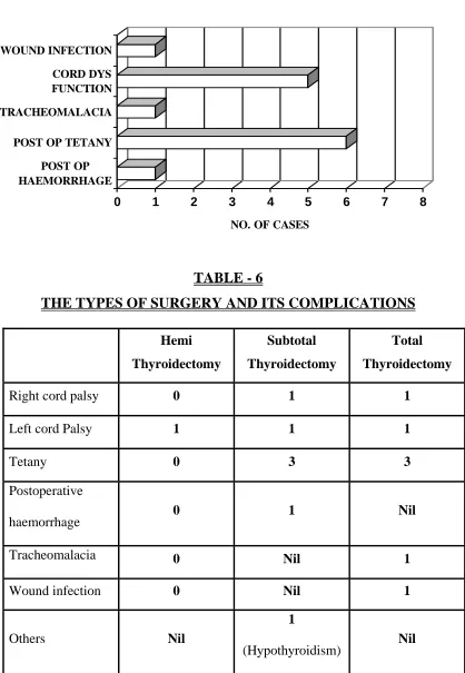
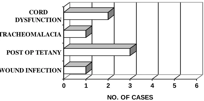
COMPLICATIONS AFTER SURGERY
Numerous complications may arise following surgical removal of the thyroid
gland. These problems often result from either the surgical technique or from
metabolic disturbances. Although the incidence of these complications is low, some
problems are seen more frequently than others (Netterville & Ossoff, 1990)14.
Complications associated with thyroid disease are,
Early Complications
1. Wound problems
Collections - Seroma formation
Infections - Subcutaneous infection
Deeper infection
2. Haemorrhage
Tension - haematoma
Subcutaneous haematoma
3. Respiratory obstruction
Laryngeal oedema
Collapse / kinking of trachea
4. Parathyroid insufficiency
Due to - removal
- infarction
5. Thyroid storm
6. Nerve Injuries
Injury to superior laryngeal nerve: Recurrent laryngeal nerve paralysis
Late complications
7. Thyroid insufficiency
8. Recurrent Thyrotoxicosis
9. Hypertrophic scar - keloid formation
Management and
Prevention of
MANAGEMENT AND PREVENTION OF COMPLICATIONS
WOUND COMPLICATIONS
Seroma is a collection of serous fluid in the subcutaneous region. Serous
oozing from the raw areas collects in the most dependent parts of the neck, usually in
the supraclavicular fossa16.
Treatment is by aspiration with a wide bore needle daily until no more serum
collects. After each aspiration apply a mild compression dressing using fluffed up
swabs held on with elastic strapping.
Avoiding unnecessary extensive dissections and obliteration of dead spaces
and achieving good haemostasis reduces this complication.
INFECTION
Infection was the major cause of death from thyroid surgery during the 1800s.
Today, infection occurs in fewer than 1-2% of all cases16. Death is unlikely if the
infection is recognized and treated promptly and appropriately.
PRESENTATION
Postthyroidectomy infection may manifest as a case of superficial cellulitis or
as an abscess. Patients with cellulitis typically present with erythema, warmth, and
tenderness of neck skin around the incision. A superficial abscess may be diagnosed
by fluctuance and tenderness. A deep neck abscess may manifest more subtly, but
signs such as fever, pain, leukocytosis, and tachycardia should raise clinical
EVALUATION
Send purulence expressed from the wound or drained from an abscess for
Gram stain and cultures to indicate the choice of antibotics. CT imaging is useful
when a deep neck abscess is thought possible. In addition, a deep neck abscess
should raise concern for a possible esophageal perforation. A gastrographing
esophageal swallow study may be useful in certain cases.
PREVENTION
The key to prevention of postoperative infection is sterile surgical technique.
The routine use of perioperative antibiotics in thyroid surgery has not been proven
beneficial. Johnson and Wagner16 performed a retrospective review of 438 patients
who had uncontaminated head and neck surgery at the Eye and Ear Hospital of
Pittsburgh. Of 113 patients who had thyroidectomies, only 12 received perioperative
antibiotics. None of the thyroidectomy patients experienced a postoperative wound
infection, perioperative antibiotics are not useful. Antibiotics should not be used
unnecessarily in this era of multidrug-resistant bacteria. Perioperative antibiotics are
not recommended for thyroid surgery.
TREATMENT
Treat cellulitis with antibiotics that have good gram-positive coverage (eg:
against staphylococci and streptococci). Drain abscesses, and direct antibiotic
coverage according to culture findings. Start patients with deep neck abscesses on
broad-spectrum antibiotics (eg: cefuroxime, clindamycin, ampicillin/sulbactam) until
HAEMORRHAGE
It may be primary haemorrhage or reactionary haemorrhage. It may produce
either a subcutaneous haemotoma or a deep tension haematoma.
Subcutaneous haematoma
Haemorrhage from smaller vessels may collect between deep fascia and the
flap and gives rise to a diffuse swelling of the neck. This can be dealt with by
evacuating the clot and insertion of sutures or by probing later after the clot has been
allowed to become softer.
Achieving good haemostasis while raising the flaps and dissection of the flaps
at the correct plane prevents this complication. Use of diathermy on the flaps carries
the risk of producing burn wound of the skin. Bipolar can be used safely on the flaps.
Tension haematoma
Usually occurs due to slipping of vascular pedicle with haemorrhage deep to
the strapmuscles. It can produce stridor and cyanosis without obvious swelling of the
neck. This is an emergency and is dealt with by promptly removing the sutures at
bedside, and evacuation of the clot from beneath the deep fascia thereby relieving the
compression on the trachea. Then the patient is taken to the operation theatre and
anaesthetised and bleeding points ligated.
Attempts to arrest the oozing haemorrhage from the wound by applying
pressure dressing to the neck or attempts to pack the bleeding sites should not be
infection, necrosis of skin flaps. Moreover the tension haematoma leads to the
development of laryngeal oedema and airway obstruction.
Ensuring perfect haemostasis, leaving the gap in the lower end while suturing
the strap muscles in the midline so as to allow the blood to come out of the relatively
closed compartment into the sub-platysmal space where it can be easily detected and
use of drainage tube wherever necessary prevents this alarming complication17,19,20.
Respiratory obstruction:
Respiratory tract obstruction following thyroid surgery can be lethal and may
be difficult to recognize.
The causes include
1. Tracheal compression due to haemorrhage - Tension haematoma
2. Laryngeal oedema
3. Recurrent laryngeal nerve injury
4. Laryngotracheal displacement from a goitre that is not improved with surgery.
5.Tracheal collapse of kinking.
Respiratory obstruction due to collapse or kinking of trachea is very rare.
Most causes are due to laryngeal oedema.
Most important cause of laryngeal oedema is a tension haematoma. If the
strap muscles are closed too tightly, haematoma may not be evident under the skin.
However, the clots may dissect below the strap muscles in the peritracheal area along
Total airway obstruction may progress within notime, once the critical
compression in this tight compartment below the strap muscles is reached which
leads to impairment of venous and lymphatic drainage leading to laryngo pharyngeal
oedema. Total compression may not be the real site of obstruction but the oedema of
laryngo-pharynx may be the site of obstruction.
Hence it is suggested not to close the strap muscles very tightly so that any
haematoma, which develops, can become apparent in the subcutaneous region17.
Trauma to the larynx by anaesthetic intubation and surgical manipulation are
important contributory factors, particularly if the goitre is very vascular and may
cause laryngeal oedema without a tension haematoma.
Unilateral or bilateral recurrent laryngeal nerve paralysis will not cause
immediate post-operative respiratory obstruction unless laryngeal oedema is also
present but they will aggravate the obstruction.
If releasing the tension haematoma does not immediately relieve airway
obstruction, the trachea should be intubated atonce. An endotracheal tube can be left
in place for several days. Steroids are given to reduce oedema and a tracheostomy is
rarely necessary. Intubation in the presence of laryngeal oedema may be very
difficult and should be carried out by an experienced anaesthetist. Repeated
unsuccessful attempts may aggravate the problem and in a crisis, it is safer for the
inexperienced surgeon to perform a needle tracheostomy as a temporary measure, a
RECURRENT LARYNGEAL NERVE INJURY
The recurrent laryngeal nerve (RLN) innervates all of the intrinsic muscles of
the larynx with the exception of the cricothyroid muscle, which is innervated by the
superior laryngeal nerve (SLN). Mechanisms of injury to the nerve include complete
or partial transection, traction, contusion, crush, burn, misplaced ligature, and
compromised blood supply. The consequence of an RLN injury is true vocal fold
paresis or paralysis42.
PRESENTATION
Patients with unilateral vocal fold paralysis present with postoperative
hoarseness or breathlessness. Presentation is often subacute. Initially, the vocal fold
usually remains in the paramedian position, thus affording a fairly normal voice. The
paralysed vocal fold will atrophy, causing the voice to worsen. Other potential
sequelae of unilateral vocal fold paralysis are dysphagia and aspiration. Bilateral
vocal fold paralysis may occur following a total thyroidectomy, and usually manifests
immediately after extubation. Both vocal folds remain in the paramedian position,
causing partial airway obstruction. These patients may have biphasic stridor and be
in respiratory distress. Occasionally, patient exhibits airway signs in the immediate
postoperative period, because the airway is sufficient despite the paralyzed vocal
folds. Such patients may present at followup with dyspnoea or stridor with
EVALUATION
Techniques for assessing vocal fold mobility include indirect and fiberoptic
laryngoscopy.
Laryngeal electromyography21 (EMG) may be useful to distinguish a vocal
fold paralysis from cricoarytenoid joint injury secondary to intubation. Furthermore,
EMG may yield information concerning the prognosis of RLN injury. Parnes et al22
performed laryngeal EMG on 24 patients with vocal fold paralysis from numerous
etiologies. Since, most of these tests were performed more than 6 months after the
onset of paralysis; this study reveals little regarding the usefulness of early EMG
testing.
The patient with bilateral true vocal fold paralysis presenting with airway
obstruction after extubation likely requires emergent reintubation or tracheotomy.
PREVENTION
Deliberate identification of the RLN minimizes the risk of injury23,25. When
the nerve is identified and dissected, the reported RLN injury rate during
thyroidectomy is 0-2.1 %. Conversely, when the nerve is not clearly identified the
reported injury rate is 6.6%24. The RLN can be located in many ways. Intraoperative
hemostasis and a thorough understanding of the anatomy are essential for nerve
identification and preservation.
The course of the RLN differs on the right and left sides of the neck7. Classic
descriptions of the RLNs hold that they ascend within the tracheoesophageal groove;
RLN is more oblique, lateral, and, probably, more prone to injury than the left RLN.
The nerve may branch multiple times before entering the larynx. Take care to
identify and preserve each branch6.
In approximately 5 of 1000 patients, a nonrecurrent laryngeal1 nerve is found
on the right23. This occurs when a retroesophageal right subclavian artery arises from
the dorsal side of the aortic arch. A left-sided nonrecurrent laryngeal nerve can occur
rarely.
The inferior thyroid artery has been described as an important landmark for
identifying the RLN24. However, its relationship to the nerve is subject to much
variation. Numerous descriptions and attempts to quantify the percentages of each
relationship of the nerve to the artery have been put forth. Percentages differ on the
right and left. On the right, the nerve runs between branches of the artery in
approximately 50% of patients. The nerve is found anterior to the artery in 25% and
posterior in 25%. On the left, the nerve courses posteriorly to the artery in 50% of
patients; in approximately 35%, the nerve runs between branches, but the exact
relationship cannot be determined with certainty. The inferior thyroid is therefore not
a dependable landmark for identifying the nerve.
Perhaps the most efficient way to identify the nerve is to locate it within the
carotid triangle24,25. The carotid artery and trachea comprise the lateral and medial
sides of the triangle, respectively. The tissue within this triangle is spread gently, a
layer at a time, until the nerve is identified. Spread in the direction of the nerve, and
take care not to disrupt the surrounding vascular network of the nerve. The inferior
identification of the nerve26. The nerve may be identified 0.5cm below the inferior
cornu.
The thyroid is attached to the trachea by thick connective tissue, called Berry
ligament, at the level of the second or third tracheal ring. This is the most common
site of injury to the nerve. The nerve may run deep to the ligament, pass through it,
or even penetrate the gland a short distance at this level. Be extremely careful in this
area during surgery. Retraction of the thyroid lobe may result in traction injury and
make the nerve more susceptible to transection. The path of the nerve must be clearly
identified.
The use of electrophysiologic monitoring of the RLN during thyroid surgery is
described in the literature27,28,29. The latest EMG devices include a postcricoid
laryngeal surface electrode and an endotracheal tube with EMG electrodes running
along the tube and exposed at the glottis to contact the vocal folds. EMG has not
been recommended for routine thyroid surgery, given the low rate of RLN injury.
EMG may be beneficial during revision thyroid surgery, with previously radiated
necks, or with very large masses, when the nerve is at increased risk29.
TREATMENT
Generally corrective procedures for unilateral vocal fold paralysis are not done
until at least 6 months after thyroidectomy. A reversible injury improves by that
time. If the nerve was definitely transected during surgery, treatment for the
paralyzed fold may be performed sooner.
Two surgical treatment options are available for patients with unilateral vocal
Medialisation of the impaired vocal fold improves contact with the
contralateral mobile fold. It may be accomplished through injection laryngoplasty or
laryngeal framework surgery. Type I thyroplasty is probably the most commonly
performed procedure. A window in the thyroid cartilage is created at the level of the
true vocal fold. An implant is then placed to push the vocal fold medially.
Medialisation with Gelfoam injection may be performed before 6 months if the
patient desires or is aspirating. Gelfoam resorbs over time and is therefore a
temporary treatment. A Silastic or Gore-Tex implant is considered permanent,
although most authorities agree that no negative consequences occur if the nerve
recovers function after a type I thyroplasty. In addition, the implant may be removed,
although this requires another surgical procedure.
A number of reinnervation procedures have been described for the
permanently injured RLN. These procedures maintain or restore tone to the intrinsic
laryngeal musculature. When the true vocal fold atrophies after denervation, it loses
contact with the contralateral fold and the voice weakens. By preventing atrophy,
reinnervation procedures may help maintain or improve voice.
Primary neurorrhaphy may be used to immediately repair the transected RLN.
This typically results in synkinesis because of nonselective reinnervation of abductor
muscles25. Reinnervation procedures have been described using the phrenic nerve,
ansa cervicalis, and preganglionic sympathetic neurons32,33. Although animal models
have demonstrated EMG and histologic evidence of reinnervation, as well as
restoration of vocal fold movement, experience in humans has not been as
impressive34. Improvement in phonation quality has been documented in humans
Neuromuscular pedicle transfers have been described and reportedly restore vocal
fold movement35, but these reports are limited and such success is not universal.
In the case of bilateral vocal cord paralysis, initial treatment involves obtaining
an adequate airway. Tracheotomy may be emergently required. If possible, first
perform endotracheal intubation. Consider exploring the neck to rule out reversible
causes of nerve injury (eg: misplaced, ligature) when certain that the RLNs are both
well. Intravenous steroids may be beneficial in this situation. Extubate over a Cook
catheter and in a controlled setting in case reintubation is necessary. Be ready to
perform an emergent tracheotomy. If nerve function has not recovered after the
second extubation trial, a tracheotomy is certainly warranted.
The principal goal for surgery in the case of bilateral vocal fold paralysis is to
improve airway patency. Cordotomy and arytenoidectomy are the most common
procedures. These procedures enlarge the airway and may permit tracheostomy
decanulation. However, the patient must be counseled that his or her voice will likely
worsen postoperatively. Neuromuscular pedicle transfer has been reported to improve
the airway in cases of bilateral true vocal fold paralysis, but again, these reports are
limited and this is not a widely accepted treatment.
HYPOPARATHYROIDISM
Hypoparathyroidism is another feared complication of thyroid surgery. The
parathyroid glands produce parathyroid hormone (PTH), which is intimately involved
in the regulation of serum calcium. PTH acts to increase serum calcium level by
causing bone resorption, increasing renal absorption of calcium, and stimulating the
Treatment Strategy for post-thyroidectomy Hypocalcemia
To be implemented if oral calcium alone is unsuccessful
Patient should be simultaneously started on a fast-acting Vitamin D Compound, such as (DHT) Hytakerol, and a slow onset Vitamin D Compound such as (D2) Calciferol. The fast acting form should
be discontinued in 2 weeks.
TREATMENT OF HYPOCALCAEMIA
Asymptomatic Normocalcemic patient Asymptomatic Hypocalcemic patient Symptomatic Hypocalcemic patient No treatment Periodic Calcium Checks No treatment
Initially Intravenous Calcium
Oral Calcium
Start Oral Calcium
Vitamin D
Magnesium
Aluminium Hydroxide Gel
Periodic Calcium Checks
Vitamin D
Magnesium Periodic Calcium Checks Persistent Hypocalcemia Symptoms abate
Aluminium
1,25-Dihydroxy vitamin D increases serum calcium level by a number of
mechanisms, including increasing intestinal absorption of calcium36.
Inadequate production of PTH leads to hypocalcemia. Risk factors for
hypocalcemia after thyroidectomy include resection for Graves disease or malignancy
and the type of procedure performed. Total thyroidectomy, thyroidectomy with neck
dessection, and repeat thyroidectomy have all been shown to increase risk for
postoperative hypocalcemia37. The more parathyroid gland inadvertently removed,
the greater the risk of hypocalcemia. PTH also increases renal excretion of
phosphorous. Thus, low PTH levels result in high serum phosphorous levels.
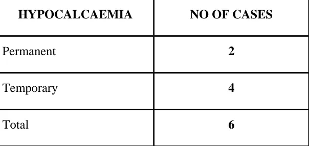
Hypoparathyroidism, and the resulting hypocalcemia, may be permanent or
transient18. The rate of permanent hypoparathyroidism is 0.4-13.8%36. The condition
may be due to direct trauma to the parathyroid glands, devascularization of the glands
or actual removal of the glands during surgery37.
The rate of temporary hypocalcemia is reportedly 2-53%37. The cause of
transient hypocalcemia postoperatively is not clearly understood. It may be
attributable to temporary hypoparathyroidism caused by reversible ischemia to the
parathyroid glands, hypothermia to the glands, or release of endothelin 2. Endothelin
1 is an acute-phase reactant known to suppress PTH production, and levels have been
found to be elevated in patients with transient hypoparathyroidsm.
Other hypotheses have been put forth to account for transient hypocalcemia
not caused by hypoparathyroidism. These include calcitonin release and "hungry
bone syndrome". Calcitonin is produced by the thyroid and inhibits bone breakdown
oppose those of PTH. Hungry bone syndrome occurs in patients with preoperative
hyperthyroidism. These patients have increased bone breakdown in their
hyperthyroid state. When a patient's thyroid hormone level drops acutely after
surgery, his or her stimulus to break down bone is removed. The bones, now
"hungry" for calcium, remove calcium from the plasma, leading to decreased serum
calcium levels49.
PRESENTATION
Most patients who are hypocalcemic after thyroidectomy are initially
asymptomatic. Symptoms and signs of hypocalcemia include circumoral
paresthesias, mental status changes, tetany, carpopedal spasm, laryngospasm,
seizures, QT prolongation on ECG, and cardiac arrest.
EVALUATION
Evaluate ionized calcium (or total calcium and albumin) in the perioperative
period in patients undergoing total thyroidectomy. If concern for iatrogenic
hypoparathyroidism exists, close follow-up care is warranted for at least 72 hours or
until the calcium levels demonstrate that parathyroid function is intact.
"Chvostek" and "Trousseau signs" may both be elicited at bedside to
confirm hypocalcemia. The Chvostek sign is elicited by tapping the facial nerve in
the preauricular area and observing for facial contractions; the Trousseau sign, by
induction of carpal spasm upon inflation of a blood pressure cuff.
PTH levels may be checked to confirm the cause of hypocalcaemia and to
elevated in patients with hypoparathyroidism secondary to decreased renal excretion,
which may help distinguish low PTH levels from other etiologies of hypocalcaemia
(eg: hungry bone syndrome). Consider other causes of hypocalcaemia as well
(eg: medications, hypomagnesemia, renal failure, pancreatitis).
PREVENTION
The best way to preserve parathyroid gland function is to identify the glands
and to maintain their blood supply38,39. A large cadaveric study to identify the most
common positions of the parathyroid glands demonstrated that 77% of superior
parathyroid glands were at the cricothyroid junction, intimately associated with the
RLN9. Twenty-two percent of the superior parathyroid glands were on the posterior
surface of the upper lobe of the thyroid. Approximately 1% of the superior glands
were located behind the junction of the hypopharynx and upper esophagus.
The study demonstrated that the location of the inferior parathyroid glands is
more variable. Forty-two percent were found on the anterior or lateral surfaces of the
lower lobe of the thyroid, often hidden by vessels or creases in the thyroid.
Thirty-nine percent were located within the superior tongue of the thymus. Fifteen percent
were extrathyroidal and lateral to the lower lobe. Two percent were within the
mediastinal thymus and another 2% were in other ectopic positions, such as the
carotid sheath. The ectopic inferior parathyroid glands were consistently associated
with remnant thymus tissue.
The inferior parathyroid glands and the thymus both develop from the third
branchial pouch, which explains the close association of these structures5. The
inferior parathyroid glands receive their blood supply from the inferior thyroid artery.
thyroid artery. However, in some cases, the superior parathyroids receive their
vascular supply from the superior thyroid artery, the anastomotic loop between the
inferior and superior thyroid arteries, or from direct branches off the thyroid gland.
The keys to parathyroid preservation are to identify the parathyroids and
preserve their blood supply by ligating all vessels distal to them39. Ligate vessels as
close to the thyroid gland as possible. Recognition of the parathyroid glands, which
appear in a variety of shapes and have a caramel-like color, is critical. When they
lose their blood supply, they turn black. The devascularized gland (confirmed
pathologically by frozen-section analysis), should be removed, cut into 1-to-2-mm
pieces, and reimplanted in the sternocleidomastoid muscle or the forearm. The
location should be marked with a permanent suture or Ligaclip. Sometimes, the
inferior glands are so anterior that preserving their blood supply is difficult. In this
case, the glands should be reimplanted as well. Once removed, examine the thyroid
gland. Any parathyroid glands in excised thyroid tissue are then reimplanted6.
TREATMENT
Patients who have asymptomatic hypocalcemia in the early postoperative
period should not be treated with supplemental calcium. Authorities believe that the
hypocalcemic state stimulates the "stunned" parathyroid glands to produce PTH14.
Patients who have symptomatic hypocalcaemia in the early postoperative
period, or whose calcium levels continue to fall require treatment14. In the
symptomatic patient replace calcium with intravenous calcium gluconate. Ten mL of
10% solution (1g) may be administered over 10 minutes. A calcium drip may be
started at 1-2 mg/kg if symptoms do not resolve. Titrate the infusion to the patient's
this. One to 2 g of elemental oral calcium should be supplied each day. Calcium
carbonate at a dose of 1250 mg provides 500 mg of elemental calcium; therefore, the
patient should take 2500-5000 mg of calcium carbonate a day. The patient will need
concomitant vitamin D replacement in the form of calcitriol at a dosage of
0.25-1 mcg/d.
The authors recommend assistance from an endocrinologist to ensure close
monitoring of calcium levels and for medical management of the sequelae of
hypoparathyroidism. In 1-2 months, an attempt to wean the patient off oral calcium
may be made to reveal if the hypoparathyroidism is temporary. Dependence on
calcium supplementation for longer than 6 months probably indicates permanent
hypoparathyroidism2,14.
THYROTOXIC STORM
Thyrotoxic storm is an unusual complication of thyroid surgery18. The
condition may occur from manipulation of the thyroid gland during surgery in the
hyperthyroid patient and can occur intraoperatively or postoperatively. Thyrotoxic
storm is potentially lethal and must be dealt with astutely.
PRESENTATION AND EVALUATION
Signs of thyrotoxic storm in the anesthetized patient include evidence of
increased sympathetic output, such as tachycardia and hypertherimia. Other
symptoms and signs in the awake patient include nausea, tremor, and altered mental
status. Cardiac arrhythmias may also occur. If treatment is not given, the patient
PREVENTION
Preoperative awareness of the hyperthyroid patient and appropriate medical
management are the keys to prevention of thyrotoxic storm, medications used in the
hyperthyroid patient include propylthiouracil, methimazole, and/or Lugol solution.
Beta-blockers are useful in controlling peripheral manifestations of hyperthyroidism.
Steroids may be useful preoperatively.
TREATMENT
The first step when faced with a thyrotoxic crisis during thyroidectomy is to
stop the procedure. Intravenous beta-blockers, propylthiouracil, sodium iodine and
steroids are administered to control sympathetic activity, thyroid hormone release,
and hyperthermia. Use cooling blankets and cooled intravenous fluids to reduce body
temperature. Carefully monitor oxygenation, because oxygen demands increase
dramatically during thyroid storm4.
SUPERIOR LARYNGEAL NERVE INJURY
The SLN has 2 divisions: internal and external. The internal branch provides
sensory innervation to the larynx. It enters the larynx through the thyrohyoid
membrane and therefore should not be at risk during thyroidectomy5. The external
branch provides motor function to the cricothyroid muscle and is at risk during
thyroidectomy8. This muscle is involved with elongation of the vocal folds. Trauma
to the nerve results in an inability to lengthen a vocal fold and thus to create a
higher-pitched sound. The rate of injury to the external branch of the SLN has been
estimated at 0-25%8. This rate is probably underestimated, because the diagnosis is
PRESENTATION
The clinical presentation of a patient with SLN paralysis may be quite subtle.
Most patients do not notice any change. Occasionally, one presents with mild
hoarseness or decreased vocal stamina. For the singer and or professional voice user,
however, paralysis of the SLN may be career threatening. The most damaging
consequence is loss of the upper register3.
EVALUATION
Diagnosing an SLN injury with indirect or fiberoptic laryngoscopy is very
difficult40. Posterior glottic rotating toward the paretic side and bowing of the vocal
fold on the weak side may be noted. In addition, the affected vocal fold may be
lower than the normal vocal fold.
The use of videostroboscopy and laryngeal EMG has increased
otolaryngologists' and speech pathologists' ability to diagnose SLN paralysis.
Videostroboscopy demonstrates an asymmetric mucosal travelling wave. EMG
demonstrates cricothyroid muscle denervation.
PREVENTION
The external SLN branch travels inferiorly along the lateral surface of the
inferior constrictor until it terminates at the cricothyroid muscle. This branch is
intimately related to the superior thyroid artery, though its exact relation to the artery
varies.
Most surgeons agree that, in contrast to the RLN, identifying the SLN is
close to the thyroid capsule as possible to avoid damaging the nerve40.
electrophysiologic monitoring of the SLN has been described but is not
recommended for routine use58.
Direct trauma to the cricothyroid muscle can cause fibrosis and poor muscle
function, which may result in a presentation similar to that of a patient with injury to
the external branch of the SLN, even when the nerve is preserved. Therefore, dissect
carefully near this muscle and avoid electrocautery damage when possible.
TREATMENT
The only treatment currently available for injury to the external branch of the
SLN is speech therapy.
HYPOTHYROIDISM PRESENTATION
Untreated hypothyroidism causes symptoms such as cold intolerance, fatigue,
constipation, muscle cramping, and weight gain. Hypothyroidism secondary to
thyroid surgery should never be left untreated long enough to elicit signs and
symptoms of myxedema (eg: hair loss, large tongue, cardiomegaly). Expect,
diagnose, and promptly treat postoperative hypothyroidism4.
EVALUATION
The most useful laboratory test for detection or monitoring of hypothyroidism
is in the postthyroidectomy patient is the thyrotropin (thyroid-stimulating hormone
[TSH]) level. Total thyroxine (T4) and triiodothyronine (T3) levels may be useful to
fine-tune levothyroxine (Synthroid) dosing but are less likely to be helpful in the
PREVENTION
Hypothyroidism is an expected sequela of total thyroidectomy. In goiter
surgery, the surgeon must balance the risks of leaving too much thyroid tissue thus
requiring repeat surgery, with excising too much thyroid tissue, which results in
hypothyroidism. This balance comes with experience and adequate follow-up care18.
TREATMENT
Start hypothyroid patients on levothyroxine (~1.7 mcg/kg/d). Check the
thyrotropin level in approximately 4-6 weeks, and adjust the dosage appropriately.
Patients who are to receive postoperative radioiodine scaning must be off
levothyroxine before the procedure.
HYPERTROPHIC SCAR OR KELOID SCAR
This is more likely to form if the incision overlies the sternum. Especially the
drain site or when the wound is complicated by wound infection and gaping.
Intralesional injections of corticosteroid should be given at once and repeated
monthly if necessary.
STITCH GRANULOMA
This may occur with or without sinus formation and is seen after the use of
non-absorbable suture material.
Absorbable ligatures and sutures must be used throughout thyroid surgery.
INFREQUENT POSTOPERATIVE COMPLICATIONS
Some postoperative complications, such as sympathetic nerve injury, chylous
years. The decrease is associated with a better understanding of thyroid anatomy, as
well as advances in the medical treatment of thyroid disease (Netterville & Ossoff14,
1990).
Injury to the sympathetic chain from stretching or compressing during thyroid
surgery may result in the development of Horner's syndrome. This condition is
characterized by such symptoms as contraction of the pupil, partial ptosis of the
eyelid, and anhydrosis of the ipsilateral side.
Damage to the thoracic duct during thyroid surgery may result in a chylous
fistula. This complication manifests itself by the presence of profuse and continuous
drainage of chyle (a milky or opaque fluid) from the operative would.
MORTALITY
The mortality rate from thyroid surgery during the 1800s was approximately
40%. Most deaths were caused by infection and hemorrhage. Sterile operative
arenas, general anesthesia, and improved surgical techniques have made death due to
FOLLICULAR CARCINOMA THYROID
MATERIALS AND METHODS
The present study included 110 patients who underwent thyroid surgery in
various surgical units from July 2004 to May 2005 in the Department of General
Surgery, Coimbatore Medical College Hospital, Coimbatore.
CRITERIA OF ELIGIBILITY
Age : All age groups from 14 to 75 years were included in this study.
Sex : Both males and females were included.
Pathology : All Benign, Malignant, Toxic and non toxic cases who underwent surgery were included.
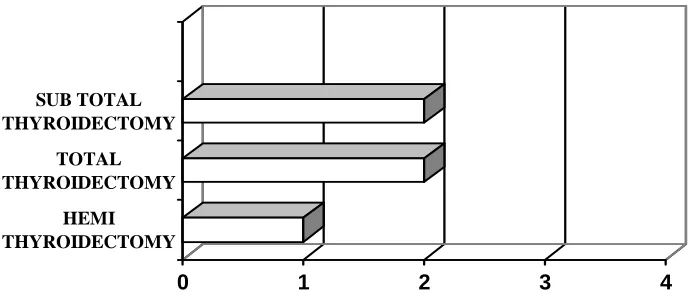
Surgery : Surgeries on thyroid included are Hemithyroidectomy, Subtotal Thyroidectomy and Total Thyroidectomy.
CRITERIA FOR EXCLUSION
Patients who had recurrent laryngeal nerve paralysis and hypocalcaemia
preoperatively were exempted from this study.
Hypothyroidism following TT was not considered as complication in this
study.
METHODS
All cases of Thyroidectomy underwent following basic investigations
preoperatively.
-Chest X-ray.
-X-ray neck - both lateral and AP view.
-ECG.
-Basic haematological and biochemical investigations.
-Vocal Cord examination by ENT surgeon with indirect laryngoscopy.
-FNAC of Thyroid nodule.
-Thyroid function tests.
All patients underwent Laryngoscopy examination as follows,