0022-538X/96/$04.0010
Copyrightq1996, American Society for Microbiology
Evidence that Two Latency-Associated Transcripts of Herpes
Simplex Virus Type 1 Are Nonlinear
TING-TING WU,1YING-HSIU SU,2TIMOTHY M. BLOCK,2ANDJOHN M. TAYLOR1*
Fox Chase Cancer Center, Philadelphia, Pennsylvania 19111-2497,1and Thomas Jefferson University Medical School
and Kimmel Cancer Institute, Philadelphia, Pennsylvania 19107-67992
Received 19 April 1996/Accepted 22 May 1996
The latency-associated transcripts (LATs) of herpes simplex virus type 1 (HSV-1) are the only viral gene products that accumulate to abundant levels in latently infected cells. Others have reported species of 2.0, 1.50, and 1.45 kb; only the 2.0-kb species is seen in productively infected cells, and there is evidence that it behaves as an intron. We examined the LATs both in trigeminal ganglia of latently infected mice and in productively infected cultures of monkey CV-1 cells. After glyoxalation, RNA was subjected to high-resolution agarose gel electrophoresis and Northern (RNA) analysis, a procedure capable of resolving linear and nonlinear RNA species. Under these conditions, we resolved the 2.0-kb LAT into two species; the slower species was much more abundant and had a mobility significantly slower than expected for a linear RNA. To test the hypothesis that this RNA was in fact nonlinear, we used partial hydrolysis by sodium carbonate and oligonucleotide-directed RNase H digestion. These procedures changed the mobility of the slower species into that of the faster species. Similarly, the mobility of the 1.50-kb LAT, which was much more abundant than the 1.45-kb LAT, was changed by these procedures to that of the 1.45-kb LAT. Our data show that the two major LAT species are nonlinear, and they support an interpretation of stable lariat structures.
All herpesviruses establish latent infections in their natural hosts (reviewed in reference 14). For example, after a period of productive infection near the portal of entry, human herpes simplex virus type 1 (HSV-1) establishes latency in those pe-ripheral neurons which send projections to the lytically in-fected region. In productively inin-fected cells, more than 70 different viral gene products are synthesized with temporally regulated kinetics. Although low levels of a variety of viral gene products may be present in latently infected cells, the latency-associated transcripts (LATs) appear to be the only transcripts which accumulate in sufficient amounts to be detected by Northern (RNA) blot analysis. Latently infected cells are therefore characterized by the accumulation of this single fam-ily of small, nuclear RNA molecules which map to the internal repeat region of the viral genome and may be present at as many as 40,000 copies per cell (29). The most abundant species is about 2.0 kb in length; also present are species of 1.50 and 1.45 kb (27, 30, 33). There is evidence that the 2.0-kb species is a stable intron and may be derived from the processing of an 8.3-kb precursor (13). Also, others have suggested that the 1.50- and 1.45-kb species might represent spliced forms of the 2.0-kb species (27).
The function, if any, of the LATs is unclear. One consistent observation is that mutant viruses with deletions of the puta-tive major LAT promoter (LAPI) and surrounding region re-activate from latent infections less efficiently than the wild-type virus (reviewed in reference 14). This finding implies that LATs are involved in either the establishment of or the reac-tivation from latent infections. However, the behaviors of LAT mutants vary with the experimental system used; also, no spe-cific coding information appears to be necessary for putative LAT function (14). For example, mutants containing a disrup-tion of the open reading frames and other posidisrup-tions within the
LAT transcripts can reactivate from explants of trigeminal ganglia derived from latently infected mice, with normal kinet-ics. Since the 2.0-kb LAT accumulates to such a high abun-dance in the nucleus and is complementary to the 39end of the ICP0 transcript, an antisense mechanism of LAT function was also hypothesized (13, 28). Recently, in a study using a rabbit in vivo system, it was found that normal reactivation pheno-types can be restored to LAT null mutants by ectopic insertion of the region corresponding to the first 1.5 kb of the putative 8.3-kb LAT primary transcript (22). Since this region does not specify RNA complementary to the ICP0 RNA, enthusiasm for the antisense mechanism has been reduced. Therefore, the putative mechanism of action of the LAT remains enigmatic. Possibly the LATs accumulate in part because they are rel-atively stable. Previous studies have provided clues as to why RNA species can possess higher stability. In the nuclei of eukaryotic cells, the major RNases have exonucleolytic rather than endonucleolytic activity (6). Since circular RNAs would not be a substrate for exonucleases, they are thus at an advan-tage. This has been demonstrated for deliberately constructed circular RNAs (16, 23). There are also naturally occurring circles that accumulate. Examples include infectious agents, such as the plant viroids (11) and human hepatitis delta virus (31). There are also certain cellular RNAs which can exist in a circular form; a process referred to as missplicing can produce circular RNAs, some of which accumulate to relatively high levels (1, 3, 9, 10, 21). In addition to circular RNAs, the RNA lariat structures produced during splicing can under special circumstances be stable. Normally lariats are unstable because of prompt debranching, which converts them to less stable linear RNAs. However, if this debranching reaction for some reason becomes inefficient, then lariats will accumulate. This has been observed for a simian virus 40 RNA artificially ex-pressed in Xenopus laevis oocytes (19). It has also been seen for certain naturally occurring immunoglobulin RNAs expressed in T cells (24). It has been shown that lariats can accumulate in yeast when the branchpoint is mutated (17) or in strains which lack debranching activity (7).
* Corresponding author. Mailing address: Fox Chase Cancer Center, 7701 Burholme Ave., Philadelphia, PA 19111-2497. Phone: (215) 728-2436. Fax: (215) 728-3616. Electronic mail address: jm_taylor@fccc .edu.
5962
on November 9, 2019 by guest
http://jvi.asm.org/
Our studies began as an attempt to determine whether the stability of HSV-1 LATs was due to a nonlinear conformation. Our initial inclination was to test for circles, just like those seen for hepatitis delta virus. On the other hand, others have shown that the 2.0-kb LAT can behave as an intron, and thus it might even be a lariat. As described here, we found that both the 2.0-and 1.50-kb LAT RNAs were primarily in a nonlinear mation, and we now favor the interpretation that this confor-mation is that of a stable lariat.
MATERIALS AND METHODS
Plasmids, probes, and primers.The relative positions of the RNA probes and oligonucleotides used are shown in Fig. 1. All Northern analyses shown were carried out with probe P, and in the RNase H studies, some were repeated with a probe spanning the whole 2.0-kb region. Positions and sequences of the oli-gonucleotides are summarized in Table 1.
Cells, viruses, and tissue culture.CV-1 cells (American Type Culture Collec-tion, Rockville, Md.) were maintained at 378C and 5% CO2as monolayers in Eagle’s minimal essential medium (GIBCO) supplemented with 5% newborn calf serum and 0.35% glucose. These cells were infected at a multiplicity of infection of 3 with HSV-1 strain 17.
Mouse infections.Female 4- to 6-week old BALB/c mice (Jackson Laboratory) were infected by corneal scarification with between 105
and 106
PFU of virus. After 30 days, a time at which latency can be established, matched mock-infected or infected mice were sacrificed, and trigeminal ganglia were excised as described previously (18).
RNA isolation and characterization.RNA was isolated from both tissue and cell culture samples by a guanidine isothiocyanate procedure (8). RNA was glyoxalated and subjected to 3% agarose gel electrophoresis and Northern anal-yses as described previously (15). RNA markers were transcribed with a Ribo-Mark labeling system (Promega). A 1-kb DNA ladder (Bethesda Research Lab-oratories) was 59 labeled by using T4 kinase. The treatments with sodium carbonate and RNase H (Bethesda Research Laboratories) were as described previously (3). For experiments summarized in Table 2, oligonucleotides D to F were 59labeled by using T4 kinase and then used as probes in Northern analyses with hybridization at 378C.
Reverse transcription-PCR (RT-PCR), cloning, and sequencing.One micro-gram of RNA from latently infected trigeminal ganglia was reverse transcribed by using random hexamer primers as described previously (27) and then PCR amplified by using oligonucleotides D and G (Table 1). The product was then cloned and sequenced as described previously (20).
RESULTS
Northern analyses of RNA from latently infected mouse
trigeminal ganglia and productively infected CV-1 cells.From
previous studies with hepatitis delta virus RNA, we had deter-mined agarose gel conditions in which the 1,679-nucleotide (nt) circular RNA migrates significantly more slowly than the corresponding linear form (15). Therefore, we applied such conditions to the RNAs from both the trigeminal ganglia of latently infected mice and productively infected monolayer cultures of CV-1 cells.
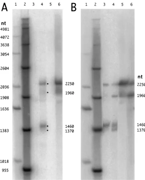
[image:2.612.318.554.68.361.2]On the basis of previous work of Spivack et al. (27), we expected that the infected trigeminal ganglia would contain species of 2.0, 1.50, and 1.45 kb. As shown in Fig. 2A, lane 4, we actually detected not three species but two major and two minor species. Assuming that these four species were linear RNAs, then relative to the size markers in lanes 1 and 2, they FIG. 1. Relationship to HSV-1 of LATs and relevant RNA probes and
[image:2.612.59.300.69.171.2]oli-gonucleotides. The locations on the HSV genome come from a published se-quence (34). The size of 1,954 nt for the major LAT was calculated from reference 13. The 1,395 nt for the smaller LAT is derived from this value, using the evidence for a 559-nt intron (27). P is the RNA probe used for Northern analyses. A to G are the oligonucleotides used in various assays; the full se-quences of these oligonucleotides are presented in Table 1.
FIG. 2. Northern analysis of RNA from latently infected mouse trigeminal ganglia and productively infected monolayers of CV-1 cells. Extracted RNA samples were glyoxalated and subjected to Northern analysis. The probe was species P, as indicated in Fig. 1. Radioactivity was detected with a Fuji phosphor imaging system. In panel A, lanes 1 and 2 are DNA and RNA markers, respec-tively; their known lengths are indicated at the left. Lanes 3 and 4 are RNAs (1
mg) from mouse trigeminal ganglia 30 days after either a mock or real infection. Lanes 5 and 6 are RNAs (0.4mg) from CV-1 cells at 4 or 23 h after the initiation of a productive infection. If the discrete species in lanes 4 and 6 are assumed to be linear, then their sizes deduced relative to the linear RNA markers in lane 2 are as indicated at the right. Panel B is a separate gel analysis. Lanes 1 and 2 are size markers as in panel A; lanes 3 and 4 are RNA from latently infected mouse trigeminal ganglia; lanes 5 and 6 are RNAs from productively infected CV-1 cells. The important difference is that prior to electrophoretic analysis, the RNAs for lanes 4 and 6 were subjected to partial hydrolysis using sodium carbonate.
TABLE 1. HSV-specific oligonucleotides
Desig-nationa Locationb Sequenceb
A 119509–119488 cggaattcCGTTTGGGTCCCCCCCCTCTAT B 120492–120468 ATGTTGGGCAGGCTCTGGTGTTAAC C 120898–120877 GGGACGAGGGAAAACAATAAGG D 121336–121315 cggaattcAGACGCGCCACGCGGAGACTTC E 121415–121394 CTGGGAGGGAGACAAGAGGAAA F 121437–121416 cggaattcCTCGGGCGGGGCCGTCGGTGCC G 120877–120898 CCTTATTGTTTTCCCTCGTCCC
aAs indicated in Fig. 1.
bBased on a published sequence (27, 34). The nucleotides indicated in
low-ercase were non-HSV sequences added to the 59ends to provide restriction enzyme sites to facilitate cDNA cloning.
on November 9, 2019 by guest
http://jvi.asm.org/
[image:2.612.59.298.601.693.2]were calculated to be equivalent to lengths of 2,250, 1,960, 1,460, and 1,370 nt. (For reasons of consistency, we will sub-sequently refer to these RNA species by these calculated equivalent linear sizes, even though the RNAs may not be linear.) Apparently, our electrophoretic conditions had re-solved what others have described as a single 2.0-kb species into two, with the 2,250-nt species being much more abundant than the 1,960-nt species. Similarly, the 1,460-nt species was much more abundant than the 1,370-nt species. None of these four species were present in RNA from the trigeminal ganglia from uninfected mice (lane 3).
Others have reported that productively infected cells contain the 2.0-kb RNA but not the 1.45- or 1.50-kb species (27). Consistent with this finding, we observed a simpler pattern for the RNAs from productively infected CV-1 cells. At 23 h after infection, we observed only two bands (Fig. 2A, lane 6). These bands corresponded to the two larger bands detected in the infected trigeminal ganglia. No species were detected after 4 h of infection (lane 5).
On the basis of these results and the following clues, we hypothesized that the two major LAT species, observed to be equivalent in migration to linear RNAs of 2,250 and 1,460 nt, were actually nonlinear and that the two minor species, ob-served to be equivalent to 1,960 and 1,370 nt in length, were more like linear forms. One clue was that the calculated size of 2,250 nt was significantly larger than the 2.0 kb reported by others (27) and larger than the predicted 1,954 nt determined in an experiment in which this region was interpreted to be-have as an intron (13); we already knew from our studies with hepatitis delta virus that under such conditions of agarose gel electrophoresis, nonlinear RNAs migrate more slowly than the corresponding linear species (15). Another clue was provided by observations relating to RNA storage. Typically, we store RNA samples in ethanol at2208C, which avoids RNA hydro-lysis. However, if samples were stored as frozen aqueous solu-tions at2708C, there was, with time, a decrease of the 2,250-nt RNA and what appeared to be a corresponding increase in the 1,960-nt species (35). One interpretation was that these ditions allowed limited hydrolysis (26), which changed the con-formation of a nonlinear RNA into a linear form.
Partial hydrolysis of LATs in sodium carbonate. Previous
studies of circular RNAs have established conditions by which treatment in sodium carbonate leads to limited hydrolysis, with the conversion of circular RNAs into linear RNAs (3). We applied these conditions to the RNAs from infected trigeminal ganglia and CV-1 cells. As shown in Fig. 2B, this treatment initially led to a decrease in the amounts of the 2,250- and 1,460-nt species, with a corresponding increase in the amounts of the 1,960- and 1,370-nt species, respectively. These changes are shown more clearly in the corresponding radioactive pro-files presented in Fig. 3. After longer times of treatment of the CV-1 RNA with sodium carbonate, although the amount of the 2,250-nt band continued to decrease, there was also a decrease in the amount of the 1,960-nt species, with the ap-pearance of a heterogeneous population of smaller RNAs (35). All of these observations are consistent with the interpretation that 2,250- and 1,460-nt LATs are nonlinear and that limited hydrolysis converted them first to linear species and later to smaller random-sized fragments.
RNase H digestion of LATs hybridized to specific
oligonu-cleotides.One way to distinguish linear from nonlinear RNAs
is to hybridize with one or two specific oligonucleotides, carry out an RNase H digestion, and then characterize the RNA products of this digestion. Such a strategy was used by others to characterize a circular RNA molecule produced from the Sry gene (3). We adapted this strategy in order to better
under-stand the putative nonlinear conformations of the 2,250- and 1,460-nt RNAs. We tested the RNAs from the infected trigem-inal ganglia and CV-1 cells, along with three specific oligonu-cleotides, B, C and E, with results as shown in Fig. 4.
[image:3.612.357.516.69.519.2]Oligonucleotides B and C, with positions indicated in Fig. 1, were able to lead to the efficient digestion of the 2,250- and 1,460-nt species, with a corresponding increase of RNAs at about 1,960 and 1,370 nt. These data indicate that the two major LATs, 2,250 and 1,460 nt, do not correspond to linear species with specific 59and 39ends; if they did, then RNase H cleavage after hybridization with either oligonucleotide B or oligonucleotide C would have produced specific discrete-sized species, which we did not detect.
FIG. 3. Phosphorimager radioactivity profiles of LATs before and after treat-ment with sodium carbonate. The tracings correspond to infected trigeminal ganglia (A and B) and CV-1 samples (C and D) from Fig. 2B, lanes 3 to 6, respectively. The samples are before (A and C) and after (B and D) treatment with sodium bicarbonate. The vertical scale represents radioactivity although the absolute scale varies between panels.
on November 9, 2019 by guest
http://jvi.asm.org/
We next used these two oligonucleotides together. In the case of the CV-1 RNA, this digestion led to the conversion of the major 2,250-nt species into a 1,520-nt species (Fig. 4, lane 10) and a 390-nt species. (The smaller species is not shown in Fig. 4; it was detected in a separate analysis using a shorter electrophoresis time and also with a probe specific for the whole 2-kb LAT region [35].) In a similar digestion of the RNA from trigeminal ganglia, the major 2,250- and 1,460-nt species were replaced by 1,520-, 950-, and 390-nt species (Fig. 4, lane 6, and reference 35). These fragments and their specific sizes are consistent with the interpretation, as represented in Fig. 5, that the two major LATs are lariats. An alternative interpre-tation, not excluded by these data, is that the two LATs are circular.
It is difficult to be conclusive about the two minor LATs of 1,960 and 1,370 nt. These species exist to a small extent before RNase H digestion (Fig. 2A, lanes 4 and 6). Our interpretation is that they represent nonspecific single nicking of the two major LATs. If they were in fact specific single openings of the major LATs, for example, a linear intron as shown in Fig. 5, then RNase H digestion following hybridization with either oligonucleotide B or oligonucleotide C would have produced specific discrete RNA fragments; we did not detect such frag-ments. (We did note that in some digestions, minor discrete-sized species were present; our interpretation is that these
species arose via nonspecific hybridization by the oligonucleo-tides.)
After the LATs were hybridized to oligonucleotide E, there was no indication of RNase H cleavage (Fig. 4, lanes 3 and 7). This was unexpected since the location of this oligonucleotide is at the extreme 39end of the predicted LAT region (Fig. 1; Table 2); actually the results were identical to those obtained with either oligonucleotide F or even in the absence of oligo-nucleotide (35). We considered three explanations for the in-ability of oligonucleotide E to lead to cleavage. First, the target RNA may not be present. Second, the target is there but hybridization may be inefficient. Third, the RNAs are in a lariat conformation, and oligonucleotide E is complementary to the 39tail region (Fig. 5), so that independent of the pres-ence or abspres-ence of hybridization and RNase H digestion, the loop of the lariat will not be opened up. In an attempt to distinguish between these interpretations, we carried out the following hybridization experiments.
Hybridization of specific oligonucleotides to LATs.We used
hybridization with three oligonucleotides: D was complemen-tary to an internal region, as a positive control; E was comple-mentary to the extreme 39end of the predicted LAT; and F was complementary to a region outside the LAT region, as a neg-ative control (Fig. 1). As a calibration of hybridization effi-FIG. 4. RNase H digestion of LATs following hybridization of specific
[image:4.612.88.266.67.362.2]oli-gonucleotides. RNAs from infected trigeminal ganglia (lanes 3 to 6) and CV-1 cells (lanes 7 to 10) were hybridized to oligonucleotides E (lanes 3 and 7), B (lanes 4 and 8), C (lanes 5 and 9), or B and C (lanes 6 and 10). The RNAs were then treated with RNase H, extracted, and subjected to glyoxalation and North-ern analysis as in Fig. 2. The DNA and RNA markers in lanes 1 and 2, respec-tively, with their sizes indicated at the left, are also as in Fig. 2. At the right are indicated the determined sizes of the observed RNA species, assuming a linear conformation.
FIG. 5. Interpretation of major LATs. The upper panel shows the predicted 1,954-nt LAT as a linear intron; our data argue against the existence of such a species. Instead, as indicated by the middle panel, our data support the inter-pretation of a lariat structure. The bottom panel shows our interinter-pretation of the other major LAT, the species which is predicted to be a 1,395-nt species, as also being a lariat. Indicated in each panel are the relative positions of three oligo-nucleotides, B, C, and E (Table 1), which were used in the RNase H studies shown in Fig. 4.
TABLE 2. Oligonucleotide hybridization to the LATs RNA of productively infected CV-1 cells
Oligonucleotidea Relative
hybridizationb
D... 1.00 E ... 0.068 F ...,0.01
aOligonucleotides, as described in Table 1 with relative positions as indicated
in Fig. 1, were 59labeled and hybridized to a Northern blot containing both CV-1 RNA (5mg) and, as a positive control, in vitro synthesized RNA (5 ng) exactly complementary to RNA R in Fig. 1.
bHybridizations were quantitated with a Fuji imager, and the signals for each
probe were first expressed relative to the hybridization to the positive control. Then these three values were normalized relative to the hybridization obtained with CV-1 RNA and oligonucleotide D.
on November 9, 2019 by guest
http://jvi.asm.org/
[image:4.612.316.555.89.144.2] [image:4.612.362.510.504.662.2]ciency, we used an in vitro RNA that should hybridize to all three oligonucleotides. These studies were carried out on the 2,250-nt LAT as detected in CV-1 cells at 23 h after infection. The results, as summarized in Table 2, indicate that both D and E hybridize to the 2,250-nt RNA. Thus, both complemen-tary regions are present, at least to some extent. However, the ability to hybridize with E was only 7% relative to the ability to hybridize to D. An interpretation of this finding is considered in Discussion.
RNA editing of LATs. Previous studies with other viral
RNAs have suggested that RNA genomes, with time, and especially during persistent infections, can accumulate changes due to posttranscriptional editing by double-stranded RNA adenosine deaminase, an activity that seems to be present in the nuclei of all animal cells (4). This activity can change adenosine to inosine. There is even a cellular RNA which is a natural substrate for this activity; the precursor in the brain to the mRNA for one of the glutamate receptors is specifically edited by double-stranded RNA adenosine deaminase (5).
Because LAT RNAs are the only transcripts detectable in HSV latent infections, we determined whether nucleotide changes could be detected on those RNAs present in infected trigeminal ganglia. We used RT-PCR with oligonucleotides G and D. The RT-PCR product was ligated into a bacterial plas-mid. Five positive clones were sequenced through a region of 519 nt. Not a single nucleotide change was found (35). Thus, editing of this RNA, if present, was below our detection level (viz.,,20% at each of about 130 adenosines).
DISCUSSION
Previous studies have shown that the 2.0-kb LAT found in both latently infected trigeminal ganglia and productively in-fected CV-1 cells accumulates in the nucleus. It has been hypothesized that it is a stable intron (13). Our studies show that this RNA can actually be separated electrophoretically into two RNA species. Our interpretation is that the more abundant is a nonlinear RNA, while the minor species is cre-ated from it by a single random nicking event.
It has previously been reported that HSV RNA from latently infected mouse trigeminal ganglia contains, in addition to the 2.0-kb LAT, smaller species designated 1.50- and 1.45-kb LATs (27). On the basis of RT-PCR cloning and sequencing, Spivack et al. have suggested that relative to the 2.0-kb species, the 1.50-kb species has undergone the removal, by RNA splic-ing, of a 559-nt region (27). Our results show that the 1.50- and 1.45-kb LATs are related. The 1.50-kb species is more abun-dant and, again, a nonlinear RNA. The 1.45-kb species is relatively minor and, again, created from the major species by a single random nicking event.
What precisely is the nonlinear conformation of the two major LATs? Our experiments, as presented here, do not distinguish between circles and lariats; however, as discussed below, in the context of experiments by others, the most likely interpretation, as represented in Fig. 5, is that the two major LATs are stable lariats.
We used two additional approaches in an attempt to distin-guish between circles and lariats. The first was an RT-PCR strategy using a pair of primers facing away from each other on the linear map; this strategy was used by others to help show that certain processed RNA transcripts from the Sry gene were circular (3). We carried out RT-PCR using oligonucleotides A and G (Fig. 1) and RNA from both infected trigeminal ganglia and infected cultured cells. We were unable to detect a prod-uct. This was not due to an inability to RT-PCR on HSV RNA; we were able to amplify the linear region between
oligonucle-otides E and G (Fig. 1). Also, it was not due to an inability to amplify circular RNA; the strategy did work on circular hep-atitis delta virus RNA. Thus, our data favor the interpretation that the HSV LAT is not circular (35).
A second approach was to debranch the putative lariat RNA. RNA from infected CV-1 cells was incubated with a crude S100 fraction from uninfected HeLa cells. We observed that the amount of nonlinear RNA progressively decreased (35). However, comparable changes also occurred for an in-ternal control of HDV RNA that was truly circular and could not be debranched. Therefore, the majority of the changes detected for the HSV RNA were probably due to random nicking and not to debranching (35). We note the alternative interpretation, that this failure to debranch in vitro might be related to the putative failure to debranch in vivo.
Even though our experiments, per se, were unable to distin-guish between circles and lariats, we can, considering our data together with the work of others, strongly argue in favor of lariats. Farrell et al. transferred the 2.0-kb region into another gene and showed that it behaved as an intron (13). They identified a splice donor and, 39of this, a splice acceptor. Thus, their data allow for formation of a lariat and at the same time exclude circle formation by missplicing, since that would de-pend on a splice acceptor being 59rather than 39of the splice donor. As mentioned in the introduction, there are several precedents for intron sequences accumulating in a stable lariat conformation. Finally, we are aware of unpublished studies by Rodahl and Haarr, who suggested that in productively infected cultured cells, the 2.0-kb LAT is nonlinear and probably a stable lariat (25).
Although Farrell et al. have attempted to locate the splice donor and acceptor sites for the large LAT intron (13), there has been no speculation as to the position of the branchpoint. We note that there is a potential branchpoint at position 121391, which is appropriately followed by a pyrimidine-rich tract and the putative splice acceptor site. We also speculate that the same branchpoint is used for the two major LATs. Mutagenesis of this putative branchpoint may provide data that further clarify the conformation, and even the function, of the LATs.
Others have seen that the short 39 tail on lariat RNAs can become trimmed, presumably by exonuclease activity (19). Relevant to this question for the HSV LATs, we carried out hybridization with oligonucleotide E, which should be 39of the putative branchpoint (Fig. 5). As summarized in Table 2, we observed hybridization to a level of 7% relative to controls. These data prove that at least some of the putative tail se-quence is present on 7% of the major 2.0-kb LAT. However, for the other 93%, we cannot distinguish between two possi-bilities: the RNA sequence is not present, presumably because of trimming, or the sequence is present but some steric block, like the nearby branchpoint, interferes with efficient hybridiza-tion. (The distance between the putative branchpoint at posi-tion 121391 and the 39end of oligonucleotide E was only 2 nt.) These two possibilities might also apply to the interpretation of our experiments when RNase H digestion was carried out following hybridization with oligonucleotide E; we observed no significant RNase H cleavage in such experiments (Fig. 4, lanes 3 and 7). However, a third possibility arises if the LATs are lariats. As can be seen in Fig. 5, even if oligonucleotide E were able to bind, then RNase H digestion would only trim the 39 tail, not open up the lariat.
Some additional comment needs to be made about the ori-gin of the 1.50-kb RNA which we interpret to be a lariat. The complication is that Spivack et al. have presented evidence that this sequence has undergone a separate splicing event, with the
on November 9, 2019 by guest
http://jvi.asm.org/
removal of an intron of about 0.5 kb (27). If this interpretation were correct, one might expect that the 0.5-kb intron was probably removed from some precursor RNA not after, but before the lariat was formed. Furthermore, according to this interpretation, this 0.5-kb intron is removed with about 50% efficiency in latently infected cells but somehow is not signifi-cantly removed in productively infected cells.
Our studies provide a clue as to why the LATs are relatively stable. However, we are still left with the question of whether this RNA has any function, especially in terms of HSV latency. As reviewed in the introduction, others have provided much evidence that the LATs are not essential for the establishment or the maintenance of latency. Recently Bloom et al. have reactivated another hypothesis, namely, that the DNA region, upstream of the 2.0-kb LAT, contains sequences (CpG islands) which provide good substrates for methylation and that this might contribute to HSV latency (2). Earlier studies by others have suggested that methylation at CpG does not extensively occur on HSV DNA derived from latently infected trigeminal ganglia (12); the possibility that specific regions were methyl-ated was not excluded. Recently a connection between specific DNA methylation and accumulations of high-copy-number RNA was observed by Wassenegger et al.; they found that expression of numerous copies of a circular viroid RNA was associated with specific methylation of corresponding inte-grated DNA sequences (32). Thus, it is tempting to speculate that the methylation of episomal HSV genomes in latently infected cells may be affected by high levels of nonlinear LATs.
ACKNOWLEDGMENTS
J.M.T. was supported by grant NP-868U from the American Cancer Society, by CORE grant CA-06927 from the National Institutes for Health, and by an appropriation from the Commonwealth of Pennsyl-vania. T.M.B. was supported by grant NS-33768 from the National Institutes for Health.
We thank Tony Yeung and the DNA Synthesis Facility for oligonu-cleotides, Nigel Fraser for some of the infected mice, and John Bouck for assistance with the debranching experiments. Comments on the manuscript were given by Glenn Rall, William Mason, and Nigel Fraser.
REFERENCES
1. Bailleul, B. 1996. During in vivo maturation of eukaryotic nuclear mRNA, splicing yields excised exon circles. Nucleic Acids Res. 24:1015–1019. 2. Bloom, D., J. Hill, G. Devi-Rao, E. Wagner, L. Feldman, and J. Stevens.
1996. A 348-base-pair region in the latency-associated transcripts facilitates herpes simplex virus type 1 reactivation. J. Virol. 70:2449–2459.
3. Capel, B., A. Swain, S. Nicolis, A. Hacker, M. Walter, P. Koopman, P. Goodfellow, and R. Lovell-Badge.1993. Circular transcripts of the testis-determining gene Sry in adult mouse testis. Cell 73:1019–1130.
4. Cattaneo, R. 1994. Biased (adenosine to inosine) hypermutation in animal virus genomes. Curr. Opin. Genet. Dev. 4:895–900.
5. Cattaneo, R. 1994. RNA duplexes guide base conversions. Curr. Biol. 4:134– 136.
6. Cecconi, F., P. Mariottini, and F. Amaldi. 1995. The Xenopus intron-en-coded U17 snoRNA is produced by exonucleolytic processing of its precur-sor in oocytes. Nucleic Acids Res. 23:4670–4676.
7. Chapman, K., and J. Boeke. 1991. Isolation and characterization of the gene encoding yeast debranching enzyme. Cell 65:483–492.
8. Chomczynski, P., and N. Sacchi. 1987. Single-step method of RNA isolation by acid guanidinium-thiocyanate-phenol-chloroform extraction. Anal. Bio-chem. 162:156–159.
9. Cocquerelle, C., P. Daubersies, M.-A. Majerus, J.-P. Kerchaert, and B.
Bailleul.1992. Splicing and inverted order of exons occurs proximal to large introns. EMBO J. 11:1095–1098.
10. Cocquerelle, C., B. Mascrez, D. Hetuin, and B. Bailleul. 1993. Mis-splicing yields circular RNA molecules. FASEB J. 7:155–160.
11. Diener, T. O. 1991. Subviral pathogens of plants: viroids and viroidlike satellite RNAs. FASEB J. 5:2808–2813.
12. Dressler, G., D. Rock, and N. Fraser. 1987. Latent herpes simplex virus type I DNA is not extensively methylated in vivo. J. Gen. Virol. 68:1761–1765. 13. Farrell, M., A. Dobson, and L. Feldman. 1991. Herpes simplex
latency-associated transcript is a stable intron. Proc. Natl. Acad. Sci. USA 88:790– 794.
14. Fraser, N., T. Block, and J. Spivack. 1992. The latency associated transcripts of herpes simplex virus: RNA in search of function. Virology 191:1–8. 15. Fu, T.-B., and J. Taylor. 1993. The RNAs of hepatitis delta virus are copied
by RNA polymerase II in nuclear homogenates. J. Virol. 67:6965–6972. 16. Harland, R., and L. Misher. 1988. Stability of RNA in developing Xenopus
embryos and the identification of a destabilizing sequence in TFIIIA. De-velopment 102:837–852.
17. Jacquier, A., and M. Rosbash. 1986. RNA splicing and intron turnover are diminished by a mutant yeast branch point. Proc. Natl. Acad. Sci. USA 83:5835–5839.
18. Maggioncalda, J., A. Mehta, N. Fraser, and T. Block. 1994. Analysis of a herpes simplex type 1 LAT mutant with a deletion between the putative promoter and the 59end of the 2.0-kb transcript. J. Virol. 68:7816–7824. 19. Michaeli, T., Z.-Q. Pan, and C. Prives. 1988. An excised SV40 intron
accu-mulates and is stable in Xenopus laevis oocytes. Genes Dev. 2:1012. 20. Netter, H. J., T.-T. Wu, M. Bockol, A. Cywinski, W.-S. Ryu, B. C. Tennant,
and J. M. Taylor.1995. Nucleotide sequence stability of the genome of hepatitis delta virus. J. Virol. 69:1687–1692.
21. Nigro, J., K. Cho, E. Fearon, S. Kern, J. Ruppert, J. Oliner, K. Kinzler, and B. Vogelstein.1991. Scrambled exons. Cell 64:607–613.
22. Perng, G.-C., H. Ghiasi, S. Slanina, A. Nesburn, and S. Wechsler. 1996. The spontaneous reactivation function of the herpes simplex virus type I LAT gene resides completely within the first 1.5 kilobases of the 8.3-kilobase primary transcript. J. Virol. 70:976–984.
23. Puttaraju, M., and M. Been. 1995. Generation of nuclease resistant circular RNA decoys for HIV-tat and HIV-rev by autocatalytic splicing. Nucleic Acids Res. 33:49–51.
24. Qian, L., M. Vu, M. Carter, and M. Wilkinson. 1992. A spliced intron accumulates as a lariat in the nucleus of T cells. Nucleic Acids Res. 20:5345– 5340.
25. Rodahl, E., and R. Haarr. Personal communication.
26. Sambrook, J., E. Fritsch, and T. Maniatis. 1989. Molecular cloning: a lab-oratory manual, 2nd ed. Cold Spring Harbor Press, Cold Spring Harbor, N.Y.
27. Spivack, J., G. Woods, and N. Fraser. 1991. Identification of a novel latency-specific splice donor signal within the herpes simplex type 1 2.0-kilobase latency-associated transcript (LAT): translation inhibition of LAT open reading frames by the intron within the 2.0-kilobase LAT. J. Virol. 65:6800– 6810.
28. Stevens, J., E. Wagner, B. Devi-Rao, M. Cook, and L. Feldman. 1987. RNA complementary to a herpes virus gene mRNA is prominent in latently in-fected neurons. Science 235:1056–1059.
29. Wagner, E., G. Devi-Rao, L. Feldman, A. Dobson, Y. Zhang, J. Hill, W. Flanagan, and J. Stevens. 1988. Physical characterization of the herpes simplex latency-associated transcripts in neurons. J. Virol. 62:1194–1202. 30. Wagner, E., W. Flanagan, G. Devi-Rao, Y. Zhang, L. Feldman, A. Dobson, J.
Hill, K. Anderson, and J. Stevens.1988. The herpes simplex virus latency-associated transcript is spliced during the latent phase of infection. J. Virol. 62:4577–4585.
31. Wang, K.-S., Q.-L. Choo, A. J. Weiner, J.-H. Ou, C. Najarian, R. M. Thayer, G. T. Mullenbach, K. J. Denniston, J. L. Gerin, and M. Houghton.1986. Structure, sequence and expression of the hepatitis delta viral genome. Nature (London) 323:508–513.
32. Wassenegger, M., S. Heimes, L. Riedel, and H. Sanger. 1994. RNA-directed de novo methylation of genomic sequences in plants. Cell 76:567–576. 33. Wechsler, S., A. Nesburn, R. Watson, S. Slanina, and H. Ghiasi. 1988. Fine
mapping of the latency-related gene of herpes simplex type I: alternate splicing produces distinct latency-related RNAs containing open reading frames. J. Virol. 62:4051–4058.
34. Wechsler, S., A. Nesburn, J. Zwaagstra, and H. Ghiasi. 1989. Sequence of the latency-related gene of herpes simplex type I. Virology 168:168–172. 35. Wu, T.-T., Y.-H. Su, T. Block, and J. Taylor. 1996. Unpublished observations.