Herpes Simplex Virus 1 Serine Protease VP24 Blocks the
DNA-Sensing Signal Pathway by Abrogating Activation of
Interferon Regulatory Factor 3
Dandan Zhang,aChenhe Su,aChunfu Zhenga,b
Institutes of Biology and Medical Sciences, Soochow University, Suzhou, Chinaa
; Department of Microbiology, Immunology and Infectious Diseases, University of Calgary, Calgary, Alberta, Canadab
ABSTRACT
The interferon (IFN)-mediated antiviral response is a central aspect of host defense; however, viruses have evolved multiple
strategies to counteract IFN-mediated responses in order to successfully infect the host. Herpes simplex virus 1 (HSV-1), a
typi-cal human-restricted DNA virus, is capable of counteracting host immune responses via several distinct viral proteins, thus
es-tablishing a lifelong latent infection. In this study, we demonstrate that the VP24 protein, a serine protease of HSV-1 essential
for the formation and maturation of capsids, is a novel antagonist of the beta interferon (IFN-

) pathway. Here, VP24 was
shown for the first time to dampen interferon stimulatory DNA (ISD)-triggered IFN-

production and inhibit IFN-

promoter
activation induced by cyclic GMP-AMP synthase (cGAS) and stimulator of interferon genes (STING) and by STING, respectively.
Further study demonstrated that ectopic expression of VP24 selectively blocked IFN regulatory factor 3 (IRF3) but not NF-
B
promoter activation. In addition, VP24 was demonstrated to downregulate ISD-induced phosphorylation and dimerization of
IRF3 during HSV-1 infection with a VP24 stable knockdown human foreskin fibroblast cell line. The underlying molecular
mechanism is that VP24 abrogates the interaction between TANK-binding kinase 1 (TBK1) and IRF3, hence impairing IRF3
acti-vation. These results illustrate that VP24 is able to block the production of IFN-

by inhibiting IRF3 activation, which may
rep-resent a critical adaptation to enable viral effective replication within the host.
IMPORTANCE
This study demonstrated that HSV-1 protein VP24 could inhibit IFN-

production and promoter activation triggered by ISD,
cGAS and STING and by STING, respectively. VP24 selectively blocked IRF3 promoter activation and ISD-induced
phosphoryla-tion and dimerizaphosphoryla-tion of IRF3 without affecting the NF-
B promoter activation during viral infection. VP24 also inhibited IRF3
activation by impeding the interaction between TBK1 and IRF3 during viral infection. This study provides new insights into the
immune evasion mediated by HSV-1 and identifies VP24 as a crucial effector for HSV-1 to evade the host DNA-sensing signal
pathway.
T
he innate immune response of the host is the first line of
de-fense against viral infection and also plays a major part in the
subsequent activation of the adaptive immune response. Viral
components, such as viral nucleic acids, can be detected by many
pathogen recognition receptors (PRRs) which subsequently lead
to the induction of type I interferons (IFNs), including alpha/beta
IFN (IFN-
␣
/

), and triggering the expression of numerous
anti-viral proteins (
1
,
2
). PRRs can be divided into
membrane-asso-ciated receptors, such as the Toll-like receptors, and cytosolic
receptors, including retinoic acid-inducible gene I (RIG-I)-like
receptors and nucleotide-binding oligomerization domain
(NOD)-like receptors (
3
). Recently, a novel class of cytosolic
DNA-sensing receptors has been identified: cyclic GMP-AMP
synthase (cGAS), IFN-
␥
-inducible protein 16 (IFI16), DEAD box
polypeptide 41 (DDX41), DNA-dependent activator of IFN
regu-latory factors (IRFs) (DAI), and several proteins involved in the
DNA damage response (
4–8
).
During the past few years, cGAS has emerged as a predominant
cytosolic DNA sensor, which induces IFN-

production by
acti-vating stimulator of interferon genes (STING) (
9
,
10
). Upon
bind-ing DNA, cGAS is activated and produces cyclic GMP-AMP also
known as cGAMP (
9–11
). The latter interacts with STING and
recruits TANK-binding kinase 1 (TBK1), which phosphorylates
interferon regulatory factor 3 (IRF3), which then leads to nuclear
localization of phosphorylated IRF3 and beta interferon (IFN-

)
production (
4
,
9
,
11–14
). In turn, newly synthesized IFN-

, which
is considered a hallmark of the antiviral response, induces the
expression of numerous interferon-stimulated genes (ISGs), such
as viperin and interferon-induced transmembrane proteins,
which are responsible for the establishment of an antiviral state in
infected cells and in neighboring noninfected cells (
2
,
15
,
16
).
Viruses have evolved various strategies to evade host innate
immune responses and establish a persistent infection. During
productive infection, cellular proteins as well as the viral gene
expression could be modulated by virus to avoid host immune
responses. Herpes simplex virus 1 (HSV-1), an archetypal
mem-Received28 January 2016 Accepted5 April 2016
Accepted manuscript posted online13 April 2016
CitationZhang D, Su C, Zheng C. 2016. Herpes simplex virus 1 serine protease
VP24 blocks the DNA-sensing signal pathway by abrogating activation of interferon regulatory factor 3. J Virol 90:5824 –5829.doi:10.1128/JVI.00186-16.
Editor:R. M. Sandri-Goldin, University of California, Irvine
Address correspondence to Chunfu Zheng, [email protected].
D.Z. and C.S. contributed equally to this article.
Copyright © 2016, American Society for Microbiology. All Rights Reserved.
on November 7, 2019 by guest
http://jvi.asm.org/
ber of the alphaherpesvirus subfamily, has evolved multiple
strat-egies to target distinct steps in the signaling events that lead to
IFN-

induction (
17–19
).
HSV-1 infection triggers a host type I IFN response, which is
subsequently shut down by HSV-encoded gene products (
20
,
21
).
Previous studies showed that ICP0, an immediate early gene
prod-uct of HSV-1, could inhibit the activation of IRF3 and NF-
B via
its RING finger domain (
22–26
). ICP27, also an immediate early
gene product, was shown to antagonize type I IFN signaling by
inhibiting STAT-1 phosphorylation and reducing the
accumula-tion of STAT-1 in the nucleus (
27
). US3, a serine/threonine
pro-tein kinase that is also conserved across the
Herpesviridae
family,
was shown to block the expression of IFN-

-dependent genes by
hyperphosphorylating IRF3 and p65, respectively (
28–30
). US11,
an RNA-binding tegument protein of HSV-1, interacts with
en-dogenous RIG-I and melanoma differentiation-associated
pro-tein 5 (MDA5), resulting in reduced production of IFN-

(
31
).
UL36, the largest tegument protein of HSV-1, could block IFN-

production via its deubiquitinase activity (
32
). UL42, a DNA
poly-merase processivity factor, was found to interact with and retain
p65 and p50 in the cytoplasm, thus inhibiting NF-
B activation
(
33
). Our previous research also revealed that VP16, an abundant
65-kDa virion phosphoprotein, could inhibit IRF3 from
recruit-ing its coactivator CREB-bindrecruit-ing protein, thereby blockrecruit-ing its
transactivation activity (
34
). Last, the virion host shutoff protein
triggers degradation of host mRNAs, for example, viperin and zinc
finger antiviral protein, and contributes to an overall decrease in
host protein synthesis (
35–39
).
The scaffold proteins of HSV-1 encoded by overlapping
in-frame UL26 and UL26.5 transcripts are essential for formation
and maturation of capsids (
40
). During capsid maturation, the
UL26-encoded (
31
) protease processes itself to release the
N-ter-minal protease domain VP24 (
40
,
41
). However, the role of VP24
in immune evasion is still unknown.
In this study, a serine protease VP24 of HSV-1 was shown for
the first time to dampen interferon stimulatory DNA (ISD)- and
cGAS-STING-mediated IFN-

production. The underlying
mo-lecular mechanism is that VP24 abrogates the interaction between
TBK1 and IRF3, thus inhibiting the activation of IRF3 by blocking
its phosphorylation and dimerization.
MATERIALS AND METHODS
Cells, viruses, and antibodies.HEK 293T cells and human foreskin fibro-blast (HFF) cells were cultured in Dulbecco’s modified Eagle medium (DMEM) (Gibco-BRL) supplemented with 10% fetal bovine serum (FBS) and 100 U/ml of penicillin and streptomycin as described previously (31,
42). The wild-type (WT) HSV-1 F strain was propagated in Vero cells and titrated as described previously (42).
The protease inhibitor mixture cocktail was purchased from Cell Sig-naling Technology (CST) (Boston, MA). RIPA lysis buffer was purchased from Beyotime (Shanghai, China). Mouse antihemagglutinin (anti-HA) monoclonal antibody (MAb) was purchased from Abmart (Shanghai, China). Mouse anti-TBK1 polyclonal antibody (PAb) and mouse anti- -actin MAb were purchased from Santa Cruz Biotechnology (Santa Cruz, CA). Rabbit anti-IRF3 PAb and rabbit antibody against HSV-1 VP24 were made by GL Biochem Ltd. (Shanghai, China). Rabbit antiserum against IRF3-S396 (IRF3 phosphorylated at Ser396) was from Rongtuan Lin (McGill University, Canada).
Plasmid construction.All enzymes for cloning procedures were pur-chased from Vazyme (Nanjing, China). To construct Flag-tagged VP24 (VP24-Flag), the VP24 gene was amplified from the HSV-1 genome as described in our previous study (43) and cloned into the HindIII and
EcoRI sites of the pCMV-Flag (CMV stands for cytomegalovirus) plasmid (Beyotime, Shanghai, China). Small hairpin RNA specific for VP24 (shVP24) or scrambled small hairpin RNA (shNC) (NC stands for negative control) was cloned into pSIREN-RetroQ-ZsGreen (Clon-tech) to yield pSIREN-shVP24 and pSIREN-shNC plasmids, respec-tively. Commercial reporter plasmids included NF-B-Luc (Luc stands for luciferase) (Stratagene, La Jolla, CA) and pRL-TK (RL stands forRenillaluciferase, and TK stands for thymidine kinase) (Pro-mega). Other plasmids used that we were given include the following: IRF3-Luc (44), pcDNA3.1-Flag-TBK1 (25), IRF3/5D (45), and IFN- promoter reporter plasmid (46).
Establishment of VP24 stably knockdowned HFF cells.HFF cells were transfected with pSIREN-shVP24 (HFF-shVP24) or pSIREN-shNC plasmid (HFF-shNC), and flow cytometry was applied to enrich for cells containing the recombinant shRNA vector. The stably transfected HFF-shNC and HFF-shVP24 cells were then cultured with G418 (500 ng/ml). RNA isolation and quantitative real-time reverse transcription-PCR (qRT-transcription-PCR).Total RNA was extracted using TRIzol (Invitrogen, Cal-ifornia) according to the manufacturer’s manual. Samples were digested with DNase I and subjected to reverse transcription as previously de-scribed (38). The cDNA was used as a template for real-time PCR to investigate the expression patterns of human IFN-. Detailed protocols have been described previously (38).
Transfection and dual-luciferase reporter (DLR) assays.HEK 293T cells were cotransfected with reporter plasmids, such as IFN--Luc,
NF-B-Luc, and IRF3-Luc, and internal control plasmid pRL-TK, with or without expression plasmids, as indicated, by standard calcium phosphate precipitation (47,48). At 24 h posttransfection, luciferase assays were performed with a dual-specific luciferase assay kit (Promega, Madison, WI) as described in our previous studies (31,49).
Coimmunoprecipitation assay and Western blot analysis. Coimmu-noprecipitation (co-IP) assays and Western blot (WB) analysis were per-formed as described in our previous studies (18,19). Briefly, HFF-shNC and HFF-shVP24 cells (⬃1⫻106) were infected with HSV-1. The cells
were then transfected with ISD at 2 h postinfection (hpi) for an additional 16 h and lysed on ice with 600l of lysis buffer. HEK 293T cells (⬃5⫻ 106) were cotransfected with 10g of each of the indicated expression
plasmids carrying HA tags. For each IP, a 500-l aliquot of lysate was incubated with the anti-IRF3 PAb, anti-TBK1 or nonspecific mouse monoclonal antibody, and 30l of a 1:1 slurry of protein A/G Plus-agarose (Santa Cruz Biotechnology, Santa Cruz, CA) for at least 4 h or overnight at 4°C. The beads were washed four times with 1 ml of lysis buffer containing 500 mM NaCl and then subjected to WB analysis. All IP assays were repeated at least three times, and similar data were obtained.
Native PAGE.Native polyacrylamide gel electrophoresis (PAGE) was performed using ReadyGels (7.5%; Bio-Rad) as described in our previous study (18). In brief, the gel was prerun with 25 mM Tris and 192 mM glycine, pH 8.4, with 1% deoxycholate (DOC) in the cathode chamber for 30 min at 40 mA. Samples in native sample buffer (10g protein, 62.5 mM Tris-Cl [pH 6.8], 15% glycerol, and 1% DOC) were size fractionated by electrophoresis for 60 min at 25 mA and transferred to nitrocellulose membranes for WB analysis.
RESULTS
VP24 inhibits ISD-mediated production of IFN-

and the
acti-vation of IFN-

promoter induced by cGAS-STING or STING
alone.
To determine whether VP24 affected the production of
IFN-

induced by ISD, HFF were transfected with a shRNA
spe-cific for VP24 (HFF-shVP24) or a scrambled shRNA as a control
(shNC) to screen for stably transfected cell lines.
HFF-shVP24 and HFF-shNC were infected with HSV-1 for 16 h, and
then the cells were harvested and subjected to WB to analyze the
knockdown efficiency of VP24 small interfering RNA (siRNA). As
shown in
Fig. 1A
, a relatively low level of VP24 was detected in
on November 7, 2019 by guest
http://jvi.asm.org/
shVP24-transfected cells (shVP24 cells) compared with
shNC-transfected cells (shNC cells) during HSV-1 infection. Protein
level of UL42 was measured in shNC and shVP24 cell lines as a
control, which indicated that other viral proteins were not affected
in VP24 knockdown cells (
Fig. 1A
). Meanwhile, HFF-shVP24 and
HFF-shNC were infected with HSV-1 for 2 h before ISD
transfec-tion. The cells were then harvested at 8 hpi and subjected to
qRT-PCR to analyze IFN-

mRNA. As a result, mRNA of endogenous
IFN-

was strongly upregulated by ISD transfection, whereas
VP24 reduced the accumulation of endogenous IFN-

mRNA
and knockdown of VP24 recovered its mRNA to a certain extent
(
Fig. 1B
) (18S rRNA was used as an internal control). In HEK
293T cells, ectopic expression of cGAS or a minimal amount of
STING alone failed to activate the IFN-

promoter, while
cotrans-fection of the same amounts of cGAS and STING plasmids
acti-vated the IFN-

promoter (
11
). However, ectopic expression of a
large amount of STING alone successfully activates the IFN-

promoter (
50
). To further determine the role of the VP24 protein
in the inhibition of cGAS-STING-induced IFN-

promoter
acti-vation, HEK 293T cells were cotransfected with a Flag-tagged
VP24 expression plasmid and an IFN-

promoter construct and
subjected to dual-luciferase reporter (DLR) assays to detect IFN-

promoter activity. As expected, transfection with cGAS and a
min-imal amount of STING plasmids or a large amount of STING
plasmid resulted in strong induction of IFN-

reporter activity; in
contrast, ectopic expression of VP24 inhibited the activation of
the IFN-

promoter (
Fig. 1C
and
D
). Taken together, these results
demonstrated that VP24 expression dramatically reduced
ISD-mediated production of endogenous IFN-

and blocked IFN-

promoter activation induced by cGAS-STING or STING.
VP24 inhibits activation of the IFN-

promoter through
IRF3 elements.
The transcription of IFN-

depends on
synergis-tic interactions of NF-
B and IRFs that bind to distinct regulatory
domains in the promoter. To examine the role of VP24 in
inhibi-tion of cGAS-STING- or STING-mediated activainhibi-tion of IRFs and
NF-
B, the expression of luciferase reporter genes driven by IRF3
or NF-
B element (IRF3-Luc or NF-
B-Luc) in the IFN-

pro-moter were measured. As a result, ectopic expression of cGAS and
a minimal amount of STING or a large amount of STING alone
resulted in strong induction of both IRF3-Luc and NF-
B-Luc
promoter activity; however, ectopic expression of VP24 inhibited
IRF3-Luc, but not NF-
B-Luc, promoter activity (
Fig. 2A
to
D
).
Taken together, these results demonstrated that VP24 selectively
blocked IRF3 promoter activation.
VP24 inhibits IFN-

production by targeting IRF3.
To
deter-mine which level in the pathway VP24 blocks IFN-

expression,
HEK 293T cells were cotransfected with VP24 plasmids and
plas-mids expressing adaptor protein downstream of STING,
includ-ing TBK1 kinase and the active form of IRF3 (IRF3/5D). All
ex-pression constructs resulted in a 180- to 600-fold induction of
IRF3-Luc reporter activity (
Fig. 3A
and
B
). Activation driven by
TBK1 was inhibited more than 90% (
Fig. 3A
), and activation
driven by IRF3/5D was not affected (
Fig. 3B
). Collectively, these
results suggested that VP24 inhibited the IFN antiviral response at
the IRF-3 level.
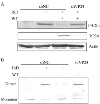
VP24 blocks the phosphorylation and dimerization of IRF3.
IRF3 is a crucial transcription factor in the IFN-

signaling
path-way; in response to cellular stimulation, IRF3 is phosphorylated
by TBK1, leading to its homodimerization and translocation into
the nucleus. To investigate whether VP24 inhibited the
phosphor-FIG 1VP24 inhibits ISD-mediated production of IFN-and the activation of IFN-promoter induced by cGAS-STING or STING alone. (A) HFF-shNC/ shVP24 cells were infected with HSV-1 for 16 h at a multiplicity of infection (MOI) of 5. Cells were harvested and subjected to WB to analyze the VP24 and UL42 protein. (B) HFF-shNC/shVP24 cells were infected with HSV-1 for 2 h before ISD transfection at an MOI of 5. Cells were harvested at 8 hpi and subjected to qRT-PCR to analyze the IFN-mRNA. (C and D) VP24-Flag plasmid was cotransfected into HEK 293T cells with an IFN-reporter plasmid (200 ng), cGAS (15 ng), and a minimal amount of STING plasmid (2.5 ng) or a large amount of STING plasmid (200 ng), respectively.Renillaluciferase reporter plasmid (pRL-TK; 50 ng) was introduced into each transfection as an internal control to normalize transfection efficiency. Cells were harvested at 24 h posttransfection and subjected to the DLR assay to detect the IFN-reporter activity. The data represent results from one of the triplicate experiments.
on November 7, 2019 by guest
http://jvi.asm.org/
[image:3.585.113.475.64.304.2]ylation of IRF-3, HFF transfected with shNC or
pSIREN-shVP24 (HFF-shNC/pSIREN-shVP24 cells) were infected with WT HSV-1
before ISD transfection, and robust phosphorylation of IRF-3 was
observed in HFF-shNC cells following ISD stimulation (
Fig. 3A
).
However, HSV-1 infection abrogated the phosphorylation of
IRF-3, and knockdown of VP24 restored its phosphorylation (
Fig.
4A
). Similarly, IRF-3 dimerization was markedly reduced during
HSV-1 infection, and knockdown of VP24 recovered its
dimeriza-tion (
Fig. 4B
). Taken together, these results demonstrated that
VP24 dampened the activation of IRF-3 induced by ISD.
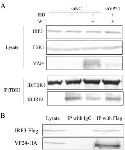
VP24 prevents the interaction between endogenous TBK1
and IRF3 during HSV-1 infection by interacting with IRF3.
On
the basis of these data, we speculate that VP24 might affect the
interaction between TBK1 and IRF3. To test our hypothesis, we
next determined ISD-induced interaction between TBK1 and
IRF3 in HFF-shNC and HFF-shVP24 cells infected with HSV-1.
HFF-shNC/shVP24 cells were infected with HSV-1 for 2 h before
ISD transfection and subjected to the co-IP assay to detect the
interaction between endogenous TBK1 and IRF3. As shown in
Fig. 5A
, HSV-1 infection reduced the interaction between TBK1
and IRF3 induced by ISD, while knockdown of VP24 restored
their interaction. These data suggested that VP24 blocked the
in-teraction of TBK1 and IRF3. In order to further explore the
un-derlying mechanisms, HEK 293T cells were transfected with
VP24-HA plasmid and 36 h after transfection, cells were harvested
and subjected to the co-IP assay to detect the interaction between
VP24 and IRF3. As a result, VP24 was efficiently co-IPed with
IRF-3 by anti-IRF3 PAb, but not by nonspecific mouse
monoclo-nal antibody IgG (
Fig. 5B
). These results demonstrated that VP24
could prevent the interaction between endogenous TBK1 and
IRF3 during HSV-1 infection by interacting with IRF3.
DISCUSSION
HSV-1 is a highly successful human pathogen that has evolved
multiple evasion mechanisms to facilitate its proliferation. To
date, a number of HSV-1 proteins have been reported to inhibit
host innate antiviral responses, and all these proteins may
func-tion cooperatively during HSV-1 infecfunc-tion. During HSV-1 capsid
maturation, the UL26-encoded protease processes itself to release
the N-terminal protease domain VP24. However, the role of VP24
in the innate immune response of the host is still largely unknown.
The type I IFN signal pathway is the first line of host innate
defense against viral infection, and it is responsible for the
induc-tion of numerous ISGs. Recent research reveals that the type I IFN
signal pathway is activated upon recognition of viral nucleic acids
by several cytosolic DNA sensors, such as cGAS, DAI, IFI16, and
DDX41 (
51
), among which cGAS was considered the major DNA
sensor. Upon binding the viral DNA segment, cGAS is activated
and produces cGAMP to activate adaptor protein STING,
fol-lowed by recruitment of TBK1, phosphorylation and nuclear
lo-calization of IRF3, and eventually, the series of events lead to
IFN-

production. Growing evidence shows that the
cGAS-STING DNA-sensing signal pathway plays a pivotal role in host
defense against HSV-1 infection. One can speculate that viruses
FIG 2VP24 inhibits the activation of IFN-promoter through IRF3 ele-ments. (A to D) VP24-Flag plasmid was cotransfected into HEK 293T cells with a NF-B reporter plasmid (200 ng) (A and C) or an IRF3 reporter plasmid (200 ng) (B and D) with cGAS (15 ng) and a minimal amount of STING (2.5 ng) plasmid or a large amount of STING plasmid (200 ng), respectively. Cells were harvested 24 h posttransfection and subjected to the DLR assay. The data represent results from one of the triplicate experiments.
FIG 3VP24 inhibits IFN-production by targeting IRF3. HEK 293T cells were transfected with plasmids expressing TBK1 (200 ng) (A) or IRF3/5D (200 ng) (B), together with an IRF3 reporter plasmid. The DLR assay was performed 24 h posttransfection. All cells were transfected with pRL-TK (50 ng) as an internal control to normalize transfection efficiency. The data represent results from one of the triplicate experiments.
FIG 4VP24 blocks the phosphorylation and dimerization of IRF3. (A) HFF-shNC/shVP24 cells were infected with WT HSV at an MOI of 5 for 2 h before transfection with ISD. Western blot analysis was performed 16 h posttransfec-tion to detect IRF3 phosphorylaposttransfec-tion. (B) HFF-shNC/shVP24 cells were mock infected or infected with WT HSV at an MOI of 5 for 2 h before transfection with ISD. Cells were harvested at 18 hpi, and native PAGE assays were per-formed to detect IRF3 dimerization. The data represent results from one of the triplicate experiments.
on November 7, 2019 by guest
http://jvi.asm.org/
[image:4.585.336.505.64.240.2] [image:4.585.39.287.65.252.2] [image:4.585.39.287.579.665.2]might have evolved certain mechanisms to impede the
cGAS-STING signal pathway. Ma et al. reported that several Kaposi’s
sarcoma-associated herpesvirus (KSHV) proteins could block
IFN-

promoter activity induced by the cGAS-STING signal
pathway. One KSHV protein, viral interferon regulatory factor 1,
was identified to inhibit the cytosol cGAS-STING signal by
block-ing the STING-TBK1 interaction (
11
). A recent study by Wu et al.
revealed that KSHV ORF52, a gammaherpesvirus-specific
tegu-ment protein, could prevent cGAS-mediated DNA-sensing signal
by directly inhibiting cGAS enzymatic activity (
52
). However,
un-til now, little has been known about the countermeasures by DNA
viruses against the cGAS-STING signal pathway, and no HSV-1
gene product has been found to target this pathway yet. IRF3 also
plays a crucial role in the cGAS-STING signal pathway, as all
sig-nals ultimately converge at IRF3 or IRF7. Therefore, it is not
sur-prising that viruses have evolved strategies to counteract innate
immune responses by targeting IRF3.
Taken together, our results provide further information on the
mechanism by which HSV-1 antagonizes the host antiviral innate
immune response. In the present study, we have demonstrated
that the HSV-1 VP24 protein inhibited cGAS-STING-mediated
IFN-

signaling pathway by blocking the interaction between
TBK1 and IRF3 during HSV-1 infection. The findings in this study
expand our knowledge on the molecular mechanisms by which
HSV-1 counteracts the antiviral innate immunity of the host to
ensure its replication and spread.
ACKNOWLEDGMENTS
We thank Rongtuan Lin for STING-HA plasmid, Yi-Ling Lin for IRF3/5D plasmid, and Takashi Fujita for IFN--Luc.
Work in the Zheng laboratory relevant to this article was supported by grants from the National Natural Science Foundation of China (81371795 and 81571974) and the Innovative Research Team at Soochow University (PCSIRT IRT1075).
C.Z. is an adjunct professor of the Department of Microbiology, Im-munology and Infectious Diseases, University of Calgary, Calgary, Al-berta, Canada.
FUNDING INFORMATION
This work, including the efforts of Chunfu Zheng, was funded by Inno-vative Research Team at Soochow University (PCSIRT IRT1075). This work, including the efforts of Chunfu Zheng, was funded by National Natural Science Foundation of China (NSFC) (81371795 and 81571974).
REFERENCES
1.Pichlmair A, Reis e Sousa C.2007. Innate recognition of viruses. Immu-nity27:370 –383.http://dx.doi.org/10.1016/j.immuni.2007.08.012. 2.Takeuchi O, Akira S.2009. Innate immunity to virus infection. Immunol
Rev227:75– 86.http://dx.doi.org/10.1111/j.1600-065X.2008.00737.x. 3.Vandevenne P, Sadzot-Delvaux C, Piette J.2010. Innate immune
re-sponse and viral interference strategies developed by human herpesvi-ruses. Biochem Pharmacol80:1955–1972.http://dx.doi.org/10.1016/j.bcp .2010.07.001.
4.Sun L, Wu J, Du F, Chen X, Chen ZJ.2013. Cyclic GMP-AMP synthase is a cytosolic DNA sensor that activates the type I interferon pathway. Science339:786 –791.http://dx.doi.org/10.1126/science.1232458. 5.Unterholzner L, Keating SE, Baran M, Horan KA, Jensen SB, Sharma S,
Sirois CM, Jin T, Latz E, Xiao TS, Fitzgerald KA, Paludan SR, Bowie AG.2010. IFI16 is an innate immune sensor for intracellular DNA. Nat Immunol11:997–1004.http://dx.doi.org/10.1038/ni.1932.
6.Zhang Z, Yuan B, Bao M, Lu N, Kim T, Liu YJ.2011. The helicase DDX41 senses intracellular DNA mediated by the adaptor STING in den-dritic cells. Nat Immunol12:959 –965.http://dx.doi.org/10.1038/ni.2091. 7.Takaoka A, Wang Z, Choi MK, Yanai H, Negishi H, Ban T, Lu Y, Miyagishi M, Kodama T, Honda K, Ohba Y, Taniguchi T.2007. DAI (DLM-1/ZBP1) is a cytosolic DNA sensor and an activator of innate immune response. Nature 448:501–505. http://dx.doi.org/10.1038 /nature06013.
8.Wu J, Chen ZJ.2014. Innate immune sensing and signaling of cytosolic nucleic acids. Annu Rev Immunol32:461– 488.http://dx.doi.org/10.1146 /annurev-immunol-032713-120156.
9.Abe T, Barber GN.2014. Cytosolic-DNA-mediated, STING-dependent proinflammatory gene induction necessitates canonical NF-kappaB acti-vation through TBK1. J Virol88:5328 –5341.http://dx.doi.org/10.1128 /JVI.00037-14.
10. Dempsey A, Bowie AG.2015. Innate immune recognition of DNA: a recent history. Virology 479-480:146 –152. http://dx.doi.org/10.1016/j .virol.2015.03.013.
11. Ma Z, Jacobs SR, West JA, Stopford C, Zhang Z, Davis Z, Barber GN, Glaunsinger BA, Dittmer DP, Damania B. 2015. Modulation of the cGAS-STING DNA sensing pathway by gammaherpesviruses. Proc Natl Acad Sci U S A112:E4306 –E4315.http://dx.doi.org/10.1073/pnas .1503831112.
12. Diner EJ, Vance RE.2014. Taking the STING out of cytosolic DNA sensing. Trends Immunol35:1–2.http://dx.doi.org/10.1016/j.it.2013.10 .011.
13. Bhat N, Fitzgerald KA.2014. Recognition of cytosolic DNA by cGAS and other STING-dependent sensors. Eur J Immunol44:634 – 640.http://dx .doi.org/10.1002/eji.201344127.
14. Wu J, Sun L, Chen X, Du F, Shi H, Chen C, Chen ZJ.2013. Cyclic GMP-AMP is an endogenous second messenger in innate immune signal-ing by cytosolic DNA. Science339:826 – 830.http://dx.doi.org/10.1126 /science.1229963.
15. Randall RE, Goodbourn S.2008. Interferons and viruses: an interplay between induction, signalling, antiviral responses and virus countermea-sures. J Gen Virol89:1– 47.
16. Sarkar SN, Sen GC.2004. Novel functions of proteins encoded by viral
FIG 5VP24 prevents the interaction between endogenous TBK1 and IRF3 during HSV-1 infection by interacting with IRF3. (A) HFF-shNC/shVP24 cells were infected with WT HSV at an MOI of 5 for 2 h before transfection with ISD. Cells were harvested at 18 hpi, and the extracts were analyzed by immu-noprecipitation using anti-TBK1 PAb (IP:TBK1), and Western blotting with anti-TBK1, anti-IRF3, and anti-VP24. The data represent results from one of the triplicate experiments. (B) HEK 293T cells were transfected with VP24-HA plasmid and harvested at 36 h posttransfection, and the extracts were analyzed by immunoprecipitation using IRF3 PAb (IP:IRF3) and WB with anti-HA and anti-IRF3. The data represent results from one of the triplicate experiments.
on November 7, 2019 by guest
http://jvi.asm.org/
[image:5.585.61.267.63.310.2]stress-inducible genes. Pharmacol Ther103:245–259.http://dx.doi.org/10 .1016/j.pharmthera.2004.07.007.
17. Leib DA.2002. Counteraction of interferon-induced antiviral responses by herpes simplex viruses. Curr Top Microbiol Immunol269:171–185. 18. Mossman KL, Ashkar AA.2005. Herpesviruses and the innate immune
response. Viral Immunol18:267–281.http://dx.doi.org/10.1089/vim .2005.18.267.
19. Paladino P, Mossman KL.2009. Mechanisms employed by herpes sim-plex virus 1 to inhibit the interferon response. J Interferon Cytokine Res
29:599 – 607.http://dx.doi.org/10.1089/jir.2009.0074.
20. Mossman KL, Macgregor PF, Rozmus JJ, Goryachev AB, Edwards AM, Smiley JR.2001. Herpes simplex virus triggers and then disarms a host antiviral response. J Virol75:750 –758.http://dx.doi.org/10.1128/JVI.75.2 .750-758.2001.
21. Nicholl MJ, Robinson LH, Preston CM. 2000. Activation of cellular interferon-responsive genes after infection of human cells with herpes simplex virus type 1. J Gen Virol81:2215–2218.http://dx.doi.org/10.1099 /0022-1317-81-9-2215.
22. Lin R, Noyce RS, Collins SE, Everett RD, Mossman KL. 2004. The herpes simplex virus ICP0 RING finger domain inhibits IRF3- and IRF7-mediated activation of interferon-stimulated genes. J Virol78:1675–1684.
http://dx.doi.org/10.1128/JVI.78.4.1675-1684.2004.
23. Melroe GT, DeLuca NA, Knipe DM.2004. Herpes simplex virus 1 has multiple mechanisms for blocking virus-induced interferon production. J Virol78:8411– 8420.http://dx.doi.org/10.1128/JVI.78.16.8411-8420 .2004.
24. Mossman K.2005. Analysis of anti-interferon properties of the herpes simplex virus type I ICP0 protein. Methods Mol Med116:195–205. 25. Paz S, Vilasco M, Arguello M, Sun Q, Lacoste J, Nguyen TL, Zhao T,
Shestakova EA, Zaari S, Bibeau-Poirier A, Servant MJ, Lin R, Meurs EF, Hiscott J.2009. Ubiquitin-regulated recruitment of IkappaB kinase epsi-lon to the MAVS interferon signaling adapter. Mol Cell Biol29:3401– 3412.http://dx.doi.org/10.1128/MCB.00880-08.
26. Zhang J, Wang K, Wang S, Zheng C.2013. Herpes simplex virus 1 E3 ubiquitin ligase ICP0 protein inhibits tumor necrosis factor alpha-induced NF-kappaB activation by interacting with p65/RelA and p50/ NF-kappaB1. J Virol87:12935–12948.http://dx.doi.org/10.1128/JVI .01952-13.
27. Johnson KE, Song B, Knipe DM.2008. Role for herpes simplex virus 1 ICP27 in the inhibition of type I interferon signaling. Virology374:487– 494.http://dx.doi.org/10.1016/j.virol.2008.01.001.
28. Liang L, Roizman B.2008. Expression of gamma interferon-dependent genes is blocked independently by virion host shutoff RNase and by US3 protein kinase. J Virol82:4688 – 4696.http://dx.doi.org/10.1128/JVI .02763-07.
29. Wang K, Ni L, Wang S, Zheng C.2014. Herpes simplex virus 1 protein kinase US3 hyperphosphorylates p65/RelA and dampens NF-kappaB ac-tivation. J Virol88:7941–7951.http://dx.doi.org/10.1128/JVI.03394-13. 30. Wang S, Wang K, Lin R, Zheng C.2013. Herpes simplex virus 1 serine/
threonine kinase US3 hyperphosphorylates IRF3 and inhibits beta inter-feron production. J Virol87:12814 –12827.http://dx.doi.org/10.1128/JVI .02355-13.
31. Xing J, Wang S, Lin R, Mossman KL, Zheng C.2012. Herpes simplex virus 1 tegument protein US11 downmodulates the RLR signaling path-way via direct interaction with RIG-I and MDA-5. J Virol86:3528 –3540.
http://dx.doi.org/10.1128/JVI.06713-11.
32. Wang S, Wang K, Li J, Zheng C.2013. Herpes simplex virus 1 ubiquitin-specific protease UL36 inhibits beta interferon production by deubiquiti-nating TRAF3. J Virol 87:11851–11860. http://dx.doi.org/10.1128/JVI .01211-13.
33. Zhang J, Wang S, Wang K, Zheng C.2013. Herpes simplex virus 1 DNA polymerase processivity factor UL42 inhibits Talpha-induced NF-kappaB activation by interacting with p65/RelA and p50/NF-NF-kappaB1. Med Microbiol Immunol202:313–325.http://dx.doi.org/10.1007/s00430 -013-0295-0.
34. Xing J, Ni L, Wang S, Wang K, Lin R, Zheng C.2013. Herpes simplex virus 1-encoded tegument protein VP16 abrogates the production of beta interferon (IFN) by inhibiting NF-kappaB activation and blocking IFN
regulatory factor 3 to recruit its coactivator CBP. J Virol87:9788 –9801.
http://dx.doi.org/10.1128/JVI.01440-13.
35. Elgadi MM, Hayes CE, Smiley JR.1999. The herpes simplex virus vhs protein induces endoribonucleolytic cleavage of target RNAs in cell ex-tracts. J Virol73:7153–7164.
36. Kwong AD, Frenkel N.1987. Herpes simplex virus-infected cells contain a function(s) that destabilizes both host and viral mRNAs. Proc Natl Acad Sci U S A84:1926 –1930.http://dx.doi.org/10.1073/pnas.84.7.1926. 37. Smiley JR.2004. Herpes simplex virus virion host shutoff protein:
im-mune evasion mediated by a viral RNase? J Virol78:1063–1068.http://dx .doi.org/10.1128/JVI.78.3.1063-1068.2004.
38. Shen G, Wang K, Wang S, Cai M, Li ML, Zheng C.2014. Herpes simplex virus 1 counteracts viperin via its virion host shutoff protein UL41. J Virol
88:12163–12166.http://dx.doi.org/10.1128/JVI.01380-14.
39. Su C, Zhang J, Zheng C.2015. Herpes simplex virus 1 UL41 protein abrogates the antiviral activity of hZAP by degrading its mRNA. Virol J
12:203.http://dx.doi.org/10.1186/s12985-015-0433-y.
40. Liu F, Roizman B. 1992. Differentiation of multiple domains in the herpes simplex virus 1 protease encoded by the UL26 gene. Proc Natl Acad Sci U S A89:2076 –2080.http://dx.doi.org/10.1073/pnas.89.6.2076. 41. Person S, Laquerre S, Desai P, Hempel J.1993. Herpes simplex virus type
1 capsid protein, VP21, originates within the UL26 open reading frame. J Gen Virol74:2269 –2273.http://dx.doi.org/10.1099/0022-1317-74-10 -2269.
42. Xing J, Wang S, Lin F, Pan W, Hu CD, Zheng C.2011. Comprehensive characterization of interaction complexes of herpes simplex virus type 1 ICP22, UL3, UL4, and UL20.5. J Virol85:1881–1886.http://dx.doi.org/10 .1128/JVI.01730-10.
43. Xing J, Wu F, Pan W, Zheng C.2010. Molecular anatomy of subcellular localization of HSV-1 tegument protein US11 in living cells. Virus Res
153:71– 81.http://dx.doi.org/10.1016/j.virusres.2010.07.009.
44. Ehrhardt C, Kardinal C, Wurzer WJ, Wolff T, von Eichel-Streiber C, Pleschka S, Planz O, Ludwig S.2004. Rac1 and PAK1 are upstream of IKK-epsilon and TBK-1 in the viral activation of interferon regulatory factor-3. FEBS Lett567:230 –238.http://dx.doi.org/10.1016/j.febslet.2004 .04.069.
45. Chang TH, Liao CL, Lin YL.2006. Flavivirus induces interferon-beta gene expression through a pathway involving RIG-I-dependent IRF-3 and PI3K-dependent NF-kappaB activation. Microbes Infect8:157–171.http: //dx.doi.org/10.1016/j.micinf.2005.06.014.
46. Lin R, Lacoste J, Nakhaei P, Sun Q, Yang L, Paz S, Wilkinson P, Julkunen I, Vitour D, Meurs E, Hiscott J. 2006. Dissociation of a MAVS/IPS-1/VISA/Cardif-IKKepsilon molecular complex from the mi-tochondrial outer membrane by hepatitis C virus NS3-4A proteolytic cleavage. J Virol80:6072– 6083.http://dx.doi.org/10.1128/JVI.02495-05. 47. Jordan M, Schallhorn A, Wurm FM.1996. Transfecting mammalian
cells: optimization of critical parameters affecting calcium-phosphate pre-cipitate formation. Nucleic Acids Res24:596 – 601.http://dx.doi.org/10 .1093/nar/24.4.596.
48. Zhong B, Yang Y, Li S, Wang YY, Li Y, Diao F, Lei C, He X, Zhang L, Tien P, Shu HB.2008. The adaptor protein MITA links virus-sensing receptors to IRF3 transcription factor activation. Immunity29:538 –550.
http://dx.doi.org/10.1016/j.immuni.2008.09.003.
49. Zhu H, Zheng C, Xing J, Wang S, Li S, Lin R, Mossman KL.2011. Varicella-zoster virus immediate-early protein ORF61 abrogates the IRF3-mediated innate immune response through degradation of activated IRF3. J Virol85:11079 –11089.http://dx.doi.org/10.1128/JVI.05098-11. 50. Ishikawa H, Barber GN.2008. STING is an endoplasmic reticulum
adap-tor that facilitates innate immune signalling. Nature455:674 – 678.http: //dx.doi.org/10.1038/nature07317.
51. Keating SE, Baran M, Bowie AG.2011. Cytosolic DNA sensors regulating type I interferon induction. Trends Immunol32:574 –581.http://dx.doi .org/10.1016/j.it.2011.08.004.
52. Wu JJ, Li W, Shao Y, Avey D, Fu B, Gillen J, Hand T, Ma S, Liu X, Miley W, Konrad A, Neipel F, Sturzl M, Whitby D, Li H, Zhu F.2015. Inhibition of cGAS DNA sensing by a herpesvirus virion protein. Cell Host Microbe18:333–344.http://dx.doi.org/10.1016/j.chom.2015.07.015.
on November 7, 2019 by guest
http://jvi.asm.org/