A Comprehensive RNA Sequencing Analysis of the Adeno-Associated
Virus (AAV) Type 2 Transcriptome Reveals Novel AAV Transcripts,
Splice Variants, and Derived Proteins
Catrin Stutika,aAndreas Gogol-Döring,b*Laura Botschen,aMario Mietzsch,aStefan Weger,aMirjam Feldkamp,cWei Chen,c Regine Heilbronna
Institute of Virology, Campus Benjamin Franklin, Charité Medical School, Berlin, Germanya; German Centre for Integrative Biodiversity Research, Halle-Jena-Leipzig, Germanyb; Laboratory for Functional Genomics and Systems Biology, Berlin Institute for Medical Systems Biology, Max-Delbrück-Centrum für Molekulare Medizin, Berlin, Germanyc
ABSTRACT
Adeno-associated virus (AAV) is recognized for its bipartite life cycle with productive replication dependent on coinfection with adenovirus (Ad) and AAV latency being established in the absence of a helper virus. The shift from latent to Ad-dependent AAV replication is mostly regulated at the transcriptional level. The current AAV transcription map displays highly expressed tran-scripts as found upon coinfection with Ad. So far, AAV trantran-scripts have only been characterized on the plus strand of the AAV single-stranded DNA genome. The AAV minus strand is assumed not to be transcribed. Here, we apply Illumina-based RNA se-quencing (RNA-Seq) to characterize the entire AAV2 transcriptome in the absence or presence of Ad. We find known and iden-tify novel AAV transcripts, including additional splice variants, the most abundant of which leads to expression of a novel 18-kDa Rep/VP fusion protein. Furthermore, we identify for the first time transcription on the AAV minus strand with clustered reads upstream of the p5 promoter, confirmed by 5' rapid amplification of cDNA ends and RNase protection assays. The p5 pro-moter displays considerable activity in both directions, a finding indicative of divergent transcription. Upon infection with AAV alone, low-level transcription of both AAV strands is detectable and is strongly stimulated upon coinfection with Ad.
IMPORTANCE
Next-generation sequencing (NGS) allows unbiased genome-wide analyses of transcription profiles, used here for an in depth analysis of the AAV2 transcriptome during latency and productive infection. RNA-Seq analysis led to the discovery of novel AAV transcripts and splice variants, including a derived, novel 18-kDa Rep/VP fusion protein. Unexpectedly, transcription from the AAV minus strand was discovered, indicative of divergent transcription from the p5 promoter. This finding opens the door for novel concepts of the switch between AAV latency and productive replication. In the absence of a suitable animal model to study
AAVin vivo, combinedin cellulaeandin silicostudies will help to forward the understanding of the unique, bipartite AAV life
cycle.
A
deno-associated viruses (AAV) are helper-dependent mem-bers of the parvovirus group that require coinfection with an unrelated helper virus, particularly adenovirus (Ad) for produc-tive replication (1). Despite of several identified AAV serotypes most research has been done with prototype AAV type 2. The AAV2 genome consists of a linear, single-stranded DNA of 4.7 kb. Both ends carry identical inverted terminal repeats (ITR) of 145 bp (2), which flank the two major open reading frames (ORF), calledrepandcap. TherepORF codes for four nonstructural pro-teins, Rep78 and a C-terminally spliced variant, Rep68. In addi-tion, N-terminally truncated versions thereof are expressed, called Rep52 and Rep40, respectively. The Rep proteins are required as regulators for various steps of the AAV life cycle. AAVcapharbors the ORF for the capsid proteins (VP1 to -3) and a separate ORF for the assembly-activating protein (AAP) (3).The current knowledge of AAV transcription dates back to early research in the 1980s when the three AAV2 promoters, p5, p19, or p40 and major transcripts derived thereof were charac-terized (4,5). All AAV transcripts identified since then map to only one DNA plus strand (6), which led to the assumption that the complementary AAV minus strand was not transcribed. Furthermore, spliced AAV transcripts were identified display-ing a sdisplay-ingle, shared intron located in the center of the AAV2
genome (4,7). Initially, a single splice donor site at AAV nu-cleotide 1906 and a single splice acceptor site located at nucle-otide (nt) 2228 were described. Later, an alternative splice ac-ceptor site at nt 2201 was detected (8), allowing the expression of VP1 protein from the same promoter (p40) as VP2 and VP3. Compared to the major splice acceptor site (nt 2228), the so-called minor splice acceptor site was more divergent from the ideal consensus sequence, likely explaining its less-frequent use
Received28 October 2015Accepted6 November 2015
Accepted manuscript posted online11 November 2015
CitationStutika C, Gogol-Döring A, Botschen L, Mietzsch M, Weger S, Feldkamp M, Chen W, Heilbronn R. 2016. A comprehensive RNA sequencing analysis of the adeno-associated virus (AAV) type 2 transcriptome reveals novel AAV transcripts, splice variants, and derived proteins. J Virol 90:1278 –1289.doi:10.1128/JVI.02750-15.
Editor:L. Banks
Address correspondence to Regine Heilbronn, [email protected].
*Present address: Andreas Gogol-Döring, Technische Hochschule Mittelhessen, Giessen, Germany.
C.S. and A.G.-D. contributed equally to this article.
Copyright © 2016, American Society for Microbiology. All Rights Reserved.
on November 7, 2019 by guest
http://jvi.asm.org/
for splicing. During AAV replication the majority of p5- or p19-initiated transcripts are not spliced, whereas transcripts initiated from p40 are almost exclusively spliced (9, 10). All AAV transcripts share a 3' end (7), coinciding with the polyad-enylation signal identified between AAV2 nt 4424 and 4429 (2). The only other AAV serotype whose transcription pattern has been analyzed is AAV5, the genetically most distant member of the AAV family. Several variations in comparison to the AAV2 transcription pattern were described. Most importantly, AAV5 reptranscripts terminate at a second, internal polyadenylation signal located in the AAV5 intron (11).
Early analysis of AAV transcription relied on S1 nuclease map-ping and reverse transcriptase-mediated primer extension (4,7,
12,13). These methods allowed the mapping and sequence anal-ysis of defined and highly expressed transcripts. Subsequently, quantitative RNase protection was used to complement the cur-rently accepted AAV transcription map (10). Using the described methods, only AAV RNAs in regions explicitly probed for could be detected. Recent NGS-based technologies allow high-through-put analysis of entire cellular transcriptomes, leading to unbiased identification and parallel quantification of all existing cellular and viral transcripts. With RNA sequencing (RNA-Seq), novel transcripts and splicing variants in human cells and of viral tran-scripts have been discovered (14,15).
In this report we used Illumina-based RNA-Seq to provide a comprehensive picture of AAV2 transcription in the presence or absence of helper adenovirus. We were able to identify known and novel AAV2 transcripts, splicing variants, and a new protein de-rived therefrom. In addition, previously unrecognized RNAs tran-scribed from the AAV minus strand were detected.
MATERIALS AND METHODS
Cell culture and viruses.HEK 293- and HeLa cells were cultivated as described previously (16). Human adenovirus type 2 (Ad2) was propa-gated and assayed in 293 cells.
Cloning and mutagenesis.For the generation of different luciferase constructs, the corresponding AAV2 wild-type promoter sequences were cloned upstream of the firefly luciferase gene in a sense orientation, AAV(⫹)190-320-luc, or in a reverse complementary orientation, AAV(–) 586-155-luc, AAV(–)320-180-luc, and AAV(–)320-256-luc, respectively. The constructs were verified by restriction analysis. For the generation of AAV mutants AAV-D1, AAV-D2, and AAV-A1, the corresponding mu-tations were introduced by the QuikChange site-directed mutagenesis protocol (Stratagene) into the AAV2reporcapgenes of pTAV2-0 (17). The mutations were verified by DNA sequencing and restriction enzyme analysis. Similarly, the nucleotide exchange G4395T and the 9 amino acid C-terminal Flag-tag insertion (DYKDDDDK*) between AAV2 nucleotide positions 4411 and 4412 led to the constructs pTAV2-0 G4395T Flag (pAAVwt-Flag), and pTAV2-0 G4137A G4395T Flag (pAAV-A1-Flag). For the primers, seeTable 1.
AAV2 production, purification, and quantification.For AAV2 pro-duction, HEK 293 cells seeded at 30% confluence were transfected 24 h later with AAV plasmids and pHelper as described previously (18). AAV2 was purified from benzonase-treated, cleared freeze-thaw supernatants by one-step AVB Sepharose affinity chromatography and quantified by qPCR as described previously (19) with primers for AAV2rep(Table 1). AAV2 infectious titers were determined by endpoint dilutions on Ad2-infected HeLa cells (20).
Virus infection and DNA plasmid transfection.HeLa cells seeded at a density of 30% were infected 20 h later with AAV2 wild-type or AAV-A1 mutant (multiplicity of infection [MOI] 250) and/or Ad2 (MOI 25). Transfection of HeLa cells with AAV plasmids was performed as described previously (21). Infection with Ad2 (MOI 25) was performed 16 h
post-TABLE 1Oligonucleotides for mutagenesis PCR, qPCR, RT-PCR, and PCRs for detection of AAV2 splice events, Northern blot hybridization, and 5=RACE
Oligonucleotide Sequence (5=-3=)
AAV-D1-Fwd CTGACGGAATGGCGCCGTCTTTCGAAGGCCCCGGAGGCCCTT AAV-D1-Rev AAGGGCCTCCGGGGCCTTCGAAAGACGGCGCCATTCCGTCAG AAV-D2-Fwd CTGACGGAATGGCGCCGTGTTTCGAAGGCCCCGGAGGCCCTT AAV-D2-Rev AAGGGCCTCCGGGGCCTTCGAAACACGGCGCCATTCCGTCAG AAV-A1-Fwd CTTAAACACCCTCCTCCACAAATTCTCATCAAGAACACC AAV-A1-Rev GGTGTTCTTGATGAGAATTTGTGGAGGAGGGTGTTTAAG
Flag-Fwd CCTGACTCGTAATCTGTAATGACTACAAGGATGACGATGACAAGTGATGCTTGTTAATCAATAAACCG Flag-Rev CGGTTTATTGATTAACAAGCATCACTTGTCATCGTCATCCTTGTAGTCATTACAGATTACGAGTCAGG Stop-Mut-Fwd CCATTGGCACCAGATACCTTACTCGTAATCTGTAATGAC
Stop-Mut-Rev GTCATTACAGATTACGAGTAAGGTATCTGGTGCCAATGG Rep2-Fwd AGAAGGAATGGGAGTTGCCG
Rep2-Rev TCTGACTCAGGAAACGTCCC
RT-Primer CAGATTACGAGTCAGGTAT-CTGGTGCCAATGGGGCG SD527-Fwd AGAAGGAATGGGAGTTGCCG
SD988-Fwd GTAAACGGTTGGTGGCGCAG
SD1906-Fwd GCCATCGACGTCAGACGCGGAAGCTTC SD3184-Fwd CTTCAACAGATTCCACTGCC
SA807-Rev GCTCCGTGAGATTCAAACAG SA985-Rev AGGCCGCATTGAAGGAGATG SA1437-Rev TTGCTTCCTCCGAGAATGGC SA1635-Rev TTGGTGACCTTCCCAAAGTC SA2228-Rev CGTAGGCTTTGTCGTGCTCG SA4138-Rev TGAAGGTGGTCGAAGGATTCGCAGGT
SDA1 CCTCGAGCCAATACGGCGCCATTC
p5-RACE-GSP1 GGTGGAGTCGTGACGTGAATT p5-RACE-GSP2 TCATAGGGTTAGGGAGGTCCT
on November 7, 2019 by guest
http://jvi.asm.org/
[image:2.585.44.550.86.364.2]transfection and cells were harvested at the indicated time points postin-fection.
RNA extraction.Total RNA was isolated by TRIzol reagent (Ambion) according to the manufacturer’s protocol. RNA samples were treated with RNase-free Turbo DNase (Ambion) to remove residual DNA, followed by phenol-chloroform extraction and precipitation. RNA quality and integ-rity was verified on 0.8% agarose gels and on bioanalyzer (Agilent). Total RNA samples of proven high quality were used for reverse transcription-PCR (RT-transcription-PCR) and for RNA library generation. Poly(A)⫹selected RNA was purified from total RNA with the Oligotex mRNA Midi purification kit (Qiagen).
RT-PCR.Newly identified AAV2 splice sites were validated using 1g of DNase-digested total RNA, reverse transcribed by SuperScript II re-verse transcriptase (Invitrogen) according to the manufacturer’s protocol with a primer that binds at the rear 3' end of the AAV2 genome (Table 1). The cDNAs were PCR amplified with AAV2-specific primer pairs (Table 1) located upstream of the predicted splice donor and downstream of the predicted splice acceptor sites.
Library preparation and Illumina sequencing.1g of each of the four different total RNA samples (cells, AAV2, AAV2⫹Ad2, and Ad2) were subjected to total RNA library generation using the TruSeq stranded total RNA sample preparation kit according to the manufacturer’s proto-col (Illumina). The libraries were pooled and sequenced on a HiSeq 2000 platform (multiplexed SR 1⫻101⫹7 high-output mode).
Read mapping.Demultiplexed sequencing reads (length, 101 bp; sin-gle end) were mapped after trimming Illumina adapters from the 3' ends on the human genome (hg19, GRCh37) and the human transcriptome by using the Bowtie 2 read mapping tool (22). The transcriptome was derived from the RefSeq gene annotation downloaded from the UCSC table browser (https://genome.ucsc.edu/cgi-bin/hgTables). All reads that could not be mapped to the human genome/transcriptome were mapped to the reference sequences of Ad2 (RefSeqAC_000007.1) and of both the flip and the flop variants of AAV2 (RefSeqNC_001401.2). In all cases, only read mappings with an alignment score (as given in the XS field of the SAM output) of at least⫺20 were accepted. A detailed analysis of the reads mapped on AAV2 revealed that the genome of the sequenced AAV2 only differs from the reference at a single nucleotide (a C instead of a G at nt 3284).
Detection of splice junctions.We generated a splice junction refer-ence, i.e., a set of sequences containing fusions of all possible combination of canonical splicing donor sites (GT dinucleotides) and downstream splicing acceptor sites (AG dinucleotides) in AAV2. A split alignment of the sequencing read onto the AAV2 genome using the Segemehl mapping tool (23) revealed no evidence of splicing at noncanonical splicing sites (data not shown). The sequencing reads were mapped to the splice junc-tion reference using Bowtie 2, where inserjunc-tions and delejunc-tions were sup-pressed by setting the gap penalties to very high values. We then deter-mined the number of uniquely mapped reads for each possible junction of donor and acceptor sites. Only reads that covered at least 20 bp upstream of the splice donor site and 20 bp downstream of the splice acceptor site were counted.
Detection of polyadenylation sites.We searched the reverse comple-ment sequencing reads for polyadenine repeats of a length of 8 (i.e., AAA AAAAA) and mapped the parts of the reads upstream of the polyadenine repeats to the AAV2 genome. Note, that AAV2 contains no polyadenine repeat longer than 6. Mappings with quality below 20 were discarded.
Promoter prediction and ORF finder. Promoters on the AAV2 wild-type genome were predicted by the Neural Network Promoter Pre-diction platform (NNPP) of the BDGP (http://www.fruitfly.org/seq_tools /promoter.html) (24). For the translation of alternative splice variants into potential new proteins an open reading frame (ORF) finder (NCBI) was used (http://www.ncbi.nlm.nih.gov/gorf/gorf.html).
Luciferase assay and Western blot analysis.Luciferase assays and Western blot analysis were performed as described previously (16). Mouse monoclonal antibodies (MAbs) and rabbit polyclonal antibodies
used for immunoblot analysis were anti-Rep MAb 303.9 (1:10; Progen), VP MAb B1 (1:10; Progen), VP1 MAb A1 (1:10; Progen), anti-VP1/2 MAb A69 (1:10; Progen) or rabbit and mouse anti-Flag (1:1,000; Sigma), respectively, followed by reaction with horseradish peroxidase-conjugated secondary antibodies and ECL detection.
Extraction of viral DNA and Southern hybridization.Extraction of viral DNA by a modified Hirt procedure and Southern blot analysis were performed as described previously (16). Samples of DpnI-digested Hirt DNA (6g) were analyzed on Southern blots for AAV replication with a 1.6-kb fragment from thecapregion of pTAV2-0 as probe labeled either with [32P]dCTP for radioactive detection or with Biotin-11-dUTP for
nonradioactive detection.
Northern hybridization with oligonucleotide probes.Northern blot hybridization of total or poly(A)⫹selected RNA samples was performed with a 24-nt oligonucleotide spanning 12 nt upstream and downstream of the AAV2 527:2228 splice junction (SDA1, seeTable 1) labeled with T4 polynucleotide kinase in the presence of [␥-32P]ATP. Hybridization was
performed for 4 h at 68°C in hybridization solution (0.5% [wt/vol] so-dium dodecyl sulfate [SDS], 6⫻SSC, 50 mM HEPES [pH 7.8], 150g/ml salmon sperm DNA, and 5⫻Denhardt solution). The filters were washed four times in 6⫻SSC (1⫻SSC is 0.15 M NaCl plus 0.015 M sodium citrate)– 0.1% SDS at room temperature for 5 min and subsequently twice with 6⫻SSC– 0.1% SDS at 68°C for 30 min.
RNase protection assay.RNase protection assays were performed with the RPA III kit (Ambion) essentially as described by the supplier. Two AAV2 sense RNA probes (nt 170 to 250 and nt 170 to 340) were radiolabeled with [␣-32P]UTP byin vitrotranscription with T7
polymer-ase (Riboprobe system; Promega) using XbaI-linearized pBluescript vec-tor containing the corresponding AAV nucleotides as the templates. The
in vitro-transcribed RPA probes were gel purified on an 8% PAA– 8 M urea denaturing gel. A total of 3⫻104cpm of purified radiolabeled probe
were hybridized with 20g of total RNA, followed by RNase A/RNase T1
treatment according to the manufacturer’s protocol with an additional final ethanol precipitation step. The protected RNA fragments were elec-trophoresed on an 8% PAA– 8 M urea gel and subsequently fixed in a solution containing 20% ethanol and 5% acetic acid for 30 min before vacuum drying for 2 h at 80°C. Gels were exposed to a storage phosphor screen and scanned by a Fujifilm FLA-3000 imager.
RACE.Rapid amplification of cDNA ends (RACE) was conducted using the 5'RACE system (version 2.0; Invitrogen) according to the man-ufacturer’s protocol. First-strand cDNA synthesis was performed on total RNA from AAV2/Ad2-coinfected HeLa cells with a primer binding to AAV2 nt 151 to 171 (p5-RACE-GSP1), followed by treatment with RNase H. The cDNA was purified and poly(C)-tailed using terminal deoxynucle-otidyltransferase (TdT). After TdT removal, PCR amplification was per-formed using the TrueStart Hot StartTaqDNA polymerase (Thermo Scientific) and a nested primer binding to AAV2 nt 175 to 195 (p5-RACE-GSP2) and the anchor primer supplied by the manufacturer. The PCR products were analyzed and excised from agarose gels and subsequently cloned into pBluescript II SK(⫹). Randomly picked clones were subjected to DNA sequencing (Eurofins Genomics). For the primer sequences, see
Table 1.
RESULTS
RNA-Seq library preparation.To identify the ideal time point for RNA-Seq analysis, time course experiments with total RNAs and proteins extracted from HeLa cells infected with AAV2 in pres-ence or abspres-ence of adenovirus (Ad2) were performed. At 27 h postinfection (hpi), AAV2 transcripts were clearly detectable on Northern blots, as were Rep and VP on Western blots. This time point was chosen for total RNA extraction from cells infected with AAV2 in presence or absence of Ad2 and from uninfected and Ad2-infected cells that served as controls. All RNA samples were analyzed by bioanalyzer tests displaying RNA integrity numbers higher than 8.90, which are indicative of highly intact RNA. Total
on November 7, 2019 by guest
http://jvi.asm.org/
RNA was rRNA depleted and digested to 100 nucleotide frag-ments. RNA libraries were sequenced on an Illumina HiSeq 2000 platform.
Illumina RNA-Seq next-generation sequencing.For each set of data, between 21.2 and 26.6 million reads were obtained by Illumina RNA-Seq (Table 2). The vast majority of reads (⬎95%) could be successfully mapped either to the human genome or to the viral genomes of AAV2 or Ad2. In AAV2-infected cells, in the absence of Ad2 ca. 97% of all RNA reads were assigned to the human genome, which is comparable to uninfected control cells. Only few AAV-specific transcripts (0.04%) could be detected at 27 hpi. In the presence of Ad, AAV-specific transcripts rose to 13.6% of the total read count (Table 2). The majority of AAV reads mapped to the coding plus strand of the AAV2 genome. However, a lower, but significant proportion of reads mapped to the AAV2 minus strand. Parallel to the Ad-induced increase of AAV-specific transcripts, a reduction of the percentage of RNAs mappable to the human genome was found. This was also seen for cells infected with Ad2 alone, suggestive of an Ad-induced effect (Table 2). AAV2/coinfected cells showed a 5-fold reduction of Ad2-specific reads compared to cells infected with Ad2 alone. This finding likely reflects the established effect that AAV inhibits ad-enoviral replication (25–28).
RNA read mapping on the AAV2 plus strand.The population of AAV2-specific RNA reads for each data set was mapped to the wild-type AAV2 genome. Based on this alignment, a coverage map of the AAV2 genome was created that displays the number of reads that map to a specific AAV2 genome position. The genome cov-erage of the data set from AAV2/Ad2-coinfected cells is based on approximately 3.0 million AAV2-specific reads (Fig. 1A). Steep increases of RNA-Seq read counts are indicative of either tran-scription initiation or of splicing. The reads assigned to the AAV2 plus strand largely confirmed the well-established transcription initiation at the p5, p19, and p40 promoters. Also in line with previous studies, transcripts initiated at the p40 promoter were more frequent compared to p5- or p19-initiated transcripts in AAV2/Ad2-coinfected cells, whereas in cells infected solely with AAV2 the p5-initiated transcripts were the most prevalent (Fig. 1B). In the absence of Ad, few AAV-specific transcripts were de-tectable. With the exception of the prevalent p5-initiated tran-scripts, the overall AAV transcription profile was very similar to that in the presence of Ad, although at a⬎200-fold reduced level. Additionally, occasional reads were detected upstream of the p5 promoter on the AAV plus strand in the absence or presence of Ad2 (Fig. 1AandB), a finding indicative of transcription initiated within the left ITR of AAV2, as described previously (11,29,30). A single polyadenylation site near the right end of the AAV2 genome was defined before (31). RNA-Seq shows that indeed the vast ma-jority of Ad2-induced AAV2 transcripts terminate by
polyadenyl-ation at nt 4450 (Fig. 1C). No other hotspots for polyadenylation were detected.
Analysis of transcription on the AAV minus strand.As stated above, a significant proportion of reads mapped to the AAV2 mi-nus strand in the absence, as well as in the presence of Ad coinfec-tion (Fig. 1AandB). These new transcripts, detected here for the first time, were largely restricted to the AAV region between the left ITR and the p5 promoter. Their abundance was about one-third compared to p5-initiated transcripts on the AAV plus strand
(Fig. 1A). To obtain hints, where transcription on the AAV2
mi-nus strand was initiated we first searched for candidate upstream promoters. In silico promoter prediction (NNPP) (24) under stringent conditions with a cutoff score above 0.9 was applied to both AAV strands. While the well-characterized AAV2 p5 and p40 promoters could be readily detected with cutoff scores of 0.95 and 1.0, respectively, the p19 promoter was only detected when low-ering the cutoff to a score of 0.53. On the AAV2 minus strand three candidate promoters were detected with cutoff scores above 0.9. Two of these are located near the left ITR, with putative transcrip-tion start sites at nt 230 (score 0.93) and nt 305 (score 0.94), re-spectively (Fig. 1D). These sites are in agreement with the assumed start sites for AAV2 minus strand transcription (Fig. 1A). The activity of the candidate promoters was tested using reporter gene constructs. Various AAV2 fragments comprising the p5 promoter region were cloned in reverse orientation upstream of the lucifer-ase gene. Ad2-infected HeLa cells were transfected with the con-structs, as depicted inFig. 1D. The luciferase activity measured at 48 h posttransfection showed strong promoter activity of AAV(–) 320-180-luc, the reverse complement of the established AAV p5 promoter AAV(⫹)190-320-luc, which was 7-fold more active. Deletion of nt 180 to 255 in the AAV(–)320-180-lucsequence led to an⬎30-fold drop in promoter activity, whereas an extended version of the p5 reverse complement, AAV(–)586-155-luc, was marginally stronger compared to AAV(–)320-180-luc. Relative promoter activities in sense and antisense orientation correlated well with the relative proportions of transcripts detected on the AAV plus and minus strands (Fig. 1A).
[image:4.585.39.544.79.151.2]To more closely map transcription start sites on the AAV mi-nus strand, we used 5'RACE with the gene-specific primer used for first-strand cDNA synthesis binding to AAV nt 151 to 171. The PCR products were cloned and individual representative clones sequenced to identify the respective transcription start site (TSS). The identified TSSs mapped to the region between AAV2 nt 227 to 320 (Fig. 1E). AAV minus strand transcription was further con-firmed by RNase protection analysis (RPA) with probes covering AAV2 nt 170 to 250 or nt 170 to 340, respectively (Fig. 1F). With both probes, a rather heterogeneous population of AAV-specific protected RNA fragments in the size range of about 40 to 80 nt was
TABLE 2RNA-Seq analysis: assignment of the reads to the species from which they originate
Data set
Total no. of reads
No. (%) of mappable reads
No. (%) of human reads
No. (%) of AAV2 reads
No. (%) of Ad2 reads Plus strand Minus strand Ambiguous
Cells 23,380,236 22,665,799 (96.94) 22,662,280 (96.93) 1,404 (0.01) 14 (0.00) 0 (0.00) 2,101 (0.01) AAV2 21,200,968 20,601,318 (97.17) 20,591,869 (97.13) 7,616 (0.04) 241 (0.00) 1 (0.00) 1,591 (0.01) AAV2⫹Ad2 23,047,796 22,167,728 (96.18) 16,327,433 (70.84) 3,061,388 (13.28) 68,099 (0.30) 184 (0.00) 2,710,624 (11.76) Ad2 26,564,586 25,351,109 (95.43) 10,920,188 (41.11) 1,614 (0.01) 16 (0.00) 0 (0.00) 14,429,291 (54.32)
on November 7, 2019 by guest
http://jvi.asm.org/
FIG 1RNA-Seq: mapping and transcription analysis on both strands of the AAV2 genome. (A) Coverage map of AAV2-specific RNA-Seq reads to the AAV2 genome of AAV2/Ad2-coinfected cells at 27 hpi. The AAV2 genome scale is displayed in the center. Reads mapped to the plus strand are presented above, and reads mapped to the minus strand are presented below. (B) Display as in panel A. RNA-Seq reads of AAV2-infected cells at 27 hpi are presented. Note the different scales of the read counts in panels A and B. (C) Mapping of RNA-Seq reads ending with a poly(A) tail. The genome position of poly(A) addition is displayed relative to the totality of polyadenylated AAV reads in the data set of AAV2/Ad2-coinfected cells. (D) Promoter activity of the AAV2 p5 region in reverse direction. The upper part shows the AAV2 genome with the nucleotide positions of the transcription start sites of the p5, p19, and p40 promoters (black arrows). In addition, putative promoters (gray arrows) originating from a promoter prediction program with a cutoff score above 0.9 are indicated on the plus or minus strand of the AAV genome with the respective nucleotide positions of the TSS. An asterisk (*) highlights the established p19 promoter, detected with a cutoff score below 0.9. The lower part shows the relative luciferase activities of the indicated candidate reverse p5 promoter sequences on the AAV2 minus strand. AAV(⫹)190-320-lucrepresents the AAV2 p5 promoter in the forward orientation serving as a positive control and empty plucserving as a negative control. The relative luciferase activities of three different experiments were determined in the presence of Ad2 and are depicted as means⫾standard deviations (SD). (E) 5'RACE analysis of AAV minus strand transcription. Displayed are the TSSs (arrows) derived from DNA sequences of cloned 5'RACE products from the AAV minus strand (nt 330 to 170). Single TSSs are displayed as black squares above the genome. The p5 promoter TSS at nt 287 (gray arrow) on the AAV plus strand is shown for orientation. (F) An RNase protection assay was performed within vitro-transcribed hybridization probes covering AAV2 nt 170 to 250 (left part) or AAV2 nt 170 to 340 (right part) in a sense orientation for the detection of AAV minus strand transcripts in total RNA from HeLa cells either mock infected, AAV2 infected, Ad2 infected, or AAV2/Ad2 coinfected for 27 h. The undigested probes (–RNase) and the probes digested in the presence of yeast t-RNA (⫹RNase) are shown as controls. Both probes contain additional non-AAV2 sequences from the pBluescript vector used for T7 polymerase-dependentin vitrotranscription. The maximal protected AAV2 regions are 81 nt for probe RPA 170-250, and 171 nt for probe RPA 170-340, respectively.
on November 7, 2019 by guest
http://jvi.asm.org/
[image:5.585.64.522.65.513.2]detected specifically in AAV2/Ad2-coinfected cells (Fig. 1F) but not in mock- or Ad-infected cells.
Analysis of AAV2 splicing.Spliced AAV transcripts were iden-tified by RNA-Seq analysis as AAV reads joined to distant, defined parts of the AAV genome. Bioinformatic tools with stringent fil-ters were utilized to exclude background noise. Only splice events above a critical abundance were investigated further. Reads cov-ering the known AAV2 splice donor site at position 1906 were highly abundant, with⬎35,000 reads in the data set for AAV2/ Ad2-coinfected cells. In ca. 72% of those reads, no splice event was detectable (Table 3). In about 23% of the reads, the splice donor site was joined to the described major splice acceptor site (nt 2228), and 3.5% of the reads were fused to the minor splice accep-tor site (nt 2201), as expected from previous studies (8). Less fre-quently used splice donor sites were identified at nt 527, 988, and 3184. In addition, occasional, alternative AAV splice acceptor sites were detected mostly at nt 4138 (Table 3).
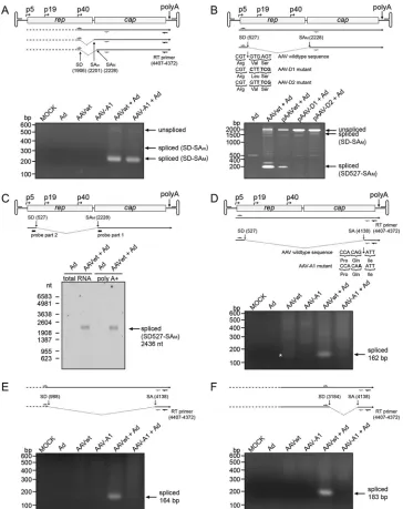
The existence of alternative splice products was verified by RT-PCR with sets of primers whose binding sites were located up-stream of the respective splice donor and downup-stream of the cor-responding splice acceptor sites (Fig. 2). Additionally, AAV mutants were generated with an inactivated splice donor site at nt 527 (Fig. 2B, D1 and D2 mutants) or splice acceptor site at nt 4138
(Fig. 2D, A1 mutant), respectively. The described splice products
1906:2201/2228 were detected by RT-PCR, as shown for AAV wild-type and mutant A1 after coinfection with Ad2 (Fig. 2A). The 527:2228 splice variant was clearly detected with the AAV wild type but, as expected, not with the D1 and D2 mutants (Fig. 2B). Northern blots with total and with poly(A)⫹-selected RNA from AAV2/Ad2-coinfected HeLa cells were probed with an oligonu-cleotide spanning the 527:2228 splice junction. A single band of about 2.4 kb was detected (Fig. 2C) corresponding to a p5-initi-ated spliced mRNA that extends to the common poly(A)⫹site (theoretical size, 2,436 nt). The splice events involving the splice acceptor site at nt 4138 leading to the splice products 527:4138, 988:4138, and 3184:4138 were detected by RT-PCR for the AAV2 wild type but, as expected, not for AAV mutant A1 (Fig. 2DtoF). Notably, splice product 527:4138 had already been faintly detected in the absence of Ad2 infection, but none of the other splice prod-ucts had (Fig. 2D). Further splice variants with lower frequencies in the RNA-Seq screen were also confirmed by RT-PCR (Table 3, values in boldface).
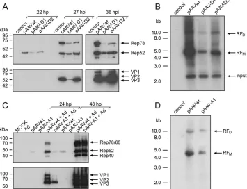
Involvement of the novel splice variants in the AAV replica-tion cycle.To address the functional significance of the newly identified splice variants for the AAV2 replication cycle, the AAV mutants for the splice donor site at nt 527 (D1 and D2) and the splice acceptor site at nt 4138 (A1) were assayed for Rep and Cap expression and for AAV DNA replication. In the D1 mutant the absolutely conserved G nucleotide at the 5' end of the intron had
been abolished, at the expense of a conservative exchange of valine to isoleucine in Rep78/68, whereas the D2 mutant maintains the amino acid sequence of Rep78/68 (Fig. 2B). On Western blots, mutant D1 showed strongly reduced Rep78 and capsid protein expression and some reduction of Rep52, whereas mutant D2 showed a less-pronounced effect on Rep, and VP expression was indistinguishable from that of AAV wild type (Fig. 3A). Also, AAV DNA replication was reduced more extensively for mutant D1 than D2 (Fig. 3B). In line with these findings, the titers of infec-tious AAV were reduced by 3 to 4 logs for mutant D1 and by 1 log for mutant D2 compared to AAV wild type (data not shown).
The AAV-A1 mutant, with inactivated splice acceptor site at nt 4138 and maintained VP amino acid sequence (Fig. 2D) showed markedly reduced Rep and Cap expression early (24 hpi), but not later (48 hpi) after infection (Fig. 3C). Furthermore, in contrast to AAV2 wild type, Rep52 was undetectable for mutant A1 in the absence of Ad2 (Fig. 3C). In line with these findings, AAV DNA replication was reduced, as evidenced by diminished monomeric and dimeric AAV2 replicative forms (Fig. 3D). In summary, the newly described, alternative splice products appear to play some role in the regulation of Ad-induced AAV replication, especially in the early stages of AAV replication.
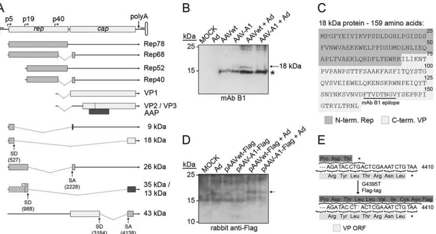
Prediction and identification of novel AAV2 proteins.Some of the alternatively spliced AAV transcripts could possibly lead to the translation of novel proteins, as predictedin silico. Candidate proteins of at least 50 amino acids, initiated by an ATG start codon or alternatively by ACG or CTG, were considered for further anal-ysis. The most abundant splice variants and the predicted proteins are depicted inFig. 4A. The splice variant 527:2228 could lead to a small protein of 9.1 kDa consisting of the N terminus of Rep78 fused to a short C terminus translated in a different reading frame as the Rep and VP proteins (Fig. 4A). Unfortunately for this pre-dicted protein, no antibody was available, nor was an antibody available for the splice variant 988:2228-derived 26-kDa protein that lacks the central portion of Rep68. However, the presumed translation product of splice variant 527:4138 could be assayed by MAb B1, which is directed against AAV2 VP1-3. Indeed, an 18-kDa protein was detected in the low-molecular-weight range in cells infected with AAV wild type but not with mutant A1 (Fig. 4B). The 15-kDa bands visible in all extracts infected with AAV most likely represent autolytic capsid cleavage products, as de-scribed previously (32). The newly identified 18-kDa protein con-sists of 159 amino acids (Fig. 4C), of which the first 69 amino acids correspond to the N-terminal part of Rep78 and the C-terminal 90 amino acids correspond to VP. As described above, the novel 18-kDa Rep/VP fusion protein might have a function on the regula-tion of early AAV gene expression (see above).
[image:6.585.40.546.78.148.2]The predicted protein of 43-kDa translated from splice variant 3184:4138, although containing the epitopes of MAb A1 and of
TABLE 3Summary of AAV2 splice events by RNA-Seq analysis: AAV2⫹Ad2
Splice donor Read count % not spliced
% spliced with acceptora
807 985 1437 1635 2201 2228 4138 Others 527 34,472 93.64 0.53 0.33 0.07 0.11 0.09 0.50 4.21 0.52
988 26,404 95.05 0.17 0.56 0.23 0.89 2.45 0.65
1906 35,746 72.25 3.57 23.42 0.36 0.40
3184 50,575 99.30 0.51 0.19
aThe displayed values give the percentages for the splice donors joined to the various splice acceptors. Values in boldface are splice variants that were validated by RT-PCR.
on November 7, 2019 by guest
http://jvi.asm.org/
MAb A69 could not be identified with either antibody. To verify expression of other predicted proteins, especially from splice vari-ant 988:4138, an in-frame Flag tag was introduced in the AAV wild-type and mutant A1 DNA sequences (Fig. 4E). Neither of the predicted 35- or 13-kDa proteins could be detected on Western blots with two alternative anti-Flag antibodies, which successfully
detected the positive control (data not shown). Furthermore, the Flag tag allowed to search for the proposed X protein (18 kDa) (33) derived from the same ORF. Indeed, on very long exposures faint bands of approximately 18 kDa were detected in AAV wild-type and in AAV-A1 transfected cells, a finding consistent with the existence of the X protein (Fig. 4D).
FIG 2Validation of RNA-Seq-derived AAV2 splice events by RT-PCR and Northern blot analysis. (A) The AAV2 genome with unspliced/spliced transcripts is displayed at the top. The primer for reverse transcription is shown with its binding position. The forward primer (nt 1862 to 1889) and reverse primer (nt 2440 to 2421) are displayed for PCR analysis of 1906:2201/2228 splicing. Below, the results of RT-PCR for AAV wild-type- and mutant A1-infected cells are displayed, separated on an agarose gel. PCR products are indicated by arrows. (B) RT-PCR of splice event 527:2228, performed as in panel A with cells transfected with plasmids for AAV wild-type or mutants D1 or D2. Recoded nucleotide sequences of AAV-D1 and AAV-D2 are displayed below the AAV genome. (C) Northern blot analysis of total and poly(A)⫹selected RNA of cells coinfected with AAV2 and Ad2 using a 24-nt oligomer as a probe that spans the AAV2 splice junction 527:2228, as depicted below the AAV genome. The arrow points to the spliced transcripts. (D to F) RT-PCR analysis of additional AAV RNA splice events, performed as in panel A. (D) Forward primer (nt 415 to 434) and reverse primer (nt 4186 to 4161) used for the analysis of 527:4138 splicing. The corresponding mutation of AAV-A1 is displayed below the AAV genome. The asterisk (*) marks a faint band of the splice product in the absence of Ad. (E) Forward primer (nt 874 to 893) and reverse primer (nt 4186 to 4161) for the analysis of 988:4138 splicing. (F) Forward primer (nt 3051 to 3070) and reverse primer (nt 4186 to 4161) for the analysis of 3184:4138 splicing.
on November 7, 2019 by guest
http://jvi.asm.org/
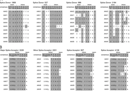
[image:7.585.111.475.65.524.2]Comparison of the splice sites to other AAV serotypes.The biological relevance of the new splice variants was further evalu-ated by comparing their conservation in the genomes of alterna-tive AAV serotypes. The genomes of AAV serotypes 1 to 9 were aligned, and the splice sites were compared to the consensus se-quence of mammalian splice donor and acceptor sites (34). The well-documented splice donor site at AAV2 nt 1906 is highly con-served among all AAV serotypes but shows minor variations com-pared to the consensus sequence (Fig. 5A). The splice donor site at nt 527 is similarly conserved among AAV serotypes and is there-fore assumed to be functional in these serotypes as well. The splice donor sites at nt 988 and 3184 are more divergent from the con-sensus sequence. Although the splice donor site nt 988 may be also functional in AAV3 and AAV4, the splice donor site at nt 3184 exists only in AAV2.
The described major splice acceptor site at nt 2228 of AAV2 is highly conserved among all AAV serotypes (Fig. 5B), as is the minor splice acceptor site at nt 2201, although with reduced py-rimidine levels indicative of lower utilization rates. The newly identified splice acceptor site at nt 4138 is also conserved, with the exception of AAV3, AAV4, and AAV5. Another splice acceptor site at nt 807 validated by PCR for splice variant 527:807 is con-served among the AAV serotypes (Fig. 5B). Unfortunately, the predicted protein of 60 kDa is very similar to the molecular mass of Rep68, and we were therefore unable to differentiate it on West-ern blots.
DISCUSSION
We present here the first NGS analysis of the AAV2 transcriptome during latency and productive AAV replication. Each of the four data sets analyzed by Illumina RNA-Seq gave rise to more than 21 million reads, more than 95% of which could be successfully mapped to the human or viral genomes. The high quality of the raw data allowed a comprehensive and comparative analysis of the transcription profiles of AAV2 with or without Ad2 infec-tion. The high sensitivity of RNA-Seq combined with unbiased RNA detection uncovered previously unrecognized transcripts, al-ternative splice variants, and a new 18-kDa protein that appears to play a role in early AAV replication. Most importantly, we are the first to document transcription from the AAV minus strand, lead-ing to an update of the current concept of AAV gene expression.
Analysis of the AAV2 transcription profile. As a single-stranded DNA virus AAV is dependent on the synthesis of the complementary strand to generate a double-stranded template for transcription initiation. In the absence of a helper virus, only lim-ited Rep78/68 expression occurs from the p5 promoter (35). This is reflected in the RNA-Seq analysis of AAV2-infected cells in the absence of Ad2, where few AAV transcripts (0.04%) are found, only⬃4-fold above background level. Of these, p5 promoter-ini-tiated transcripts are the most abundant RNAs, but transcripts originating from p19 and p40 are also detected. The findings are in line with AAV2 biology, where Rep78/68 was shown to repress
FIG 3Influence of alternative splicing on AAV replication. (A) Analysis of AAV2 Rep and VP expression in AAV wild type-, splice mutant AAV-D1-, or AAV-D2 (seeFig. 2B)-transfected HeLa cells at the indicated time points after Ad infection. AAV Rep and VP proteins are detected by MAb 303.9 or MAb B1, respectively. (B) Southern blot analysis of Hirt extracts of AAV wild type-, AAV-D1-, or AAV-D2-transfected and Ad-infected HeLa cells. AAV-specific monomeric (RFM) and
dimeric (RFD) replicative forms, detected by probing with a labeled 1.6-kbcap-derived DNA sequence, are indicated by arrows. Input refers to undigested input
DNA. (C) Western blot analysis as described in panel A with mutant AAV-A1 (seeFig. 2D). Mock- and Ad-infected cells served as controls. (D) Southern blot analysis as performed for panel B with mutant AAV-A1.
on November 7, 2019 by guest
http://jvi.asm.org/
[image:8.585.115.470.70.342.2]AAV2 promoter activity, which is viewed as a mechanism to es-tablish and maintain the latent state (35,36).
In the presence of Ad2, AAV2-specific reads are increased by
⬎200-fold, with a shift of transcription toward p40 expressed genes, which is consistent with previous reports (10). RNA-Seq also detects more-abundant transcription from the p5 promoter than from the p19 promoter, a finding that is at variance with the described 1:3 p5/p19 ratio found by RNase protection-based quantification late in AAV infection (10) and is likely explained by experimental differences. Furthermore, RNA-Seq identified reads mapping to the AAV plus strand upstream of the p5 promoter. These transcripts appear to be derived from the described tran-scription initiation site (Inr) located within the ITR, observed be-fore for AAV5 and for recombinant AAV2-based vectors (11,29,
30). In addition, RNA-Seq precisely mapped the previously de-fined, unique AAV2 polyadenylation site (31). Mammalian mRNAs typically cleave the RNA molecule 10 to 30 nt behind the polyadenylation signal (37). In the case of AAV2, we show that cleavage occurs exactly 21 nt behind the AAUAAA polyadenyla-tion signal at posipolyadenyla-tion 4450 (2).
Previously, a candidate promoter (p81) was described on the AAV plus strand near genome position 3800 (38). We were unable to detect this promoterin silicounder the stringent conditions applied. Furthermore, RNA-Seq did not detect an increase of AAV-specific reads around nt 3800, as would have been expected downstream of an active promoter. However, on Western blots with Flag-tagged constructs a faint 18-kDa protein became visible consistent with the proposed X protein (33). Its very weak
expres-sion likely explains the difficulties in confirming its existence by alternative methods but also poses questions as to itsin vivo sig-nificance.
Transcription on the AAV2 minus strand.All AAV2 tran-scripts described in previous reports were derived from a single DNA strand, referred to as the AAV plus strand. Therefore, the complementary AAV2 minus strand has not been considered transcribed until now (6, 7, 12). RNA-Seq analysis, however, showed that a small proportion of AAV-specific reads from AAV2/Ad2-coinfected cells (0.30%) map to the AAV minus strand. AAV minus strand reads map to a region between the left ITR and the p5 promoter and have an abundance of approxi-mately one-third of the transcripts initiated at the p5 promoter of the AAV2 plus strand.In silico, candidate promoters are predicted on the AAV2 minus strand with transcription start sites at nt 230 and 305, respectively. Reporter gene assays revealed that the AAV2 sequence from nt 320 to 180 was sufficient to initiate transcription in minus strand direction. An extended sequence from nt 586 to 155 only marginally enhanced transcriptional activity. Obviously, the p5 promoter region is also active in opposite direction, which likely explains transcription of the AAV2 minus strand. This find-ing is unique for p5; transcripts in the opposite direction were not detected upstream of p19 or p40.
The finding is reminiscent of a mechanism called divergent transcription (39–42) that is related to transcription initiation in opposite directions from the same promoter. The rather short transcripts initiated at nonuniform start sites that have been de-scribed for the upstream direction in divergent transcription are
FIG 4Detection of novel AAV proteins derived from alternative splicing. (A) The AAV2 genome and current transcription/translation map is represented at the top. The different shading patterns indicate alternative reading frames. Below, the newly identified spliced transcripts are displayed with the deduced novel fusion ORFs. The molecular mass of the derived protein candidates is indicated to the right in kilodaltons. (B) Analysis of the predicted protein (18 kDa) expressed from splice variant 527:4138 on a Western blot reacted with MAb B1. AAV-A1 represents the AAV2 mutant at nt 4138 (seeFig. 2D). An asterisk (*) marks the previously described 15- to 17-kDa band, which is indicative of degraded input capsids. (C) Amino acid sequence of the novel 18-kDa protein. The first 69 amino acids correspond to the N-terminal part of Rep78; the last 90 amino acids correspond to the C terminus of the VP proteins, including the epitope for MAb B1. (D) Detection of AAVwt-Flag and AAV-A1-Flag constructs on a Western blot reacted with rabbit anti-Flag upon extensive exposure. The arrow marks a faint band of⬃18 kDa consistent with the previously proposed X protein (38). (E) Displayed is the proposed X-gene-derived C terminus of the resulting protein (dark gray) before and after insertion of the Flag tag in comparison to the confirmed capsid (VP) protein sequence (light gray). The asterisk (*) denotes the stop codon in the wild-type sequence.
on November 7, 2019 by guest
http://jvi.asm.org/
[image:9.585.80.506.65.294.2]in line with the heterogeneous population of AAV2 minus strand transcripts that we identified both by 5'RACE and by RNase pro-tection. One assumed role for divergent transcription is to facili-tate DNA unwinding, so that transcription initiation rates and expression of the downstream gene are enhanced (43). Further-more, the short antisense transcripts are believed to regulate the transcription of downstream genes by affecting the chromatin and thereby modulating promoter activity (44). An analogous mech-anism may be advantageous for the regulation of AAV p5-initi-ated transcripts, since these transcripts are the first AAV2 genes to be transcribed both in the presence and in the absence of adeno-virus. It will be interesting to see whether divergent transcription at p5 serves as a key for the switch between the latent and the lytic AAV replication cycles.
AAV splicing.Splicing of AAV2 transcripts is essential for the generation of Rep68 and Rep40 and for the correct stoichiometry of VP1, VP2, and VP3. The corresponding splice sites have been known for nearly 2 decades and are located at AAV2 nt 1906 (the splice donor) and nt 2201 and 2228 (the splice acceptor) (7,8). The majority of splice products detected by RNA-Seq correspond
to these sites. The ratio of usage for the established minor or major splice acceptor site, respectively, joined to the splice donor site at nt 1906, was roughly 1:7, a finding consistent with previous re-ports (31). The proportion of unspliced versus spliced transcripts at these sites is higher than previously reported (10) and is likely explained by the use of total cellular RNA for RNA-Seq compared to cytoplasmic RNA in previous studies. In addition, splicing en-hances the export of mRNAs (45). Therefore, the total cellular RNA used here may be enriched for unspliced transcripts still located in the nucleus.
The first indication of additional splice products came from the genome coverage of total AAV2-specific reads. The read count at the far right end of the AAV genome, downstream of nt 4000, was significantly increased, despite the absence of known or pre-dicted promoters in this region. Of several newly detected splice donor and acceptor sites utilized at variable efficiencies, the splice products involving a splice donor site at nt 527 and a splice accep-tor site at nt 4138 were particularly noteworthy. Reads covering the 527:4138 splice variant were even more abundant than the known splice event 1906:2201, allowing expression of the newly
FIG 5Alignment of splice donor and acceptor sites for AAV serotypes 1 to 9. (A) Splice donor sites at nt positions 1906,⫺527,⫺988, and⫺3184 of AAV2 were aligned to the corresponding sites of AAV serotypes 1 to 9 and compared to the mammalian consensus sequence of splice donor sites (34). Exon-intron boundaries are indicated by dashed lines. Below the consensus sequence the frequency (%) of a particular nucleotide in average splice donor sites is displayed. White areas indicate aberrations to the consensus sequence in the given AAV serotype. (B) Splice acceptor sites at nt positions 2228,⫺2201,⫺807, and⫺4138 of AAV2 were aligned to the corresponding sites of AAV serotypes 1 to 9 and compared to the mammalian consensus sequence of splice acceptor sites. The display is as described for panel A. Py, pyrimidine.
on November 7, 2019 by guest
http://jvi.asm.org/
[image:10.585.42.543.77.428.2]described 18-kDa Rep/VP protein. Mutants involving these par-ticular splice donor (AAV2 nt 527) or splice acceptor (AAV2 nt 4138) sites showed reduced expression levels of Rep and Cap and also some decrease in DNA replication.
Unlike the AAVs, most parvovirus genera exhibit more than one intronic sequence to generate alternative transcripts. The au-tonomous parvovirus minute virus of mice (MVM) possesses a large intron on the left-hand side of the genome, in addition to a short intron analogous to the single intron of the AAVs (46–48). For MVM, splicing of the large intron generates a transcript, which is translated into the nonstructural protein NS2. NS2 is required for viral growth in its natural murine host, while it is dispensable in many nonmurine cells (49). Furthermore, the hu-man parvovirus B19 contains two introns and the simian parvo-virus (SPV) contains three introns, which leads to various spliced transcripts with partially unknown protein function (50,51). The newly identified AAV2 splice donor site at nt 527 appears to be analogous to the described splice donor site of MVM (nt 514) and the erythroviruses B19 (nt 531) and SPV (nt 279). All of these are located close to the left ends of their genomes. Similarly, the newly identified splice acceptor site at AAV2 nt 4138 may have counter-parts in B19 (nt 4883) and SPV (nt 4638) at the right ends of the respective genomes. For parvovirus B19, a multiply spliced tran-script involving the above-described splice sites is translated to a small protein of 11 kDa, which appears to increase viral infectivity in vivo(52) and may play a role in apoptosis and cell signaling pathways (53,54). In cell culture the novel 18-kDa Rep/VP pro-tein only had a minor effect on AAV replication. However, anal-ogous to NS2 of MVM, the 18-kDa protein may have a more pronounced role for the AAV2 life cyclein vivo. Unfortunately,in vivomodels of wild-type AAV replication do not exist, and in humans and primates the site(s) and mechanisms of AAV latency and productive replication are largely unknown. It will be chal-lenging to further unravel possible functions of the novel RNAs and derived proteins during the AAV life cycle in its natural host.
ACKNOWLEDGMENTS
We thank Eva-Maria Hammer of the Heilbronn lab for help with cells and virus propagation and Madlen Sohn of the Chen lab for assistance during RNA library preparation.
C.S. acknowledges the receipt of a Charité-funded Ph.D. fellowship.
FUNDING INFORMATION
Charité Universitätsmedizin Berlin provided funding to Catrin Stutika.
REFERENCES
1.Blacklow NR, Hoggan MD, Rowe WP.1967. Isolation of adenovirus-associated viruses from man. Proc Natl Acad Sci U S A58:1410 –1415.
http://dx.doi.org/10.1073/pnas.58.4.1410.
2.Srivastava A, Lusby EW, Berns KI. 1983. Nucleotide sequence and organization of the adeno-associated virus 2 genome. J Virol45:555–564. 3.Sonntag F, Schmidt K, Kleinschmidt JA.2010. A viral assembly factor promotes AAV2 capsid formation in the nucleolus. Proc Natl Acad Sci U S A107:10220 –10225.http://dx.doi.org/10.1073/pnas.1001673107. 4.Green MR, Roeder RG.1980. Definition of a novel promoter for the major adenovirus-associated virus mRNA. Cell22:231–242.http://dx.doi .org/10.1016/0092-8674(80)90171-3.
5.Lusby EW, Berns KI.1982. Mapping of the 5' termini of two adeno-associated virus 2 RNAs in the left half of the genome. J Virol41:518 –526. 6.Jay FT, de la Maza LM, Carter BJ.1979. Parvovirus RNA transcripts containing sequences not present in mature mRNA: a method for isola-tion of putative mRNA precursor sequences. Proc Natl Acad Sci U S A
76:625– 629.http://dx.doi.org/10.1073/pnas.76.2.625.
7. Laughlin CA, Westphal H, Carter BJ. 1979. Spliced
adenovirus-associated virus RNA. Proc Natl Acad Sci U S A76:5567–5571.http://dx .doi.org/10.1073/pnas.76.11.5567.
8.Trempe JP, Carter BJ.1988. Alternate mRNA splicing is required for synthesis of adeno-associated virus VP1 capsid protein. J Virol62:3356 – 3363.
9.Marcus CJ, Laughlin CA, Carter BJ.1981. Adeno-associated virus RNA transcription in vivo. Eur J Biochem121:147–154.http://dx.doi.org/10 .1111/j.1432-1033.1981.tb06443.x.
10. Mouw MB, Pintel DJ.2000. Adeno-associated virus RNAs appear in a temporal order and their splicing is stimulated during coinfection with adenovirus. J Virol 74:9878 –9888. http://dx.doi.org/10.1128/JVI.74.21 .9878-9888.2000.
11. Qiu J, Nayak R, Tullis GE, Pintel DJ.2002. Characterization of the transcription profile of adeno-associated virus type 5 reveals a number of unique features compared to previously characterized adeno-associated viruses. J Virol76:12435–12447.http://dx.doi.org/10.1128 /JVI.76.24.12435-12447.2002.
12. Green MR, Straus SE, Roeder RG.1980. Transcripts of the adenovirus-associated virus genome: multiple polyadenylated RNAs including a po-tential primary transcript. J Virol35:560 –565.
13. Green MR, Roeder RG.1980. Transcripts of the adeno-associated virus genome: mapping of the major RNAs. J Virol36:79 –92.
14. Djebali S, Davis CA, Merkel A, Dobin A, Lassmann T, Mortazavi A, Tanzer A, Lagarde J, Lin W, Schlesinger F, Xue C, Marinov GK, Khatun J, Williams BA, Zaleski C, Rozowsky J, Roder M, Kokocinski F, Abdel-hamid RF, Alioto T, Antoshechkin I, Baer MT, Bar NS, Batut P, Bell K, Bell I, Chakrabortty S, Chen X, Chrast J, Curado J, Derrien T, Drenkow J, Dumais E, Dumais J, Duttagupta R, Falconnet E, Fastuca M, Fejes-Toth K, Ferreira P, Foissac S, Fullwood MJ, Gao H, Gonzalez D, Gordon A, Gunawardena H, Howald C, Jha S, Johnson R, Kapranov P, King B.2012. Landscape of transcription in human cells. Nature489:101– 108.http://dx.doi.org/10.1038/nature11233.
15. Zhao H, Chen M, Pettersson U. 2014. A new look at adenovirus splicing. Virology456-457:329 –341.http://dx.doi.org/10.1016/j.virol .2014.04.006.
16. Winter K, von Kietzell K, Heilbronn R, Pozzuto T, Fechner H, Weger S.2012. Roles of E4orf6 and VA I RNA in adenovirus-mediated stimula-tion of human parvovirus B19 DNA replicastimula-tion and structural gene ex-pression. J Virol86:5099 –5109.http://dx.doi.org/10.1128/JVI.06991-11. 17. Heilbronn R, Bürkle A, Stephan S, zur Hausen H.1990. The
adeno-associated virusrepgene suppresses herpes simplex virus-induced DNA-amplification. J Virol64:3012–3018.
18.Hüser D, Gogol-Doring A, Chen W, Heilbronn R. 2014. Adeno-associated virus type 2 wild-type and vector-mediated genomic integra-tion profiles of human diploid fibroblasts analyzed by third-generaintegra-tion PacBio DNA sequencing. J Virol88:11253–11263.http://dx.doi.org/10 .1128/JVI.01356-14.
19. Mietzsch M, Broecker F, Reinhardt A, Seeberger PH, Heilbronn R.
2014. Differential adeno-associated virus serotype-specific interaction patterns with synthetic heparins and other glycans. J Virol88:2991–3003.
http://dx.doi.org/10.1128/JVI.03371-13.
20. Weindler FW, Heilbronn R.1991. A subset of herpes simplex virus replication genes provides helper functions for productive adeno-associated virus replication. J Virol65:2476 –2483.
21. Stutika C, Hüser D, Weger S, Rutz N, Hessler M, Heilbronn R.2015. Definition of herpes simplex virus helper functions for the replication of adeno-associated virus type 5. J Gen Virol96:840 – 850.http://dx.doi.org /10.1099/vir.0.000034.
22. Langmead B, Salzberg SL.2012. Fast gapped-read alignment with Bowtie 2. Nat Methods9:357–359.http://dx.doi.org/10.1038/nmeth.1923. 23. Hoffmann S, Otto C, Kurtz S, Sharma CM, Khaitovich P, Vogel J,
Stadler PF, Hackermuller J.2009. Fast mapping of short sequences with mismatches, insertions and deletions using index structures. PLoS Com-put Biol5:e1000502.http://dx.doi.org/10.1371/journal.pcbi.1000502. 24. Reese MG.2001. Application of a time-delay neural network to promoter
annotation in theDrosophila melanogastergenome. Comput Chem26:51– 56.http://dx.doi.org/10.1016/S0097-8485(01)00099-7.
25. Casto BC, Atchison RW, Hammon WM.1967. Studies on the relation-ship between adeno-associated virus type I (AAV-1) and adenoviruses. I. Replication of AAV-1 in certain cell cultures and its effect on helper ade-novirus. Virology32:52–59.
26. Casto BC, Armstrong JA, Atchison RW, Hammon WM.1967. Studies on the relationship between adeno-associated virus type 1 (AAV-1) and
on November 7, 2019 by guest
http://jvi.asm.org/
adenoviruses. II. Inhibition of adenovirus plaques by AAV; its nature and specificity. Virology33:452– 458.
27. Weitzman MD, Fisher KJ, Wilson JM.1996. Recruitment of wild-type and recombinant adeno-associated virus into adenovirus replication cen-ters. J Virol70:1845–1854.
28. Timpe JM, Verrill KC, Trempe JP.2006. Effects of adeno-associated virus on adenovirus replication and gene expression during coinfection. J Virol80:7807–7815.http://dx.doi.org/10.1128/JVI.00198-06.
29. Flotte TR, Afione SA, Solow R, Drumm ML, Markakis D, Guggino WB, Zeitlin PL, Carter BJ.1993. Expression of the cystic fibrosis transmem-brane conductance regulator from a novel adeno-associated virus pro-moter. J Biol Chem268:3781–3790.
30. Haberman RP, McCown TJ, Samulski RJ.2000. Novel transcriptional regulatory signals in the adeno-associated virus terminal repeat A/D junc-tion element. J Virol74:8732– 8739.http://dx.doi.org/10.1128/JVI.74.18 .8732-8739.2000.
31. Qiu J, Pintel D.2008. Processing of adeno-associated virus RNA. Front Biosci13:3101–3115.http://dx.doi.org/10.2741/2912.
32. Salganik M, Venkatakrishnan B, Bennett A, Lins B, Yarbrough J, Muzyczka N, Agbandje-McKenna M, McKenna R.2012. Evidence for pH-dependent protease activity in the adeno-associated virus capsid. J Virol86:11877–11885.http://dx.doi.org/10.1128/JVI.01717-12. 33. Cao M, You H, Hermonat PL.2014. The X gene of adeno-associated
virus 2 (AAV2) is involved in viral DNA replication. PLoS One9:e104596.
http://dx.doi.org/10.1371/journal.pone.0104596.
34. Zhang MQ.1998. Statistical features of human exons and their flanking regions. Hum Mol Genet7:919 –932.http://dx.doi.org/10.1093/hmg/7.5 .919.
35. Pereira DJ, McCarty DM, Muzyczka N. 1997. The adeno-associated virus (AAV) Rep protein acts as both a repressor and an activator to reg-ulate AAV transcription during a productive infection. J Virol71:1079 – 1088.
36. Beaton A, Palumbo P, Berns KI. 1989. Expression from the adeno-associated virus p5 and p19 promoters is negatively regulated in trans by the rep protein. J Virol63:4450 – 4454.
37. Proudfoot NJ.2011. Ending the message: poly(A) signals then and now. Genes Dev25:1770 –1782.http://dx.doi.org/10.1101/gad.17268411. 38. Hermonat PL, Santin AD, De Greve J, De Rijcke M, Bishop BM, Han
L, Mane M, Kokorina N.1999. Chromosomal latency and expression at map unit 96 of a wild-type plus adeno-associated virus (AAV)/Neo vector and identification of p81, a new AAV transcriptional promoter. J Hum Virol2:359 –368.
39. Seila AC, Core LJ, Lis JT, Sharp PA.2009. Divergent transcription: a new feature of active promoters. Cell Cycle8:2557–2564.http://dx.doi.org/10 .4161/cc.8.16.9305.
40. Flynn RA, Chang HY.2012. Active chromatin and noncoding RNAs: an intimate relationship. Curr Opin Genet Dev22:172–178.http://dx.doi.org /10.1016/j.gde.2011.11.002.
41. Duttke SH, Lacadie SA, Ibrahim MM, Glass CK, Corcoran DL, Benner C, Heinz S, Kadonaga JT, Ohler U.2015. Human promoters are intrin-sically directional. Mol Cell 57:674 – 684. http://dx.doi.org/10.1016/j .molcel.2014.12.029.
42. Scruggs BS, Gilchrist DA, Nechaev S, Muse GW, Burkholder A, Fargo DC, Adelman K.2015. Bidirectional transcription arises from two dis-tinct hubs of transcription factor binding and active chromatin. Mol Cell
58:1101–1112.http://dx.doi.org/10.1016/j.molcel.2015.04.006. 43. Kouzine F, Sanford S, Elisha-Feil Z, Levens D.2008. The functional
response of upstream DNA to dynamic supercoiling in vivo. Nat Struct Mol Biol15:146 –154.http://dx.doi.org/10.1038/nsmb.1372.
44. Seila AC, Calabrese JM, Levine SS, Yeo GW, Rahl PB, Flynn RA, Young RA, Sharp PA. 2008. Divergent transcription from active promoters. Science322:1849 –1851.http://dx.doi.org/10.1126/science.1162253. 45. Valencia P, Dias AP, Reed R.2008. Splicing promotes rapid and efficient
mRNA export in mammalian cells. Proc Natl Acad Sci U S A105:3386 – 3391.http://dx.doi.org/10.1073/pnas.0800250105.
46. Pintel D, Dadachanji D, Astell CR, Ward DC. 1983. The genome of minute virus of mice, an autonomous parvovirus, encodes two overlap-ping transcription units. Nucleic Acids Res11:1019 –1038.http://dx.doi .org/10.1093/nar/11.4.1019.
47. Jongeneel CV, Sahli R, McMaster GK, Hirt B.1986. A precise map of splice junctions in the mRNAs of minute virus of mice, an autonomous parvovirus. J Virol59:564 –573.
48. Haut DD, Pintel DJ.1998. Intron definition is required for excision of the minute virus of mice small intron and definition of the upstream exon. J Virol72:1834 –1843.
49. Ruiz Z, D’Abramo A, Jr, Tattersall P.2006. Differential roles for the C-terminal hexapeptide domains of NS2 splice variants during MVM in-fection of murine cells. Virology349:382–395.http://dx.doi.org/10.1016 /j.virol.2006.01.039.
50. Ozawa K, Ayub J, Hao YS, Kurtzman G, Shimada T, Young N.1987. Novel transcription map for the B19 (human) pathogenic parvovirus. J Virol61:2395–2406.
51. Liu Z, Qiu J, Cheng F, Chu Y, Yoto Y, O’Sullivan MG, Brown KE, Pintel DJ.2004. Comparison of the transcription profile of simian parvovirus with that of the human erythrovirus B19 reveals a number of unique features. J Virol 78:12929 –12939. http://dx.doi.org/10.1128/JVI.78.23 .12929-12939.2004.
52. Zhi N, Mills IP, Lu J, Wong S, Filippone C, Brown KE.2006. Molecular and functional analyses of a human parvovirus B19 infectious clone dem-onstrates essential roles for NS1, VP1, and the 11-kilodalton protein in virus replication and infectivity. J Virol80:5941–5950.http://dx.doi.org /10.1128/JVI.02430-05.
53. Chen AY, Zhang EY, Guan W, Cheng F, Kleiboeker S, Yankee TM, Qiu J.2010. The small 11 kDa nonstructural protein of human parvovirus B19 plays a key role in inducing apoptosis during B19 virus infection of pri-mary erythroid progenitor cells. Blood115:1070 –1080.http://dx.doi.org /10.1182/blood-2009-04-215756.
54. Fan MM, Tamburic L, Shippam-Brett C, Zagrodney DB, Astell CR.
2001. The small 11-kDa protein from B19 parvovirus binds growth factor receptor-binding protein 2 in vitro in a Src homology 3 domain/ligand-dependent manner. Virology 291:285–291. http://dx.doi.org/10.1006 /viro.2001.1217.