Progenitor Cells Confers Functional and Persistent HIV-1 Resistance
in Humanized Mice
Renier Myburgh,a,bSandra Ivic,aMichael S. Pepper,bGustavo Gers-Huber,aDuo Li,aAnnette Audigé,aMary-Aude Rochat,a Vincent Jaquet,cStephan Regenass,dMarkus G. Manz,ePatrick Salmon,fKarl-Heinz Krause,c Roberto F. Specka
Division of Infectious Diseases and Hospital Epidemiology, University Hospital of Zurich, University of Zurich, Zurich, Switzerlanda; Department of Immunology and Institute for Cellular and Molecular Medicine, Faculty of Health Sciences, University of Pretoria, Pretoria, South Africab; Department of Pathology and Immunology, Faculty of Medicine, University of Geneva, Geneva, Switzerlandc; Clinic of Immunology, University Hospital Zurich, Zurich, Switzerlandd; Division of Hematology, University Hospital Zurich, Zurich, Switzerlande; Department of Neurosciences, Faculty of Medicine, University of Geneva, Geneva, Switzerlandf
ABSTRACT
Gene-engineered CD34ⴙhematopoietic stem and progenitor cells (HSPCs) can be used to generate an HIV-1-resistant immune system. However, a certain threshold of transduced HSPCs might be required for transplantation into mice for creating an HIV-resistant immune system. In this study, we combined CCR5 knockdown by a highly efficient microRNA (miRNA) lentivector with pretransplantation selection of transduced HSPCs to obtain a rather pure population of gene engineered CD34ⴙcells. Low-level transduction of HSPCs and subsequent sorting by flow cytometry yielded>70% transduced cells. Mice transplanted with these cells showed functional and persistent resistance to a CCR5-tropic HIV strain: viral load was significantly decreased over months, and human CD4ⴙT cells were preserved. In one mouse, viral mutations, resulting presumably in a CXCR4-tropic strain, overcame HIV resistance. Our results suggest that HSPC-based CCR5 knockdown may lead to efficient control of HIVin vivo. We overcame a major limitation of previous HIV gene therapy in humanized mice in which only a proportion of the cells in chimeric micein vivoare anti-HIV engineered. Our strategy underlines the promising future of gene engineering HIV-resistant CD34ⴙcells that produce a constant supply of HIV-resistant progeny.
IMPORTANCE
Major issues in experimental long-termin vivoHIV gene therapy have been (i) low efficacy of cell transduction at the time of transplantation and (ii) transduction resulting in multiple copies of heterologous DNA in target cells. In this study, we demon-strated the efficacy of a transplantation approach with a selection step for transduced cells that allows transplantation of an en-riched population of HSPCs expressing a single (low) copy of a CCR5 miRNA. Efficient maintenance of CD4ⴙT cells and a low viral titer resulted only when at least 70% of the HIV target cells were genetically modified. These findings imply that clinical protocols of HIV gene therapy require a selective enrichment of genetically targeted cells because positive selection of modified cells is likely to be insufficient below this threshold. This selection approach may be beneficial not only for HIV patients but also for other patients requiring transplantation of genetically modified cells.
C
ombined antiretroviral therapy (cART) changed the face ofHIV medicine: patients have a life expectancy close to that of
uninfected people (1). However, cART has major disadvantages,
including adverse events, emergence of drug-resistant strains in patients with poor adherence, a need for lifelong intake, psycho-logical dependence, and cost. Thus, cART has not halted the
pan-demic (http://www.who.int/hiv/en/), and alternative therapies are
needed to cure HIV.
Gene therapy has been widely discussed as a possible strategy to cure HIV and has been tested in phase I and II clinical trials.
Autolo-gous CD4⫹T cells (2,3) or CD34⫹cells (4,5) were gene engineered to
express various anti-HIV moieties, including a combination of three RNA-based anti-HIV moieties (tat/rev short hairpin RNA [shRNA],
TAR decoy, and CCR5 ribozyme) (4), a tat-vpr-specific anti-HIV
ribozyme (5), and a conditionally replicating lentiviral vector
ex-pressing a long antisense to HIV (3), or were gene edited by zinc
finger nucleases for CCR5 knockout (2). Gene engineering also
gen-erated HIV-specific CD4⫹or CD8⫹T cells (6,7). Overall, the effects
on HIV infection were modest, but importantly, gene engineering proved to be safe in humans.
The concept of engineering an HIV-resistant immune system
received new impetus from the “Berlin patient,” who was infected with HIV and was treated with hematopoietic stem cell transplan-tation for acute myeloid leukemia. He received bone marrow from
a donor homozygous for the⌬32 CCR5 mutation, and thus, the
progeny cells did not express CCR5. His case was the first in which
a cure for HIV was documented (8) and provided hope that
elim-Received4 February 2015Accepted30 March 2015
Accepted manuscript posted online22 April 2015
CitationMyburgh R, Ivic S, Pepper MS, Gers-Huber G, Li D, Audigé A, Rochat M-A, Jaquet V, Regenass S, Manz MG, Salmon P, Krause K-H, Speck RF. 2015. Lentivector knockdown of CCR5 in hematopoietic stem and progenitor cells confers functional and persistent HIV-1 resistance in humanized mice. J Virol 89:6761– 6772.doi:10.1128/JVI.00277-15.
Editor:F. Kirchhoff
Address correspondence to Roberto F. Speck, [email protected].
K.-H.K. and R.F.S. contributed equally to this article.
Copyright © 2015, American Society for Microbiology. All Rights Reserved.
doi:10.1128/JVI.00277-15
on November 7, 2019 by guest
http://jvi.asm.org/
inating CCR5 from the cell surface would be the “Holy Grail” for the cure of HIV. However, another HIV-infected patient suffering from anaplastic large-cell lymphoma also received a stem cell transplant from a homozygous CCR5-null donor. Unfortunately, in that case, X4-tropic HIV strains emerged that necessitated the
reinitiation of cART (9).
In view of the modest success of phase I and II clinical trials and the data from stem cell transplantation, preclinical studies are needed to define the best anti-HIV moieties and the minimal number of gene-engineered cells required to advance gene therapy in HIV. Humanized (hu) mice, which are generated by the
trans-plantation of CD34⫹cells, are of particular value in this context.
These mice excel in their multilineage hematopoiesis (10), are
highly permissive to HIV (11), and allow for the gene engineering
of human CD34⫹cells before transplantation (12). Indeed,
vari-ous anti-HIV moieties have been investigated in hu mice as gene therapy options, including cellular factors, boosting the anti-HIV
immune response, and the HIV genome itself (12). These mice
were usedin extensoto investigate the effects of targeting CCR5 by
shRNA (13–15) or zinc finger nucleases (ZNF) (16). All these
studies reported a decrease in CCR5 expression in circulating and
tissue leukocytes, which were not permissive to HIVex vivo, but
only the study by Holt et al. reported a significant decrease of HIV
RNA copy numberin vivo(16). The other studies either did not
analyze the effects on HIV infectionin vivo(14) or demonstrated
no effect on viral load (15). The results of Holt et al. revealed
disruption of CCR5 in only⬃20% of all CD4⫹T cells. The
fol-low-up in that study was only 8 weeks. Gene engineering of hema-topoietic stem and progenitor cells (HSPCs) with a lentiviral vec-tor encoding the broadly neutralizing anti-HIV human antibody 2G12 showed suppression of HIV RNA but was studied only 7 days after a challenge with a virus containing the corresponding
epitope (17); gene engineering of an HIV-specific T-cell receptor
also lowered the HIV RNA but only modestly (18). A very elegant
study with CD34⫹cells edited with an HIV-1 long terminal repeat
(LTR)-specific Tre recombinase showed a potent lowering of HIV
RNA activity after HIV challenge in hu mice (19). All these data
are promising; however, we lack a long-term follow-up of the effects of anti-HIV gene therapy in hu mice, the number of
gene-engineered CD34⫹cells needed in the various studies to obtain an
HIV RNA lowering effect, and a detailed characterization of the hematopoietic system. A major advance was recently presented by
Barclay et al., who purified gene-engineered CD34⫹cells via the
expression of a truncated version of CD25 (20).
Various means are available for gene engineering of CD34⫹
cells; each has its pros and cons. A great deal of experience exists
with shRNA (21); potential cons may be its potential to trigger the
innate immune system (22) and its less-than-absolute
downregu-lation of the target gene. Targeted gene disruption by ZFN, Talen, or Crispr/cas has the advantage of complete disruption of the gene
of interest (23–25); however, the modest rate of gene engineering
of CD34⫹cells (26), the potential of off-target effects (27–29), and
the lack of clinical experience represent substantial hurdles for
wider usein vivo.
We recently reported a novel microRNA (miRNA)-based gene knockdown (KD) strategy with improved knockdown relative to
that obtained by methods conventionally used (30). A
triple-hair-pin cassette targeting CCR5 resulted in⬎90% CCR5 knockdown
upon single-copy transduction in HeLa cells. The aim of the
pres-ent study was to assess whether gene engineering of CD34⫹cells
with this vector construct results in downregulation of CCR5 in progeny cells in hu mice and whether it could protect against HIV
challengeex vivoas well asin vivo. Since there is some evidence in
the literature that the number of gene-engineered CD34⫹cells is a
major determinant of the success of the anti-HIV moieties (15,
16), we also made a major effort to generate hu mice with a very
high number of gene-engineered CD34⫹cells. Finally, we
charac-terizedin extenso the hematopoietic system subsequent to HIV
infection in hu mice with gene-engineered CD34⫹cells.
MATERIALS AND METHODS
Ethics statement.The procurement and use of CD34⫹cells from human cord blood were approved by the Cantonal Ethical Committee of Zurich (EK-1103). All adult subjects provided written informed consent. Animal care and experimental protocols were in accordance with the “Swiss Ethical Principles and Guidelines for Experiments on Animals” (http://www .akademien-schweiz.ch/en/index/Portrait/Kommissionen-AG/Kommission -fuer-Tierversuchsethik.html) and approved by the Veterinary Office of the Canton of Zurich, permit 26/2011. Manipulations of mice were in accordance with the regulations of the Veterinary Office of the Canton of Zurich (http://www.veta.zh.ch/internet/gesundheitsdirektion/veta/de /home.html).
Lentiviral vector production and titration.Lentiviral vector stocks were generated using transient transfection of HEK 293T/17 cells with the self-inactivating vector pCLX-R4-DEST-R2 encoding the microRNA to CCR5 (30), the psPAX2 plasmid encoding gag/pol, and the pCAG-VSVG envelope plasmid, as described previously (31). Lentivector titration was performed using transduction of HeLa cells, followed by quantification of green fluorescent protein (GFP)-positive cells 5 days after infection by flow cytometry as described previously (31).
Generation of humanized mice. NOD.scid.IL2R⫺/⫺ (NSG) mice
were bred and maintained in individual ventilated cages and were fed autoclaved food and water. Mice with a human immune system (human-ized [hu] mice) were generated as described previously (32). Briefly, new-born (⬍5 days old) NSG mice received sublethal (1 Gy) total-body irra-diation with a Cs source and then received 2⫻ 105 transduced or untransduced CD34⫹human HSPCs with a 50-l Hamilton syringe via the intrahepatic (i.h.) route. For the fluorescence-activated cell sorter (FACS)-sorted R5 knockdown animals, CD34⫹ cells were sorted postransduction into GFP-positive and -negative fractions, and then 2⫻ 105CD34⫹GFP-positive or GFP-negative cells were injected i.h. into
respective cohorts. All manipulations of hu mice were performed under laminar flow. Cell suspensions of the hu mouse organs were prepared in RPMI 1640 medium supplemented with 2% fetal calf serum.
HIV stock and infection of mice.Viral stocks were obtained by poly-ethylenimine (PEI)-mediated transfection (Polysciences) of 293T cells with either pYU-2 (R5 tropic) or JRCSF (R5 tropic) (provided through the NIH AIDS Research and Reference Reagent Program). At 48 h after transfection, virus was harvested, filtered (0.45m), and frozen at ⫺80°C until use. Viral titers were determined as described previously (33). Briefly, 50% tissue culture infectious dose (TCID50) was deter-mined by infecting human CD8⫹T-cell-depleted peripheral blood mononuclear cells (PBMCs) from three donors that were stimulated by adding interleukin-2 (IL-2), phytohemagglutinin (PHA), and anti-CD3 beads (Dynal 11131D; Life Technologies). Mice were infected intraperitoneally (i.p.) with either HIV YU-2 or JRCSF at 2⫻105 TCID50s per mouse. Plasma HIV RNA levels were measured by reverse transcription-PCR (RT-PCR) (AmpliPrep/COBAS TaqMan HIV-1 test; Roche) at various times after infection.
Flow cytometry.The cells in whole blood were counted in a Beck-man cell counter. Cell suspensions were labeled with anti-huBeck-man monoclonal antibodies (MAb) targeting the following cell surface markers: CD45-peridinin chlorophyll protein (PerCP), CD3-allophy-cocyanin (APC), CD4-Pe Cy7, CD8-BVa, CCR5-phycoerythrin (PE), CD34-APC, CD45RA-APC, and CCR7-PE (all from BD Biosciences or Myburgh et al.
on November 7, 2019 by guest
http://jvi.asm.org/
Biolegend). Washing and reagent dilutions were done with FACS buf-fer (phosphate-bufbuf-fered saline [PBS] containing 2% fetal calf serum and 0.05% sodium azide). All acquisitions were performed on a Cyan ADP (Beckman Coulter) flow cytometer. Data were analyzed with FlowJo software (Ashland, OR). Cellular debris and dead cells were excluded by their light-scattering characteristics. Transduced CD34⫹ cells were sorted according to intrinsic GFP expression as measured by a BD FACSARIA III cell sorter.
HIV challengeex vivo.Spleens of five hu mice transplanted with HSPCs gene engineered with the microRNA to CCR5 were dissociated through a nylon mesh, and red blood cells were lysed with ACK buffer (Lonza) for 3 min. Cells were sorted (ⱖ99% pure) with an ARIA sorter (BD Bioscience) into GFP-positive and -negative cells and were subse-quently activated for 24 h with PHA (4 mg/ml) in RPMI 1640 culture medium supplemented with IL-2 (100 U/ml) and 10% fetal calf serum. Thereafter, cells were infected with a TCID50of 3.3⫻105/ml of YU-2 for 6 h and washed three times. The supernatant of the last wash was used as the baseline p24 antigen level measured by an in-house enzyme-linked immunosorbent assay (ELISA) (34). Virus spread was then monitored at days 1, 4, 6, 8, and 10 postinfection.
Analysis of HIV envelope sequences. (i) Nucleic acid extraction.For viral envelope sequencing, total nucleic acid was extracted from 60l of mouse plasma on an EasyMag extractor (bioMérieux, Switzerland) ac-cording to the manufacturer’s instructions. The elution volume was 50l.
(ii) Reverse transcription and PCR.For cDNA synthesis, 9l of ex-tracted nucleic acid was reverse transcribed in a total reaction volume of 20l at 42°C for 30 min using a sequence-specific primer, MSR5 (35), and the PrimeScript One Step RT-PCR kit (TaKaRa Bio Europe/SAS, France). After heat inactivation at 96°C for 5 min, amplification primers were added to the reaction mixture, and DNA corresponding to positions 5956 to 8535 in isolate HXB2 (GenBank accession numbers K03455 and
M38432) was amplified for 20 cycles. Nested PCR in a total reaction vol-ume of 40l with Phusion Hot Start II DNA polymerase (Thermo Scien-tific, Switzerland) was carried out to amplify gp120 (positions 6817 to 7812) and gp41 (positions 7789 to 8382).
(iii) DNA sequencing.Before sequencing, amplified DNA was treated with Illustra ExoStar 1-step reagent (Fisher Scientific, Switzerland). For cycle sequencing, the BigDye Terminator v3.1 cycle sequencing kit (LifeTechnologies, Switzerland) and specific sequencing primers were used. Twenty-five cycles of heat denaturation at 96°C for 10 s, annealing at 50°C for 5 s, and synthesis at 60°C for 4 min were carried out on a 2720 thermal cycler (Life Technologies). Samples were further processed by ethanol precipitation, followed by capillary electrophoresis on a model 3130xl genetic analyzer (Applied Biosystems, Switzerland). The sequences were assembled and edited with SeqMan Pro from the Lasergene 11 pack-age (DNAStar).
Statistical analyses.Statistical analyses were performed using Graph-Pad Prism 5.04 (GraphGraph-Pad Software). Data were subjected to either un-pairedttests or pairedttests, as indicated in the figure legends. TheP
values obtained were considered significant when⬍0.05. Statistical out-lier analysis was performed using the GraphPad Outout-lier calculator with an alpha of 0.01 (http://graphpad.com/quickcalcs/Grubbs1.cfm).
RESULTS
Transplanting CD34ⴙcells with partial CCR5 knockdown does not hinder HIV replication.We previously developed a highly efficient microRNA called mirGE that allows efficient knockdown
by single-copy transduction (30). In this study, we explored the
po-tential of a mirGE lentivector targeting CCR5 to produce an HIV-resistant immune system in hu mice. The construct consists of a triple hairpin, and the vector cassette contains GFP driven by the same promoter as the miRNA that allows transduced cells to be identified directly. To minimize possible cellular perturbations from multiple vector inserts, we established a protocol that gave us a transduction rate of 20 to 30%. This transduction rate was based on previous work
and should correlate with single-copy integration (36). In the first
series of experiments, mice were transplanted with
mirGE-trans-duced CD34⫹cells without further manipulation (R5 knockdown
mice); the CD34⫹cells were a mixture of transduced (20 to 30%) and
untransduced (70 to 80%) cells (data not shown). As controls, we
used mice transplanted with CD34⫹cells transduced either with a
control GFP lentivector (control-transduced mice) (30) or with
un-transduced CD34⫹cells (untransduced mice). Upon infection with
an R5-tropic HIV (YU-2), the percentage and absolute number of
GFP-positive CD4⫹T cells were increased in R5 knockdown cohorts
and not in control-transduced cohorts (Fig. 1a; see alsoFig. 3gandh).
The CD4⫹cell population, however, remained the same over the
observation period of 92 days (cohort 1) or showed a CD4⫹T-cell
loss only at day 134 days (cohort 2) (Fig. 1b). We explain this increase
in GFP⫹HIV-resistant CD4⫹cells as the result of preferential
expan-sion at the cost of untransduced CD4⫹T cells, while the lymphoid
system tries to keep the lymphoid T-cell number constant. However, the rates of HIV replication were similar in R5 knockdown mice and
control-transduced mice at 92 and 134 days (Fig. 1c). Analysis of
absolute numbers of CD4⫹T cells (cohort 2) indicated that the
con-trol-transduced mice lost CD4⫹T cells, whereas CD4⫹T cells were
more or less maintained in the R5 knockdown mice (data not shown).
In the R5 knockdown mice, CCR5 was downregulated in the
GFP-positive CD4⫹T cells, but CCR5 was detected on the
GFP-negative CD4⫹T cells in blood and spleen (Fig. 1d). In the
con-trol-transduced mice, CCR5 was detected on GFP-negative and
-positive CD4⫹T cells. We verified the efficacy of our gene
engi-neering approach by separating transduced from untransduced splenocytes (R5 knockdown mice) by FACS and infected the
pop-ulationsex vivowith R5-tropic HIV. GFP-positive splenocytes had
no HIV replication (Fig. 1e).
These results suggest that CCR5 knockdown efficiently
pro-tects CD4⫹T cells from HIV infection, while CCR5-expressing
CD4⫹T cells are eradicated. In our mice, despite HIV challenge, at
least a proportion of the⬃70 to 80% untransduced hematopoietic
stem cells survived and continued to produce HIV-permissive
CD4⫹T cells, which sustained high HIV titers. It is still not
en-tirely clear what effect HIV infection has on CD34⫹cells and to
what extent they are depleted, if at all (37).
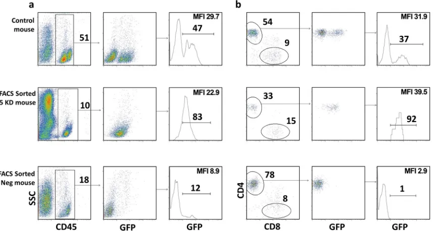
Transplantation of purified CCR5 knockdown CD34ⴙcells results in mice with “pure” populations of transduced cellsin
vivo.The lack of resistance to HIV infection was likely due to the
chimerism of transduced and untransduced CD34⫹cells in our
initial experiments. Therefore, we sorted the CD34⫹cells after
transduction into CCR5 knockdown, GFP-positive and -negative
fractions obtaining a ⬎90% pure population of GFP-positive
CD34⫹cells. Mice transplanted with GFP-sorted cells were called
FACS-sorted R5 knockdown mice. Analysis of the peripheral blood in six of the FACS-sorted R5 knockdown animals showed a
single GFP-positive peak for human CD45⫹and CD4⫹T cells,
sug-gesting that only transduced CD34⫹cells were engrafted in these
mice (Fig. 2); the level of GFP-positive cells was a major criterion for
successful gene engineering and engraftment. Mice transplanted with the GFP-negative fraction were called FACS-sorted negative mice.
They developed CD4⫹and CD8⫹T-cell populations with no GFP
expression (Fig. 2). In contrast, the control-transduced mice had two
distinct GFP-negative and -positive populations for CD45⫹ and
CD4⫹T cells (Fig. 2). A summary of mice used in this study is
on November 7, 2019 by guest
http://jvi.asm.org/
vided inTable 1. Mice with less than 5% human engraftment in the peripheral blood before HIV infection were excluded.
Transplanting purified CCR5 knockdown CD34ⴙcells dra-matically lowered viral load and protected HIV target cellsin
vivo.The FACS-sorted R5 knockdown mice had markedly lower
viral loads than the FACS-sorted negative mice over 28 weeks (Fig.
3a). Peak viremia for the FACS-sorted R5 knockdown mice was,
on average, 4.2⫻103copies/ml, and FACS-sorted negative mice
had 3.5⫻105copies/ml. Viral loads for the untransduced,
con-trol-transduced, R5 knockdown, and FACS-sorted negative mice were similar. The FACS-sorted R5 knockdown mice had lower viral loads than all cohorts.
CD4⫹ T cells (percentage of total CD3⫹ T cells) from the
FACS-sorted negative mice declined steadily upon infection
(day 0, 55%; day 134, 20%) (Fig. 3b). In contrast, the
FACS-sorted R5 knockdown mice showed a steady increase in CD4⫹
T cells (day 0, 33%; day 196, 65%). Furthermore, the absolute
numbers of CD4⫹T cells increased for the FACS-sorted R5
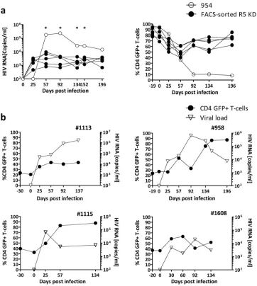
FIG 1Homeostatic expansion of GFP-positive CD4⫹T cells in R5 knockdown mice despite sustained R5-tropic (YU-2) HIV infection. (a) Percent change of GFP-positive CD4⫹T cells in the peripheral blood of control-transduced mice (cohort 1,n⫽8; cohort 2,n⫽4) and R5 knockdown mice (cohort 1,n⫽6; cohort 2,n⫽5). Values are means⫾SEMs. *,P⫽0.0152; **,P⫽0.0034; ***,P⫽0.0002.Pvalues were determined by two-tailed unpairedttest. Insets show the results for individual mice. (b) Frequency of total CD4⫹T cells (percentage of total CD3 T cells) for the control-transduced and R5 knockdown cohorts 1 and 2. Values are means⫾SEMs. *,P⫽0.0146.Pvalues were determined by two-tailed unpairedttest. (c) HIV RNA copies per milliliter of blood plasma collected for the control-transduced and R5 knockdown cohort 1 and cohort 2 over 92 and 134 days, respectively. Time of termination was chosen at random for the various groups. The dashed line indicates 400 copies/ml, the detection limit of the HIV RNA assay. Values are means⫾SEMs. (d) Percentage CCR5 expression on total CD4⫹T cells in peripheral blood and spleens of various cohorts of mice. Values are means⫾SEMs. For blood, asterisks indicatePvalues as follows: *,P⫽0.0237; *=,P⫽0.0121;**,P⫽0.0035;***,P⫽0.0007;****,P⫽0.0001. For spleens, asterisks indicatePvalues as follows:*,P⫽0.041;***,P⫽0.0003.Pvalues were determined by two-tailed unpairedttest. (e) HIV replication is inhibitedex vivoin GFP-positive sorted splenocytes from R5 knockdown mice. Splenocytes were isolated from R5 knockdown mice 20 weeks after CD34⫹cell injection. Splenocytes were sorted into GFP-positive (n⫽5) and GFP-negative (n⫽5) fractions at 99% purity and challenged with R5-tropic HIV (YU-2). The amount of HIV production in the culture supernatant was monitored by HIV p24 ELISA. Viral loads were normalized to the peak viral load under each condition (means⫾SEMs).
Myburgh et al.
on November 7, 2019 by guest
http://jvi.asm.org/
[image:4.585.51.542.66.480.2]knockdown mice but decreased for the FACS-sorted negative mice (data not shown).
At euthanasia, for the FACS-sorted R5 knockdown mice, 70%
of CD4⫹T cells in the blood and 83% of CD4⫹T cells in the spleen
were GFP positive (Fig. 3candd). In the control-transduced and
R5 knockdown groups, the values were, on average, 20% and 18%
in the blood and 21% and 20% in the spleen, respectively (Fig. 3c
andd). Similarly, the CD4⫹/CD8⫹T-cell ratios in the blood (end/
preinfection) and spleen were very low in the various groups
ex-cept for the FACS-sorted R5 knockdown group (Fig. 3eandf).
Absolute numbers of GFP-positive CD4⫹T cells expanded
signif-icantly upon HIV challenge in the FACS-sorted R5 KD mice (Fig.
3i), and CCR5 expression was downregulated in the blood and
spleens of the FACS-sorted R5 knockdown mice (Fig. 1d).
In summary, transplantation of GFP-positive sorted CD34⫹
cells produced mice with high levels of gene-engineered CCR5
knockdown CD4⫹T cellsin vivo. This resulted in long-term
inhi-bition of HIV replicationin vivoand preservation of HIV target
cells in the blood and spleen.
Outlier analysis and follow-up of mice that did not meet ac-ceptance criteria.The FACS-sorted R5 knockdown mice typically
controlled the virus long term (titers ⬍ 104 copies/ml) while
maintaining a high level of GFP-positive CD4⫹T cells in the blood
(Fig. 4a). In contrast, a single FACS-sorted R5 knockdown mouse (number 954) showed an unexpected decline in GFP-positive
CD4⫹T cells with high HIV copy numbers,⬎105copies/ml, on
days 57 and 92 (Fig. 4a). We performed an outlier analysis (see
Materials and Methods), and at four different times, viral loads
detected in mouse 954 were statistical outliers (Fig. 4a). Based on
this, mouse 954 was not included in the mean values and was analyzed separately (see below). At termination, this mouse had much lower splenic engraftment of total human splenocytes, with 31% human cells, than did the FACS-sorted R5 knockdown mice,
which had 54% (standard error of the mean [SEM],⫾11%).
[image:5.585.81.510.68.297.2]GFP-FIG 2GFP-positive CCR5 knockdown sorted CD34⫹HSPCs produced mice with “pure” populations of transduced cellsin vivo. (a) FACS plots showing the percent human engraftment of CD45⫹(percentage of live cells) and GFP-positive CD45⫹(percentage of human CD45) cells for representative control-transduced, FACS-sorted R5 knockdown, and FACS-sorted negative mice before HIV infection. (b) FACS plots showing the percentage CD4⫹T cells (percent CD3⫹), CD8⫹(percent CD3⫹), and GFP-positive CD4⫹T cells (percent CD4⫹) in the peripheral blood before HIV infection of the same mice as in panel a. For all cell subset analyses, the subgating was done as follows: total live cells and CD45⫹, CD3⫹, CD4⫹, and CD8⫹cells. Mean fluorescence intensity (MFI) for GFP is indicated.
TABLE 1Mice used in this study
Trait
% GFP⫹CD34⫹cells pretransplantation
Total no. of mice
No. of mice with:
% engraftment (⬎5% hCD45⫹)
% GFP⫹CD4⫹T cells (⬎70%)
% GFP⫹CD4⫹T cells (20–70%)
% GFP⫹CD4⫹T cells (⬍20%)
Untransduced NAa 15 11 NA NA NA
Control transduced 15–40 17 12 0 5 7
R5 knockdown 17–20 15 11 0 0 11
FACS GFP positiveb 12–32 20 10 6 4 0
FACS GFP negative NA 26 15 NA NA NA
a
NA, not applicable.
bFACS GFP-positive cells prior to FACS cell sorting for GFP-positive cells.
on November 7, 2019 by guest
http://jvi.asm.org/
[image:5.585.43.545.603.707.2]positive CD4⫹T cells were barely detected in the spleen of mouse 954 (6%), while the FACS-sorted R5 knockdown mice had, on
average, 83% (SEM,⫾2%) GFP-positive CD4⫹ T cells in the
spleen (Fig. 3d).
The inclusion criterion we defined as successful reconstitution
for FACS-sorted R5 knockdown mice was 70% GFP⫹CD4⫹T
cells in the peripheral blood before infection. Four mice did not meet this criterion despite being transplanted with GFP-positive
sorted CD34⫹ cells (Fig. 4b). Mouse 1113 had no protection
against HIV and had a limited expansion of GFP-positive CD4⫹T
cells (23% on day 0 to 43% on day 137). Mice 958 and 1115 had
massive expansions of GFP-positive cells, reaching close to 100%
of the total CD4⫹T-cell population, which went in parallel with a
decrease in the viral load (Fig. 4b). For mouse 1608, the dynamics
of recovery of GFP-positive CD4⫹ T cells and viral load were
slower, and recovery of GFP-positive CD4⫹T cells on day 134 was
less extensive (Fig. 4b). These three mice (958, 1115, and 1608)
maintained a high CD4/CD8 ratio, and the percentage of
GFP-positive CD4⫹T cells in the blood increased upon infection from
24 to 80, 39 to 82, and 37 to 52%, respectively. These animals also
had high frequencies of GFP-positive CD4⫹T cells in the spleens,
i.e., 59, 60, and 83%, respectively.
FIG 3Sustained HIV load inhibition in FACS-sorted R5 knockdown mice. (a) HIV RNA copies per milliliter in blood plasma from FACS-sorted R5 knockdown (YU-2 [n⫽5]) and FACS-sorted negative (n⫽15 [YU-2,n⫽9, and JRCSF,n⫽6]) mice collected over 134 and 196 days, respectively. The viral load detection limit is indicated by the dashed line (400 copies/ml). Values are means⫾SEMs. Single asterisks indicatePvalues of 0.0486, 0.0188, and 0.0391 for days 57, 92, and 134, respectively.Pvalues were determined by a two-tailed unpairedttest. (b) Percent CD4⫹T cells of FACS-sorted R5 knockdown (n⫽5) and FACS-sorted negative (n⫽15) mice. Values are means⫾SEMs. Double asterisks indicatePvalues of 0.0017 and 0.0018 for days 92 and 134, respectively.Pvalues were determined by two-tailed unpairedttest. (c) Percent GFP-positive CD4⫹T cells in the peripheral blood at termination. Values are means⫾SEMs. ****,P⫽
0.0001.Pvalues were determined by two-tailed unpairedttest. (d) Percent GFP-positive CD4⫹T cells in the spleen at termination. Values are means⫾SEMs. ****,P⫽0.0001.Pvalues were determined by two-tailed unpairedttest. (e) Change of CD4⫹/CD8⫹T-cell ratio in the peripheral blood, comparing the CD4⫹/CD8⫹T-cell ratio of each cohort end and preinfection. Values are means⫾SEMs. **,P⫽0.0026.Pvalues were determined by two-tailed unpairedttest. (f) CD4⫹/CD8⫹T-cell ratio at termination in the spleens of various cohorts of mice. Values are means⫾SEMs. **,P⫽0.0059; *,P⫽0.039.Pvalues were determined by two-tailed unpairedttest. (g to i) Absolute numbers of GFP⫹CD4⫹T cells or total CD4⫹T cells/microliter of blood of representative mice from the control-transduced (n⫽5), R5 knockdown (n⫽8), and FACS-sorted R5 knockdown (n⫽5) cohorts are shown. Values are means⫾SEMs. Single asterisks in panels h and i indicatePvalues of 0.0194 and 0.0369, respectively.Pvalues were determined by pairedttest.
Myburgh et al.
on November 7, 2019 by guest
http://jvi.asm.org/
[image:6.585.42.537.66.458.2]Strikingly, we observed this level of expansion (Fig. 4b) of
HIV-resistant GFP-positive CD4⫹T cells and a concomitant inhibitory
effect on HIV only in mice transplanted with CCR5 knockdown
GFP-sorted CD34⫹cells (these animals had 30% [⫾4% SEM]
GFP⫹CD4⫹T cells on day 0). This degree of expansion was not
seen in R5 knockdown mice (which had 5%⫾2% GFP⫹CD4⫹T
cells before HIV infection). Based on these results, we estimate
that at least 20% of CD4⫹T cells need to be CCR5 repressed to
observe homeostatic expansion and relevant effects on viremia.
Preserved engraftment and preferential expansion of central memory T cells in FACS-sorted R5 knockdown mice upon HIV infection.Engraftment as reflected in peripheral blood decreased in all control cohorts but increased in the FACS-sorted R5
knock-down mice (Fig. 5a). This effect on total engraftment was even
more impressive in the spleen. FACS-sorted R5 knockdown mice
had 10 times more human cells than control cohorts (Fig. 5b).
We evaluated the CD4⫹ and CD8⫹ effector (CD45RApos
CCR7neg), effector memory (CD45RAneg CCR7neg), naive
(CD45RApos CCR7pos), and central memory (CD45RAneg
CCR7pos) T-cell subsets in the blood and spleens of the
FACS-sorted R5 knockdown and representative FACS-FACS-sorted negative
mice (Fig. 5ctof). In the peripheral blood of the FACS-sorted
negative mice, the frequency of central memory CD4⫹T cells was
significantly decreased, and the CD8⫹central memory T-cell
sub-set was unchanged (Fig. 5cande). In contrast, central memory
CD4⫹and CD8⫹T cells were increased in the FACS-sorted R5
knockdown mice. Similarly, more CD4⫹and CD8⫹central
mem-ory T cells were present in the spleens of the FACS-sorted R5
knockdown mice (Fig. 5dandf). We observed no differences
be-tween the cohorts for effector and effector memory cells (data not shown). Notably, however, there was a trend toward a decrease in
naive CD4⫹and CD8⫹T cells in all cohorts (data not shown).
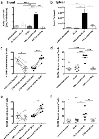
FIG 4Outlier analysis and examples of viral load control due to homeostatic expansion of transduced cells. (a) Left graph, viral load of the FACS-sorted R5 knockdown mice (YU-2) and viral load of outlier FACS-sorted R5 knockdown mouse 954 (YU-2). Right graph, percent GFP-positive CD4⫹T cells of the FACS-sorted R5 knockdown mice and mouse 954. Percent GFP-positive CD4⫹T cells is shown as a percentage of total CD4⫹T cells.Pvalues were determined by GraphPad Outlier calculator. *,P⬍0.001. (b) Viral load and percent GFP-positive CD4⫹T cells of four individual FACS-sorted R5 knockdown mice (mouse 1113, JRCSF; mouse 958, YU-2; mouse 1115, JRCSF; and mouse 1608, JRCSF) that were excluded due to not reaching the inclusion criteria of⬎70% GFP-positive CD4⫹T cells before HIV infection.
on November 7, 2019 by guest
http://jvi.asm.org/
[image:7.585.111.473.66.470.2]Evidence of shift from R5- to X4-tropic strain in one mouse.
As described above (Fig. 4), FACS-sorted R5 knockdown mice had
a low viral load and maintained high levels of CCR5 knockdown
CD4⫹T cells. Mouse 954 was clearly an outlier: it had high viral titers
and lost GFP-positive CD4⫹T cells (Fig. 6a). We hypothesized that
this mouse might have had a tropism shift of the virus from R5 to X4. We performed HIV population sequencing (from plasma) on days 57
and 196. As a control, we analyzed mouse 958 (Fig. 6b). Sequencing
revealed no mutations in mouse 958. Mouse 954 had mutations in
the V3 loop of the HIV envelope sequence (Fig. 6c), resulting in
amino acid substitutions to basic amino acids as indicated (Fig. 6d).
Substitutions in the V3 loop to basic amino acids have been reported
to result in a switch from R5 to X4 tropism (38).
DISCUSSION
In this study, we investigated a lentiviral vector-based, CCR5-tar-geting miRNA as a tool for engineering an HIV-resistant human immune system. We show that (i) the miRNA-based vector was very efficient in downregulating CCR5 on T cells and prevented
their infection by HIV ex vivo, (ii) only mice that were
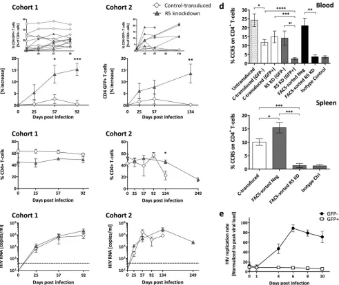
trans-FIG 5Increased engraftment and central memory T cells in blood and spleens of FACS-sorted R5 knockdown mice. (a) Change in level of peripheral blood engraftment expressed as the ratio of total CD45⫹at the end and preinfection. Values are means⫾SEMs. **,P⫽0.0031; ***,P⫽0.0006; ****,P⬍0.0001.P
values were determined by two-tailed unpairedttest. (b) Absolute numbers of human cells (CD45⫹) in the spleen at termination. Values are means⫾SEMs. *,
P⫽0.0229; **,P⬍0.0019.Pvalues were determined by two-tailed unpairedttest. (c and e) Percent CD4⫹and CD8⫹central memory T cells in the peripheral blood, preinfection and at the end time point. Values are means and SEMs. *,P⫽0.0153; **,P⫽0.0042; ***,P⫽0.0004.Pvalues were determined by paired
ttest. (d and f) Percent CD4⫹and CD8⫹central memory T cells in the spleen at termination. Values are means⫾SEMs. *=,P⫽0.0472; *,P⫽0.0106; **,P⫽
0.0022; ***,P⫽0.0001; ****,P⬍0.0001.Pvalues were determined by two-tailed unpairedttest. Myburgh et al.
on November 7, 2019 by guest
http://jvi.asm.org/
[image:8.585.134.453.67.515.2]planted with a preselected population of transduced CD34⫹cells
and maintained gene-engineered CD4⫹T cells had a dramatically
reduced viral load (functional cure), and (iii) the HIV-infected
mice transplanted with miRNA CCR5 gene-engineered CD34⫹
cells showed a dramatic expansion of memory T cells (i.e., the miRNA-edited T cells were mainly of this phenotype). Thus, we provide here preclinical proof of concept for gene engineering of an HIV-resistant immune system through the use of vector-me-diated miRNA expression and the need for a certain threshold of
gene-engineered CD34⫹cells for functional cure of HIV.
While gene engineering of HIV-resistant cells is a viable option for cure of HIV, major issues remain to be solved. These include finding the best antiviral moiety or combination, the most
effica-cious way to gene engineer the CD34⫹cells, and the threshold of
gene-engineered CD34⫹cells needed for functional cure.
Lentivirus-based transduction has been supplemented by gene-targeting methods, such as ZNF or Talen nucleases, or the
Crispr/Cas system for gene editing (23–25). However, off-target
effects of these methods are still unknown, and gene engineering
in primary cells is only modestly effective (26). And even though
no adverse events have been reported, there is less experience in
clinical trials with gene-targeting methods than with lentivirus-based transduction. Thus, we opted for lentivirus-lentivirus-based gene
en-gineering (39–41). Furthermore, we are the first to engage in
miRNA technology for knocking down the HIV coreceptor CCR5
in CD4⫹T cells via gene engineering of CD34⫹cells. miRNAs
closely mimic naturally occurring pri-miRNAs and thus are less likely to cause oversaturation of the RNA interference pathway and to affect cellular homeostasis than the widely used shRNAs
(42,43). However, miRNAs are thought to have a lower capacity
to downregulate target genes than shRNAs. In this study, we used a miRNA we developed with optimized features that efficiently knock down target genes upon single-copy vector transduction (30).Ex vivo-sorted cells were indeed resistant to a challenge with CCR5-tropic strains. However, bulk transplantation of
trans-duced CD34⫹cells into mice resulted in a human lymphoid
sys-tem that replicated HIV similarly to untransduced hu mice but
preserved CD4⫹T-cell counts. Similar data have been reported
previously (15).
We hypothesized that the majority of HSPCs needs to be gene engineered to see an effect on the HIV load; otherwise, the
HIV-permissive CD4⫹T cells that originated from the untransduced
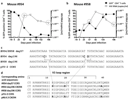
FIG 6Population sequencing of HIV in plasma: evidence for gene therapy failure. (a and b) Plots showing the percent GFP-positive CD4⫹T cells in peripheral blood (as a percentage of CD3⫹T cells) on the leftyaxis and HIV load on the rightyaxis for mice 954 and 958, respectively. Mouse 954 had a high and sustained viral load over time, with a complete loss of GFP-positive CD4⫹T cells. Mouse 958 experienced an expansion of GFP-positive CD4⫹T cells from less than 30% on day 19 to more than 80% on day 196. (c) On day 57, both animals had a homogenous HIV population in the peripheral blood; sequencing data of HIV envelope V3 loop are consistent with an R5-tropic HIV strain. On day 196, mouse 954, which experienced a complete loss of GFP-positive CD4⫹T cells (blood and spleen), had detectable mutations within the V3 loop of HIV. For mouse 958, no mutations were detected in the V3 loop on day 196, indicating the presence of a homogenous HIV population. (d) Changes to basic amino acids H and R. According to a Geno2Pheno (http://www.geno2pheno.org/) analysis of the obtained sequence in panel c, there is only 18% confidence that the virus at day 196 of mouse 954 was not an X4 variant.
on November 7, 2019 by guest
http://jvi.asm.org/
[image:9.585.74.507.62.400.2]CD34⫹cells would “outnumber” the HIV-resistant cells. Thus, we
used GFP to allow for efficient sorting of transduced CD34⫹cells
before transplantation. By doing so, we found that to achieve
long-term suppression of viral load, more than 70% of CD34⫹
transplanted should be gene engineered. Walker et al. obtained an
average engraftment (⫾standard deviation [STD]) level of
anti-HIV vector-transduced cells of 17.5%⫾8% in the peripheral
blood and argued that these numbers of cells were insufficient to
see any decrease in plasma viremia (15). Furthermore, a very
re-cent publication from the same group sorted the gene-engineered
CD34⫹cells with a truncated version of CD25 (tCD25) before
their transplantation into 2- to 5-day-old NRG mice (20). They
found that mice transplanted with tCD25-purified CD34⫹cells
had normal multilineage hematopoiesis, similar to mice
trans-planted with untransduced CD34⫹cells. Upon HIV challenge,
tCD25-transplanted mice did not suffer from HIV-induced CD4⫹
T-cell depletion as did the untransduced mice, and tCD25 mice had a 1.5-log inhibition in plasma viremia compared to that of
mice with untransduced CD34⫹cells. Our data nicely
comple-ment the data provided by Walker et al. and Barclay et al. and underline the importance of the number of transduced cells that are required for efficient HIV gene therapy. Notably, three hu
mice transplanted with purified gene-engineered CD34⫹ cells
showed at baseline⬃30% GFP⫹cells which expanded
substan-tially upon HIV infection; the expansion went along with a de-crease in viral load. The data for these three mice were reminiscent of the data reported by Holt et al. showing that disruption of
CCR5 by zinc finger nucleases was achieved in⬃20% of CD34⫹
cells and resulted in viral repression over time (16).
Obviously, in humans, GFP-based sorting would not be an option, given the xenogeneic nature of the protein. However,
novel strategies for sorting of transduced CD34⫹cells based on the
expression of truncated cellular surface receptors, such as CD25
(20), the epidermal growth factor receptor (44), or the nerve
growth factor receptor (45), are very promising for achieving high
numbers of engrafted gene-engineered cells. An alternative
ap-proach to pretransplantation sorting would bein vivoselection of
transduced cells (46, 47). Regrettably, currentin vivo selection
methods use potentially carcinogenic compounds, such as
myco-phenolate, methotrexate, or alkylating agents (i.e., O6
-benzylgua-nine/bis-chloroethylnitrosourea), that offset their use in a disease, such as HIV, that is amenable to an efficient and well-tolerated cART. We want to emphasize that in our gene-engineering efforts, we aimed for single lentiviral copy integration. The two recent phase I clinical trials used gene engineering protocols that resulted in vector copy numbers ranging from 2 to 4 per genome of bone marrow cells prior to transplantation without documenting any
adverse events over an observation period of⬎20 months (39,
40). Thus, ensuring CD34⫹transduction might present another
alternative for increasing the number of gene-engineered CD34⫹
cells. These protocols appeared not to affect the long-term en-graftment negatively in these phase I clinical trials.
In fact, we do not know the number of gene-engineered HSPCs needed to render the immune system resistant to HIV. As outlined above, we aimed for a rather pure population of gene-engineered HSPCs as proof of preclinical concept. However, we observed HIV-lowering effects in some mice with 20 to 40% engraftment of
transduced cells, data similar to those reported by Holt et al. (16).
HIV, certainly by killing untransduced cells via its cytopathic ef-fects, will promote the expansion of HIV-resistant cells. To what
extent the HIV-resistant cells will foster an efficient HIV-specific immune response and thereby constrain HIV remains unknown. Whether additional factors contribute to HIV-lowering effects re-mains unknown.
White blood cell counts from HIV-infected mice generated with FACS-sorted R5 KD cells showed an expansion of central
memory CD4⫹and CD8⫹T cells, while all other groups showed a
progressive loss of these CD4⫹memory T cells and no change in
CD8⫹T cells. This pattern was also evident when looking at the
splenocytes. These memory CD8⫹T cells might have contributed
to the control of HIV. There was a decrease in the frequency of
naive CD4⫹and CD8⫹T cells in the peripheral blood in both
FACS-sorted negative and FACS-sorted R5 KD mice, whereas in
the spleen, the naive CD4⫹and CD8⫹T cells in the FACS-sorted
R5 KD mice tended to be higher (data not shown). The expansion of central memory T cells is reminiscent of the immune
reconsti-tution seen in HIV-infected patients on ART (48).
HIV is known for its high mutational rate. In this respect, we observed one mouse (mouse 954) with an escape mutation. De-spite high levels (day 0) of engraftment of CCR5 knockdown cells, this mouse had a high viral load and a complete loss of circulating
CD4⫹T cells. Population sequencing of the V3 loop indicated a
likely shift to X4 tropism, which might explain the uncontrolled infection. The mutations were detectable in the blood only at rel-atively late time points. This might be due to an initial compart-mentalized replication of the X4-tropic strains in the thymus. We previously showed that X4-HIV NL4-3 severely depleted the
thy-mus, whereas YU-2 had only minor effects (49). However, we do
not know whether the potential emergence was due to the CCR5 knockdown or was just a coincidence. Indeed, emergence of CXCR4-tropic strains may occur without any immune or drug
pressure in hu mice infected with CCR5-tropic strains (50). In any
case, CCR5 knockdown should be done in concert with another strategy to constrain HIV (i.e., including another anti-HIV moi-ety, combining with efficient antiretrovirals, or boosting the im-mune response in parallel to transplantation). Indeed, the solid-ness of successful gene engineering by the expression of more than
one antiviral moiety may prevent HIV evolution (51). Gene
engi-neering could be combined with conventional ART: combining
two treatment modalities was efficient in cell lines (52), as
induc-tion therapy (53) or with anti-PD1 antibodies that decrease viral
load and increase the level of CD4⫹T cells in HIV-infected mice
(54). In any case, gene engineering efforts cannot promote more
virulent HIV strains, either for the individual patient or for the general population.
In summary, our results provide the first preclinical proof of concept that transplantation of miRNA CCR5 knockdown
CD34⫹cells can lead to long-term control of HIV viremia.
Trans-lation of our results to the clinical setting is relatively straightfor-ward but will require the implementation of existing strategies for pre- and posttransplantation selection compatible with human use. At this point, our strategy demonstrates long-term viral control but not yet a cure. However, while a cure remains the ultimate goal, long-term viral control independent of antiretrovirals is a relevant inter-mediate step, worth translating to the clinical setting. We believe that a definitive cure of HIV might indeed come from a combination of different approaches such as CCR5 knockdown combined with drug therapy, vaccination, or a second gene therapy target.
Myburgh et al.
on November 7, 2019 by guest
http://jvi.asm.org/
ACKNOWLEDGMENTS
We thank Astrid Koster, Xenia Rüfenacht, and Fatbardha Dautaj (Clinic of Immunology, University Hospital Zurich, Zurich, Switzerland) for their work in obtaining the HIV loads of the mice. We thank Jürg Böni, Caroline Käser, and Cyril Shah (Institute of Medical Virology, University of Zurich) for performing the sequencing of the HIV genome and analyz-ing the data.
This work was supported by the clinical research focus program Hu-man Hemato-Lymphatic Diseases of the University of Zurich and the Swiss South African Research Joint Program, the Swiss National Science Foundation, a University of Zurich Candoc grant, the South African Na-tional Research Foundation, the Medical Research Council of South Af-rica, and the Wolfermann Nägeli Stiftung.
R.M. designed and performed experiments, analyzed data, and wrote the paper. R.F.S. and K.-H.K. designed the experiments, supervised the work, analyzed the data, and wrote the paper. M.S.P. was involved in the initial conceptualization of the study, supervised aspects of the work, and helped to write the paper. A.A. helped to coordinate the work with the mice and transplantation of newborn mice, process cord blood, and write the paper. P.S. helped supervise aspects of the work and designed certain experiments. V.J. helped to write the paper and analyze data. G.G.-H., D.L., and M.-A.R. helped to terminate experiments and process cord blood samples. S.I. helped in processing of cord blood samples. S.R. pro-vided the expertise and obtained the viral loads for HIV-infected mice. M.G.M. gave highly valuable input in the entire study.
We declare no conflicts of interest.
REFERENCES
1.Lohse N, Gerstoft J, Kronborg G, Larsen CS, Pedersen C, Pedersen G, Nielsen L, Sorensen HT, Obel N.2011. Comorbidity acquired before HIV diagnosis and mortality in persons infected and uninfected with HIV: a Danish population-based cohort study. J Acquir Immune Defic Syndr 57:334 –339.http://dx.doi.org/10.1097/QAI.0b013e31821d34ed. 2.Tebas P, Stein D, Tang WW, Frank I, Wang SQ, Lee G, Spratt SK,
Surosky RT, Giedlin MA, Nichol G, Holmes MC, Gregory PD, Ando DG, Kalos M, Collman RG, Binder-Scholl G, Plesa G, Hwang WT, Levine BL, June CH.2014. Gene editing of CCR5 in autologous CD4 T cells of persons infected with HIV. N Engl J Med370:901–910.http://dx .doi.org/10.1056/NEJMoa1300662.
3.Tebas P, Stein D, Binder-Scholl G, Mukherjee R, Brady T, Rebello T, Humeau L, Kalos M, Papasavvas E, Montaner LJ, Schullery D, Shaheen F, Brennan AL, Zheng Z, Cotte J, Slepushkin V, Veloso E, Mackley A, Hwang WT, Aberra F, Zhan J, Boyer J, Collman RG, Bushman FD, Levine BL, June CH.2013. Antiviral effects of autologous CD4 T cells genetically modified with a conditionally replicating lentiviral vector ex-pressing long antisense to HIV. Blood121:1524 –1533.http://dx.doi.org /10.1182/blood-2012-07-447250.
4.DiGiusto DL, Krishnan A, Li L, Li H, Li S, Rao A, Mi S, Yam P, Stinson S, Kalos M, Alvarnas J, Lacey SF, Yee JK, Li M, Couture L, Hsu D, Forman SJ, Rossi JJ, Zaia JA.2010. RNA-based gene therapy for HIV with lentiviral vector-modified CD34(⫹) cells in patients undergoing transplantation for AIDS-related lymphoma. Sci Transl Med2:36ra43.
http://dx.doi.org/10.1126/scitranslmed.3000931.
5.Mitsuyasu RT, Merigan TC, Carr A, Zack JA, Winters MA, Workman C, Bloch M, Lalezari J, Becker S, Thornton L, Akil B, Khanlou H, Finlayson R, McFarlane R, Smith DE, Garsia R, Ma D, Law M, Murray JM, von Kalle C, Ely JA, Patino SM, Knop AE, Wong P, Todd AV, Haughton M, Fuery C, Macpherson JL, Symonds GP, Evans LA, Pond SM, Cooper DA.2009. Phase 2 gene therapy trial of an anti-HIV ribozyme in autologous CD34⫹cells. Nat Med15:285–292.http://dx.doi.org/10 .1038/nm.1932.
6.Deeks SG, Wagner B, Anton PA, Mitsuyasu RT, Scadden DT, Huang C, Macken C, Richman DD, Christopherson C, June CH, Lazar R, Broad DF, Jalali S, Hege KM.2002. A phase II randomized study of HIV-specific T-cell gene therapy in subjects with undetectable plasma viremia on com-bination antiretroviral therapy. Mol Ther5:788 –797.http://dx.doi.org/10 .1006/mthe.2002.0611.
7.Riddell SR, Elliott M, Lewinsohn DA, Gilbert MJ, Wilson L, Manley SA, Lupton SD, Overell RW, Reynolds TC, Corey L, Greenberg PD.1996.
T-cell mediated rejection of gene-modified HIV-specific cytotoxic T lym-phocytes in HIV-infected patients. Nat Med2:216 –223.http://dx.doi.org /10.1038/nm0296-216.
8.Hütter G, Nowak D, Mossner M, Ganepola S, Mussig A, Allers K, Schneider T, Hofmann J, Kucherer C, Blau O, Blau IW, Hofmann WK, Thiel E.2009. Long-term control of HIV by CCR5 Delta32/Delta32 stem-cell transplantation. N Engl J Med 360:692– 698.http://dx.doi.org/10 .1056/NEJMoa0802905.
9.Kordelas L, Verheyen J, Beelen DW, Horn PA, Heinold A, Kaiser R, Trenschel R, Schadendorf D, Dittmer U, Esser S, Essen HIVAG.2014. Shift of HIV tropism in stem-cell transplantation with CCR5 Delta32 mu-tation. N Engl J Med371:880 – 882.http://dx.doi.org/10.1056/NEJMc140 5805.
10. Rongvaux A, Takizawa H, Strowig T, Willinger T, Eynon EE, Flavell RA, Manz MG.2013. Human hemato-lymphoid system mice: current use and future potential for medicine. Annu Rev Immunol31:635– 674.http: //dx.doi.org/10.1146/annurev-immunol-032712-095921.
11. Nischang M, Gers-Huber G, Audige A, Akkina R, Speck RF. 2012. Modeling HIV infection and therapies in humanized mice. Swiss Med Wkly142:w13618.http://dx.doi.org/10.4414/smw.2012.13618. 12. Bennett MS, Akkina R. 2013. Gene therapy strategies for HIV/AIDS:
preclinical modeling in humanized mice. Viruses5:3119 –3141.http://dx .doi.org/10.3390/v5123119.
13. Shimizu S, Hong P, Arumugam B, Pokomo L, Boyer J, Koizumi N,
Kittipongdaja P, Chen A, Bristol G, Galic Z, Zack JA, Yang O, Chen IS, Lee B, An DS.2010. A highly efficient short hairpin RNA potently down-regulates CCR5 expression in systemic lymphoid organs in the hu-BLT mouse model. Blood 115:1534 –1544. http://dx.doi.org/10.1182/blood-2009-04 -215855.
14. Ringpis GE, Shimizu S, Arokium H, Camba-Colon J, Carroll MV,
Cortado R, Xie Y, Kim PY, Sahakyan A, Lowe EL, Narukawa M, Kandarian FN, Burke BP, Symonds GP, An DS, Chen IS, Kamata M. 2012. Engineering HIV-1-resistant T-cells from short-hairpin RNA-expressing hematopoietic stem/progenitor cells in humanized BLT mice. PLoS One7:e53492.http://dx.doi.org/10.1371/journal.pone.0053492. 15. Walker JE, Chen RX, McGee J, Nacey C, Pollard RB, Abedi M, Bauer
G, Nolta JA, Anderson JS.2012. Generation of an HIV-1-resistant im-mune system with CD34(⫹) hematopoietic stem cells transduced with a triple-combination anti-HIV lentiviral vector. J Virol86:5719 –5729.http: //dx.doi.org/10.1128/JVI.06300-11.
16. Holt N, Wang J, Kim K, Friedman G, Wang X, Taupin V, Crooks GM,
Kohn DB, Gregory PD, Holmes MC, Cannon PM.2010. Human
hema-topoietic stem/progenitor cells modified by zinc-finger nucleases targeted to CCR5 control HIV-1 in vivo. Nat Biotechnol28:839 – 847.http://dx.doi .org/10.1038/nbt.1663.
17. Joseph A, Zheng JH, Chen K, Dutta M, Chen C, Stiegler G, Kunert R, Follenzi A, Goldstein H.2010. Inhibition of in vivo HIV infection in humanized mice by gene therapy of human hematopoietic stem cells with a lentiviral vector encoding a broadly neutralizing anti-HIV antibody. J Virol84:6645– 6653.http://dx.doi.org/10.1128/JVI.02339-09.
18. Kitchen SG, Levin BR, Bristol G, Rezek V, Kim S, Aguilera-Sandoval C, Balamurugan A, Yang OO, Zack JA.2012. In vivo suppression of HIV by antigen specific T cells derived from engineered hematopoietic stem cells. PLoS Pathog8:e1002649.http://dx.doi.org/10.1371/journal.ppat.1002649. 19. Hauber I, Hofmann-Sieber H, Chemnitz J, Dubrau D, Chusainow J,
Stucka R, Hartjen P, Schambach A, Ziegler P, Hackmann K, Schrock E, Schumacher U, Lindner C, Grundhoff A, Baum C, Manz MG, Buchholz F, Hauber J.2013. Highly significant antiviral activity of HIV-1 LTR-specific tre-recombinase in humanized mice. PLoS Pathog9:e1003587.
http://dx.doi.org/10.1371/journal.ppat.1003587.
20. Barclay SL, Yang Y, Zhang S, Fong R, Barraza A, Nolta JA, Torbett BE, Abedi M, Bauer G, Anderson JS.2015. Safety and efficacy of a tCD25 pre-selective combination anti-HIV lentiviral vector in human hemato-poietic stem and progenitor cells. Stem Cells33:870 – 879.http://dx.doi .org/10.1002/stem.1919.
21. Berkhout B, Liu YP.2014. Towards improved shRNA and miRNA re-agents as inhibitors of HIV-1 replication. Future Microbiol9:561–571.
http://dx.doi.org/10.2217/fmb.14.5.
22. Jackson AL, Linsley PS.2004. Noise amidst the silence: off-target effects of siRNAs? Trends Genet 20:521–524. http://dx.doi.org/10.1016/j.tig .2004.08.006.
23. Bibikova M, Golic M, Golic KG, Carroll D.2002. Targeted chromosomal
on November 7, 2019 by guest
http://jvi.asm.org/
cleavage and mutagenesis in Drosophila using zinc-finger nucleases. Ge-netics161:1169 –1175.
24. Cho SW, Kim S, Kim JM, Kim JS.2013. Targeted genome engineering in human cells with the Cas9 RNA-guided endonuclease. Nat Biotechnol 31:230 –232.http://dx.doi.org/10.1038/nbt.2507.
25. Miller JC, Tan S, Qiao G, Barlow KA, Wang J, Xia DF, Meng X,
Paschon DE, Leung E, Hinkley SJ, Dulay GP, Hua KL, Ankoudinova I, Cost GJ, Urnov FD, Zhang HS, Holmes MC, Zhang L, Gregory PD, Rebar EJ.2011. A TALE nuclease architecture for efficient genome edit-ing. Nat Biotechnol29:143–148.http://dx.doi.org/10.1038/nbt.1755. 26. Genovese P, Schiroli G, Escobar G, Di Tomaso T, Firrito C, Calabria A,
Moi D, Mazzieri R, Bonini C, Holmes MC, Gregory PD, van der Burg M, Gentner B, Montini E, Lombardo A, Naldini L. 2014. Targeted genome editing in human repopulating haematopoietic stem cells. Nature 510:235–240.http://dx.doi.org/10.1038/nature13420.
27. Sander JD, Ramirez CL, Linder SJ, Pattanayak V, Shoresh N, Ku M, Foden JA, Reyon D, Bernstein BE, Liu DR, Joung JK.2013. In silico abstraction of zinc finger nuclease cleavage profiles reveals an expanded landscape of off-target sites. Nucleic Acids Res41:e181.http://dx.doi.org /10.1093/nar/gkt716.
28. Gabriel R, Lombardo A, Arens A, Miller JC, Genovese P, Kaeppel C, Nowrouzi A, Bartholomae CC, Wang J, Friedman G, Holmes MC, Gregory PD, Glimm H, Schmidt M, Naldini L, von Kalle C.2011. An unbiased genome-wide analysis of zinc-finger nuclease specificity. Nat Biotechnol29:816 – 823.http://dx.doi.org/10.1038/nbt.1948.
29. Cradick TJ, Ambrosini G, Iseli C, Bucher P, McCaffrey AP. 2011. ZFN-site searches genomes for zinc finger nuclease target sites and off-target sites. BMC Bioinformatics12:152.http://dx.doi.org/10.1186/1471 -2105-12-152.
30. Myburgh R, Cherpin O, Schlaepfer E, Rehrauer H, Speck RF, Krause KH, Salmon P.2014. Optimization of critical hairpin features allows miRNA-based gene knockdown upon single-copy transduction. Mol Ther Nucleic Acids3:e207.http://dx.doi.org/10.1038/mtna.2014.58. 31. Giry-Laterrière M, Verhoeyen E, Salmon P.2011. Lentiviral vectors.
Methods Mol Biol737:183–209.http://dx.doi.org/10.1007/978-1-61779 -095-9_8.
32. Nischang M, Sutmuller R, Gers-Huber G, Audige A, Li D, Rochat MA, Baenziger S, Hofer U, Schlaepfer E, Regenass S, Amssoms K, Stoops B, Van Cauwenberge A, Boden D, Kraus G, Speck RF.2012. Humanized mice recapitulate key features of HIV-1 infection: a novel concept using long-acting anti-retroviral drugs for treating HIV-1. PLoS One7:e38853.
http://dx.doi.org/10.1371/journal.pone.0038853.
33. McDougal JS, Cort SP, Kennedy MS, Cabridilla CD, Feorino PM,
Francis DP, Hicks D, Kalyanaraman VS, Martin LS.1985. Immunoassay for the detection and quantitation of infectious human retrovirus, lymph-adenopathy-associated virus (LAV). J Immunol Methods76:171–183.
http://dx.doi.org/10.1016/0022-1759(85)90489-2.
34. Moore JP, Sattentau QJ, Clapham PR.1990. Enhancement of soluble CD4-mediated HIV neutralization and gp 120 binding by CD4 autoanti-bodies and monoclonal antiautoanti-bodies. AIDS Res Hum Retroviruses6:1273– 1279.
35. Salminen MO, Koch C, Sanders-Buell E, Ehrenberg PK, Michael NL, Carr JK, Burke DS, McCutchan FE. 1995. Recovery of virtually full-length HIV-1 provirus of diverse subtypes from primary virus cultures using the polymerase chain reaction. Virology213:80 – 86.http://dx.doi .org/10.1006/viro.1995.1548.
36. Fehse B, Kustikova OS, Bubenheim M, Baum C.2004. Pois(s)on—it’s a question of dose. Gene Ther11:879 – 881.http://dx.doi.org/10.1038/sj.gt .3302270.
37. De Luca A, Teofili L, Antinori A, Iovino MS, Mencarini P, Visconti E, Tamburrini E, Leone G, Ortona L.1993. Haemopoietic CD34⫹progenitor cells are not infected by HIV-1 in vivo but show impaired clonogenesis. Br J Haematol 85:20–24.http://dx.doi.org/10.1111/j.1365-2141.1993.tb08640.x.
38. Milich L, Margolin B, Swanstrom R.1993. V3 loop of the human im-munodeficiency virus type 1 Env protein: interpreting sequence variabil-ity. J Virol67:5623–5634.
39. Biffi A, Montini E, Lorioli L, Cesani M, Fumagalli F, Plati T, Baldoli C, Martino S, Calabria A, Canale S, Benedicenti F, Vallanti G, Biasco L, Leo S, Kabbara N, Zanetti G, Rizzo WB, Mehta NA, Cicalese MP, Casiraghi M, Boelens JJ, Del Carro U, Dow DJ, Schmidt M, Assanelli A, Neduva V, Di Serio C, Stupka E, Gardner J, von Kalle C, Bordignon C, Ciceri F, Rovelli A, Roncarolo MG, Aiuti A, Sessa M, Naldini L.2013. Lentiviral
hemato-poietic stem cell gene therapy benefits metachromatic leukodystrophy. Sci-ence341:1233158.http://dx.doi.org/10.1126/science.1233158.
40. Aiuti A, Biasco L, Scaramuzza S, Ferrua F, Cicalese MP, Baricordi C, Dionisio F, Calabria A, Giannelli S, Castiello MC, Bosticardo M, Evangelio C, Assanelli A, Casiraghi M, Di Nunzio S, Callegaro L, Benati C, Rizzardi P, Pellin D, Di Serio C, Schmidt M, Von Kalle C, Gardner J, Mehta N, Neduva V, Dow DJ, Galy A, Miniero R, Finocchi A, Metin A, Banerjee PP, Orange JS, Galimberti S, Valsecchi MG, Biffi A, Montini E, Villa A, Ciceri F, Roncarolo MG, Naldini L.2013. Lentiviral hematopoietic stem cell gene therapy in patients with Wiskott-Aldrich syndrome. Science341:1233151.
http://dx.doi.org/10.1126/science.1233151.
41. McGarrity GJ, Hoyah G, Winemiller A, Andre K, Stein D, Blick G, Greenberg RN, Kinder C, Zolopa A, Binder-Scholl G, Tebas P, June CH, Humeau LM, Rebello T.2013. Patient monitoring and follow-up in lentiviral clinical trials. J Gene Med15:78 – 82.http://dx.doi.org/10.1002/jgm.2691. 42. Grimm D, Streetz KL, Jopling CL, Storm TA, Pandey K, Davis CR,
Marion P, Salazar F, Kay MA.2006. Fatality in mice due to oversatura-tion of cellular microRNA/short hairpin RNA pathways. Nature441:537– 541.http://dx.doi.org/10.1038/nature04791.
43. An DS, Qin FX, Auyeung VC, Mao SH, Kung SK, Baltimore D, Chen IS.2006. Optimization and functional effects of stable short hairpin RNA expression in primary human lymphocytes via lentiviral vectors. Mol Ther 14:494 –504.http://dx.doi.org/10.1016/j.ymthe.2006.05.015.
44. Wang X, Chang WC, Wong CW, Colcher D, Sherman M, Ostberg JR,
Forman SJ, Riddell SR, Jensen MC. 2011. A transgene-encoded cell surface polypeptide for selection, in vivo tracking, and ablation of engi-neered cells. Blood118:1255–1263.http://dx.doi.org/10.1182/blood-2011 -02-337360.
45. Dupré L, Trifari S, Follenzi A, Marangoni F, Lain de Lera T, Bernad A, Martino S, Tsuchiya S, Bordignon C, Naldini L, Aiuti A, Roncarolo MG.2004. Lentiviral vector-mediated gene transfer in T cells from Wis-kott-Aldrich syndrome patients leads to functional correction. Mol Ther 10:903–915.http://dx.doi.org/10.1016/j.ymthe.2004.08.008.
46. Phaltane R, Lachmann N, Brennig S, Ackermann M, Modlich U,
Moritz T.2014. Lentiviral MGMT(P140K)-mediated in vivo selection employing a ubiquitous chromatin opening element (A2UCOE) linked to a cellular promoter. Biomaterials35:7204 –7213.http://dx.doi.org /10.1016/j.biomaterials.2014.05.001.
47. Jonnalagadda M, Brown CE, Chang WC, Ostberg JR, Forman SJ, Jensen MC.2013. Engineering human T cells for resistance to methotrexate and mycophenolate mofetil as an in vivo cell selection strategy. PLoS One 8:e65519.http://dx.doi.org/10.1371/journal.pone.0065519.
48. Muñoz-Calleja C, Costantini A, Silvestri G, Butini L, Regnery CM, Man-cini S, Montroni M.2001. Highly active antiretroviral therapy induces spe-cific changes in effector and central memory T cell sub-populations. AIDS 15:1887–1890.http://dx.doi.org/10.1097/00002030-200109280-00022. 49. Baenziger S, Tussiwand R, Schlaepfer E, Mazzucchelli L, Heikenwalder
M, Kurrer MO, Behnke S, Frey J, Oxenius A, Joller H, Aguzzi A, Manz MG, Speck RF. 2006. Disseminated and sustained HIV infection in CD34⫹cord blood cell-transplanted Rag2⫺/⫺␥
c⫺/⫺mice. Proc Natl Acad
Sci U S A103:15951–15956.http://dx.doi.org/10.1073/pnas.0604493103. 50. Ince WL, Zhang L, Jiang Q, Arrildt K, Su L, Swanstrom R. 2010.
Evolution of the HIV-1envgene in the Rag2⫺/⫺␥
C⫺
/⫺humanized mouse
model. J Virol84:2740 –2752.http://dx.doi.org/10.1128/JVI.02180-09. 51. Herrera-Carrillo E, Liu YP, Berkhout B.2014. The impact of
unpro-tected T cells in RNAi-based gene therapy for HIV-AIDS. Mol Ther22: 596 – 606.http://dx.doi.org/10.1038/mt.2013.280.
52. Boutimah F, Eekels JJ, Liu YP, Berkhout B.2013. Antiviral strategies combining antiretroviral drugs with RNAi-mediated attack on HIV-1 and cellular co-factors. Antiviral Res98:121–129.http://dx.doi.org/10.1016/j .antiviral.2013.02.011.
53. Horwitz JA, Halper-Stromberg A, Mouquet H, Gitlin AD, Tretiakova A, Eisenreich TR, Malbec M, Gravemann S, Billerbeck E, Dorner M, Buning H, Schwartz O, Knops E, Kaiser R, Seaman MS, Wilson JM, Rice CM, Ploss A, Bjorkman PJ, Klein F, Nussenzweig MC.2013. HIV-1 suppression and durable control by combining single broadly neutralizing antibodies and antiretroviral drugs in humanized mice. Proc Natl Acad Sci U S A110:16538 –16543.http://dx.doi.org/10.1073/pnas.1315295110. 54. Palmer BE, Neff CP, Lecureux J, Ehler A, Dsouza M, Remling-Mulder
L, Korman AJ, Fontenot AP, Akkina R.2013. In vivo blockade of the PD-1 receptor suppresses HIV-1 viral loads and improves CD4⫹T cell levels in humanized mice. J Immunol190:211–219.http://dx.doi.org/10 .4049/jimmunol.1201108.
Myburgh et al.
![FIG 3 Sustained HIV load inhibition in FACS-sorted R5 knockdown mice. (a) HIV RNA copies per milliliter in blood plasma from FACS-sorted R5 knockdownin panels h and i indicate(f) CD4(YU-2 [ �the control-transduced (n 5]) and FACS-sorted negative (n � 15 [Y](https://thumb-us.123doks.com/thumbv2/123dok_us/142996.20140/6.585.42.537.66.458/sustained-inhibition-knockdown-milliliter-knockdownin-indicate-transduced-negative.webp)