Distinct Escape Pathway by Hepatitis C Virus Genotype 1a from a
Dominant CD8
ⴙT Cell Response by Selection of Altered Epitope
Processing
Andreas Walker,aKathrin Skibbe,bEike Steinmann,cStephanie Pfaender,cThomas Kuntzen,dDominik A. Megger,eSvenja Groten,b Barbara Sitek,eGeorg M. Lauer,fArthur Y. Kim,gThomas Pietschmann,cTodd M. Allen,dJoerg Timma
Institute for Virology, Heinrich-Heine-University, University Hospital, Düsseldorf, Germanya
; Institute of Virology, University Hospital Essen, University of Duisburg-Essen, Essen, Germanyb
; Institute of Experimental Virology, Twincore Centre for Experimental and Clinical Infection Research, a joint venture between the Medical School Hanover and the Helmholtz Centre for Infection Research, Hanover, Germanyc; Ragon Institute of MGH, MIT, and Harvard, Charlestown, Massachusetts, USAd;
Medizinisches Proteom-Center, Ruhr-Universität Bochum, Bochum, Germanye; Gastrointestinal Unit, Massachusetts General Hospital and Harvard Medical School, Boston, Massachusetts, USAf
; Division of Infectious Diseases, Massachusetts General Hospital and Harvard Medical School, Boston, Massachusetts, USAg
ABSTRACT
Antiviral CD8ⴙT cells are a key component of the adaptive immune response against HCV, but their impact on viral control is influenced by preexisting viral variants in important target epitopes and the development of viral escape mutations. Immuno-dominant epitopes highly conserved across genotypes therefore are attractive for T cell based prophylactic vaccines. Here, we characterized the CD8ⴙT cell response against the highly conserved HLA-B*51-restricted epitope IPFYGKAI1373–1380located in
the helicase domain of NS3 in people who inject drugs (PWID) exposed predominantly to HCV genotypes 1a and 3a. Despite this epitope being conserved in both genotypes, the corresponding CD8ⴙT cell response was detected only in PWID infected with genotype 3a and HCV-RNA negative PWID, but not in PWID infected with genotype 1a. In genotype 3a, the detection of strong CD8ⴙT cell responses was associated with epitope variants in the autologous virus consistent with immune escape. Analysis of viral sequences from multiple cohorts confirmed HLA-B*51-associated escape mutations inside the epitope in genotype 3a, but not in genotype 1a. Here, a distinct substitution in the N-terminal flanking region located 5 residues upstream of the epitope (S1368P;Pⴝ0.00002) was selected in HLA-B*51-positive individuals. Functional assays revealed that the S1368P substitution impaired recognition of target cells presenting the endogenously processed epitope. The results highlight that, despite an epitope being highly conserved between two genotypes, there are major differences in the selected viral escape pathways and the corre-sponding T cell responses.
IMPORTANCE
HCV is able to evolutionary adapt to CD8ⴙT cell immune pressure in multiple ways. Beyond selection of mutations inside tar-geted epitopes, this study demonstrates that HCV inhibits epitope processing by modification of the epitope flanking region un-der T cell immune pressure. Selection of a substitution five amino acids upstream of the epitope unun-derlines that efficient antigen presentation strongly depends on its larger sequence context and that blocking of the multistep process of antigen processing by mutation is exploited also by HCV. The pathways to mutational escape of HCV are to some extent predictable but are distinct in different genotypes. Importantly, the selected escape pathway of HCV may have consequences for the destiny of antigen-specific CD8ⴙT cells.
B
ased on phylogenetic analysis, hepatitis C virus (HCV) can beclassified into at least seven genotypes and multiple subtypes
that differ up to 20% at the amino acid level (1). The HCV
geno-types have distinct epidemiological characteristics, they are asso-ciated with different transmission risk factors, and their frequen-cies in a population are regionally different. In Europe and North
America the HCV genotypes 1 and 3 are most common (2,3).
Since routine screening via nucleic acid amplification for HCV eliminated the risk for infection through blood products, the most important risk group for incident HCV infection are people who inject drugs (PWID). The high prevalence of HCV infection with seroprevalence rates up to 80% paired with frequent risk practices for HCV transmissions in PWID results in incidence rates
be-tween 8 and 25% per year in young adult injectors (4,5), and there
is strong evidence that multiple exposures are common in this risk
group (6,7). The degree of sequence diversity between genotypes
and subtypes precludes broad protection against reinfection.
Ac-cordingly, multiple infections of the same individual with
differ-ent viruses have been reported (7).
Even at the subtype level HCV isolates typically differ between hosts. Lack of a proof reading function of the virus encoded
RNA-Received7 August 2015 Accepted1 October 2015
Accepted manuscript posted online7 October 2015
CitationWalker A, Skibbe K, Steinmann E, Pfaender S, Kuntzen T, Megger DA, Groten S, Sitek B, Lauer GM, Kim AY, Pietschmann T, Allen TM, Timm J. 2016. Distinct escape pathway by hepatitis C virus genotype 1a from a dominant CD8⫹ T cell response by selection of altered epitope processing. J Virol 90:33– 42.
doi:10.1128/JVI.01993-15.
Editor:M. S. Diamond
Address correspondence to Joerg Timm, [email protected].
A.W. and K.S. contributed equally to this article.
Copyright © 2015, American Society for Microbiology. All Rights Reserved.
on November 7, 2019 by guest
http://jvi.asm.org/
dependent RNA polymerase results in a high error rate during RNA replication. As a consequence, HCV exists in chronically infected patients as a quasispecies of closely related but genetically distinct viral variants. There is now strong evidence that viral ge-netic variation between hosts is the product of continuous
selec-tion of mutaselec-tions by host immune pressure (8–14). Collectively,
this inherent sequence diversity of HCV at the genotype, subtype,
and quasispecies level is a major obstacle to vaccine design (15).
Strategies that aim to develop prophylactic vaccines against HCV have to cope with this genetic heterogeneity either by inducing
immune responses with a high degree of cross-reactivity (16,17),
by inducing multiple responses against different sequence variants
(18), or by focusing the immune response on highly conserved
regions of the virus.
Dominant CD8⫹T cell epitopes that are conserved across
dif-ferent HCV genotypes are rare (19,20). Here, we characterized a
highly conserved dominant HLA-B*51-restricted CD8⫹ T cell
epitope (IPFYGKAI1373–1380) in HCV NS3 in PWID
predomi-nantly exposed to genotype 1a and 3a. Vigorous responses were detected in PWID with spontaneous immune control of HCV and in PWID with genotype 3a infection, but not in PWID infected with genotype 1a. Although selection of mutations inside the epitope was overall rare, there was evidence for mutational escape in HCV genotype 1b and 3a by population sequence analysis. In-terestingly, HCV genotype 1a followed a distinct escape pathway by selecting a substitution (S1368P) located five amino acids up-stream of the epitope. Further analysis of the functional relevance revealed that the S1368P substitution altered epitope processing. The results demonstrate that beyond selection of mutations inside
CD8⫹epitopes HCV adapts to immune pressure by selecting
mu-tations in the epitope flanking region. The evolutionary escape pathways differ between HCV genotypes, indicating distinct ge-netic plasticity.
MATERIALS AND METHODS
Patients.Blood samples from patients with a history of injection drug use were collected from the ward for inpatient detoxification treatment of drug addicts or the clinic for opioid maintenance treatment at the Depart-ment of Addictive Behavior and Addiction Medicine, Rhine State Hospi-tal Essen, HospiHospi-tal of the University of Duisburg-Essen. Written informed consent was obtained from all study participants and the study was ap-proved by the ethics committee of the Medical Faculty of the University of Duisburg-Essen in accordance with the Declaration of Helsinki. In total 43 HLA-B*51-positive treatment-naive subjects who were HCV antibody positive (by anti-HCV chemiluminescent microparticle immunoassay from Abbott) were analyzed, including 15 HCV-RNA-negative patients and 28 subjects with detectable HCV-RNA. Peripheral blood mononu-clear cells (PBMCs) were isolated from blood via Ficoll gradient centrif-ugation and subsequently cryopreserved.
Analysis and alignment of HCV sequences.The frequency of HLA-B*51-associated sequence polymorphisms was analyzed in 405 HCV ge-notype 1a sequences and 145 HCV gege-notype 1b sequences from a multi-center cohort (21) and in a large HCV genotype 1b outbreak (22). A region covering the epitope IPFYGKAI1373–1380was amplified and se-quenced from additional 37 HCV genotype 1a isolates, as well as 102 HCV genotype 3a isolates collected in Germany. All sequences have been sub-mitted to GenBank (accession no.FJ864775toFJ864816,KJ130249to
KJ130317, andKJ668232toKJ668268).
Analysis of the CD8ⴙT cell response.Antigen-specific T cells were expanded from cryopreserved HLA-B*51-positive PBMCs utilizing syn-thetic peptides (⬎70% purity) purchased from EMC, Tübingen, Ger-many. After thawing, the PBMCs were cultured in RPMI 1640 medium
containing 10% fetal calf serum, 100 U of penicillin/ml, 100g of strep-tomycin/ml, 10 mM HEPES buffer, and 25 U of recombinant interleu-kin-2 (IL-2)/ml and then stimulated with HLA-B*51 peptide 1373 (1g/ ml) and 0.1g of anti-CD28 and anti-CD49d/ml. After 7 days medium containing IL-2 was added. On day 10 the cells were restimulated with the same peptide (10g/ml) in the presence of brefeldin A (100 ng/ml) for 4 h and then analyzed for their CD4⫹, CD8⫹ and gamma interferon (IFN-␥) expression via flow cytometry. To determine the degree of cross-reactivity between different B*51-1373 variants, PBMCs were cultivated in the presence of the HLA-B*51-1373 prototype or the variant peptide. After 10 days, both cultures were restimulated with the prototype and the variant peptide at different concentrations before intracellular IFN-␥ staining. For the detection of HCV-specific cellsex vivoand after peptide-specificin vitroexpansion, thawed PBMCs were stained with phycoeryth-rin-labeled IPFYGKAI-specific HLA-B*5101 dextramer (Immudex, Den-mark), followed by surface staining with CD8⫹PerCP-Cy5.5 (eBioscience). All samples were acquired using a FACSCanto (BD), and the data were analyzed by using FlowJo software (Tree Star, Inc.).
Expression plasmids for HCV-GFP fusion proteins.All constructs were based on the parental plasmids pEGFP-N1 (Clontech, Germany) featuring a cytomegalovirus promoter-controlled enhanced green fluo-rescent protein for N-terminal fusion. Fragments of NS3 were generated by amplifying NS3 1329-1433 by nested PCR with the primers 4b-F (CC TACGGCAAGTTCCTTGC) and 4b-R_new (GCAGTCTATCACCGAG TCG) and the primers B51-for-EcoRI (CCAAGGGAATTCTTGGCT TCGTCTTCACCCTCGGCATCGGCACYGTCCTTGACCAAG) and B51-Rev-SalI (CGGATACCGTCGACCCGCGGTARTAMGCCACGGC). Subsequently, the fragment was cloned via EcoRI/SalI into pEGFP-N1. The substitution S1368P was introduced via site-directed mutagenesis (Stratagene) using the primer B51-S1368P-for (GGAGGTTGCTCTGCC CACCACCGGAGA) and B51-S1368P-rev (TCTCCGGTGGTGGGCAG AGCAACCTCC) according to the manufacturer’s instructions. All con-structs were verified by DNA sequencing.
Analysis of the CD8ⴙT cell response against endogenously pro-cessed antigens.HLA-B*51 PBMCs from healthy donors were obtained from buffy coats (Department of Transfusion Medicine, University Hos-pital Essen, Essen, Germany). For analysis of endogenously processed antigens, HLA-B*51-positive cells were electroporated with plasmids pac-NS3-S1368S-GFP or pac-NS3-S1368P-GFP encoding an NS3-GFP fusion protein utilizing the Amaxa T Cell Nucleofector kit (vpa-1002; Lonza). In brief, after thawing PBMCs were cultured in RPMI medium. After 24 h, 7⫻106cells were resuspended in 100l of Nucleofector solution, mixed with 7.5g of DNA and pulsed with the optimized protocol for unstimu-lated human T cells (Program v024; Amaxa2b) with an Amaxa apparatus (Lonza). After electroporation, the PBMCs were cultured in RPMI me-dium for 24 h cells before GFP-positive cells were sorted using a FACSAria II (BD). Subsequently, 105B*51-1373-specific cells from a 10-day culture were restimulated with GFP-positive cells in a 1:1 ratio for 4 h before intracellular IFN-␥staining.
In vitroproteasome digestion.The synthetic peptides EVALSTTGEI PFYGKAIPLEAIKGG and EVALPTTGEIPFYGKAIPLEAIKGG (ⱖ95% purity) were purchased from EMC, Tübingen, Germany, and digested using a 20S proteasome assay kit complemented with human constitutive or immunoproteasome (Boston Biochem). In brief, 20g of peptide was digested with 4g of sodium dodecyl sulfate-activated proteasome in a 500-l reaction. The reaction was stopped on indicated time points by adding 3 volumes of ice-cold acetone. For precipitation of proteasome, the samples were subsequently frozen at⫺20°C for 30 min and then centrifuged for 30 min at 4°C with 15,000 rpm. The supernatants were collected, evaporated to dryness, and dissolved in 50l of 0.1% trifluoro-acetic acid. The peptide concentration of the resulting solution was deter-mined by amino acid analysis as previously described (23). For the subse-quent liquid chromatography-tandem mass spectrometry experiment, the sample was diluted to a concentration of 0.33 pmol/l, and 5 pmol was analyzed using an UltiMate 3000 RSLCnano system online coupled to a
on November 7, 2019 by guest
http://jvi.asm.org/
Velos Pro linear ion trap mass spectrometer (both from Thermo Scien-tific, Bremen, Germany) as described earlier (23). The acquired raw files were further analyzed with Proteome Discoverer software (Thermo Sci-entific, v1.3.0.339) and searched with Sequest (24) against a self-written database containing the investigated prototype sequence (pt) and S1368P peptides. Precursor and fragment ion mass tolerance was set to 0.4 Da, and the confidence level of peptide identification was set to a false discov-ery rate of 1%. Relative peptide quantification was carried out via spectral counting. Then, a normalized spectral index was calculated for each of the identified peptides by dividing the number of acquired peptide spectrum matches (PSMs) of a particular peptide by the number of PSMs acquired in the whole sample.
Generation and analysis of TNcc viruses.Plasmid pTNcc encoding for the GT1a virus TNcc was kindly provided by Jens Bukh (25). Muta-tions S1368P, I1373V, and I1380L were introduced by site-directed mu-tagenesis, and all constructs were verified by DNA sequencing.In vitro transcription and electroporation of Huh-7.5 cells was performed as de-scribed before (26). At 4 h posttransfection, the medium was changed with fresh Dulbecco modified Eagle medium (2 mML-glutamine, nones-sential amino acids, 100 U of penicillin/ml, 100g of streptomycin/ml, 10% fetal calf serum) supplemented with or without 10 mM 2=CMA (kindly provided by T. Tellinghuisen [27]). At 72 h posttransfection, cell-free supernatant was filtered through 0.45-m-pore-size filters and 10⫻ concentrated through Amicon centrifugal filters (Millipore). Virus titers were determined as described elsewhere (28) with slight modifications. In brief, Huh-7.5 cells were seeded in 96-well plates at a density of 104cells per well 24 h prior to inoculation with dilutions of filtered cell culture supernatant (at least six wells were used per dilution). After 3 days, cells were washed with phosphate-buffered saline (PBS), fixed for 20 min with ice-cold methanol at⫺20°C, and washed three times with PBS. HCV-infected cells were detected with anti-core C7.50 (1:300) (29) and anti-NS5A 9E10 (1:1,000) (28) antibody in PBS for 45 min at room tempera-ture. The cells were washed as described above, and bound antibodies were detected by incubation with horseradish peroxidase-conjugated antibodies specific to murine IgG (Sigma-Aldrich, Steinheim, Ger-many) diluted 1:200 in PBS. After 1 h of incubation at room temper-ature, the cells were washed as specified above. The peroxidase activity was detected by using carbazole substrate (0.32% [wt/vol] of 3-amino-9-ethylcarazole [Sigma] inN,N-dimethyl-formamide was diluted at a ratio of 1:3.3 with 15 mM acetic acid, 35 mM sodium acetate, [pH 8.0], and 0.4% H2O2). Virus titers (50% tissue culture infective doses [TCID50]/ml) were calculated based on the method of Spearman and Kärber. HCV RNA was quantified by X-tail reverse transcriptase-PCR as described previously (22). For the detection of HCV core protein, virus-containing supernatant was inactivated by the addition of Triton X-100 to a final concentration of 1% (vol/vol) and the amount of released core protein was determined by a commercially available core enzyme-linked immunosorbent assay (Architect HCV Core AG test; Abbott, Wiesbaden, Germany).
Statistical analysis.All statistical tests were performed using Graph-Pad Prism 5.0 software (GraphGraph-Pad Software, San Diego, CA).
RESULTS
CD8ⴙT cells directed against the epitope IPFYGKAI1373–1380
are detected after spontaneous resolution and in chronic ge-notype 3a infection but not in chronic gege-notype 1a infection.
We previously reported an HLA-B*51-restricted epitope in
NS3 (IPFYGKAI1373–1380) that is⬎90% conserved across the
HCV genotypes 1a, 1b, and 3a (19). The CD8⫹T cell immune
response directed against the epitope IPFYGKAI1373–1380was
analyzed in a cohort of anti-HCV-positive PWID carrying the HLA-B*51 allele. A total of 43 subjects were analyzed, includ-ing 15 anti-HCV-positive PWID with undetectable HCV-RNA, 13 PWID infected with GT1a, 3 PWID infected with GT1b, and
12 PWID infected with GT3a. After 10 days ofin vitroexpansion
IPFYGKAI-specific CD8⫹ T cells were detectable in 12 of 15
(80%) individuals with undetectable viremia and in 6 of 12 (50%)
patients chronically infected with genotype 3a (Fig. 1A). In
con-trast, CD8⫹T cells directed against this epitope were not
detect-able after antigen-specific expansion in patients with genotype 1a
or 1b infection (Fig. 1A). Similar results were obtained when
CD8⫹T cell were directly analyzedex vivowith HLA class
I/pep-tide dextramers. IPFYGKAI-specific CD8⫹T cells were detectable
in most HCV-RNA-negative PWID and in some PWID with a genotype 3a infection. Notably, the four genotype 3a-infected pa-tients with high frequencies were identical to those where
antigen-specific CD8⫹T cells were efficiently expandedin vitro(Fig. 1A).
In turn, the analysis with HLA class I/peptide dextramers
con-firmed lack of detection of IPFYGKAI-specific CD8⫹T cells in
patients infected with genotype 1 (Fig. 1B). The CD8⫹T cell
re-sponse against the epitope IPFYGKAI1373–1380was also studied in
a patient with acute HCV genotype 1a infection. The patient was previously reported in a study on the impact of HLA-B*57 on
HCV infection outcome (30) and was also HLA-B*51 positive.
Interestingly, here, the response was detectable by week 11 after
infection but became completely undetectable in an IFN-␥
en-zyme-linked immunospot (ELISpot) assay by week 47 (Fig. 1C).
The autologous virus of 22 PWID from the total of 26 with detectable HCV-RNA was sequenced. The sequences of the
epitope and the flanking region are shown inTable 1and are
indicated inFig. 1Afor PWID with genotype 3a infection with a
detectable CD8⫹T cell response (colored inFig. 1A). In 3 of 10
HCV genotype 3a sequences, the virus harbored substitutions in the epitope region (I1373V, K1377R, or I1380L). Of note, all three patients mounted robust responses against the prototype
se-quence (Fig. 1A). In functional assays the I1380L variant impaired
recognition by antigen-specific CD8⫹T cells, whereas the I1373V
variant was cross-reactive in HCV-RNA-negative PWID and to a
lesser extent in GT3a-infected PWID (Fig. 1DandE). Notably, the
three remaining GT3a-infected patients with detectable CD8⫹T
cell responses and no evidence for escape mutations had a history of injection drug use of less than 6 months, consistent with more recent exposure and infection with HCV, possibly at a stage prior to mutational escape. One of nine GT1a sequences and one of three GT1b sequences harbored the I1373V substitution in
posi-tion 1 of the epitope. In both patients no CD8⫹T cell response was
detectable. One PWID with genotype 1a infection harbored a S1368P substitution. Notably, this substitution was also selected in the patient with acute HCV genotype 1a infection and with the
rapid decline of the CD8⫹T cell response (Fig. 1Cand data not
shown).
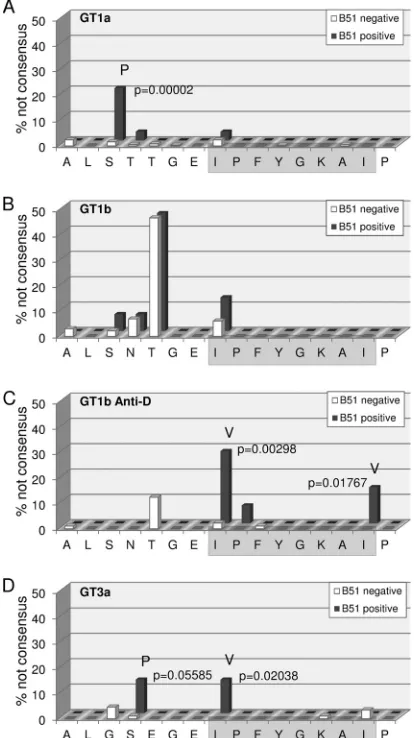
The residue under selection pressure in HLA-B*51-positive individuals depends on the viral genotype.Given the relatively
high reproducibility of IPFYGKAI-specific CD8⫹T cell responses
in HCV-RNA negative PWID, we aimed to address whether there is evidence for immune escape in patients with persistent HCV infection at a population level. We therefore analyzed the impact of HLA-B*51 expression on the frequency of sequence polymor-phisms in this epitope in different cohorts. In an analysis of 442
genotype 1a sequences from a multicenter cohort (Fig. 2A), there
was no evidence for mutational escape inside the epitope. Al-though an I1373V polymorphism was observed, this substitution was not enriched in HLA-B*51-positive patients. In contrast, the I1373V polymorphism was slightly enriched in
on November 7, 2019 by guest
http://jvi.asm.org/
tive patients from a multicenter cohort of 145 genotype
1b-in-fected patients (Fig. 2B), although this difference was not
statisti-cally significant. However, in a large genotype 1b single source outbreak there was statistical evidence for selection of polymor-phisms in position 1 (I1373V) and 8 (I1380L) of the epitope in the
presence of HLA-B*51 (Fig. 2C). Finally, we also analyzed a local
cohort of 102 patients infected with genotype 3a (Fig. 2D). Here,
there was statistical evidence for selection of the I1373V polymor-phism inside the epitope. Importantly, even though in genotype 1a there was no evidence for mutational escape inside the epitope, there was strong statistical evidence for HLA-B*51-associated se-lection pressure on a residue located five amino acids upstream of
the epitope (S1368) (Fig. 2A). In genotype 1a, 20.7% of the
HLA-B*51-positive patients carried a S1368P substitution; in contrast, only 0.8% of the HLA-B*51-negative patients carried any
substi-tutions in this position (P⫽0.00002). Notably, in genotype 3a an
S1369P substitution was also enriched in HLA-B*51-positive in-dividuals; however, the difference was not statistically significant (Fig. 2D). Taken together, by analyses of different cohorts we found statistical evidence for mutational escape inside the HLA-B*51-restricted epitope (IPFYGKAI) in genotype 1b and 3a and evidence for the selection of a distinct substitution in the epitope flanking region in genotype 1a.
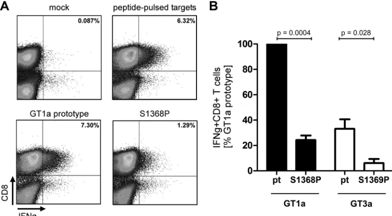
The S1368P substitution impairs targeting of the endogenously processed HLA-B*51-restricted epitope IPFYGKAI1373–1380.HCV genotype 1a was unique because evidence for HLA-B*51-associ-ated selection pressure was only observed in the epitope flanking region. Of note, this region does not contain a second HLA-B*51 binding motif by analysis with prediction algorithms for HLA
class I binding (www.immuneepitope.org). We therefore
hypoth-esized that the S1368P may impair processing and presentation of the epitope. To address this experimentally, an assay that allows analysis of endogenously processed antigens was established.
Ef-fector CD8⫹ T cells directed against the HLA-B*51-restricted
epitope IPFYGKAI were obtained by 10 days of antigen-specific expansion from different PWID with spontaneous immune con-trol of HCV. To generate target cells that present the endoge-nously processed epitope HLA-B*51-positive buffy coat cells were transfected with expression plasmids encoding a fusion protein of a short fragment of NS3 containing the epitope fused to GFP. At 24 h after transfection GFP-positive cells were sorted and used as
targets for specific CD8⫹T. As a positive-control native target cells
were pulsed with the synthetic peptide overnight.Figure 3Ashows
a representative result of one experiment. Upon stimulation with
peptide-pulsed targets 6.3% IFN-␥⫹CD8⫹T cells were
detect-able. When targets were transfected with a genotype 1a prototype
FIG 1CD8⫹T cell response against the epitope IPFYGKAI1373–1380in PWID exposed to HCV. (A) T cells were expanded for 10 days from PBMCs in the presence
of the peptide IPFYGKAI. Afterin vitroexpansion, the cells were restimulated with the peptide before intracellular IFN-␥staining. The autologous viral epitope sequences from GT3a-infected PWID with detectable responses are indicated. (B) HLA-B*511373–1380-specific T cells were detected exvivovia IPFYGKAI-specific
HLA-B*5101 dextramer staining. (C) The HLA-B*511373–1380specific T cell response was determined by ELISpot assay at the indicated time points in a patient
with acute HCV infection. (D and E) Serial peptide dilutions of the prototype (green), I1373V (blue), or I1380L (red) sequence of the B51-1373 peptide were tested in RNA-negative (D) or GT3a-infected (E) patients. Statistical comparisons between groups in panels A and B were done with a Kruskal-Wallis test, and significantPvalues are indicated.
on November 7, 2019 by guest
http://jvi.asm.org/
sequence 7.3% of CD8⫹T cells secreted IFN-␥. In contrast, when targets were transfected with the plasmid harboring the S1368P
substitution the number of IFN-␥⫹CD8⫹T cells was reduced to
1.3%. In seven independent experiments the CD8⫹ T cell
re-sponse against targets transfected with the S1368P variant was reproducibly reduced to levels of 24% compared to prototype 1a (Fig. 3B). This suggests that less antigen was presented on S1368P-transfected target cells consistent with impaired endogenous processing associated with this substitution. To compare the processing efficiency between genotype 1a and genotype 3a, the same fusion proteins were constructed with the 3a prototype sequence and the genotype 3a S1369P substitution. In three
inde-pendent experiments the CD8⫹T cell response against targets
transfected with prototype 3a were reproducibly weaker (33%) compared to prototype 1a. The S1369P substitution further reduced the response to 6% compared to prototype 1a. This suggests that the epitope is less efficiently processed in a geno-type 3a context compared to a genogeno-type 1a context. Moreover, both substitutions in the epitope flanking region (S1368P in
genotype 1a and S1369P in genotype 3a) further reduce the antigen processing efficiency.
Differential proteasomal cleavage of peptides with the S1368P substitution.It was next addressed whether the S1368P substitution selected in genotype 1a has an impact on proteasomal cleavage consistent with altered processing. Therefore, synthetic peptides 25 amino acids in length either with the prototype se-quence (pt) or harboring the S1368P substitution (S1368P) were digested with constitutive or immune proteasome, and the
cleav-age products were analyzed by mass spectrometry. InFig. 4the
normalized spectral indices for cleavage products containing the full epitope sequence are shown at different time points after
di-gestion with the constitutive proteasome (Fig. 4A) or the immune
proteasome (Fig. 4B). There was no significant difference in the
[image:5.585.318.525.69.438.2]relative frequency of epitope containing peptides upon digestion of the prototype or the S1368P variant at any time. In fact, there
TABLE 1Patient characteristics
Patient ID Genotype
Viral load (IU/ml)
Sequencea
A L S T T G E I P F Y G K A I P 138 1a 2,148,000 . . . . 154 1a 777,200 . . . . 175 1a 2,200,000 . . P . . . . 251 1a 61,790 – – – – – – – – – – – – – – – – 278 1a 3,015,000 . . . V . . . . 283 1a 2,422,000 . . . . 299 1a 275,700 . . . . 324 1a 2,469,000 . . . . 348 1a 83,080 . . . . 393 1a 5,282,000 – – – – – – – – – – – – – – – – 408 1a 1,562,000 . . . . 423 1a 320,900 – – – – – – – – – – – – – – – – 581 1a 3,942,000 – – – – – – – – – – – – – – – – 084 1b 61,550 . . . N I . . . . 117 1b 1,033,000 . . . N I . . V . . . . 332 1b 220,000 . . . N . . . . 096 3a 247,700 . . G S E . . . . 113 3a 2258,000 . . G S E . . . . 122 3a 87,400 . . G S E . . . . 137 3a 1,046,000 . . G S E . . . . 176 3a 924,700 . . G S E . . . . 206 3a 368,000 . . G S E . . . L . 240 3a 154,500 . . G S E . . . . 257 3a 3,642 – – – – – – – – – – – – – – – – 274 3a 146,300 . . G S E . . V . . . . 292 3a 80,690 – – – – – – – – – – – – – – – – 466 3a 34,010,000 . . G S E . . . . 533 3a 511,500 . . G S D . . . R . . . 042 RNA negb
062 RNA neg 110 RNA neg 161 RNA neg 196 RNA neg 242 RNA neg 264 RNA neg 286 RNA neg 344 RNA neg 365 RNA neg 417 RNA neg 418 RNA neg 474 RNA neg 574 RNA neg 587 RNA neg
aThe H77 prototype sequence is indicated in the column subheadings. Differences
from the prototype sequence are specified by the appropriate letter in the table. A period indicates no difference from the prototype sequence. –, Not done.
b
RNA neg, RNA negative.
FIG 2Frequency of HLA-B*51-associated viral polymorphisms in the epitope region. The frequency of variations from the reported prototype sequence of the epitope region in GT1a (A), GT1b (B), the East-German Anti-D cohort (C), and GT3a (D) are shown for patients carrying the HLA-B*51 allele (red) and patients not carrying the HLA-B*51 allele (green). Positions with signifi-cant differences in polymorphism frequencies in the absence or presence of HLA-B*51 are marked, and thePvalues (Fisher exact test) and the most fre-quent variant amino acid are indicated.
on November 7, 2019 by guest
http://jvi.asm.org/
[image:5.585.41.311.71.457.2]was a minor trend toward higher frequencies of epitope contain-ing peptides after 24 h when the S1368P variant was digested (Fig. 4AandB). Since carboxypeptidase activity is basically absent from the endoplasmic reticulum (ER), antigen presentation re-quires peptides with correct C-terminal ends after proteasomal
cleavage (31). We therefore focused the analysis on peptide
prod-ucts ending with the epitope sequence IPFYGKAI.Figure 4Cand
Dshow the normalized spectral indices of individual peptides
af-ter digestion by the constitutive proteasome (Fig. 4C) or the
im-mune proteasome (Fig. 4D) at different time points. After
diges-tion of the prototype peptide the most prevalent cleavage product was STTGEIPFYGKAI carrying the residue that was under selec-tion pressure in HLA-B*51-positive patients in genotype 1a at its
N-terminal end (boxed inFig. 4CandD). Also highly prevalent were
N-extended peptides with three (TGEIPFYGKAI) or four (TTGEIPF YGKAI) additional residues. When the S1368P variant was digested the most prevalent cleavage product (PTTGEIPFYGKAI) carried the substitution selected in HLA-B*51-positive patients in genotype 1a at its N-terminal end. Importantly, shorter N-extended cleav-age products with three or four additional residues were nearly absent after digestion of the S1368P variant. Similar results were
obtained after digestion by the constitutive proteasome (Fig. 4C)
or the immune proteasome (Fig. 4D).
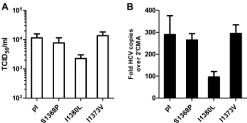
The S1368P substitution does not impair viral fitness in HCV genotype 1a.We hypothesized that fitness constraints caused pre-ferred selection of the S1368P substitution in genotype 1a. To address this experimentally, the recently described genotype 1a
full genome virus TNcc (25) was utilized to study the replication
fitness and infectivity associated with the substitutions S1368P, I1373V or I1380L. At 72 h after the electroporation of virus RNA into Huh-7.5 cells, the supernatants were used for infection of
naive Huh-7.5 cells (Fig. 5A). Upon transfection with the parental
HCV TN genome the TCID50was 1.1⫻104infectious particles
per ml. Approximately the same amount of infectious particles
was released upon transfection with the S1368P variant (0.8⫻104
TCID50/ml) or the I1373V variant (1.3⫻104TCID50/ml). In
con-trast, the I1380L substitution was associated with a decreased
TCID50of 0.2⫻104per ml. The same hierarchy was observed
at the replication level, with only a slight reduction of replication for the S1368P and I1373V substitution and a 3-fold reduced rep-lication for the I1380L substitution. This indicates that only the substitution I1380L impairs viral fitness in genotype 1a, whereas the substitutions S1368P and I1380L do not.
DISCUSSION
Here, we characterized a highly conserved HLA-B*51-restricted
CD8⫹T cell epitope located in the helicase domain of HCV NS3.
The high frequency of CD8⫹T cells specific for this epitope in
PWID who achieved spontaneous immune control of HCV
infec-tion suggests that this CD8⫹ T cell response is reproducibly
mounted in HLA-B*51-positive patients. Given the high degree of conservation of the epitope, it was therefore surprising that in
chronic infection CD8⫹T cell responses against this epitope were
detectable only in PWID infected with genotype 3a and were com-pletely absent in patients infected with genotype 1a. Although vi-ral sequence polymorphisms were ovevi-rall rare, there was selection pressure on the epitope containing region, as evidenced by HLA-B*51-associated viral sequence polymorphisms in all studied ge-notypes. Notably, in genotype 1b there was statistical support for the selection of escape mutations inside the epitope in the single-source outbreak, whereas there was only a nonsignificant trend in the genotype 1b multicenter cohort. It is possible that escape pat-terns inside this epitope differ between cohorts. Alternatively, be-cause the natural sequence variability in this epitope in the
geno-FIG 3CD8⫹T cell response against the endogenously processed epitope IPFYGKAI1373–1380. Effector T cells were expanded for 10 days from PBMCs in the
presence of the peptide IPFYGKAI. HLA-B*51-positive target cells were generated by electroporation with a NS3(aa1330-2420)-GFP fusion protein. GFP-positive cells were sorted and used as targets for restimulation of IPFYGKAI-specific effector CD8⫹T cells in an effector/target ratio of 1:1 for 4 h, followed by an intracellular cytokine staining (ICS). Mock transfected targets and peptide-pulsed targets served as negative or positive controls, respectively. (A) Represen-tative fluorescence-activated cell sorting results of one ICS. (B) The IFN-␥response against targets transfected with GT1a prototype NS3 was normalized to 100% and compared to the response against other targets as indicated. The data represent results from at least three independent experiments. ThePvalues were calculated using a one-samplettest with a hypothetical value of 100 (S1368P versus pt1a) or an unpairedttest (pt GT3a versus S1369P).
on November 7, 2019 by guest
http://jvi.asm.org/
[image:6.585.100.482.67.278.2]type 1b multicenter cohort is higher than in the single-source outbreak, the size of the multicenter cohort may be too small to unmask an existing effect of HLA-B*51-associated selection pres-sure.
The predominant escape mechanisms of substitutions selected
by CD8⫹T cell immune pressure in HCV epitopes are impaired
binding of the variant epitope to the HLA class I-molecule or impaired binding of the T cell receptor to the HLA class
I/peptide-FIG 4Impact of the S1368P substitution on proteasomal degradation. (A and B) The relative abundance of epitope-containing cleavage products after digestion of 25mer peptides with the prototype sequence (pt) or carrying the S1368P substitution (S1368P) with constitutive proteasome (A) or immune proteasome (B) is shown by normalized spectral indices. (C and D) The relative abundance of cleavage products with the correct C-terminal epitope end is shown by normalized spectral indices of individual peptides after digestion with constitutive proteasome (C) or immune proteasome (D).
on November 7, 2019 by guest
http://jvi.asm.org/
[image:7.585.40.545.64.603.2]complex. As an alternative mechanism, processing of the variant epitope can be affected by substitutions in the epitope region. We
previously reported CD8⫹T cell selection of a substitution inside
the HLA-B*08-restricted epitope NS3 HSKKKCDEL1395–1403
lo-cated in position 9 of the epitope (9). Functional analyses revealed
that the selected leucine to valine or phenylalanine substitution impaired presentation of the endogenously processed epitope.
Similarly, Kimura et al. (32) described substitutions selected
in-side targeted CD8⫹T cell epitopes that impaired proteasomal
processing in the chimpanzee model. In contrast, CD8⫹T cell
selection of substitutions in the epitope flanking region has not
been described yet in HCV. Seifert et al. (33) demonstrated that a
tyrosine to phenylalanine polymorphism located in the
C-termi-nal position⫹1 of the HLA-A*02-restricted epitope NS3 CVNG
VCWTV1073–1081 influenced carboxy-terminal cleavage of the
epitope by the proteasome; however, there was no conclusive
ev-idence for immune selection pressure on this residue (22,33). In
HIV larger population studies of HLA class I-associated viral ad-aptation suggested that selection of processing mutations in the epitope flanking region contributes to mutational immune escape
(34). This included substitutions in the C-terminal and
N-termi-nal epitope flanking regions. Only a few studies directly addressed the functional consequences of such putative processing
muta-tions (35,36). The example of selection of altered processing in
HLA-B*51-positive patients infected with HCV genotype 1a sup-ports that similar immune escape strategies are utilized by HCV.
Processing and presentation of viral epitopes is a multistep process starting with production of peptide precursors by
protea-somal degradation of viral proteins (reviewed in reference37).
These peptide precursors undergo N-terminal trimming by differ-ent endopeptidases and aminopeptidases in the cytosol and by the aminopeptidases ERAP1 and ERAP2 after the peptides have been
transported by TAP to the ER (38,39). Importantly, since
car-boxypeptidase activity is absent from the ER, production of pep-tide precursors with correct C-terminal ends by the proteasome is
required for subsequent HLA class I presentation (31). It has been
highlighted that the trimming efficiency of N-extended epitopes
strongly depends on the amino acid composition of the epitope flanking region. Some amino acids, such as leucine, lysine, phe-nylalanine, and methionine, are cleaved with high activity, whereas amino acids such as proline and glutamic acid are only
poorly cleaved (40). Accordingly, these latter amino acids are
un-derrepresented in flanking regions of known CD8⫹T cell epitopes
(41). In HCV genotype 1a there was HLA-B*51-associated
selec-tion pressure on posiselec-tion⫺5 of the epitope with a substitution
from serine to proline. Digestion of the N- and C-extended variant peptide by the constitutive and the immune proteasome yielded predominantly precursor peptides starting with proline, whereas digestion of the prototype yielded also shorter N-extended pre-cursor peptides. The shorter peptides are advantageous for the
transport into the ER (42), and more importantly, N-terminal
trimming of the peptide precursor starting with proline is
inhib-ited (31). This suggests that the serine to proline substitution
al-tered proteasomal production of epitope precursors with the con-sequence of a very low abundance of cleavable N-extended epitopes in the ER, although the latter was not formally tested.
Selection of CD8⫹T cell escape mutations is a trade-off
be-tween functional escape and viral constraints. Indeed, HCV may explore its replication space with different substitutions in
tar-geted epitopes before some mutations reach fixation (9,43). The
example presented here underlines that even despite an epitope being conserved across HCV genotypes the selected escape
path-ways and the CD8⫹T cell response can substantially differ.
Al-though not fully conclusive, our data suggest that selection of the S1368P substitution represents the optimal trade-off between functional immune escape and viral fitness in genotype 1a. The I1380L substitution may be disadvantageous due to its fitness costs despite efficient immune escape. In turn, the degree of cross-reactivity of the I1373V variant may prevent fixation in genotype 1a despite its low fitness costs. Interestingly, we observed interin-dividual differences in the level of cross-reactivity of the I1373V substitution with high cross-reactivity in HCV-RNA-negative PWID and lower levels of cross-reactivity in PWID infected with
genotype 3a (Fig. 1DandE), suggesting that the degree of
cross-reactivity may influence the outcome of infection as previously
reported (17). Absence of the CD8⫹T cell response in PWID with
chronic genotype 1a infection is unlikely caused by lack of prim-ing. The rapid decline of the response in the patient with acute HCV genotype 1a infection rather suggests secondary failure. This patient harbored a virus with the S1368P substitution. Although
the CD8⫹T cell response was clearly impaired against
endoge-nously processed antigens carrying the S1368P substitution, there was still some degree of T cell reactivity in our assays. This residual
epitope presentation may cause continuous CD8⫹T cell
stimula-tion associated with progressive dysfuncstimula-tion or exhausstimula-tion of
an-tigen-specific CD8⫹T cells (44–46) and possibly ultimate
extinc-tion of these cells (46).
The degree of CD8⫹T cell reactivity against the S1368P
sub-stitution in the context of genotype 1a was comparable to the reactivity against the endogenously processed genotype 3a anti-gen, suggesting that the epitope is less efficiently processed in the context of genotype 3a. Notably, in genotype 3a the substitution S1369P also impaired recognition of the endogenously processed antigen; however, here the I1373V was preferentially selected. One possibility is that in combination with the lower processing effi-ciency of the epitope in genotype 3a the I1373V substitution is sufficient to escape from the immune response. A second plausible
FIG 5Infectivity and replication of wild-type and mutant TNcc strains. RNA transcripts of the parental HCV TN genome and TN mutants were transfected into Huh-7.5 cells. (A) Seventy-two hours later, cell-free supernatant was used for inoculation of naive Huh-7.5 cells. The TCID50of the variants was
deter-mined by a limiting-dilution assay and staining with core and NS5A-specific antibodies. (B) RNA levels at 72 h posttransfection were measured by quanti-tative real-time reverse transcription-PCR. The RNA levels in the presence of 2=methyladenosin (2=CMA) was used for normalization. The ratio of mea-sured HCV RNA to meamea-sured RNA levels in the presence of 2=CMA was cal-culated for each construct. The data from four independent experiments are shown.
on November 7, 2019 by guest
http://jvi.asm.org/
[image:8.585.39.285.65.188.2]explanation is that the I1373V is associated with lower fitness costs compared to S1369P and I1380L in genotype 3a. However, this cannot be addressed until robust genotype 3a infectious viruses are publicly available. In either case, at least in some individuals
the corresponding CD8⫹T cell response was preserved in our
cohort of PWID infected with genotype 3a, a finding consistent with a memory T cell phenotype after mutational escape of the
targeted antigen as previously reported (44,45).
Taken together, HCV is able to evolutionarily adapt to CD8⫹T
cell immune pressure in multiple ways. The pathways to muta-tional escape are predictable but are distinct in different geno-types. Beyond selection of mutations inside targeted epitopes that impair HLA class I binding of the variant peptide ligand or TCR binding to the variant HLA class I/peptide complex HCV also inhibits epitope processing by modification of the epitope flank-ing region under T cell immune pressure. Importantly, the se-lected escape pathway of HCV may have consequences for the
destiny of antigen-specific CD8⫹T cells.
ACKNOWLEDGMENTS
We thank Jens Bukh for the TNcc strain, Darius Moradpour and Charles Rice for monoclonal antibodies, and Michael Engelmann and Lejla Tim-mer for technical assistance.
FUNDING INFORMATION
Deutsche Forschungsgemeinschaft (DFG) provided funding to Joerg Timm under grant numbers TRR60 and RTG1045. The Helmholtz Association provided funding to Thomas Pietschmann under grant num-ber SO-024.
REFERENCES
1.Pybus OG, Cochrane A, Holmes EC, Simmonds P.2005. The hepatitis C virus epidemic among injecting drug users. Infect Genet Evol5:131– 139.http://dx.doi.org/10.1016/j.meegid.2004.08.001.
2.Esteban JI, Sauleda S, Quer J. 2008. The changing epidemiology of hepatitis C virus infection in Europe. J Hepatol48:148 –162.http://dx.doi .org/10.1016/j.jhep.2007.07.033.
3.Rustgi VK.2007. The epidemiology of hepatitis C infection in the United States. J Gastroenterol42:513–521.http://dx.doi.org/10.1007/s00535-007 -2064-6.
4.Page K, Hahn JA, Evans J, Shiboski S, Lum P, Delwart E, Tobler L, Andrews W, Avanesyan L, Cooper S, Busch MP.2009. Acute hepatitis C virus infection in young adult injection drug users: a prospective study of incident infection, resolution, and reinfection. J Infect Dis200:1216 – 1226.http://dx.doi.org/10.1086/605947.
5.Mehta SH, Astemborski J, Kirk GD, Strathdee SA, Nelson KE, Vlahov D, Thomas DL. 2011. Changes in blood-borne infection risk among injection drug users. J Infect Dis203:587–594.http://dx.doi.org/10.1093 /infdis/jiq112.
6.Micallef JM, Macdonald V, Jauncey M, Amin J, Rawlinson W, van Beek I, Kaldor JM, White PA, Dore GJ.2007. High incidence of hepatitis C virus reinfection within a cohort of injecting drug users. J Viral Hepat
14:413– 418.http://dx.doi.org/10.1111/j.1365-2893.2006.00812.x. 7.Aitken CK, Lewis J, Tracy SL, Spelman T, Bowden DS, Bharadwaj M,
Drummer H, Hellard M.2008. High incidence of hepatitis C virus rein-fection in a cohort of injecting drug users. Hepatology48:1746 –1752.
http://dx.doi.org/10.1002/hep.22534.
8.von Hahn T, Yoon JC, Alter H, Rice CM, Rehermann B, Balfe P, McK-eating JA.2007. Hepatitis C virus continuously escapes from neutralizing antibody and T-cell responses during chronic infection in vivo. Gastroenter-ology132:667– 678.http://dx.doi.org/10.1053/j.gastro.2006.12.008. 9.Timm J, Lauer GM, Kavanagh DG, Sheridan I, Kim AY, Lucas M, Pillay
T, Ouchi K, Reyor LL, Schulze zur Wiesch J, Gandhi RT, Chung RT, Bhardwaj N, Klenerman P, Walker BD, Allen TM.2004. CD8 epitope escape and reversion in acute HCV infection. J Exp Med200:1593–1604.
http://dx.doi.org/10.1084/jem.20041006.
10. Ruhl M, Knuschke T, Schewior K, Glavinic L, Neumann-Haefelin C,
Chang DI, Klein M, Heinemann FM, Tenckhoff H, Wiese M, Horn PA, Viazov S, Spengler U, Roggendorf M, Scherbaum N, Nattermann J, Hoffmann D, Timm J.2011. CD8⫹T-cell response promotes evolution of hepatitis C virus nonstructural proteins. Gastroenterology140:2064 – 2073.http://dx.doi.org/10.1053/j.gastro.2011.02.060.
11. Erickson AL, Kimura Y, Igarashi S, Eichelberger J, Houghton M, Sidney J, McKinney D, Sette A, Hughes AL, Walker CM.2001. The outcome of hepatitis C virus infection is predicted by escape mutations in epitopes targeted by cytotoxic T lymphocytes. Immunity15:883– 895.http://dx.doi .org/10.1016/S1074-7613(01)00245-X.
12. Cox AL, Mosbruger T, Mao Q, Liu Z, Wang XH, Yang HC, Sidney J, Sette A, Pardoll D, Thomas DL, Ray SC.2005. Cellular immune selec-tion with hepatitis C virus persistence in humans. J Exp Med201:1741– 1752.http://dx.doi.org/10.1084/jem.20050121.
13. Ray SC, Fanning L, Wang XH, Netski DM, Kenny-Walsh E, Thomas DL.2005. Divergent and convergent evolution after a common-source outbreak of hepatitis C virus. J Exp Med201:1753–1759.http://dx.doi.org /10.1084/jem.20050122.
14. Farci P, Alter HJ, Wong DC, Miller RH, Govindarajan S, Engle R, Shapiro M, Purcell RH.1994. Prevention of hepatitis C virus infection in chimpanzees after antibody-mediated in vitro neutralization. Proc Natl Acad Sci U S A91:7792–7796.http://dx.doi.org/10.1073/pnas.91.16.7792. 15. Fauvelle C, Lepiller Q, Felmlee DJ, Fofana I, Habersetzer F, Stoll-Keller F, Baumert TF, Fafi-Kremer S.2013. Hepatitis C virus vaccines: progress and perspectives. Microb Pathog58:66 –72.http://dx.doi.org/10.1016/j .micpath.2013.02.005.
16. Ziegler S, Skibbe K, Walker A, Ke X, Heinemann FM, Heinold A, Mok JY, van Esch WJ, Yang D, Wolfl M, Timm J.2014. Impact of sequence variation in a dominant HLA-A*02-restricted epitope in hepatitis C virus on priming and cross-reactivity of CD8⫹T cells. J Virol88:11080 –11090.
http://dx.doi.org/10.1128/JVI.01590-14.
17. Yerly D, Heckerman D, Allen TM, Chisholm JV, III, Faircloth K, Linde CH, Frahm N, Timm J, Pichler WJ, Cerny A, Brander C.2008. In-creased cytotoxic T-lymphocyte epitope variant cross-recognition and functional avidity are associated with hepatitis C virus clearance. J Virol
82:3147–3153.http://dx.doi.org/10.1128/JVI.02252-07.
18. Yusim K, Dilan R, Borducchi E, Stanley K, Giorgi E, Fischer W, Theiler J, Marcotrigiano J, Korber B, Barouch DH.2013. Hepatitis C genotype 1 mosaic vaccines are immunogenic in mice and induce stronger T-cell responses than natural strains. Clin Vaccine Immunol20:302–305.http: //dx.doi.org/10.1128/CVI.00605-12.
19. Giugliano S, Oezkan F, Bedrejowski M, Kudla M, Reiser M, Viazov S, Scherbaum N, Roggendorf M, Timm J.2009. Degree of cross-genotype reactivity of hepatitis C virus-specific CD8⫹T cells directed against NS3. Hepatology50:707–716.http://dx.doi.org/10.1002/hep.23096.
20. von Delft A, Humphreys IS, Brown A, Pfafferott K, Lucas M, Klener-man P, Lauer GM, Cox AL, Gaudieri S, Barnes E.2015. The broad assessment of HCV genotypes 1 and 3 antigenic targets reveals limited cross-reactivity with implications for vaccine design. Guthttp://dx.doi .org/10.1136/gutjnl-2014-308724.
21. Kuntzen T, Timm J, Berical A, Lennon N, Berlin AM, Young SK, Lee B, Heckerman D, Carlson J, Reyor LL, Kleyman M, McMahon CM, Birch C, Schulze Zur Wiesch J, Ledlie T, Koehrsen M, Kodira C, Roberts AD, Lauer GM, Rosen HR, Bihl F, Cerny A, Spengler U, Liu Z, Kim AY, Xing Y, Schneidewind A, Madey MA, Fleckenstein JF, Park VM, Gala-gan JE, Nusbaum C, Walker BD, Lake-Bakaar GV, Daar ES, Jacobson IM, Gomperts ED, Edlin BR, Donfield SM, Chung RT, Talal AH, Marion T, Birren BW, Henn MR, Allen TM.2008. Naturally occurring dominant resistance mutations to hepatitis C virus protease and polymer-ase inhibitors in treatment-naive patients. Hepatology 48:1769 –1778.
http://dx.doi.org/10.1002/hep.22549.
22. Ruhl M, Chhatwal P, Strathmann H, Kuntzen T, Bankwitz D, Skibbe K, Walker A, Heinemann FM, Horn PA, Allen TM, Hoffmann D, Piet-schmann T, Timm J.2012. Escape from a dominant HLA-B*15-restricted CD8⫹T cell response against hepatitis C virus requires compensatory mutations outside the epitope. J Virol86:991–1000.http://dx.doi.org/10 .1128/JVI.05603-11.
23. Megger DA, Bracht T, Kohl M, Ahrens M, Naboulsi W, Weber F, Hoffmann AC, Stephan C, Kuhlmann K, Eisenacher M, Schlaak JF, Baba HA, Meyer HE, Sitek B. 2013. Proteomic differences between hepatocellular carcinoma and nontumorous liver tissue investigated by a combined gel-based and label-free quantitative proteomics study. Mol
on November 7, 2019 by guest
http://jvi.asm.org/
Cell Proteomics12:2006 –2020.http://dx.doi.org/10.1074/mcp.M113 .028027.
24. Borisenko IA, Viazovichenko Iu E, Gudkov VI.1994. An improvement in information support in the interests of the epidemiological health wel-fare of the troops. Voenno-Meditsinskii Zhurnal37– 42:80. (In Russian.) 25. Li YP, Ramirez S, Jensen SB, Purcell RH, Gottwein JM, Bukh J.2012. Highly efficient full-length hepatitis C virus genotype 1 (strain TN) infec-tious culture system. Proc Natl Acad Sci U S A109:19757–19762.http://dx .doi.org/10.1073/pnas.1218260109.
26. Steinmann E, Brohm C, Kallis S, Bartenschlager R, Pietschmann T.
2008. Efficient trans-encapsidation of hepatitis C virus RNAs into infec-tious virus-like particles. J Virol82:7034 –7046.http://dx.doi.org/10.1128 /JVI.00118-08.
27. Marukian S, Jones CT, Andrus L, Evans MJ, Ritola KD, Charles ED, Rice CM, Dustin LB.2008. Cell culture-produced hepatitis C virus does not infect peripheral blood mononuclear cells. Hepatology48:1843–1850.
http://dx.doi.org/10.1002/hep.22550.
28. Lindenbach BD, Evans MJ, Syder AJ, Wolk B, Tellinghuisen TL, Liu CC, Maruyama T, Hynes RO, Burton DR, McKeating JA, Rice CM.
2005. Complete replication of hepatitis C virus in cell culture. Science
309:623– 626.http://dx.doi.org/10.1126/science.1114016.
29. Moradpour D, Wakita T, Tokushige K, Carlson RI, Krawczynski K, Wands JR.1996. Characterization of three novel monoclonal antibodies against hepatitis C virus core protein. J Med Virol48:234 –241.http://dx .doi.org/10.1002/(SICI)1096-9071(199603)48:3⬍234::AID-JMV4⬎3.0 .CO;2-9.
30. Kim AY, Kuntzen T, Timm J, Nolan BE, Baca MA, Reyor LL, Berical AC, Feller AJ, Johnson KL, Schulze zur Wiesch J, Robbins GK, Chung RT, Walker BD, Carrington M, Allen TM, Lauer GM.2011. Spontane-ous control of HCV is associated with expression of HLA-B 57 and pres-ervation of targeted epitopes. Gastroenterology140:686 – 696 e681.http: //dx.doi.org/10.1053/j.gastro.2010.09.042.
31. van Endert P.2011. Post-proteasomal and proteasome-independent gen-eration of MHC class I ligands. Cell Mol Life Sci68:1553–1567.http://dx .doi.org/10.1007/s00018-011-0662-1.
32. Kimura Y, Gushima T, Rawale S, Kaumaya P, Walker CM.2005. Escape mutations alter proteasome processing of major histocompatibility com-plex class I-restricted epitopes in persistent hepatitis C virus infection. J Virol79:4870 – 4876.http://dx.doi.org/10.1128/JVI.79.8.4870-4876.2005. 33. Seifert U, Liermann H, Racanelli V, Halenius A, Wiese M, Wedemeyer H, Ruppert T, Rispeter K, Henklein P, Sijts A, Hengel H, Kloetzel PM, Rehermann B. 2004. Hepatitis C virus mutation affects proteasomal epitope processing. J Clin Invest114:250 –259.http://dx.doi.org/10.1172 /JCI200420985.
34. Carlson JM, Brumme ZL, Rousseau CM, Brumme CJ, Matthews P, Kadie C, Mullins JI, Walker BD, Harrigan PR, Goulder PJ, Heckerman D.2008. Phylogenetic dependency networks: inferring patterns of CTL escape and codon covariation in HIV-1 Gag. PLoS Comput Biol
4:e1000225.http://dx.doi.org/10.1371/journal.pcbi.1000225.
35. Allen TM, Altfeld M, Yu XG, O’Sullivan KM, Lichterfeld M, Le Gall S, John M, Mothe BR, Lee PK, Kalife ET, Cohen DE, Freedberg KA, Strick
DA, Johnston MN, Sette A, Rosenberg ES, Mallal SA, Goulder PJ, Brander C, Walker BD.2004. Selection, transmission, and reversion of an antigen-processing cytotoxic T-lymphocyte escape mutation in human immunodeficiency virus type 1 infection. J Virol78:7069 –7078.http://dx .doi.org/10.1128/JVI.78.13.7069-7078.2004.
36. Milicic A, Price DA, Zimbwa P, Booth BL, Brown HL, Easterbrook PJ, Olsen K, Robinson N, Gileadi U, Sewell AK, Cerundolo V, Phillips RE.
2005. CD8⫹T cell epitope-flanking mutations disrupt proteasomal pro-cessing of HIV-1 Nef. J Immunol175:4618 – 4626.http://dx.doi.org/10 .4049/jimmunol.175.7.4618.
37. Sijts EJ, Kloetzel PM.2011. The role of the proteasome in the generation of MHC class I ligands and immune responses. Cell Mol Life Sci68:1491– 1502.http://dx.doi.org/10.1007/s00018-011-0657-y.
38. Hammer GE, Kanaseki T, Shastri N. 2007. The final touches make perfect the peptide-MHC class I repertoire. Immunity26:397– 406.http: //dx.doi.org/10.1016/j.immuni.2007.04.003.
39. Serwold T, Gonzalez F, Kim J, Jacob R, Shastri N. 2002. ERAAP customizes peptides for MHC class I molecules in the endoplasmic retic-ulum. Nature419:480 – 483.http://dx.doi.org/10.1038/nature01074. 40. Zhang SC, Martin E, Shimada M, Godfrey SB, Fricke J, Locastro S, Lai
NY, Liebesny P, Carlson JM, Brumme CJ, Ogbechie OA, Chen H, Walker BD, Brumme ZL, Kavanagh DG, Le Gall S.2012. Aminopepti-dase substrate preference affects HIV epitope presentation and predicts immune escape patterns in HIV-infected individuals. J Immunol188:
5924 –5934.http://dx.doi.org/10.4049/jimmunol.1200219.
41. Schatz MM, Peters B, Akkad N, Ullrich N, Martinez AN, Carroll O, Bulik S, Rammensee HG, van Endert P, Holzhutter HG, Tenzer S, Schild H.2008. Characterizing the N-terminal processing motif of MHC class I ligands. J Immunol 180:3210 –3217. http://dx.doi.org/10.4049 /jimmunol.180.5.3210.
42. van Endert PM, Tampe R, Meyer TH, Tisch R, Bach JF, McDevitt HO.
1994. A sequential model for peptide binding and transport by the trans-porters associated with antigen processing. Immunity1:491–500.http: //dx.doi.org/10.1016/1074-7613(94)90091-4.
43. Uebelhoer L, Han JH, Callendret B, Mateu G, Shoukry NH, Hanson HL, Rice CM, Walker CM, Grakoui A. 2008. Stable cytotoxic T cell escape mutation in hepatitis C virus is linked to maintenance of viral fitness. PLoS Pathog4:e1000143.http://dx.doi.org/10.1371/journal.ppat .1000143.
44. Kasprowicz V, Kang YH, Lucas M, Schulze zur Wiesch J, Kuntzen T, Fleming V, Nolan BE, Longworth S, Berical A, Bengsch B, Thimme R, Lewis-Ximenez L, Allen TM, Kim AY, Klenerman P, Lauer GM.2010. Hepatitis C virus (HCV) sequence variation induces an HCV-specific T-cell phenotype analogous to spontaneous resolution. J Virol84:1656 – 1663.http://dx.doi.org/10.1128/JVI.01499-09.
45. Bengsch B, Seigel B, Ruhl M, Timm J, Kuntz M, Blum HE, Pircher H, Thimme R.2010. Coexpression of PD-1, 2B4, CD160 and KLRG1 on exhausted HCV-specific CD8⫹T cells is linked to antigen recognition and T cell differentiation. PLoS Pathog6:e1000947.http://dx.doi.org/10.1371 /journal.ppat.1000947.
46. Wherry EJ.2011. T cell exhaustion. Nat Immunol12:492– 499.