Replication-Competent NYVAC-KC Yields Improved
Immunogenicity to HIV-1 Antigens in Rhesus Macaques
Compared to Nonreplicating NYVAC
Karen V. Kibler,
aBenedikt Asbach,
bBeatriz Perdiguero,
cJuan García-Arriaza,
cNicole L. Yates,
dRobert Parks,
dSherry Stanfield-Oakley,
dGuido Ferrari,
dDavid C. Montefiori,
dGeorgia D. Tomaras,
dMario Roederer,
eKathryn E. Foulds,
eDonald N. Forthal,
fMichael S. Seaman,
gSteve Self,
hRaphael Gottardo,
hSanjay Phogat,
iJames Tartaglia,
iSusan Barnett,
jAnthony D. Cristillo,
kDeborah Weiss,
kLindsey Galmin,
kSong Ding,
lJonathan L. Heeney,
mMariano Esteban,
cRalf Wagner,
b,nGiuseppe Pantaleo,
oBertram L. Jacobs
a,paBiodesign Institute, Arizona State University, Tempe, Arizona, USA
bInstitute of Medical Microbiology and Hygiene, University of Regensburg, Regensburg, Germany
cDepartment of Molecular and Cellular Biology, Centro Nacional de Biotecnología, Consejo Superior de Investigaciones Científicas, Madrid, Spain
dDuke University Medical Center, Durham, North Carolina, USA
eVaccine Research Center, National Institute of Allergy and Infectious Diseases, National Institutes of Health, Bethesda, Maryland, USA
fDivision of Infectious Diseases Department of Medicine, University of California, Irvine School of Medicine, Irvine, California, USA
gCenter for Virology and Vaccine Research, Beth Israel Deaconess Medical Center, Boston, Massachusetts, USA
hStatistical Center for HIV/AIDS Research and Prevention, Fred Hutchinson Cancer Research Center, Seattle,
Washington, USA
iSanofi Pasteur, Swiftwater, Pennsylvania, USA
jNovartis Vaccines and Diagnostics, Inc., Cambridge, Massachusetts, USA
kAdvanced BioScience Laboratories, Inc., Rockville, Maryland, USA
lEuroVacc Foundation, Lausanne, Switzerland
mLab of Viral Zoonotics, Department of Veterinary Medicine, University of Cambridge, Cambridge, United Kingdom
nInstitute of Clinical Microbiology and Hygiene, University of Regensburg, Regensburg, Germany
oDivision of Immunology and Allergy, Department of Medicine, Centre Hospitalier Universitaire Vaudois,
University of Lausanne, Lausanne, Switzerland
pSchool of Life Sciences, Arizona State University, Tempe, Arizona, USA
ABSTRACT
As part of the continuing effort to develop an effective HIV vaccine, we
generated a poxviral vaccine vector (previously described) designed to improve on
the results of the RV144 phase III clinical trial. The construct, NYVAC-KC, is a
replication-competent, attenuated recombinant of the vaccinia virus strain NYVAC.
NYVAC is a vector that has been used in many previous clinical studies but is
repli-cation deficient. Here, we report a side-by-side comparison of replirepli-cation-restricted
NYVAC and replication-competent NYVAC-KC in a nonhuman primate study, which
utilized a prime-boost regimen similar to that of RV144. NYVAC-C and NYVAC-C-KC
express the HIV-1 antigens gp140, and Gag/Gag-Pol-Nef-derived virus-like particles
(VLPs) from clade C and were used as the prime, with recombinant virus plus
enve-lope protein used as the boost. In nearly every T and B cell immune assay against
HIV-1, including neutralization and antibody binding, NYVAC-C-KC induced a greater
immune response than NYVAC-C, indicating that replication competence in a
poxvi-rus may improve upon the modestly successful regimen used in the RV144 clinical
trial.
IMPORTANCE
Though the RV144 phase III clinical trial showed promise that an
ef-fective vaccine against HIV-1 is possible, a successful vaccine will require
improve-ment over the vaccine candidate (ALVAC) used in the RV144 study. With that goal in
mind, we have tested in nonhuman primates an attenuated but
replication-competent vector, NYVAC-KC, in direct comparison to its parental vector, NYVAC,
CitationKibler KV, Asbach B, Perdiguero B,
García-Arriaza J, Yates NL, Parks R, Stanfield-Oakley S, Ferrari G, Montefiori DC, Tomaras GD, Roederer M, Foulds KE, Forthal DN, Seaman MS, Self S, Gottardo R, Phogat S, Tartaglia J, Barnett S, Cristillo AD, Weiss D, Galmin L, Ding S, Heeney JL, Esteban M, Wagner R, Pantaleo G, Jacobs BL. 2019. Replication-competent NYVAC-KC yields improved immunogenicity to HIV-1 antigens in rhesus macaques compared to nonreplicating NYVAC. J Virol 93:e01513-18. https://doi.org/10.1128/JVI.01513-18.
EditorGuido Silvestri, Emory University
Copyright© 2019 Kibler et al. This is an
open-access article distributed under the terms of theCreative Commons Attribution 4.0 International license.
[This article was published on 17 January 2019 with a standard copyright line (“© 2019 American Society for Microbiology. All Rights Reserved.”). The authors elected to pay for open access for the article after publication, necessitating replacement of the original copyright line with the one above, and this change was made on 21 August 2019. The authors intend to publish an Author Correction announcing this change in the Journal of Virology, vol. 93, no. 21, 2019.]
Address correspondence to Bertram L. Jacobs, [email protected].
For a companion article on this topic, see https://doi.org/10.1128/JVI.01529-18.
Received13 September 2018
Accepted31 October 2018
Accepted manuscript posted online14
November 2018
Published
VACCINES AND ANTIVIRAL AGENTS
crossm
February 2019 Volume 93 Issue 3 e01513-18 Journal of Virology jvi.asm.org 1
17 January 2019
on November 6, 2019 by guest
http://jvi.asm.org/
Downloaded from
on November 6, 2019 by guest
http://jvi.asm.org/
Downloaded from
on November 6, 2019 by guest
http://jvi.asm.org/
which is replication restricted in human cells, similar to the ALVAC vector used in
RV144. We have utilized a prime-boost regimen for administration of the vaccine
candidate that is similar to the one used in the RV144 study. The results of this
study indicate that a replication-competent poxvirus vector may improve upon the
effectiveness of the RV144 clinical trial vaccine candidate.
KEYWORDS
Gag-Pol-Nef, HIV, NYVAC, NYVAC-KC, T cell response, antibody
responses, gp140, nonhuman primates, vaccines
T
hough recombinant poxviruses have long been used to express foreign genes in
the development of vaccines against multiple disease-causing organisms (1, 2),
most of the candidates have been based on the modified and highly attenuated
poxviruses: NYVAC (3), MVA (4), and ALVAC (3, 5). Replication of these viruses is
restricted in human cells, thus offering the advantage of greater safety than use of
replication-competent poxviruses. The safety of attenuation, however, has frequently
been achieved at the expense of decreased immunogenicity, driving the field of human
immunodeficiency virus (HIV) vaccine development to explore the use of attenuated,
replication-competent vectors (reviewed in reference 6).
The first HIV-1 vaccine candidate to demonstrate partial success was the
ALVAC-based regimen used in the phase III RV144 clinical trial conducted in Thailand (7).
ALVAC is a canary poxvirus and does not replicate in human cells. The results of RV144
were released in 2009, and since then the HIV vaccine field has been working to
improve the modest 31% efficacy achieved by the ALVAC vector combined with
administration of HIV-1 gp120 protein in a prime-boost model. With this goal in mind,
we based our HIV vaccine vector on NYVAC rather than ALVAC and made it replication
competent in human cells. This was accomplished by reinsertion of the
K1L
and
C7L
genes, which were two host range viral genes of the 18 open reading frames originally
deleted from the Copenhagen strain to create NYVAC (3). The resulting construct,
NYVAC-KC, has been previously described (8). In the newborn-mouse model of
patho-genesis, NYVAC-KC constructs clustered near those of the replication-deficient NYVAC
and MVA vectors, and the 50% lethal dose (LD
50) was about 4 logs higher than that of
wild-type vaccinia virus. This model is the most sensitive measure we have of poxvirus
pathogenicity and demonstrates that NYVAC-KC is highly attenuated even though it is
replication competent in human cells. Enhanced expression of the antigen in human
cells from this replication-competent vector has been verified (8). Here, we report
immunogenicity study results in nonhuman primates (NHPs), comparing immunization
with NYVAC versus that with NYVAC-KC. Both viruses expressed novel HIV-1 clade C
proteins (9) and therefore are identified as NYVAC-C and NYVAC-C-KC in this NHP study.
The study design has combined the advantages of a prime-boost regimen, which were
demonstrated in the RV144 trial, with the enhanced antigen expression of a
replication-competent vector. The replication-replication-competent vector demonstrated greater
HIV-1-specific T cell responses, greater IgG binding to both autologous and heterologous
envelope glycoproteins (Envs) and to the V1-V2 loop, and greater neutralization of a
panel of pseudotyped tier 1 virus-like particles (VLPs) in TZM-bl assays. Combined, these
results provide evidence that the NYVAC-KC construct holds promise as an improved
HIV vaccine vector.
RESULTS
Study plan.
Eight rhesus macaques were assigned to each of two groups. The
macaques were randomized, including by weight and Mamu allele status (two in
each group were A
*
01-positive, and one in group 1 was also B
*
17-positive; all were
B
*
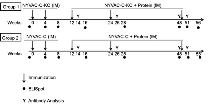
08-negative). As shown in Fig. 1, group 1 was immunized at weeks 0 and 4 with
NYVAC-C-KC combined viruses [mixture of NYVAC-KC-Envgp140(96ZM651) plus
NYVAC-KC-Gag(96ZM651)-Pol-Nef(97CN54)], and group 2 was immunized at weeks
0 and 4 with C combined viruses ([Envgp140(96ZM651) plus
NYVAC-Gag(96ZM651)-Pol-Nef(97CN54)]. Both groups were then boosted with the
on November 6, 2019 by guest
http://jvi.asm.org/
tive virus plus protein (gp120 plus MF59 adjuvant in place of gp120 plus Rehydragel
as used in the RV144 trial) on weeks 12, 24, and 49. All inoculations were
intra-muscular (i.m.).
The animals were monitored for any adverse events throughout the study. As in
previous studies (10–15), the immunizations were well tolerated. In group 1, one animal
following the first virus-only immunization and one following the second virus-only
immunization had mild erythema at the inoculation site (both were grade 1). Animals
gained weight normally, and there were no noted abnormalities in appetite.
T cell responses.
PBMCs were restimulated with peptide sets providing coverage of
the HIV-1 antigens, and gamma interferon (IFN-
␥
)-producing cells were quantified in an
enzyme-linked immunosorbent spot (ELISpot) assay. At all time points beginning with
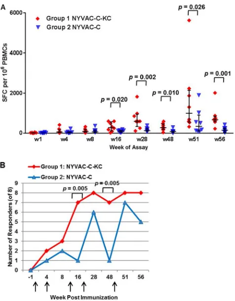
week 16, after the third immunization, NYVAC-C-KC induced a statistically significant
greater response to the peptides than NYVAC-C (Fig. 2A). In general, the response from
both groups was low using this immunization regimen compared to that with the
regimen used in the companion paper in this issue (16), and the fold difference
between groups, even when significant, ranged from 2 to 8. No animal had a response
above 2,000 spot-forming cells (SFC) per 10
6cells until week 51, with earlier responses
clustering in the range of 400 to 1,000 SFC (Fig. 2A). In group 1, which consisted of the
animals immunized with NYVAC-C-KC, there was an increase in both the range of SFC
counts and total number of SFC at 4 weeks following the immunizations done at weeks
12 and 24, and the greatest increase for both groups came with the sample taken 2
weeks following the week 49 immunization. These results are in contrast to what was
observed with the regimen in a parallel study (see companion manuscript of Asbach
et al. [16]) in which the prime was DNA, and the response in each of the groups
reached above 10,000 SFC after the animals were boosted with C or
NYVAC-C-KC. Figure 2B demonstrates the analysis of responders to either immunization at
various time points after immunization. While animals immunized with NYVAC-C-KC
had T cell responses after the third immunization, appreciable T cell responses were
induced with NYVAC-C only after the fourth immunization. While the number of
responders in the NYVAC-C-KC arm was stable (7 or 8 responders) from week 16
through the end of the study at week 56, the number of responders in the NYVAC-C
arm of the study waned with time after immunization and the number of responders
was boosted, even by the fifth immunization at week 48. Thus, animals immunized with
NYVAC-C-KC responded earlier than animals immunized with NYVAC-C, and the
re-sponse was sustained throughout the course of the study.
FIG 1Immunization schedule for the virus prime and virus/protein boost regimen. Two groups of 8 macaques were each immunized twice with virus alone (either NYVAC-C-KC or NYVAC-C), followed by three immunizations with virus plus protein. All immunizations were by the intramuscular (i.m.) route. Blood was collected for ELISpot analysis or antibody analysis at the indicated time points.
Immunogenicity of a Replicating Vaccinia Virus Vector Journal of Virology
February 2019 Volume 93 Issue 3 e01513-18 jvi.asm.org 3
on November 6, 2019 by guest
http://jvi.asm.org/
[image:3.585.43.397.70.246.2]Characteristics of T cell responses.
We then examined the polyfunctionality of the
CD4
⫹and CD8
⫹T cell responses. Peripheral blood mononuclear cells (PBMCs) from
each of the time points indicated in Fig. 3 were stimulated with nine peptide pools.
Staining for surface markers and for cytokine production (IFN-
␥
, interleukin-2 [IL-2], and
tumor necrosis factor alpha [TNF-
␣
]) provided a polyfunctionality profile to compare
results with the replication-deficient NYVAC-C to those with the replication-competent
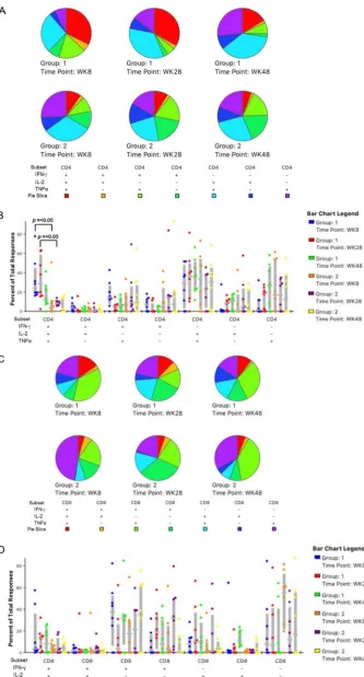
NYVAC-C-KC. For CD4 T cells, at all three time points (weeks 8, 28, and 48), the
percentage of responses that included all three of the assayed cytokines was higher for
group 1 animals than for group 2 (Fig. 3A). The polyfunctionality of the CD4
⫹responses
at weeks 8 and 28 was significantly higher for the NYVAC-C-KC-inoculated animals than
for those inoculated with NYVAC-C (Fig. 3B), with a 5-fold maximum difference. The
CD8
⫹responses were similar in distribution to those of CD4
⫹responses, with the triple
response greater for group 1 than for group 2 (Fig. 3C) though these were not
statistically significant differences (Fig. 3D).
Magnitude of Ab responses.
Antibody (Ab) responses to HIV-1 Env and Gag
antigens in each group were analyzed in serum samples taken 2 weeks after the first
virus/protein immunization done at week 12, 2 and 24 weeks after the second virus/
protein immunization done at week 24, and 2 weeks after the third virus/protein
immunization done at week 49 (week 51) (Fig. 4). The magnitude of both IgG and IgA
binding activities to a set of eight different envelope-derived and two Gag-Pol-derived
FIG 2The competent NYVAC-C-KC induces a greater T cell response does replication-deficient NYVAC-C. PBMCs were freshly isolated at the indicated time points and restimulated with a pool of peptides in an ELISpot assay. The number of spot-forming cells (producing IFN-␥) per 106cells is
shown. (A) Each value is the sum of the responses from the nine peptides per each of the 8 animals. Statistical significance was determined using the Mann Whitney method, with a confidence interval of 95%. w, week. (B) The number of animals in each group (n⫽8) that were responders is shown for each time point tested. Criteria for responders were defined asⱕ50 SFC/106cells and a 4-fold increase above
the baseline level for any pool of peptides. Immunizations were at weeks 0, 4, 12, 24, and 49 (indicated by arrows). Statistical significance was determined using a Wilcoxon rank sum test (A) and by a Fisher exact test (B).
on November 6, 2019 by guest
http://jvi.asm.org/
[image:4.585.87.325.70.374.2]FIG 3The replication-competent NYVAC-C-KC (group 1) induces more polyfunctional CD4⫹and CD8⫹T cell
responses than replication-deficient NYVAC-C (group 2). PBMCs were obtained at the indicated time points, stimulated with the nine peptide pools, and stained for CD3, CD4, and CD8, as well as intracellular IFN-␥, IL-2,
(Continued on next page)
Immunogenicity of a Replicating Vaccinia Virus Vector Journal of Virology
February 2019 Volume 93 Issue 3 e01513-18 jvi.asm.org 5
on November 6, 2019 by guest
http://jvi.asm.org/
[image:5.585.44.377.69.688.2]antigens was measured by a binding antibody multiplex assay (17, 18). Antibody
responses were detected at all time points and in similar patterns for both groups.
However, with few exceptions, the antibody responses from group 1 animals,
immu-nized with NYVAC-C-KC, were significantly greater than those from group 2 animals,
immunized with NYVAC-C; for these responses, the fold change between the groups
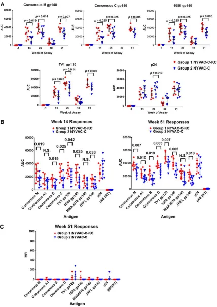
averaged 1.5. The IgG responses to five of the antigens are shown in Fig. 4A: consensus
M gp140, consensus C gp140, 1086 gp140, TV1 gp120, and p24. Response patterns
were similar following both of the first two virus/protein immunizations (weeks 12 and
24) though the second response was slightly improved. Responses then declined
during the period of weeks 26 to 48 and rebounded following the week 49
immuni-zation. In a comparison of the responses of all tested antigens at weeks 14 and 51 (Fig.
4B), the trend in the sera suggested that the reactivity is greatest against autologous
antigens: the protein immunizations were derived from TV1, and the 1086 isolate is
closely related. The next greatest reactivity was to the consensus sequences (M and C),
which would be expected for group C because the immunization antigens are derived
from subtype C isolates; however, the response to consensus M gp140 was nearly
identical to that of consensus C, indicative of at least some level of cross-reactivity.
Other indications of cross-reactivity are the responses to the B and A1 subtype
consensus proteins, as well as to other isolates from these subtypes, which were robust
although reduced compared to those of the autologous antigens. At all time points
postimmunization, NYVAC-C-KC induced greater IgG titers to the HIV-1 envelope than
NYVAC-C. Overall, the results demonstrated an improved response with the
replication-competent NYVAC-C-KC constructs. Antibodies to Gag, expressed in the vector
prime-only regimen, were also elicited. There was no response to reverse transcriptase.
Both groups’ responses to IgA were very low (Fig. 4C), and the responses of the two
groups were comparable. There were no statistical differences between the responses
of either group at any time point and those of the prebleed. Since certain specifications
of plasma IgA responses correlated directly with HIV-1 risk in the RV144 study, one
hypothesis is that a low plasma Env IgA response, as observed in this study, may be
desired as part of a protective immune response (19, 20).
In contrast to IgA responses, the response to the V1-V2 region of Env was strong at
a dilution of 1:2430 in an enzyme-linked immunosorbent assay (ELISA), as shown in Fig.
5A, and at the greater dilution of 1:7290 (Fig. 5B) the response was still detectable. At
both dilutions, NYVAC-C-KC induced greater reactivity than NYVAC-C.
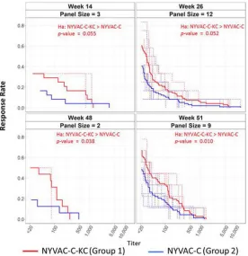
Functional characteristics of antibody responses.
Serum samples were also
measured for HIV-1 neutralizing antibody responses in a TZM-bl assay. A profile was
created based on results from a panel of viruses (nine tier 1 and three tier 2) with
pseudo-Envs (12 pseudo-Envs were measured at week 26, and various combinations of
up to 9 were measured at the other time points). Results are shown on a
magnitude-breadth (MB) curve in Fig. 6, which indicates that at all time points the NYVAC-C-KC group
had an overall broader neutralizing response per animal than the NYVAC-C group. During
the span of the time course, the responses among the 8 animals of each group became
more consistent, resulting in lower
P
values over time. By week 48, just prior to the final
immunization, and by week 51, 2 weeks following the final immunization on week 49, the
difference in the neutralizing antibody responses between the replication-competent
NYVAC-C-KC and the replication-deficient NYVAC-C was statistically significant. For
both NYVAC-C and NYVAC-C-KC, most of the positive activity was detected against tier
1 viruses (see Data Set S1 in the supplemental material).
FIG 3Legend (Continued)
and TNF-␣. Responding CD4⫹(A and B) and CD8⫹(C and D) T cells were quantified by flow cytometry. (A) The
fraction of triple-positive (red) responses for NYVAC-C-KC vaccinees (group 1) was greater at every time point. (B) Responses at weeks 8 and 28 were significantly higher for NYVAC-C-KC vaccinees than for NYVAC-C vaccinees (group 2). (C) The fraction of triple-positive (red) responses for NYVAC-C-KC vaccinees was greater at every time point. (D) Though not statistically significant, the responses at nearly all time points were higher for those animals vaccinated with NYVAC-C-KC than with NYVAC-C. Statistics calculated by Wilcoxon signed rank test using SPICE software (NIAID).
on November 6, 2019 by guest
http://jvi.asm.org/
FIG 4NYVAC-C-KC (group 1) gave improved IgG antibody responses to HIV-1 antigens and both groups had low responses to IgA. (A) A customized binding antibody multiplex assay was used to quantify IgG antibodies that bound to antigen-coated beads. The binding response for each antigen per each animal is shown as the area under the curve (AUC) of the titration curve, and medians and interquartile ranges are shown as horizontal lines. (B) Comparative responses of the week 14 and week 51 sera toward all antigens assessed. Statistical significance was determined using a Wilcoxon rank sum test (for panels A and B). (C) IgA responses of the week 51 sera toward all antigens assessed.
Immunogenicity of a Replicating Vaccinia Virus Vector Journal of Virology
February 2019 Volume 93 Issue 3 e01513-18 jvi.asm.org 7
on November 6, 2019 by guest
http://jvi.asm.org/
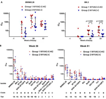
[image:7.585.43.471.71.676.2]We also examined individual responses to the specific pseudotyped virus-like
particles. Responses to MW965.26 (tier 1, clade C) and MN.3 (tier 1 A, clade B) at all time
points are shown for neutralization in TZM-bl cells (Fig. 7A). For MW965.26 and MN.3,
at all responsive time points postimmunization, the NYVAC-C-KC group demonstrated
greater responses than the NYVAC-C group; the differences were statistically significant
for MN.3, with fold differences of about 1.5. The values of the 50% infective doses (ID
50)
for all isolates at weeks 26 and 51 are shown in Fig. 7B. There was neutralizing activity
against tier 1 A viruses (MW965.26, MN.3, SF162, and SHIV-SF162P4 SHIV is simian-human
immunodeficiency virus), and in each case, animals immunized with NYVAC-C-KC
demon-strated a trend toward greater neutralization.
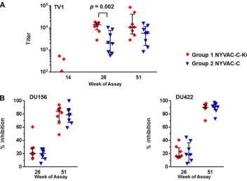
Serum from immunized animals was also tested for ADCC and ADCVI activity.
The animals immunized with the replication-competent NYVAC-C-KC (group 1)
re-sponded to a greater extent (about 4-fold higher) in an antibody-dependent
cell-mediated cytotoxicity (ADCC) assay (Fig. 8A) at week 26 than those receiving the
replication-deficient NYVAC-C (group 2). This assay measures the release of granzyme
B from natural killer cells (NK cells); such an Ab response has been associated with
protective responses and with delayed onset of AIDS in HIV-infected individual patients
and infected macaques (21–25). In the antibody-dependent cell-mediated virus
inhibi-tion (ADCVI) assay, the two groups had very similar, and strong, responses (Fig. 8B). In
this assay, PBMCs were used as effector cells, with serum added to CEM.NKR
CCR5cells
that had been infected with infectious HIV-1 strain DU156 or DU422. The strong
responses indicate that both vaccine candidates induced antibodies capable of binding
to HIV-1 peptides, thus making the cells coated with viral proteins a target of NK cells.
DISCUSSION
In this study, we have examined the immune response of the replication-restricted
poxvirus vector NYVAC in a head-to-head comparison with the replication-competent
NYVAC-KC vector in an NHP model and have shown that the replication-competent
NYVAC-C-KC expressing HIV-1 antigens induces greater HIV-1-specific T cell responses
in NHPs than NYVAC expressing the same HIV-1 antigens, as well as a greater
HIV-1-specific humoral response in the regimen that was utilized. Our hypothesis was that the
results of RV144 (19, 26) could be improved upon by using a vector that achieved
higher levels of antigen expression than levels attained with a replication-deficient
vector and that the higher levels could be reached through use of a vector that is
replication competent in human cells (8). Each of the two groups in this study was
FIG 5Antibody response to the V1-V2 region was strong for both NYVAC-C-KC and NYVAC-C groups. Response was measured by an ELISA, and the results for dilutions of 1:2430 (A) and 1:7290 (B) are shown. At both dilutions, the antibody levels following the week 24 boost were significantly higher for NYVAC-C-KC (group 1) than for NYVAC-C (group 2).Pvalues were calculated by the Wilcoxon rank-sum test. OD, optical density.
on November 6, 2019 by guest
http://jvi.asm.org/
[image:8.585.41.401.70.261.2]primed and boosted (once) with virus only and then boosted three times more but with
virus plus protein at each of the final three time points. This regimen was based on that
of RV144, which was the same schedule with the single exception that this study had
a fifth (final) inoculation whereas the RV144 trial had only the first four administered.
NYVAC-C has been studied previously in a head-to-head comparison with ALVAC-C,
with both vector mixtures expressing the same antigens and used in identical regimens
(11); in that study, NYVAC-C was found to induce improved T cell and humoral
responses compared to those of ALVAC-C. Therefore, this current report focused on the
comparison between NYVAC-C and NYVAC-C-KC in our efforts to improve upon the
RV144 immune responses; we started with the premise that if NYVAC has an improved
response compared to that of ALVAC, then a response that exceeds that of NYVAC is
also an improvement compared to that of RV144. Retrospective analyses of RV144 have
shown that the correlates of protection are complex and most likely will be dependent
on many variables and combinations of variables (27–38). Therefore, with this study we
have tried to gain perspective based on some of the ideas brought forth in
retrospec-tive reviews of what was learned from RV144.
The T cell responses with the poxvirus/protein regimen in this report were modest,
and while differences between the two groups were statistically significant, they
ranged from minimal to 5-fold changes. In contrast, in the companion study in this
issue (16), the magnitude of the T cell response was greater, whether measuring IFN-
␥
responses, HIV-1-specific CD4
⫹and CD8
⫹T cell responses, or polyfunctionality of the
responses; this is most likely a result of the DNA prime used in the companion study.
FIG 6Neutralizing antibody panel potency subject-specific and average magnitude breadth curves for TZM-bl assay. A magnitude-breadth (MB) curve of 50% inhibitory dose (ID50) neutralizing antibody titers
against pseudoenvelopes shown over time (x axis, neutralization titers; y axis, fraction of viruses neutralized). Isolates were the following: MN.3, MW965.26, and TV1.21 for week 14; TV1.21, BaL.26, Bx08.16, MN.3, MW965.26, SF162.LS, SHIV SF162P4, SHIV-SF162.P3, SS1196.1, SHIV 1157ipEL-p, SHIV 1157ipd3N4, and SHIV BAL-P4 for week 26; MN.3 and MW965.26 for week 48; TV1.21, BaL.26, Bx08.16, MN.3, MW965.26, SF162.LS, SHIV SF162P4, SHIV-SF162.P3, and SS1196.1 for week 51. Values of⬍20 (limit of detection of the assay) were assigned a value of 10. Dashed lines represent subject-specific responses. Solid lines represent group averages.Pvalues were derived from a Wilcoxon rank sum test.
Immunogenicity of a Replicating Vaccinia Virus Vector Journal of Virology
February 2019 Volume 93 Issue 3 e01513-18 jvi.asm.org 9
on November 6, 2019 by guest
http://jvi.asm.org/
[image:9.585.69.345.72.357.2]For a more in-depth comparison of the T cell responses, please see the companion
paper by Asbach et al. (16).
IgG antibody responses in the study reported here were more robust than the T cell
responses, and the replication-competent NYVAC-C-KC responses at most time points
were significantly higher than those of the replication-restricted NYVAC-C. In many
cases, direct comparison of the results in this report to RV144 humoral response assays
is difficult because the assays were not performed identically between the clinical trial
and this NHP study. While it is difficult to directly compare responses in humans to
responses in macaques, it would appear that NYVAC-C-KC induced a much stronger
binding response to V1-V2 than ALVAC did in RV144. We have previously shown that
NYVAC was an improved vector over ALVAC in a head-to-head comparison in NHPs
(11). In this study, we show that in IgG binding assays, in V1-V2 loop-binding assays, in
neutralizing antibody assays, and in the ADCC assay, NYVAC-C-KC induced a greater
response than the replication-restricted NYVAC-C. Thus, we believe that NYVAC-KC will
likely be superior to ALVAC as a vector for humans.
We also compared the prime-boost regimen used in this study to that of a
DNA-primed study, which is described in the companion paper in this issue (16). Though the
DNA-primed regimen clearly and predictably produced a greater T cell response than
the regimen described here of poxvirus and protein components, the reverse was true
for antibody responses, with mostly higher HIV-1 antibody responses in terms of IgG
binding, V1-V2 binding, and neutralization in the TZM-bl assay in the virus-primed
FIG 7Macaque sera neutralization in TZM-bl assay. (A) Serum samples were diluted and tested for neutralization against the indicated pseudoviruses. The value shown is the reciprocal of dilution at which 50% inhibition (ID50)
was observed for each animal; median and interquartile range are shown at the indicated time points of the pseudoviruses carrying MW965.26 or MN.3. Statistical significance was determined using a Mann-Whitney method. (B) ID50values of the serum samples from week 26 and week 51 for pseudoviruses carrying Envs from the indicated
isolates.
on November 6, 2019 by guest
http://jvi.asm.org/
[image:10.585.42.400.69.403.2]groups. This was especially clear in a comparison of the response kinetics of the two
groups, where there were major differences. Except for ADCVI activity, peak responses
were mostly already achieved at week 26 in this study (following the second poxvirus/
protein boost) and were similar again at week 51 following the final boost (e.g., IgG
responses) (Fig. 6). In contrast, in the DNA-primed groups, responses peaked only at the
final time point assessed; however, by then they often achieved levels similar to those
of this study. In IgG binding, the week 14 results of this study, after one protein boost,
were similar to the week 36 results in the companion study after two protein boosts.
In this report, with this regimen, we observed a boosting effect of p24 binding even
after the fifth and final virus administration at week 49, suggesting that multiple doses
of the poxvirus vector did not prevent expression of the antigens (Fig. 4A). The
companion study did not include administration of p24 after week 20. For the V1-V2
data, the response in the ELISA for NYVAC-C-KC at week 26, after two protein boosts,
was comparable to that of the highest group of the companion study at week 51 (after
three protein boosts). In the neutralization assay, the response to pseudoviruses
carrying MW965.26 (the highest responder in both studies) was 1 log higher at week 26
with NYVAC-C-KC, after two protein boosts, than at week 51 for the groups in Asbach
et al. (16) after three protein boosts. Interestingly, robust ADCC activity was observed
in this poxvirus-primed study already after the second boost in which protein was
included, while there was no consistent activity for the DNA-primed groups following
the second protein boost. However, at week 51 after the last protein boost, the ADCC
activity was conversely slightly higher in the DNA-primed groups than in the
NYVAC-C-KC-group (1.2-fold) and the NYVAC-C-group (2.3-fold). ADCVI activity also showed a
trend to be slightly superior in the poxvirus/protein groups to that in the DNA-primed
FIG 8Serum samples from macaques collected at week 51 demonstrated a strong HIV-1 response in both ADCC and ADCVI assays. (A) ADCC assay. CEM.NKRCCR5 target cells coated with TV1 gp120 were coincubated with PBMC preparations (including NK cells) at an effector/target ratio of 30:1 in the presence of plasma dilutions from the immunized macaques. The amount of granzyme B released by NK cells was measured using a fluorescent substrate. The plasma dilution was calculated to match the granzyme B activity cutoff, and the reciprocals are given as titers for values above the assay’s cutoff value of 100, with median and interquartile range also shown. (B) ADCVI assay. CEM.NKRCCR5 cells were infected with infectious HIV-1 strain DU156 or DU422 and then coincubated with PBMC effector cells in the presence of plasma at an effector/target ratio of 10:1. The amount of virus released was measured by a p24 ELISA, and the inhibition of virus production compared to that of samples lacking plasma is given as a percentage.
Immunogenicity of a Replicating Vaccinia Virus Vector Journal of Virology
February 2019 Volume 93 Issue 3 e01513-18 jvi.asm.org 11
on November 6, 2019 by guest
http://jvi.asm.org/
[image:11.585.43.395.71.329.2]groups for week 26 (second protein boost) of this study in comparison to that of week
36 (second protein boost) of the DNA-primed study (1.4-fold for NYVAC-C-KC). One
notable distinction between the two studies that should be considered is the contrast
in the regimens: there were a total of seven immunizations done in the companion
study (one DNA prime and two DNA boosts, one virus boost, and three protein boosts),
while only five were done in the study reported here (one virus prime and one virus
boost, followed by three virus/protein boosts); the greater humoral responses in this
study were achieved in most cases with only two protein boosts (week 26), while the
peak responses in the DNA-primed study required a third protein boost (week 51).
The immunogenicity profile was undoubtedly influenced by the adjuvant used in
this study, which was MF59. However, other adjuvants might also be appropriate for
use in similar studies conducted in the future. An example is an NHP study that
demonstrated greater efficacy with alum than with MF59 (39).
For most assays in this study, not only were the responses greater in the
replication-competent NYVAC-C-KC immunized animals than in the replication-deficient NYVAC-C
immunized animals in magnitude (T cells, binding, neutralization, and ADCC), but they
were also greater in breadth, with more animals responding or with a greater range of
response. We already know that NYVAC-KC is attenuated in the newborn-mouse model
(8), the most sensitive pathogenesis model we have for poxviruses, and that
NYVAC-KC’s reactogenicity in rabbits is comparable to that of NYVAC (B. L. Jacobs, K. V. Kibler,
and K. Denzler, unpublished data). In this study, neither of the vectors caused any
adverse effects in the NHPs. Future studies will ascertain if the differences in
immuno-genicity in this study compared to levels of previous HIV-1 vaccine candidates (40–43)
will contribute to vaccine efficacy. Though no macaque SHIV challenge study was done
in connection with this immunogenicity study, there is an NHP challenge study
currently taking place that includes an appropriate group size for measuring efficacy
and a regimen that is partially based on the results of this study and that of the
companion paper.
Together, the two outcomes of improved immunogenicity and apparent safety
suggest that NYVAC-KC is a promising candidate for an HIV vaccine as well as for
vaccines against other infectious agents.
MATERIALS AND METHODS
Data availability. Assay data presented here can be accessed through the DataSpace sharing platform of the Collaboration for AIDS Vaccine Discovery (https://dataspace.cavd.org).
Ethics statement.The study was performed with male Indian rhesus macaques (Macaca mulatta mulatta) that were housed, fed, given environmental enrichment, and handled at ABL, Inc.’s animal facility (Rockville, MD), as described previously (10). The study was approved by the Advanced BioScience Laboratories, Inc., Institutional Animal Care and Use Committee (animal use protocol number 444). All procedures were carried out as previously described (10). A total of 16 animals were divided into two groups of 8 animals each.
Antigens.The Gag sequence is derived from the HIV-1 C-clade isolate 96ZM651 (GenBank accession numberAF286224). The Pol-Nef sequence is derived from the HIV C/B-clade isolate 97CN54 (GenBank accession numberAX149647.1) and consists of p6*(amino acids [aa] 1 to 56), followed by protease (aa 57 to 155; inactivated by mutation D81N), the N-terminal part of reverse transcriptase (RT) (aa 156 to 320 and 357 to 360), scrambled Nef (aa 101 to 206 followed by aa 1 to 100), the C-terminal part of RT (aa 361 to 715), and finally the middle part of RT (aa 321 to 356). The GagFSPolNef (where FS is frameshift) cassette inserted into NYVAC (see below) contains identical sequences on the amino acid level though the two reading frames are connected, employing the natural ribosomal frameshift (thus keeping the wild-type codon usage for the region from the slippery site to the stop codon of Gag), leading to expression of Gag and Gag-Pol-Nef presumably in a ratio of about 10:1 (32). The gp140 sequence is derived from strain 96ZM651 and consists of amino acids 1 to 673, including the autologous signal sequence (incorporation of a strong Kozak initiation site leads to the mutation R2G within the signal sequence), and contains a mutated gp120-gp41 cleavage site (R516S).
All antigen open reading frames were codon optimized for human expression using the GeneOpti-mizer algorithm (44), supplemented with a strong Kozak initiation site, and synthesized by GeneArt AG (Regensburg) with suitable restriction sites for insertion into the respective plasmids.
Gag-Pol-Nef and gp140 were inserted into the New York vaccinia virus (NYVAC) (45) as described in Kibler et al. (8). Previously, we reported that cells infected with replication-deficient NYVAC produced trimeric gp140 and Gag-derived VLPs, which activate HIV-1-specific B and T cell immune responses in mice (9). Recombinant clade C gp120 protein (isolate TV1) (46) was expressed from stably transfected CHO cell lines, purified, and characterized as previously described (47).
on November 6, 2019 by guest
http://jvi.asm.org/
Vaccines and immunizations.NYVAC-based recombinant viruses were grown in BHK-21 cells and purified by sucrose cushion centrifugation twice. Titers were determined by plaque immunostaining in BSC-40 cells. Each animal received two recombinant viruses, one expressing Envgp140(96ZM651) and one expressing Gag(96ZM651)-Pol-Nef(97CN54), each at 1⫻108PFU in a total volume of 1 ml of 1⫻
Tris-buffered saline (TBS). Group 1 received the replication-competent NYVAC-C-KC combined viruses [NYVAC-KC-Envgp140(96ZM651) plus NYVAC-KC-Gag(96ZM651)-Pol-Nef(97CN54)], and group 2 received the replication-deficient NYVAC-C combined viruses [NYVAC-Envgp140(96ZM651) plus NYVAC-Gag(96ZM651)-Pol-Nef(97CN54)]. One milliliter was injected intramuscularly into the deltoid of the right arm of each animal. The subtype C gp120 protein boost of HIV-1 consisted of 100g of the TV1 isolate with MF59 as an adjuvant, at a 0.1-mg/ml final protein concentration. One milliliter was injected intramuscularly into the deltoid of the left arm.
Blood samples were taken at the indicated time points shown in Fig. 1 as described previously (10). Briefly, EDTA-blood samples were collected for T cell assays, and plasma or clotted blood was collected for the antibody analyses.
Immunological analyses. (i) Peptides.Nine different peptide pools were used for T cell stimulations (described in reference 10).
(ii) IFN-␥ELISpot.Freshly isolated PBMCs were stimulated and processed as described previously (10). Briefly, the stimulated PBMCs were used to probe IFN-␥-coated plates. Results were visualized and counted with an automated ELISpot reader (ImmunoSpot v5 Reader; CTL). Animals with more than 50 SFC/106cells and four times the week 0 background values for any one of the peptide pools were
considered responders (40).
(iii) Intracellular cytokine staining (ICS).The assay was carried out by the Nonhuman Primate Immunogenicity Core (M. Roederer lab), Vaccine Research Center, NIAID, as described previously (10, 48). Briefly, cells were stained with fluorescence-labeled antibodies directed against CD3, CD4, CD8, IFN-␥, IL-2, and TNF-␣for flow cytometric analysis.
(iv) HIV-1-specific binding antibody assay.HIV-1-specific IgG and IgA antibodies to gp120/gp140 proteins, p24, and p66 (RT) were measured by an HIV-1 binding antibody multiplex assay (G. D. Tomaras lab [10]) as previously described (22, 24). All assays were run under conditions compliant with good clinical laboratory practice, including tracking of positive controls by Levy-Jennings charts. Briefly, positive controls included an HIVIG and CH58 monoclonal antibody (MAb) IgG titration. Negative controls included in every assay were blank beads and HIV-1-negative sera. Antibody measurements were acquired on a Bio-Plex instrument (Bio-Rad, Hercules, CA) using 21CFR Part 11-compliant software, and the primary readout is in mean fluorescence intensity (MFI). Samples not matching the assay’s predefined positivity criteria were considered nonresponders of value zero. For IgG, MFI data were transformed to values of the area under the curve (AUC) (41). The following antigens were examined: (i) consensus gp140 proteins of group M (Con S gp140 CF [49, 50]), clade A (A1.con.env03 140 CF), clade B (B.con.env03 140 CF), and clade C (C.con.env03 140 CF); (ii) the primary Env variants 1086 gp120 (clade C), TV1 gp120 (clade C), JRFL gp140 (clade B), MSA4076 gp140 (clade A1, also called OOMSA); (iii) Gag p24 and RT p66.
(v) Env-V1-V2-specific antibodies. Antibodies binding the variable loops 1 and 2 of Env were measured by an ELISA. ELISA plates (384-well) (3700; Costar) were coated with 2g/ml MLV gp70-scaffolded Case-A2 V1-V2 in 0.1 M sodium bicarbonate and blocked with assay diluent (4% [wt/vol] whey protein, 15% normal goat serum, 0.5% Tween 20, 0.05% sodium azide in phosphate-buffered saline [PBS]). Plasma samples were applied in quadruplicates as eight 3-fold serial dilutions starting at 1:90. Plates were incubated for 1.5 h and then washed with PBS– 0.1% Tween 20. Ten microliters of horseradish peroxidase (HRP)-conjugated goat anti-rhesus secondary antibody (617-103-012 [Rockland],1:10,000 in assay diluent without azide) was added for 1 h. The plates were washed again and developed with 20l of SureBlue Reserve (53-00-03; KPL) for 15 min. The reaction was stopped by addition of 20l of HCl stop solution. Absorbance was measured at 450 nm.
(vi) Neutralization of HIV-1.Neutralization of sera against HIV-1 was assessed using a TZM-bl assay by the Comprehensive Antibody Vaccine Immune Monitoring Consortium (CA-VIMC). Pseudoviruses were incubated, and procedures were done as described previously (10). Neutralization titers are given as the serum dilution leading to a 50% decrease (ID50) in relative light units for the respective virus
compared to the value for the control after subtraction of the background values.
(vii) ADCC assay.An antibody-dependent cellular cytotoxicity (ADCC) assay was carried out by the CA-VIMC ADCC core laboratory as previously described (10, 51). Briefly, CEM.NKRCCR5cells were used as
target cells and labeled with fluorescence markers to assess viability. The target cells were mixed with a granzyme B (GrB) substrate prior to addition of human effector cells. The plasma samples were tested starting at 1:100 dilutions and using six 4-fold dilution schemes. Cells were analyzed by flow cytometry for the fluorescent signal arising from cleavage of the GrB substrate in the target cells. The percentages of GrB-positive viable target cells minus background, represented by the effector and target cell conditions in the absence of any Ab, were calculated for each dilution. The titers of the ADCC-mediating antibodies were then calculated by interpolation of the dilution exhibiting a GrB activity matching the cutoff value for positivity of 8%.
(viii) ADCVI assay.An antibody-dependent cell-mediated virus inhibition (ADCVI) assay (D. N. Forthal lab) was carried out as described previously (10, 52). Briefly, target and effector cells were mixed at an effector/target cell ratio of 10:1; supernatants were harvested and assayed for p24 by ELISA. The virus inhibition in percent is calculated as [1⫺(c(p24)_v/c(p24)_c)]⫻100, where c(p24)_v is the concentration of p24 in tests with plasma from vaccinated animals and c(p24)_c is the concentration in control plasma (average of week 0 samples).
Immunogenicity of a Replicating Vaccinia Virus Vector Journal of Virology
February 2019 Volume 93 Issue 3 e01513-18 jvi.asm.org 13
on November 6, 2019 by guest
http://jvi.asm.org/
Statistics.The method of analysis is identified in each graph. Graphical distributions of the magni-tude of responses were plotted for NYVAC-C-KC and NYVAC-C at each time point. The midline in the box plots denotes the median, and the ends of the whiskers denote the 25th and 75th percentiles. If necessary, the plots were drawn on a log10scale. Only significantPvalues (i.e., less than 0.05) are
displayed above the plots in comparisons of two groups.
To determine a difference in response magnitudes between groups at specific time points, a Wilcoxon rank sum test was used, based on the exact reference distribution and using the mid-ranks method for ties (53). All tests were one-sided (alternative hypothesis [Ha], NYVAC-C-KC⬎NYVAC-C) and
considered significant at an alpha of 0.05. Due to the exploratory nature of this analysis, multiple comparison adjustments were not made here. R (version 3.3.1) and GraphPad Prism, version 7, were used to create all box plots, and the R coin package was used for testing.
Individual-specific and group-average magnitude-breadth (MB) curves (54) were used to display the breadth of binding antibody activity in terms of the percentage of antigens with log10titers of⬍xfor a
range of log10titers,x. The group differences of area under the MB curve (AUC-MB) were tested using
a Wilcoxon rank sum test.
Intracellular cytokine staining was graphed using SPICE software (NIAID). The percentages of total responses between two groups were compared using a Wilcoxon rank sum test for each cytokine combination at each time point.
SUPPLEMENTAL MATERIAL
Supplemental material for this article may be found at
https://doi.org/10.1128/JVI
.01513-18
.
SUPPLEMENTAL FILE 1
, XLSX file, 0.03 MB.
ACKNOWLEDGMENTS
This investigation was funded by the Bill & Melinda Gates Foundation Poxvirus T Cell
Vaccine Discovery Consortium (PTVDC) (38599). The Vaccine Immune Monitoring
Cen-ters (OPP1032144 and OPP1032325) and the Vaccine Immunology Statistical Center
(OPP1032317), as part of the Collaboration for AIDS Vaccine Discovery (CAVD), were
funded by the Bill & Melinda Gates Foundation. Novartis Vaccines received support for
this work under contract number HHSN266200500007C from DAIDS-NIAID-NIH.
The funders had no role in study design, data collection and interpretation, or the
decision to submit the work for publication.
We thank Kelli Greene and Hongmei Gao from the Vaccine Immune Monitoring
Centers, Eva Chung from the Vaccine Immunology Statistical Center for program
management, Sheetal Sawant for data management, and Hua-Xin Liao and Barton
Haynes for ELISAs and recombinant envelope proteins.
REFERENCES
1. Moss B. 1996.Poxviridae: the viruses and their replication, p 2637–2671.
InFields BN, Knipe DM, Howley PM, Chanock RM, Melnick JL, Monath TP, Roizman R, Straus SE (ed), Fields virology, 3rd ed, vol 2. Lippincott-Raven, Philadelphia, PA.
2. Paoletti E. 1996. Applications of pox virus vectors to vaccination: an update. Proc Natl Acad Sci U S A 93:11349 –11353.https://doi.org/10 .1073/pnas.93.21.11349.
3. Paoletti E, Tartaglia J, Taylor J. 1994. Safe and effective poxvirus vectors–NYVAC and ALVAC. Dev Biol Stand 82:65– 69.
4. Gomez CE, Perdiguero B, Garcia-Arriaza J, Esteban M. 2013. Clinical applications of attenuated MVA poxvirus strain. Expert Rev Vaccines 12:1395–1416.https://doi.org/10.1586/14760584.2013.845531. 5. Franchini G, Gurunathan S, Baglyos L, Plotkin S, Tartaglia J. 2004.
Poxvirus-based vaccine candidates for HIV: two decades of experience with special emphasis on canarypox vectors. Expert Rev Vaccines 3(4 Suppl):S75–S88.https://doi.org/10.1586/14760584.3.4.S75.
6. Parks CL, Picker LJ, King CR. 2013. Development of replication-competent viral vectors for HIV vaccine delivery. Curr Opin HIV AIDS 8:402– 411.https://doi.org/10.1097/COH.0b013e328363d389.
7. Rerks-Ngarm S, Pitisuttithum P, Nitayaphan S, Kaewkungwal J, Chiu J, Paris R, Premsri N, Namwat C, de Souza M, Adams E, Benenson M, Gurunathan S, Tartaglia J, McNeil JG, Francis DP, Stablein D, Birx DL, Chunsuttiwat S, Khamboonruang C, Thongcharoen P, Robb ML, Michael NL, Kunasol P, Kim JH. 2009. Vaccination with ALVAC and AIDSVAX to prevent HIV-1 infection in Thailand. N Engl J Med 361:2209 –2220.
https://doi.org/10.1056/NEJMoa0908492.
8. Kibler KV, Gomez CE, Perdiguero B, Wong S, Huynh T, Holechek S, Arndt W, Jimenez V, Gonzalez-Sanz R, Denzler K, Haddad EK, Wagner R, Sekaly RP, Tartaglia J, Pantaleo G, Jacobs BL, Esteban M. 2011. Improved NYVAC-based vaccine vectors. PLoS One 6:e25674.https://doi.org/10 .1371/journal.pone.0025674.
9. Perdiguero B, Gomez CE, Cepeda V, Sanchez-Sampedro L, Garcia-Arriaza J, Mejias-Perez E, Jimenez V, Sanchez C, Sorzano CO, Oliveros JC, Delaloye J, Roger T, Calandra T, Asbach B, Wagner R, Kibler KV, Jacobs BL, Pantaleo G, Esteban M. 2015. Virological and immunological characterization of novel NYVAC-based HIV/AIDS vaccine candidates expressing clade C trimeric soluble gp140(ZM96) and Gag(ZM96)-Pol-Nef(CN54) as virus-like particles. J Virol 89:970 –988.https://doi.org/10.1128/JVI.02469-14.
10. Asbach B, Kliche A, Kostler J, Perdiguero B, Esteban M, Jacobs BL, Montefiori DC, LaBranche CC, Yates NL, Tomaras GD, Ferrari G, Foulds KE, Roederer M, Landucci G, Forthal DN, Seaman MS, Hawkins N, Self SG, Sato A, Gottardo R, Phogat S, Tartaglia J, Barnett SW, Burke B, Cristillo AD, Weiss DE, Francis J, Galmin L, Ding S, Heeney JL, Pantaleo G, Wagner R. 2016. Potential to streamline heterologous DNA prime and NYVAC/ protein boost HIV vaccine regimens in rhesus macaques by employing improved antigens. J Virol 90:4133– 4149. https://doi.org/10.1128/JVI .03135-15.
11. Garcia-Arriaza J, Perdiguero B, Heeney J, Seaman M, Montefiori DC, Labranche C, Yates NL, Shen X, Tomaras GD, Ferrari G, Foulds KE, McDermott A, Kao SF, Roederer M, Hawkins N, Self S, Yao J, Farrell P, Phogat S, Tartaglia J, Barnett SW, Burke B, Cristillo A, Weiss D, Lee C, Kibler K, Jacobs B, Asbach B, Wagner R, Ding S, Pantaleo G, Esteban M.
on November 6, 2019 by guest
http://jvi.asm.org/
2015. Head-to-head comparison of poxvirus NYVAC and ALVAC vectors expressing identical HIV-1 clade C immunogens in prime-boost combi-nation with Env protein in nonhuman primates. J Virol 89:8525– 8539.
https://doi.org/10.1128/JVI.01265-15.
12. Hulot SL, Korber B, Giorgi EE, Vandergrift N, Saunders KO, Balachandran H, Mach LV, Lifton MA, Pantaleo G, Tartaglia J, Phogat S, Jacobs B, Kibler K, Perdiguero B, Gomez CE, Esteban M, Rosati M, Felber BK, Pavlakis GN, Parks R, Lloyd K, Sutherland L, Scearce R, Letvin NL, Seaman MS, Alam SM, Montefiori D, Liao HX, Haynes BF, Santra S. 2015. Comparison of immunogenicity in rhesus macaques of transmitted-founder, HIV-1 group M consensus and trivalent mosaic Envelope vaccines formulated as a DNA prime, NYVAC and Envelope protein boost. J Virol 89: 6462– 6480.https://doi.org/10.1128/JVI.00383-15.
13. Mooij P, Koopman G, Drijfhout JW, Nieuwenhuis IG, Beenhakker N, Koestler J, Bogers WM, Wagner R, Esteban M, Pantaleo G, Heeney JL, Jacobs BL, Melief CJ. 2015. Synthetic long peptide booster immunization in rhesus macaques primed with replication-competent NYVAC-C-KC induces a balanced CD4/CD8 T-cell and antibody response against the conserved regions of HIV-1. J Gen Virol 96:1478 –1483.https://doi.org/ 10.1099/vir.0.000074.
14. Zurawski G, Zurawski S, Flamar AL, Richert L, Wagner R, Tomaras GD, Montefiori DC, Roederer M, Ferrari G, Lacabaratz C, Bonnabau H, Klucar P, Wang Z, Foulds KE, Kao SF, Yates NL, LaBranche C, Jacobs BL, Kibler K, Asbach B, Kliche A, Salazar A, Reed S, Self S, Gottardo R, Galmin L, Weiss D, Cristillo A, Thiebaut R, Pantaleo G, Levy Y. 2016. Targeting HIV-1 Env gp140 to LOX-1 elicits immune responses in rhesus macaques. PLoS One 11:e0153484.https://doi.org/10.1371/journal.pone.0153484.
15. Garcia-Arriaza J, Perdiguero B, Heeney JL, Seaman MS, Montefiori DC, Yates NL, Tomaras GD, Ferrari G, Foulds KE, Roederer M, Self SG, Borate B, Gottardo R, Phogat S, Tartaglia J, Barnett SW, Burke B, Cristillo AD, Weiss DE, Lee C, Kibler KV, Jacobs BL, Wagner R, Ding S, Pantaleo G, Esteban M. 2017. HIV/AIDS vaccine candidates based on replication-competent recombinant poxvirus NYVAC-C-KC expressing trimeric gp140 and Gag-derived virus-like particles or lacking the viral molecule B19 that inhibits type I interferon activate relevant HIV-1-specific B and T cell immune functions in nonhuman primates. J Virol 91:e02182-16.
https://doi.org/10.1128/JVI.02182-16.
16. Asbach B, Kibler KV, Köstler J, Perdiguero B, Yates NL, Stanfield-Oakley S, Tomaras GD, Kao S-F, Foulds KE, Roederer M, Seaman MS, Montefiori DC, Parks R, Ferrari G, Forthal DN, Phogat S, Tartaglia J, Barnett SW, Self SG, Gottardo R, Cristillo AD, Weiss DE, Galmin L, Ding S, Heeney JL, Esteban M, Jacobs BL, Pantaleo G, Wagner R. 2018. Priming with a potent HIV-1 DNA vaccine frames the quality of immune responses prior to a poxvirus and protein boost. J Virol 93:e01529-18. https://doi.org/10.1128/JVI .01529-18.
17. Tomaras GD, Yates NL, Liu P, Qin L, Fouda GG, Chavez LL, Decamp AC, Parks RJ, Ashley VC, Lucas JT, Cohen M, Eron J, Hicks CB, Liao HX, Self SG, Landucci G, Forthal DN, Weinhold KJ, Keele BF, Hahn BH, Greenberg ML, Morris L, Karim SS, Blattner WA, Montefiori DC, Shaw GM, Perelson AS, Haynes BF. 2008. Initial B-cell responses to transmitted human immu-nodeficiency virus type 1: virion-binding immunoglobulin M (IgM) and IgG antibodies followed by plasma anti-gp41 antibodies with ineffective control of initial viremia. J Virol 82:12449 –12463. https://doi.org/10 .1128/JVI.01708-08.
18. Yates NL, Lucas JT, Nolen TL, Vandergrift NA, Soderberg KA, Seaton KE, Denny TN, Haynes BF, Cohen MS, Tomaras GD. 2011. Multiple HIV-1-specific IgG3 responses decline during acute HIV-1: implications for detection of incident HIV infection. AIDS 25:2089 –2097.https://doi.org/ 10.1097/QAD.0b013e32834b348e.
19. Haynes BF, Gilbert PB, McElrath MJ, Zolla-Pazner S, Tomaras GD, Alam SM, Evans DT, Montefiori DC, Karnasuta C, Sutthent R, Liao HX, DeVico AL, Lewis GK, Williams C, Pinter A, Fong Y, Janes H, DeCamp A, Huang Y, Rao M, Billings E, Karasavvas N, Robb ML, Ngauy V, de Souza MS, Paris R, Ferrari G, Bailer RT, Soderberg KA, Andrews C, Berman PW, Frahm N, De Rosa SC, Alpert MD, Yates NL, Shen X, Koup RA, Pitisuttithum P, Kaewkungwal J, Nitayaphan S, Rerks-Ngarm S, Michael NL, Kim JH. 2012. Immune-correlates analysis of an HIV-1 vaccine efficacy trial. N Engl J Med 366:1275–1286.https://doi.org/10.1056/NEJMoa1113425. 20. Tomaras GD, Ferrari G, Shen X, Alam SM, Liao HX, Pollara J, Bonsignori M,
Moody MA, Fong Y, Chen X, Poling B, Nicholson CO, Zhang R, Lu X, Parks R, Kaewkungwal J, Nitayaphan S, Pitisuttithum P, Rerks-Ngarm S, Gilbert PB, Kim JH, Michael NL, Montefiori DC, Haynes BF. 2013. Vaccine-induced plasma IgA specific for the C1 region of the HIV-1 envelope blocks
binding and effector function of IgG. Proc Natl Acad Sci U S A 110: 9019 –9024.https://doi.org/10.1073/pnas.1301456110.
21. Ahmad R, Sindhu ST, Toma E, Morisset R, Vincelette J, Menezes J, Ahmad A. 2001. Evidence for a correlation between antibody-dependent cellular cytotoxicity-mediating anti-HIV-1 antibodies and prognostic predictors of HIV infection. J Clin Immunol 21:227–233.https://doi.org/10.1023/ A:1011087132180.
22. Banks ND, Kinsey N, Clements J, Hildreth JE. 2002. Sustained antibody-dependent cell-mediated cytotoxicity (ADCC) in SIV-infected macaques correlates with delayed progression to AIDS. AIDS Res Hum Retroviruses 18:1197–1205.https://doi.org/10.1089/08892220260387940.
23. Gomez-Roman VR, Patterson LJ, Venzon D, Liewehr D, Aldrich K, Florese R, Robert-Guroff M. 2005. Vaccine-elicited antibodies mediate antibody-dependent cellular cytotoxicity correlated with significantly reduced acute viremia in rhesus macaques challenged with SIVmac251. J Immu-nol 174:2185–2189.https://doi.org/10.4049/jimmunol.174.4.2185. 24. Nag P, Kim J, Sapiega V, Landay AL, Bremer JW, Mestecky J, Reichelderfer
P, Kovacs A, Cohn J, Weiser B, Baum LL. 2004. Women with cervicova-ginal antibody-dependent cell-mediated cytotoxicity have lower genital HIV-1 RNA loads. J Infect Dis 190:1970 –1978.https://doi.org/10.1086/ 425582.
25. Scott-Algara D, Truong LX, Versmisse P, David A, Luong TT, Nguyen NV, Theodorou I, Barré-Sinoussi F, Pancino G. 2003. Cutting edge: increased NK cell activity in HIV-1-exposed but uninfected Vietnamese intravascu-lar drug users. J Immunol 171:5663–5667. https://doi.org/10.4049/ jimmunol.171.11.5663.
26. de Souza MS, Ratto-Kim S, Chuenarom W, Schuetz A, Chantakulkij S, Nuntapinit B, Valencia-Micolta A, Thelian D, Nitayaphan S, Pitisuttithum P, Paris RM, Kaewkungwal J, Michael NL, Rerks-Ngarm S, Mathieson B, Marovich M, Currier JR, Kim JH, Ministry of Public Health-Thai AIDS Vaccine Evaluation Group Collaborators. 2012. The Thai phase III trial (RV144) vaccine regimen induces T cell responses that preferentially target epitopes within the V2 region of HIV-1 envelope. J Immunol 188:5166 –5176.https://doi.org/10.4049/jimmunol.1102756.
27. Auclair S, Liu F, Niu Q, Hou W, Churchyard G, Morgan C, Frahm N, Nitayaphan S, Pitisuthithum P, Rerks-Ngarm S, Kimata JT, Soong L, Franchini G, Robb M, Kim J, Michael N, Hu H. 2018. Distinct susceptibility of HIV vaccine vector-induced CD4 T cells to HIV infection. PLoS Pathog 14:e1006888.https://doi.org/10.1371/journal.ppat.1006888.
28. Corey L, Gilbert PB, Tomaras GD, Haynes BF, Pantaleo G, Fauci AS. 2015. Immune correlates of vaccine protection against HIV-1 acquisition. Sci Transl Med 7:310rv317.https://doi.org/10.1126/scitranslmed.aac7732. 29. Edlefsen PT, Rolland M, Hertz T, Tovanabutra S, Gartland AJ, deCamp AC,
Magaret CA, Ahmed H, Gottardo R, Juraska M, McCoy C, Larsen BB, Sanders-Buell E, Carrico C, Menis S, Kijak GH, Bose M, Team RVS, Arroyo MA, O’Connell RJ, Nitayaphan S, Pitisuttithum P, Kaewkungwal J, Rerks-Ngarm S, Robb ML, Kirys T, Georgiev IS, Kwong PD, Scheffler K, Pond SL, Carlson JM, Michael NL, Schief WR, Mullins JI, Kim JH, Gilbert PB. 2015. Comprehensive sieve analysis of breakthrough HIV-1 sequences in the RV144 vaccine efficacy trial. PLoS Comput Biol 11:e1003973.https://doi .org/10.1371/journal.pcbi.1003973.
30. Esparza J. 2013. A brief history of the global effort to develop a preventive HIV vaccine. Vaccine 31:3502–3518. https://doi.org/10 .1016/j.vaccine.2013.05.018.
31. Gartland AJ, Li S, McNevin J, Tomaras GD, Gottardo R, Janes H, Fong Y, Morris D, Geraghty DE, Kijak GH, Edlefsen PT, Frahm N, Larsen BB, Tovanabutra S, Sanders-Buell E, deCamp AC, Magaret CA, Ahmed H, Goodridge JP, Chen L, Konopa P, Nariya S, Stoddard JN, Wong K, Zhao H, Deng W, Maust BS, Bose M, Howell S, Bates A, Lazzaro M, O’Sullivan A, Lei E, Bradfield A, Ibitamuno G, Assawadarachai V, O’Connell RJ, deSouza MS, Nitayaphan S, Rerks-Ngarm S, Robb ML, Sidney J, Sette A, Zolla-Pazner S, Montefiori D, McElrath MJ, Mullins JI, Kim JH, Gilbert PB, Hertz T. 2014. Analysis of HLA A*02 association with vaccine efficacy in the RV144 HIV-1 vaccine trial. J Virol 88:8242– 8255.https://doi.org/10 .1128/JVI.01164-14.
32. Girard MP, Osmanov S, Assossou OM, Kieny MP. 2011. Human immuno-deficiency virus (HIV) immunopathogenesis and vaccine development: a review. Vaccine 29:6191– 6218.https://doi.org/10.1016/j.vaccine.2011.06 .085.
33. Kim JH, Excler JL, Michael NL. 2015. Lessons from the RV144 Thai phase III HIV-1 vaccine trial and the search for correlates of protec-tion. Annu Rev Med 66:423– 437. https://doi.org/10.1146/annurev -med-052912-123749.
34. Lewis GK. 2016. The first 24 h: targeting the window of opportunity for
Immunogenicity of a Replicating Vaccinia Virus Vector Journal of Virology
February 2019 Volume 93 Issue 3 e01513-18 jvi.asm.org 15
on November 6, 2019 by guest
http://jvi.asm.org/
antibody-mediated protection against HIV-1 transmission. Curr Opin HIV AIDS 11:561–568.https://doi.org/10.1097/COH.0000000000000319. 35. Pegu P, Vaccari M, Gordon S, Keele BF, Doster M, Guan Y, Ferrari G, Pal
R, Ferrari MG, Whitney S, Hudacik L, Billings E, Rao M, Montefiori D, Tomaras G, Alam SM, Fenizia C, Lifson JD, Stablein D, Tartaglia J, Michael N, Kim J, Venzon D, Franchini G. 2013. Antibodies with high avidity to the gp120 envelope protein in protection from simian immunodefi-ciency virus SIV(mac251) acquisition in an immunization regimen that mimics the RV-144 Thai trial. J Virol 87:1708 –1719.https://doi.org/10 .1128/JVI.02544-12.
36. Rao M, Alving CR. 2016. Adjuvants for HIV vaccines. Curr Opin HIV AIDS 11:585–592.https://doi.org/10.1097/COH.0000000000000315. 37. Tomaras GD, Haynes BF. 2014. Advancing toward HIV-1 vaccine efficacy
through the intersections of immune correlates. Vaccines (Basel) 2:15–35.https://doi.org/10.3390/vaccines2010015.
38. Tuero I, Mohanram V, Musich T, Miller L, Vargas-Inchaustegui DA, Dem-berg T, Venzon D, Kalisz I, Kalyanaraman VS, Pal R, Ferrari MG, LaBranche C, Montefiori DC, Rao M, Vaccari M, Franchini G, Barnett SW, Robert-Guroff M. 2015. Mucosal B cells are associated with delayed SIV acqui-sition in vaccinated female but not male rhesus macaques following SIVmac251 rectal challenge. PLoS Pathog 11:e1005101.https://doi.org/ 10.1371/journal.ppat.1005101.
39. Vaccari M, Gordon SN, Fourati S, Schifanella L, Liyanage NPM, Cameron M, Keele BF, Shen X, Tomaras GD, Billings E, Rao M, Chung AW, Dowell KG, Bailey-Kellogg C, Brown EP, Ackerman ME, Vargas-Inchaustegui DA, Whitney S, Doster MN, Binello N, Pegu P, Montefiori DC, Foulds K, Quinn DS, Donaldson M, Liang F, Loré K, Roederer M, Koup RA, McDermott A, Ma Z-M, Miller CJ, Phan TB, Forthal DN, Blackburn M, Caccuri F, Bissa M, Ferrari G, Kalyanaraman V, Ferrari MG, Thompson DVon, Robert-Guroff M, Ratto-Kim S, Kim JH, Michael NL, Phogat S, Barnett SW, Tartaglia J, Venzon D, Stablein DM, Alter G, Sekaly R-P, Franchini G. 2016. Adjuvant-dependent innate and adaptive immune signatures of risk of SIVmac251 acquisition. Nat Med 22:762–770.https://doi.org/10.1038/nm.4105. 40. Bart PA, Goodall R, Barber T, Harari A, Guimaraes-Walker A, Khonkarly M,
Sheppard NC, Bangala Y, Frachette MJ, Wagner R, Liljestrom P, Kraehen-buhl JP, Girard M, Goudsmit J, Esteban M, Heeney J, Sattentau Q, McCormack S, Babiker A, Pantaleo G, Weber J. 2008. EV01: a phase I trial in healthy HIV negative volunteers to evaluate a clade C HIV vaccine, NYVAC-C undertaken by the EuroVacc Consortium. Vaccine 26: 3153–3161.https://doi.org/10.1016/j.vaccine.2008.03.083.
41. Harari A, Bart PA, Stohr W, Tapia G, Garcia M, Medjitna-Rais E, Burnet S, Cellerai C, Erlwein O, Barber T, Moog C, Liljestrom P, Wagner R, Wolf H, Kraehenbuhl JP, Esteban M, Heeney J, Frachette MJ, Tartaglia J, McCor-mack S, Babiker A, Weber J, Pantaleo G. 2008. An HIV-1 clade C DNA prime, NYVAC boost vaccine regimen induces reliable, polyfunctional, and long-lasting T cell responses. J Exp Med 205:63–77.https://doi.org/ 10.1084/jem.20071331.
42. McCormack S, Stohr W, Barber T, Bart PA, Harari A, Moog C, Ciuffreda D, Cellerai C, Cowen M, Gamboni R, Burnet S, Legg K, Brodnicki E, Wolf H, Wagner R, Heeney J, Frachette MJ, Tartaglia J, Babiker A, Pantaleo G, Weber J. 2008. EV02: a phase I trial to compare the safety and immu-nogenicity of HIV DNA-C prime-NYVAC-C boost to NYVAC-C alone. Vaccine 26:3162–3174.https://doi.org/10.1016/j.vaccine.2008.02.072. 43. Mooij P, Balla-Jhagjhoorsingh SS, Beenhakker N, van Haaften P, Baak I,
Nieuwenhuis IG, Heidari S, Wolf H, Frachette MJ, Bieler K, Sheppard N, Harari A, Bart PA, Liljestrom P, Wagner R, Pantaleo G, Heeney JL. 2009.
Comparison of human and rhesus macaque T-cell responses elicited by boosting with NYVAC encoding human immunodeficiency virus type 1 clade C immunogens. J Virol 83:5881–5889.https://doi.org/10.1128/JVI .02345-08.
44. Raab D, Graf M, Notka F, Schodl T, Wagner R. 2010. The GeneOptimizer algorithm: using a sliding window approach to cope with the vast sequence space in multiparameter DNA sequence optimization. Syst Synth Biol 4:215–225.https://doi.org/10.1007/s11693-010-9062-3. 45. Tartaglia J, Perkus ME, Taylor J, Norton EK, Audonnet JC, Cox WI, Davis
SW, van der Hoeven J, Meignier B, Riviere M. 1992. NYVAC: a highly attenuated strain of vaccinia virus. Virology 188:217–232. https://doi .org/10.1016/0042-6822(92)90752-B.
46. Zambonelli C, Dey AK, Hilt S, Stephenson S, Go EP, Clark DF, Wininger M, Labranche C, Montefiori D, Liao HX, Swanstrom RI, Desaire H, Haynes BF, Carfi A, Barnett SW. 2016. Generation and characterization of a bivalent HIV-1 subtype C gp120 protein boost for proof-of-concept HIV vaccine efficacy trials in southern Africa. PLoS One 11:e0157391.https://doi.org/ 10.1371/journal.pone.0157391.
47. Srivastava IK, Kan E, Sun Y, Sharma VA, Cisto J, Burke B, Lian Y, Hilt S, Biron Z, Hartog K, Stamatatos L, Diaz-Avalos R, Cheng RH, Ulmer JB, Barnett SW. 2008. Comparative evaluation of trimeric envelope glyco-proteins derived from subtype C and B HIV-1 R5 isolates. Virology 372:273–290.https://doi.org/10.1016/j.virol.2007.10.022.
48. Donaldson MM, Kao SF, Eslamizar L, Gee C, Koopman G, Lifton M, Schmitz JE, Sylwester AW, Wilson A, Hawkins N, Self SG, Roederer M, Foulds KE. 2012. Optimization and qualification of an 8-color intracellular cytokine staining assay for quantifying T cell responses in rhesus ma-caques for pre-clinical vaccine studies. J Immunol Methods 386:10 –21.
https://doi.org/10.1016/j.jim.2012.08.011.
49. Gaschen B, Taylor J, Yusim K, Foley B, Gao F, Lang D, Novitsky V, Haynes B, Hahn BH, Bhattacharya T, Korber B. 2002. Diversity consid-erations in HIV-1 vaccine selection. Science 296:2354 –2360.https:// doi.org/10.1126/science.1070441.
50. Liao HX, Sutherland LL, Xia SM, Brock ME, Scearce RM, Vanleeuwen S, Alam SM, McAdams M, Weaver EA, Camacho Z, Ma BJ, Li Y, Decker JM, Nabel GJ, Montefiori DC, Hahn BH, Korber BT, Gao F, Haynes BF. 2006. A group M consensus envelope glycoprotein induces antibodies that neu-tralize subsets of subtype B and C HIV-1 primary viruses. Virology 353:268 –282.https://doi.org/10.1016/j.virol.2006.04.043.
51. Pollara J, Hart L, Brewer F, Pickeral J, Packard BZ, Hoxie JA, Komoriya A, Ochsenbauer C, Kappes JC, Roederer M, Huang Y, Weinhold KJ, Tomaras GD, Haynes BF, Montefiori DC, Ferrari G. 2011. High-throughput quantita-tive analysis of HIV-1 and SIV-specific ADCC-mediating antibody responses. Cytometry A 79:603– 612.https://doi.org/10.1002/cyto.a.21084.
52. Bialuk I, Whitney S, Andresen V, Florese RH, Nacsa J, Cecchinato V, Valeri VW, Heraud JM, Gordon S, Parks RW, Montefiori DC, Venzon D, Demberg T, Guroff MR, Landucci G, Forthal DN, Franchini G. 2011. Vaccine induced antibodies to the first variable loop of human immunodeficiency virus type 1 gp120, mediate antibody-dependent virus inhibition in ma-caques. Vaccine 30:78 –94.https://doi.org/10.1016/j.vaccine.2011.10.040. 53. Hollander MW, Wolfe DA. 1999. Nonparametric statistical methods, 2nd
ed. John Wiley & Sons, New York, NY.
54. Huang Y, Gilbert PB, Montefiori DC, Self SG. 2009. Simultaneous evalu-ation of the magnitude and breadth of a left and right censored multi-variate response, with application to HIV vaccine development. Stat Biopharm Res 1:81–91.https://doi.org/10.1198/sbr.2009.0008.
on November 6, 2019 by guest
http://jvi.asm.org/
Correction for Kibler et al., “Replication-Competent NYVAC-KC
Yields Improved Immunogenicity to HIV-1 Antigens in Rhesus
Macaques Compared to Nonreplicating NYVAC”
Karen V. Kibler,
aBenedikt Asbach,
bBeatriz Perdiguero,
cJuan García-Arriaza,
cNicole L. Yates,
dRobert Parks,
dSherry Stanfield-Oakley,
dGuido Ferrari,
dDavid C. Montefiori,
dGeorgia D. Tomaras,
dMario Roederer,
eKathryn E. Foulds,
eDonald N. Forthal,
fMichael S. Seaman,
gSteve Self,
hRaphael Gottardo,
hSanjay Phogat,
iJames Tartaglia,
iSusan Barnett,
jAnthony D. Cristillo,
kDeborah Weiss,
kLindsey Galmin,
kSong Ding,
lJonathan L. Heeney,
mMariano Esteban,
cRalf Wagner,
b,nGiuseppe Pantaleo, Bertram L. Jacobs
a,paBiodesign Institute, Arizona State University, Tempe, Arizona, USA
bInstitute of Medical Microbiology and Hygiene, University of Regensburg, Regensburg, Germany
cDepartment of Molecular and Cellular Biology, Centro Nacional de Biotecnología, Consejo Superior de Investigaciones Científicas, Madrid, Spain
dDuke University Medical Center, Durham, North Carolina, USA
eVaccine Research Center, National Institute of Allergy and Infectious Diseases, National Institutes of Health, Bethesda, Maryland, USA
fDivision of Infectious Diseases Department of Medicine, Irvine School of Medicine, University of California, Irvine, California, USA
gCenter for Virology and Vaccine Research, Beth Israel Deaconess Medical Center, Boston, Massachusetts, USA
hStatistical Center for HIV/AIDS Research and Prevention, Fred Hutchinson Cancer Research Center, Seattle, Washington, USA
iSanofi Pasteur, Swiftwater, Pennsylvania, USA
jNovartis Vaccines and Diagnostics, Inc., Cambridge, Massachusetts, USA
kAdvanced BioScience Laboratories, Inc., Rockville, Maryland, USA
lEuroVacc Foundation, Lausanne, Switzerland
mLab of Viral Zoonotics, Department of Veterinary Medicine, University of Cambridge, Cambridge, United Kingdom
nInstitute of Clinical Microbiology and Hygiene, University of Regensburg, Regensburg, Germany
oDivision of Immunology and Allergy, Department of Medicine, Centre Hospitalier Universitaire Vaudois, University of Lausanne, Lausanne, Switzerland
pSchool of Life Sciences, Arizona State University, Tempe, Arizona, USA
Volume 93, no. 3, e01513-18, 2019,
https://doi.org/10.1128/JVI.01513-18
. The article
was originally published on 17 January 2019 with a standard copyright line (“© 2019
American Society for Microbiology. All Rights Reserved.”). We elected to pay for open
access for the article after publication, necessitating replacement of the original