A DISSERTATION ON
PT AND APTT AS AN EARLY MARKER IN ASSESSMENT OF
HEMATOTOXIC SNAKE BITE AS COMPARED TO WHOLE BLOOD
CLOTTING TIME IN A TERTIARY CARE HOSPITAL
COIMBATORE-641018.
Submitted to
THE TAMILNADU DR. M.G.R. MEDICAL UNIVERSITY CHENNAI – 600032
In partial fulfilment of the Regulations for the Award of the Degree of
M.D. BRANCH - I GENERAL MEDICINE
DEPARTMENT OF GENERAL MEDICINE COIMBATORE MEDICAL COLLEGE HOSPITAL
DECLARATION
I solemnly declare that this dissertation entitled
“PT and aPTT as an early
marker in assessment of hematotoxic snake bite as compared to
whole blood clotting time”
bonafide and genuine research work carriedout by me at Coimbatore Medical College and Hospital, from (Feb 2017 to Feb
2018), during the academic year 2016-2019, under the guidance and supervision
of Dr. SWAMINATHAN, MD., Professor, Department of Medicine, Coimbatore Medical College Hospital, Coimbatore.
This dissertation is submitted to the Tamil Nadu Dr.M.G.R. Medical
University, towards the partial fulfilment of requirement for the award of
M.D.Degree in General Medicine (Branch-I).
Place:Coimbatore Dr. MHASISIELIE ZUMU
CERTIFICATE I
Certified that this is the bonafide dissertation done by Dr. MHASISIELIE ZUMU from Feb 2017 to Feb 2018 during the academic year 2016 to 2018 and submitted in partial fulfillment of the requirements for the
Degree of M.D.,General Medicine, Branch I of The Tamil Nadu
Dr. M.G.R. Medical University, Chennai.
Date: Prof.Dr.K.SWAMINATHAN MD.,
Guide, Professor & Chief
Department Of General Medicine
Date: Prof.Dr. KUMAR NATARAJAN MD.,
Professor & Head Of Department Department Of General Medicine
Date: Prof.DR.B.ASOKAN MS.,Mch.,
Dean
Coimbatore Medical College
CERTIFICATE-II
This is to certify that this dissertation work titled “PT and a PTT
as an early marker in assessment of hematotoxic snake bite as compared to whole blood clotting time ” of the candidate
DR MHASISIELIE ZUMU with registration Number 201611308
for the award of MD. GENERAL MEDICINE in the branch of
GENERAL MEDICINE. I personally verified the urkund.com website for
the purpose of plagiarism Check. I found that the uploaded thesis file
contains from introduction to conclusion pages and result shows 6
percentage of plagiarism in the dissertation.
ACKNOWLEDGEMENT
At the outset I thank our dean Prof. Dr. ASHOKAN MS. McH., for
permitting me to carry out this study in our hospital. I express my
profound thanks to my esteemed Professor and Teacher
Prof. Dr. KUMAR NATARAJAN MD, Professor and HOD of General Medicine, Coimbatore Medical College Hospital, for encouraging and
extending invaluable guidance to perform and complete this dissertation.
I immensely thank my Guide Prof. Dr. SWAMINATHAN M.D
Associate Professor of Medicine for his constant encouragement and
guidance throughout the study. I also would like to thank my Unit chief
Prof. DR. K.SIVAKUMAR M.D for his constant support, and valuable
advices. I also thank Prof. Dr. LALITHA MD. Professor and HOD of
Dept. of Pathology CMCH and Prof. Dr. MANIMEGALAI MD. ,
Professor and HOD Dept. of Biochemistry CMCH for their support and
help. I wish to thank Dr. UVARAJ MURUGANANDAM MD., Dr.
AVUDAIAPPAN MD., Dr. BALAJI MD. and Dr. RAMESH MD.,
Assistant Professors of my unit Department of Medicine, Coimbatore
Medical College Hospital for their valuable suggestions, encouragement
and advice. I sincerely thank the members of Institutional Ethical
Committee, Coimbatore Medical College Hospital for approving my
I thank all my colleagues, House Surgeons, and Staff nurses and other
para-medical workers for their support. Lastly but not the least, I would
like to thank my parents for his constant words of encouragement.
ABBREVIATIONS
1. ASV - Anti-Snake Venom
2. ADR - Adverse Drug Reactions
3. ADE - Adverse Drug Event
4. AKI - Acute Kidney Injury
5. WHO - World Health Organization
6. AChE - Acetylcholinesterase
7. GSI - Geographical Survey of India
8. CPR - Cardio Pulmonary Resuscitation
9. WBCT - Whole Blood Clotting Count
10. aPTT - Activated Prothrombin Thromboplastin Time
11. PT - Prothrombin Time
12. RBC - Red Blood Cells
13. ABG - Arterial Blood Gas
14. DIC - Disseminated Intravascular Coagulation
15. ATIII - Antithrombin III
16. WBCT - Whole Blood Clotting Time
17. IM - Intramuscular
18. IV - Intravenous
19. M - Male
20. F - Female
21. D - Day
22. N - Night
23. SHTN - Systemic Hypertension
CONTENTS
Sr. No. Topic Page No.
1 Introduction 1
2 Aim and Objective 3
3 Review of literature 4
4 Materials and Methods 57
5 Results and observation 61
6 Discussion 79
7 Conclusion & Limitation 81
8 Annexures
9 Bibliography
Profoma
LIST OF FIGURES
SERIAL NO. FIGURES
1
IDENTIFICATION OF POISONOUS AND
NON-POISONOUS SNAKE
2 IDENTIFICATION OF SNAKE SPECIES
3 SAW SCALE VIPER
4 RUSSELL'S VIPER
5 COMMON KRAIT
6 COMMON COBRA
7 SNAKE VENOM APPARATUS
8 FEATURES OF DIFFERENT SNAKE BITES
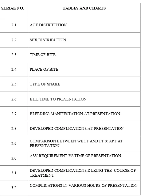
[image:12.595.99.528.132.523.2]LIST OF TABLES AND CHARTS
SERIAL NO. TABLES AND CHARTS
2.1 AGE DISTRIBUTION
2.2 SEX DISTRIBUTION
2.3 TIME OF BITE
2.4 PLACE OF BITE
2.5 TYPE OF SNAKE
2.6 BITE TIME TO PRESENTATION
2.7 BLEEDING MANIFESTATION AT PRESENTATION
2.8 DEVELOPED COMPLICATIONS AT PRESENTATION
2.9 COMPARISON BETWEEN WBCT AND PT & APT AT
PRESENTATION
3.0 ASV REQUIREMENT VS TIME OF PRESENTATION
3.1 DEVELOPED COMPLICATIONS DURING THE COURSE OF
TREATMENT
[image:13.595.88.547.97.737.2]3.3 DURATION OF HOSPITAL STAY
3.4 MORTALITY AMONG THE VARIOUS GROUPS
3.5 STATISTIC CORRELATION BETWEEN PT ,APTT AND
WBCT
3.6 STATISTIC RELATION BETWEEN THE STUDY GROUPS
WITH THE COMPLICATIONS
3.7 STATISTIC CORRELATION BETWEEN THE STUDY
GROUPS AND NUMBER OF ASV ADMINISTERED
3.8 STATISTIC CORRELATION BETWEEN THE STUDY
1
INTRODUCTION
Snake bite is of significant health concern, especially in rural
are-as of tropical and subtropical countries. Snake bite is common amongst
farm-ers, plantation workfarm-ers, construction workfarm-ers, snake charmfarm-ers, huntfarm-ers, snake
rescuers, and migrant populations in tropical and subtropical countries.
Urbani-zation and deforestation has made snake bite an important public health
prob-lem. In India, snake bite takes a lot of human lives, and therefore warrant
ur-gent attention. High mortality is due to poor rural health services and delay in
initiating anti snake venom. About 49 000 people in India die every year of
snake-bite[31], although the figure is probably under-rated because most rural
Indian population consults a traditional healers and so goes unreported. And
also Doctors at primary health centres in India have less experience in,
man-agement of snake bites. Many victims die on the journey to tertiary hospitals.
WHO’s inclusion of snake-bite envenoming in the list of category A as
ne-glected tropical diseases in early 2009[28]. It has been reported that in up to 80%
of snake bite cases in developing countries, first attend a traditional healers
be-fore consulting a medical centre [2,3]. Maharashtra has reported an incidence of
70 snake bites per 100,000 population and a mortality rate of 2.4 per 100,000
per year [4]. High incidence of snakebites cases among Indian states are Tamil
Nadu, West Bengal, Uttar Pradesh, Maharashtra, and Kerala.[1]. Though
Snake bites are observed in all age groups, but males between the aged 15-45
2
Indian Krait( Bungarus caeruleus), Russells’ viper (Daboia russelii), Saw
scale viper( Echis carinatus) are commonly encountered in India.
3
AIM AND OBJECTIVES
AIM
This study mainly focuses on the PT and aPTT as an early marker for detection
of snake bite, hematotoxicity as compared to Whole blood clotting time
OBJECTIVES
1. PT and aPTT as an early marker for detection of snake bite, hemotoxici
ty as compared to Whole blood clotting time .
2. Early administration of anti snake venom can avoid hemotoxic compli
cations, excess loading of anti snake venom and anti snake venom com
4
REVIEW OF LITERATURE
Snake bite poisoning is known to man since antiquity.
All snakes are predatory carnivores. Since snakes are preyed upon by
other animals they tend to be secretive and have evolved many survival
strate-gies. Many species are mainly nocturnal (night hunters) e.g. kraits, but other
species are mainly diurnal (day understanding about the habits of snakes,
sim-ple precautions can be adopted to reduce the incidence of snake bites. About
2500-3000 species of snakes exist in the world. Most venomous snakes are
found in Asia as compared to the other area of the world, 250 species and
sub-species, out of which 50 are poisonous. In India, out of the 216 species of
snakes, 60 are considered poisonous[36]. The families of poisonous snakes
in-clude Elapidae, Viperidae and Hydrophidae which are responsible for
neuro-toxicity, vasculotoxicity and myotoxicity respectively. Viperidae family
in-cludes two subfamilies: Viperinae (classic vipers) and Crotalinae (pit vipers).
Elapidae family includes cobras and common kraits. Hydrophidae are the sea
snakes Viper bites are more common than the other venomous snake bites in
human beings. Viper bites are responsible for vasculotoxicity leading to
bleed-ing tendencies and coagulation defects. These bleedbleed-ing diatheses are mostly
caused by consumptive coagulopathy, anti coagulation, fibrinolytic or may be
due to direct effect of snake venom on platelet aggregation. Early detection of
vasculotoxicity and prompt institution of anti venom therapy can prevent
5
absolute indication for anti-venom administration, which is the specific
treat-ment of snake bite.
Case Fatality due to snake bite can be attributed to a wide species
varia-tion, shortage of anti-snake venom(ASV), poor treatment protocols, lack of
public education and lack of proper trained health professionals.It is estimated
that there are over 1,000,000 snakebites in India alone leading to between 45
000 and 50 000 deaths annually[51] . Reliance on traditional healers and false
believe further compound the problem. High death rate cannot be attributed to
superstition and lack of awareness only, as there are a number of victims who
die after seeking medical treatment, the reason being lack of experience health
provider and non-compliance with the existing treatment protocol. Snake bite is
a medical emergency, where timely intervention can reduce both morbidity and
mortality. Lack of knowledge about simple measures of prevention,
occupa-tional hazard and inappropriate first-aid measures all magnify the problem.
Dif-ficulty in accessibility to health care services, difDif-ficulty in transportation and
subsequent delay in Anti Snake Venom administration result in high mortality.
The time since snake bite is of vital importance, because with time more venom
gets bound to the tissues, making it less manageable for neutralization by Anti
Snake Venom. Further, use of Anti Snake Venom may be avoided due to
inex-perience and fear of anaphylaxis or irrationally use of ASV when not indicated,
resulting in total wastage of resources and also exposing the patient to toxicity
risk. There is also lot of uncertainty in the dosage of Anti Snake Venom,
6
MOHFW(Ministry of Health & Family Welfare, Government of India) is in
place besides the WHO Guidelines[33,34].
Hematotoxic abnormalities are the most common manifestation of snake
envenoming global. Most of the viperid, and some elapid, venoms activate
common pathways of coagulation resulting in consumptive coagulopathy.
Snake Venom induced coagulopathy is brought about by activation of the
clot-ting pathway by procoagulant toxins, resulclot-ting in clotclot-ting factor consumption
and coagulopathy. The type of procoagulant toxin differs between snakes and
can activate prothrombin, factor V, and factor X or consume fibrinogen.
Fi-brinogen, which is the common final substrate of the coagulation cascade, is
invariably low in this condition. However, fibrinogen assay is not generally
available, especially in remote areas where snakebites are prevalent. Therefore,
alternative blood tests are required for initial evaluation, follow-ups and
moni-toring responses after antivenom therapy in snakebite victims.
A simple 20-min whole-blood clotting test (20WBCT) has been
shown to correlate with fibrinogen levels in patients bitten by Bothrops sp. in
South America[30]. Consequently, the WHO recommended this method for
evaluation of coagulopathy in snakebite patients. In addition, a standard Lee
and White venous clotting time (VCT), in conjunction with platelet counts, was
shown to be predictive for systemic bleeding in a multivariate analysis of green
pit viper bitten patients. While these tests are relatively rapid and simple, their
accuracy has rarely been evaluated. Furthermore, they are usually performed at
7
they are subject to error. Training of the treatment team is critical for the
relia-bility of these tests.
PT(Prothrombin time) and aPTT(activated partial thromboplastin
time) are better standardised tests with well-established quality control systems.
They are available in many hospitals and can be automated, thus assuring their
precision. In addition, the international normalized ratio (INR) values
harmo-nise PT tests performed in different laboratories using different commercial
re-agents. However, data on their uses in viper bitten patients are lacking. We
hy-pothesised that PT and APTT would be effective for the assessment of viper
bitten patients.
Delayed administration of ASV in case of hematotoxic snake bite
can lead to complication of consumptive coagulopathy, acute renal failure,
ex-cess administration of anti snake venom , complications of anti snake venom
administration, fresh frozen plasma transfusion and death .
While snake bite is observed in all ages, the large majority are in
males between the age of 15-45 years. The predominance of male victims
sug-gests a special risk of outdoor works.
SPECIES
There are some 2700 described species of snakes in the world
coming under 402 genera and 18 families, of these some 500 species are
ven-omous. 275 species of snakes have been described from India belonging
cate-8
gorized as ‘Venomous’ 42 as ‘mildly venomous’ and 171 as ‘non venomous’.
Of the 62 venomous species, 42 are seen on land and 20 in the sea.
In India the poisonous snakes belong to three broad families.
Family –Elapidae – (Cobras, Kraits & Coral Snakes)
Family –Viperidae –The family of Viperidae has two subfamilies , Viperidae
(Rus sell’s Viper, Saw Scaled Viper) and Crotalinae (Pit Viper)
Family – Hydrophidae –Sea Snakes
In India, ‘Four’ among the poisonous snakes are highly venomous.
1. Cobra–Najanaja(Spectacled,Cobra),
2. Russell’sViper-(Daboiarusselli )
3. Saw Scaled Viper (EchisCarinatus),
4. Krait (BungarusCaerules)
Other deadly snakes may be going unnoticed and causing death and
disability. The recent discovery of the Hump nosed Pit Viper (Hypnale
Hyp-nale) as a species capable of causing life threatening symptoms
In order to determine the actual list of medically significant species in
India, the old concept of “The Big 4” is to be abandoned for a newer more
flex-ible model that enables better classification of species. In the Indian
set-ting, almost two-thirds of bites are attributed to saw-scaled viper (as high as
95% in some areas like Jammu and Kashmir) , about one fourth to Russell's
vi-per and smaller proportions to cobra and kraits. In Sri Lanka, Daboia russellii
9
accounts for 70% bites in Myanmar. However, clinical features and outcomes
are not as simple to predict because every bite does not result in complete
en-venomation.
IDENTIFICATION
[image:23.595.114.510.239.668.2]POISONOUS OR NON-POISONOUS SNAKES
10
[image:24.595.88.525.104.661.2]Identification of Snake species
11
INDIVIDUAL SPECIES IDENTIFICATION:
SAW SCALED VIPER
Scientific Name
Echis carinatus.
Other Common Names
Carpet viper; “Phoorsa”.
Geographical Distribution
All over India (especially plains and deserts).
Physical Appearance
1. Saw-scaled viper grows upto 1½ to 2 feet long, they are usually brown in colour, with a wavy white line across the entire length of each flank, a
dia-mond-shaped markings over the back, usually numbering 25 to 30.
2. Triangular head with small scales. Whitish color arrow-shaped mark is of-ten present on the head with pupils vertical in shape.
3. Saw-scaled viper is named so because its serrated scales. When agitated, the snake throws itself into a double coil (“figure of eight”), and rubs the coils
together vigorously. At the same time by exhaling forcefully through the
nostrils producing a loud hisse.
4. Saw-Scaled viper is viviparous.
12
Habitat
This snake prefers desert regions, and is often found basking in the sun
during the daytime, among rocks or in sandy soil. It may enter human
habita-tions especially tents, in search of prey. In some parts of peninsular India, it is
very uncommon, particularly in most parts of Kerala.
Nature of Venom
Vasculo- and haemotoxic. 0.0046 gram of venom is injected at the time
13
Fig 3 : Saw Scale viper
RUSSELL’S VIPER:
This snake got its name from a Scottish herpetologist Patrick Russell
who was the first person to describe many Indian snakes. Daboia is derived
14
Scientific Name:-
Vipera russelli, Daboia russelli.
Geographical Distribution:-
All over India.
Physical Appearance
1. It is a brownish, stout snake and grows up to several feet in length. 2. The snake has a triangular head, with a ‘V’ shaped mark (apex forward
pointing), which are covered with small scales and has vertical pupil.
3. Fangs of the snake are long, channelised, and hinged .
4. The snake has 3 rows of chained dark spots over the entire body. It is known to hiss loudly when agitated. It is viviparous.
5. They are nocturnal snake, but during the daytime, it often rests up under bushes, base of trees or in leaf litter.
Nature of Venom
Predominantly vasculo- and haemotoxic, but also can produce
neuro-toxic effects . The snake has also been associated with Acute renal failure and
15
16 COMMON KRAIT:
Scientifi c Name:-
Bungarus caeruleus.
Other Common Names:-
Indian krait.
Geographical Distribution:-
All over India.
Physical Appearance
1. The snake is steel-blue in colour and can grow up to 3 to 4 feet in length, with whitish bands brown in colour.
2. They have a small dark eyes with almost invisible pupils. The upper lip is yellow or white in colour and the belly is very white.
3. A chain of hexagonal large scales is seen throughout the mid doral as-pect of the body. The ventral scales are undivided distal to the vent,
un-like other elapids. The fourth infralabial scale is the largest of the
in-fralabial.
Habitat
The common krait is a reclusive snake which prefers to reside in crevices of rocks or logs of wood, and being nocturnal, emerges only during
the night to hunt for prey. Its primary diet is other snakes. It can be found all
over Peninsular India and often seeks habitation near human dwellings.
The common krait may enter houses and hide in dark corners,
whis-17
tling sound. These snakes prowl on hot humid nights; they often do not strike,
butmake a quick snapping bite.
Nature of Venom
Predominantly neurotoxic. It is the most venomous snake of India.
[image:31.595.108.542.171.693.2]
18 INDIAN COBRA :
Scientific Name:-
Naja naja.
Other Common Names:-
Indian Cobra.
Geographical Distribution
All over India.
Physical Appearance
1) It is usually brown or black in colour.
2) It can grow up to 5 to 6 feet in length. The neck is distensible that can be expanded into a hood. Dorsal side of the hood, there may be a
mo-nocellate or bimo-nocellate mark. It has alternate wide and narrow,
trans-verse, dark bands. Dorsal hood is mark by a pale circle edged with black
and has 1 to 3 spots; ventral hood mark has a pair of dark spots, or a
wide dark band.
3) The hood markings is the hallmark of cobra, and its habit of rearing up when alarmed.
4) Ventral surface of the hood are faint, broad, black stripes above which are two dark spots that extend over 3 to 4 scales.
19
Habitat
Grassy plains, fields, and mountainous regions (up to 15000 feet).
They usually reside among piles of bricks, termite mounds, tangles of roots at
the base of trees, and old masonry constructions. The spectacled cobra is
en-countered virtually over the whole of mainland India except the north-east. The
black cobra (Naja oxiana) occurs in the extreme north of India around Jammu
and Kashmir, and also in Gujarat and Rajasthan, although these may be
pattern-less versions of the spectacled cobra. The cobra is diurnal, but bites from
co-bras occur during both the day and the night. The cobra’s principal diet is rats.
It is known to enter human habitations in search of prey.
Nature of Venom
Cobra venom is cardio toxic, neurotoxic, haematotoxic and cytotoxic.
20
[image:34.595.108.507.70.474.2]
Fig 6 : Common Cobra
SNAKE VENOM APPARATUS
The typical snake venom delivery apparatus consists of bilateral venom
glands situated below and behind the eyes and connected by ducts to hollow
anterior maxillary fangs. In viperids (vipers and pit vipers), these fangs are long
and highly mobile; they are retracted against the roof of the mouth when the
snake is at rest and brought to an upright position for a strike. In elapids, the
fangs are smaller and are relatively fixed in an erect position. Approximately
21
for sea snakes) are “dry” bites, meaning no venom is released. Significant
en-venomation probably occurs in ~50% of all venomous snakebites. Elapidae and
Viperidae venom glands are located behind the eye which are surrounded by
compressor muscles. At the base of fangs, venom duct opens where the venom
are transported to its tip within a canal like a hypodermic needle. The snake can
introduce the venom deep into the tissues of its prey with the help of its fang.
In case of human bite, venom is usually injected subcutaneously or
intramuscu-larly .Spitting cobras can squeeze its venom from their fangs like a fine spray
[image:35.595.177.455.345.690.2]directed towards the victim’s eye.
22
VENOMS
Snake Venom is a complex fluid with powerful ingredients and is
secreted by the parotid glands, mainly for immobilizing, killing & digesting
small animals like rats. Snake venoms are highly variable and complex
mix-tures of enzymes, low-molecular-weight polypeptides, glycoproteins, and other
constituents. Among the deleterious components are hemorrhagins that
pro-mote vascular leakage and cause both local and systemic bleeding Proteolytic
enzymes cause local tissue necrosis, affect the coagulation pathway at various
steps, and impair organ function. Hyaluronidases promote the spread of venom
through connective tissue. Myocardial depressant factors reduce cardiac output,
and bradykinins cause vasodilation and hypotension. Neurotoxins act either
pre- or postsynaptically to block transmission at the neuromuscular junction,
causing muscle paralysis. Most snake venoms have multisystem effects on their
victims. Snake can bite once and continuously secrete the venom a number of
times in succession. The lethal dose of the venoms for a man (Deoras 1965).
Cobra - 0.12 g
Krait–0.06g
Russell’sViper - 0.15 g
EchisCarinatus- 0.08 g
The concentration of venom shows diurnal and seasonal variation.
Bites inflicted at night and immediately after hibernation are the most severe.
In summer months the output of venom is more than in the winter, when the
high-23
er mortality rate by Snake bites in the summer months including the monsoon.
Most snakes inject about 10% of the available venom in single strike. It has the
most complex of all venoms. More than 90% dry weight is protein comprising
a rich variety of enzymes, non-enzymatic polypeptide toxins, and non-toxic
proteins. Non-protein ingredients of venom include carbohydrates and metals
(often in the form of glycoprotein metalloprotein enzymes), lipids, free amino
acids, nucleotides, and biogenic amines. The lethal and more deleterious
frac-tions of snake venoms are certain peptides and proteins of relatively low
mo-lecular weight (6,000 to 30,000). The peptides appear to have very specific
re-ceptor sites, both chemically and physiologically.
The polypeptide toxins (often called neurotoxins) are found most
abundantly in elapid and hydrophid venoms. Postsynaptic alpha neurotoxins
such as alpha bungarotoxin and cobrotoxin contain about 60 to 70 amino acid
residues, and bind to acetylcholine receptors on the motor end-plate.
Pre-synaptic beta neurotoxins such as beta-bungarotoxin, cobrotoxin, and taipoxin
contain about 120–140 amino acid residues, and a phospholipase A subunit,
and prevent release of acetylcholine at the neuromuscular junction. Cobra’s
al-pha bungarotoxin, binds to the acetylcholine receptors and inhibits neural
transmission at the neuromuscular junction. Krait’s beta bungarotoxin causes
an initial release of acetylcholine, but then damages the nerve terminal and
prevents any further release. It is for this reason that krait victims often take
longer to recover than cobra victims. The acetylcholinesterase found in most
24
Enzyme function and path physiological disturbances are most
clearly related in the case of viper venom pro-coagulants. For instance,
Rus-sell’s viper venom (RVV) contains at least two proteases, which activate the
blood-clotting cascade. RVV-X, a glycoprotein, activates factor X by a
calci-um-dependant reaction, and also acts on factor IX and protein C. RVV-V, an
arginine ester hydrolase, activates factor V. Echis venom contains a zinc
metal-loprotein “ecarin” which activates prothrombin. Russell’s viper can induce
neu-rotoxic symptoms in addition to haematological abnormalities. Many species of
Russell’s viper have this ability, and it is particularly evident in Southern India
and Sri Lanka.
Hyaluronidase may serve to promote the spread of venom through
tissues. Proteolytic enzymes (hydrolases) may be responsible for local changes
in vascular permeability leading to oedema, blistering, and bruising, and to
ne-crosis. Biological amines such as histamine and 5-hydroxytryptamine may
con-tribute to local pain and permeability changes at the site of a snakebite.
Non protein ingredients of venom include carbohydrate and
met-als (often part of glycoproteins & metalloprotein enzymes) lipids, free amino
acids, nucleosides and biogenic amines such as serotonin and Acetylcholine.
About 80–90% of Viperidae & 25%to70% of Elapidae Venom consists of
en-zymes. The role of enzymes in envenoming is most clearly seen in the case of
25
Enzymatic components in Snake Venom
ENZYME EFFECTS
Arginin hydrolase Bradykinin release, interference with clotting
Collagenase Digestion of Collagen
Hyaluronidase A
Reduction of Collagen Viscosity. Promotes the
spread of venon through tissues
Phospholipase A Uncoupling of oxidative phosphorilation
Phospholipase B Hydrolysis of lysophosphatids
Phosphodiesterase Inhibition of DNA, RNA, arabinose derivatives
Acetylcholinesterases
Catalysis hydrolysis of Ach. But this is no longer
thought to contribute to their neurotoxicity
5’ nucleotidiase
Specific hydrolysis of Phosphate mono-esterases
which links with 5 position of DNA, RNA
L-aminoacid oxidase
Catalysis of aminoacid oxidation gives colour of
venom.
26
Non-Enzymatic components in Snake Venom
COMPONENT EFFECTS
Neurotoxins (elapidae) Post synaptic non depolarizing neuromuscular.
Cobrotoxin blockade of long duration, acting only on nicotinic acetyl choline receptors to some extend cardiotoxic.
Erabutoxin Acetyl choline receptors to some extend cardiotoxic
Alpha Bungarotoxin Haemotoxic and anticoaguland-blinds to receptors.
Cerelotoxin Similar post synaptic block but without binding to receptors
BetaBungarotoxin Crotoxin
Taipoxin
Pre-synaptic motor nerve end blockade
Haemorrhagins Direct disruption of vessel endothelium
(HR-1,HR-2)
Viperidae, Crotalidae
27
Fig 8 : features of different snake bite
ALTERATION IN COAGULATION FOLLOWING SNAKE BITE:
Snake venom is complex toxin or poison. But it’s mixture of
vari-ous components such as proteins, enzymes, non-toxic proteins, nucleotides,
carbohydrates, lipids, biogenic amines and nucleotides.
Russell’s viper and Echis carinatus bites can result in defects in
co-agulation and bleeding. Viper venom contains many active substances, which
can induce bleeding and induce clotting. Snake venom containing Hemorrhagin
can damages the blood vessels directly, by loosening the gaps between the
en-dothelial cells and thereby leading to the injury of the capillary basement
28
In-vivo, large amount of venom leads to massive intravascular
clotting, which can stop circulation and results in rapid death. But in case of
snake bite the venom is less in quantity and it leads to continuous activation of
fibrinogen which in turn produces fibrin which is fragile and more susceptible
to lysis than the ordinary fibrin. As the venom destroys fibrinogen at a faster
pace then liver produces, the blood tends to clot poorly or it fails entirely. The
balance between this anticoagulants, procoagulants, fibrinogenolytic and
fibri-nolytic components of the injected venom determines the final state of
coagula-tion disturbance.
Russell’s viper venom selectively activates Factor X. Echis
car-inatus venom accelerate the conversion of prothrombin into abnormal thrombin
and also activates Factor X. Thus the abnormal thrombin prevents the
stabiliza-tion of fibrin and also promotes coagulastabiliza-tion which is achieved by stimulastabiliza-tion
of plasminogen system and by inhibition of factor XIII activity. The result is
similar to that of DIC, fibrinolysis and increased Factor V consumption. Viper
poisoning usually shock and hemorrhage resolves within a week, but
coagula-tion changes tend to persist for 2 weeks or longer in case the specific anti snake
venom is not given on time. In more than 50 % of patients, Intravascular
he-molysis was present amongst patient who presented with ARF- Acute renal
failure, jaundice, anemia, reticulocytosis and hemoglobinuria and raised plasma
29
Platelet dysfunction and thrombocytopenia are commonly seen
be-cause of the various proteins present in this venom which can directly destroy
the platelets and also can cause a functional impairment of the platelets.
Haemorrhagins (HR –1 & HR – 2)
Haemorrhagins, two immunologically distinct non enzymatic
haemorrhagic principles (HR – 1 & HR – 2) are typical components of
Crotal-id(Pit viper) and Viperid (true Viper) venoms. They cause acute, rapid
haemor-rhage. In many cases of serve envenomation, haemorrhagins play a lethal
role by causing haemorrhage in the kidneys, heart, Brain, Lungs, and
gastro-intestinal tract. Pharmacologically, the haemorrhagins have been demonstrated
to be separate entities from proteolytic enzymes in the venom.
The haemorrhagins act by directly disrupting the endothelial
lin-ing and by inhibitlin-ing platelet aggregation. Pharmacological studies have further
shown that the haemorrhagic principles induce the release of certain
autophar-macological mediators such as histamine and 5-HT from various tissues which
open up endothelial cell junction and disrupt the isolated basement membrane,
presumably in an enzymatic mode of action thus causing vascular damage and
haemorrhage. Similarily vasculotoxic changes have also been observed in the
renal and cerebral vessels with crotalid venom. The observed vasculotoxic
changes resulting in severe cutaneous and systemic haemorrhage particularly in
kidneys, lungs and brain bear a close resemblance to that of experimental
30
PATHOGENESIS OF SNAKE BITE:
31
NON-VENOMOUS SNAKEBITE
A significant proportion of snakebites is said to be due to
non-venomous snakes. Since the question of envenomation does not arise in such
cases, systemic manifestations are nonexistent, except those due to
psychologi-cal shock. As a result of the fear and apprehension associated with snakes,
eve-ry bite (venomous or otherwise) is attended by some degree of shock
character-ised by giddiness, syncope, sweating, palpitation, tachycardia, and hypotension.
These emotional manifestations develop almost instantaneously and may
pro-duce psychological shock and even death. Fear of snake bite may cause also
transient pallor, sweating and vomiting. Consequent upon reassurance
especial-ly by a doctor, about the non-venomous nature of the bite, these symptoms
usually resolve rapidly.
VENOMOUS SNAKEBITE
Based on the predominant constituents of venom, snakes were
loosely classified as neurotoxic (notably cobras and kraits), vasculotoxic
(vi-pers) and myotoxic (sea snakes). However it is now well recognized that such a
strict categorization is not valid as each species can result in any kind of
mani-festations.
Without Envenomation:
a. 20 to 50% of venomous bites does not have serious toxicity.
b. The reasons for lack of envenomation in bites of venomous snakes are as
32
Dry bite: venoms are not always injected in every bite.
Protective gear: bites inflicted on shod feet and heavily clothed parts,
Envenomation may not occur .
Leakage of venom: all or some of the venom escape outside the bite site
when snake sidewipes.
Superficial bite:. The snake often does not bite deeply deliberately, but
instead only strikes superficially because humans are not normal prey
for most of the snakes, thereby conserving precious venom for its
genu-ine prey and they bite only to defend itself.
With Envenomation:
a. Elapid Bite
Local Effects: Pain and swelling, serosanguinous oozing from the bite
site with mild tenderness, and blistering. Cobras can sometimes cause
significant local swelling, pain, blistering, and regional
lymphadenopa-thy.
Systemic Effects: The dominant clinical feature of elapid bites is
Neuro-toxicity. It can occur within 15 minutes to ½ hour in cobra bite, while
krait bite it is often delayed up to several hours.
Pre-paralytic Stage
Vomiting Ptosis
33 Headache, myalgia
Vertigo
Paraesthesiae around the mouth Hypersalivation.
Paralytic Stage
progressively flaccidly paralysed of facial muscles, vocal cords, neck muscles, palate, jaws, tongue, and muscles of deglutition.
Respiratory arrest can occur due to paralysed tongue or inhaled vomitus, or paralysis of intercostals muscles and diaphragm.
Bedside tests to identify impending respiratory failure .
1. SINGLE BREATH COUNT : in one exhalation the number of digits
counted - should be > 30 .
2. BREATH HOLDING TIME : Breath held in inspiration - normal count
is > 45 second.
3. Ability to complete one sentence in one breath .
Convulsions and loss of consciousness as result of chronic hypoxaemia. Roughly 50% of patients bitten by monocellate cobra(Naja kaouthia )
do not sustain envenomation.
Rarely elapid bites cause renal failure.
34 b. Viperid Bite
Local Effects:
local manifestations usually occurs within half an hour of bite, but may be
delayed for several hours as well.
First Swelling will appears around the snake bite site, and then spreads to
the adjacent area and quickly to involve the entire limb and adjacent trunk,
as-sociated with pain, tenderness, and regional lymphadenopathy.
In about 12 hours Blisters begin to appears around the bite site which
subsequently progress to involve the entire limb. About 10 to 15% of the cases
extensive necrosis of skin, subcutaneous tissues, and muscles can develope.
Raised intracompartmental pressure , causing compartment syndrome.
Systemic Effects:
Haemostatic abnormalities are very characteristic. Evidence is first seen
as persistent bleeding from the bite site. Within a few hours of the bite
hematu-ria develops then gingival bleeding, then followed by epistaxis. Hours later
bleeding into the floor of the mouth, haemoptysis, haematemesis, ecchymoses,
intracranial and sub-conjuctival haemorrhages, and tympanic membrane,
gas-trointestinal tract, and genito-urinary tract bleed can occur. Few cases of
Ante-rior pituitary bleeding has been reported. Hemiplegia, loss of consciousness,
and convulsions can occur as a result of Subarachnoid and intracerebral
haem-orrhage may. Cases of Retroperitoneal and intraperitoneal haemhaem-orrhages have
35
Intravascular haemolysis producing haemoglobinuria and renal failure is a
frequent occurrence, especially in Russell’s viper bite .
Hypotension accompanied by tachycardia, unless the snake venom has
affected the heart directly or reflexly.
Like Cardiotoxicity seen in elapid , it can also produces a wide variety of
ECG.
Neurological symptoms may occur in the case of Russell’s viper bite like
ptosis, respiratory failure and paralysis.
LIFE THREATENING COMPLICATIONS :
ACUTE KIDNEY INJURY
Acute kidney injury (AKI) is an important complication of snake
bite and a major cause of mortality. AKI is common after bites from myotoxic
or hemotoxic snakes. These snakes are Russell’s viper, saw-scaled viper,
hump-nosed pit viper, green pit viper, and sea-snake. Renal pathologic changes
include tubular necrosis, cortical necrosis, interstitial nephritis,
glomerulone-phritis, and vasculitis. Hemodynamic alterations caused by vasoactive
media-tors and cytokines and direct nephrotoxicity account significantly for the
de-velopment of nephropathy. Hemorrhage, hypotension, disseminated
intravascu-lar coagulation (DIC), intravascuintravascu-lar hemolysis, and rhabdomyolysis enhance
renal ischemia leading to AKI[23]. The incidence of AKI caused by these
snakes varies from 5% to 29% depending on the species of snake and the
36
96 h after the bite. The duration of AKI after snake bite generally ranges from 2
to 3 wk. Tubular necrosis is an important pathological correlate of AKI.
Pro-longed AKI with oligoanuria after snake bite is indicative of cortical necrosis
or acute tubular necrosis associated with interstitial nephritis or extracapillary
glomerulonephritis[23].
LONG TERM COMPLICATIONS OF SNAKEBITE
1. Chronic ulceration, osteomyelitis, arthritis.
2. Malignant transformation (Marjolin’s ulcer) .
3. Chronic kidney disease.
4. Hypopituitarism or Diabetes Insipidus may occur after Russell’s viper
bite.
5. Chronic neurological deficits in patients who survive intracranial
haem-orrhages and thrombosis.
6. Psychological disturbances such as depression, anxiety, post traumatic
stress disorder.
7. Chronic musculoskeletal disabilities such as muscle wasting, stiff joints,
37
OCCULT SNAKE BITE :
Identification of Krait bite marks is difficult as Krait has fine slender
teeth and a nocturnal habitat. Sometimes in the early morning patient may
come with complains of paralysis with no other symptoms in Krait bite cases.
They will have typical history of going to bed at night and getting up in the
morning with severe epigastric pain and vomiting following which the patient
developed neuroparalytic symptoms with no history of snake bite. Krait bite
produces descending neuroparalysis whereas GBS produces ascending
paraly-sis.
FIRST AID MEASURES :
History of snake bite should be checked and evidence of snake bite like fang marks , bleeding , swelling of bitten part should be check .
HISTORY :
a) history about the events occurred during bite and the progression of
lo-cal and systemic symptoms and signs.
b) Many of the non venomous species leave just two fang-like marks on
biting, so Bite marks are of little importance.
c) To Determine the exact time of snake bite. As in case of early
presenta-tion of snake bite, the patient may have only few symptoms and signs
38
d) Patient should be asked about the quantity of urine he passed. Patients
with neurotoxic envenomation may complains of drooping of eyelids,
blurring of vision, double vision or sleepiness.
e) As far as possible the snake responsible must be identified.
f) History of any traditional medicine was used for snake bites before
reaching hospital as it can cause problems and produce symptoms that
confuse the diagnosis.
Reassured to patients that 70% of all snakebites are non-venomous snakes.
Limb rest and immobilization using bandage or cloth to hold the splint without blocking the blood supply.
Discard Traditional first aid measures as it will do more harm to pa-tients.
Loosening and removing all Tight objects like clothing, shoes, watches, rings since it can increase the swelling.
Anti Snake Venom should not be injected locally.
39
The mnemonic recommended for snake bite, first aid “ Do it R.I.G.H.T ” which
is
R = Reassure the patient as 70% snake bites are non venomous and
only 50% of bites by venomous species envenomate the patient.
I = Immobilize the limb in the same way for fractured limb.
GH = Get to Hospital immediately. Traditional remedies should be
avoided.
T = Tell the doctor about any systemic symptoms such as ptosis that
manifest on the way to hospital.
ASSESSMENT:
Time since the snake bite should be determined.
Any Medical history like presence of systemic diseases, allergy, use of
medica-tion.
PHYSICAL EXAMINATION:
Patient should be monitored closely, the following parameters:
o Pulse rate
o Respiratory rate
o Blood pressure
o Oxygen saturation
o 20 minutes Whole Blood Clotting Time, every hour for first 3 hours
40
o Examination of distal pulses and capillary filling time in the presence
of gross swelling. Pain on passive movement, pallor , pulse less limb,
hypoesthesia one should suspect the diagnosis of compartment
syn-drome . Compartment pressure is measured by inserting a 16 G IV
cannula and connecting it to manometer if it reads above 40 cm
wa-ter then the diagnosis of compartment syndrome should be made.
EARLY SIGNS OF SEVERE ENVENOMING:
Rapid extension of local swelling.
Early tender enlarged lymph nodes, indicates the spread of venom to
lymphatic.
Neurological signs like ptosis, neck muscle weakness and respiratory
distress .
Bleeding manifestations like bleeding from bite site, gums, ecchymoses,
haematuria, hemoptysis and epistaxis.
Dark brown urine.
LAB TESTS:
20 MINUTE WHOLE BLOOD CLOTTING TIME (20 WBCT):
Prothrombin time
OTHER TESTS:
a. Complete Blood Count
41 c. Liver function test
d. Renal Function test
e. Blood sugar
f. ECG
g. Abdominal ultrasound
Anti Snake Venom(ASV):
1. ASV is the only specific antidote for snake bite.
2. No absolute contraindication to ASV.
3. ASV may reverse systemic envenomation.
4. Polyvalent ASV is available in India which is effective against all four
common species (Russell’s Viper, Common Cobra , saw -scaled viper and
common Krait).
5. ASV are produced in two forms liquid and lyophilized forms. Liquid ASV
should be stored in cold chain and has shelf life of 2 years. Lyophilised
ASV, in powder form, has shelf life of 5 years and can be stored in cool
place.
6. Anti Snake Venom contains equine immunoglobulin fragments F(ab’)2
prepared from plasma of horses immunised with venom of one or more
42
7. Anti Snake Venom can be either Monovalent or Polyvalent. Monovalent
ASV neutralizes, venom of only one snake species whereas Polyvalent
ASV neutralizes the venom of several different snake species.
8. One ml of polyvalent ASV has the capacity of neutralizing the venomof
the following:
a. 0.60mg of dried Indian Cobra (Naja naja) venom.
b. 0.45mg of dried Common Krait (Bungarus Caeruleus )
ven-om .
c. 0.60mg of dried Russell’s Viper (Daboia russelii ) venom.
d. 0.45mg of dried Saw-scaled viper (Echis carinatus) venom .
9. The antitoxic equine Immunoglobulin are obtained from the serum of
healthy equines which are immunized against venoms of various snake species.
10. ASV must be given only by Intravenous route, and should be given slowly.
11. Adrenaline should always be kept ready before ASV is administered.
12. ASV should not be given by Intramuscular route due to poor
bioavailabil-ity.
DOSE OF ASV FOR NEUROPARALYSIS:
For Neuroparalysis 10 vials of Anti Snake Venom is given as IV
infu-sion over 30 minutes. Second dose of 10 vials can be given if there is no
43
DOSE OF ASV FOR VASCULOTOXIC SNAKE BITE.
Two regimens are followed for vasculotoxic snake bite.
o Low Dose infusion therapy:
10 vials of ASV for Russel’s Viper ; 6 vials of ASV for saw scaled
vi-per as a stat infusion over 30 minutes followed by 2 vials of ASV for every 6
hours as an infusion in 100 ml of normal saline , till the clotting time
normaliz-es or for 3 days whichever is earlier.
o High bolus therapy:
10 vials of ASV as a stat over 30 minutes as an infusion followed by 6
vials of ASV every 6 hourly as a bolus until clotting time normalizes .The
amount of venom injected is about 5mg to 14.7mg. The maximum Anti Snake
Venom dose is 30 vials since each vial neutralizes 6mg of Russell’s viper
ven-om .
ASV REACTION :
Anti Snake Venom test dose is not needed.
Some patients may develop anaphylaxis, characterized by hypotension,
bron-chospasm, and angioedema .
EARLY ANAPHYLACTIC REACTION TO ASV
Occurs within 10 to 180 min of starting ASV. They may present with
itching, urticaria, dry cough, nausea, vomiting, abdominal colic and
44
PYROGENIC REACTIONS
Develops within 1 to 2 hours after treatment, patient may present with
fever, chills and rigors and hypotension.
Any new onset of sign or symptom after starting the Anti Snake Venom should
be considered as ASV reaction.
LATE REACTIONS TO ASV
It develops within 1 to 12 days of treatment initiation. They presents
with fever, vomiting, diarrhea, urtricaria, arthralgia, myalgia, nephritis and
en-cephalopathy rarely.
TREATMENT OF EARLY ASV REACTION :
1. Adrenaline must be given intramuscular, an initial dose of 0.5mg for
adults. Adrenaline has been proven safe in pregnant women, but
anaphy-laxis can induce abortion. Since life threatening anaphyanaphy-laxis evolve very
rapidly, adrenaline must be given at the very first sign of ASV reaction.
Dose must be repeated in every 5 to 10 minutes, if the reaction persists
or worsen.
2. Antihistamine like Chlorpheniramine maleate should be given through
intravenous route over few minutes.
45
UNRESPONSIVE TO INTRAMUSCULAR ADRENALINE IN
ANA-PHYLAXIS
1. Some patients who don’t respond to the repeated doses of adrenaline,
and who remain shocked must be made to laid supine and, with their
legs elevated above the ground, the patient also be given intravenous
volume replacement with 0.9% saline , usually 1-2 liters rapidly .
2. Adrenaline should be given as IV infusion [adult dose 1 mg in 250ml of
5% dextrose / 0.9% saline in 4 ugm /ml concentration; infused at 1-4
ugm/minute at 15 to 60 drops/min). Infusion rate can be increased up to
10 ugm/min.
3. If the patient still remains in shock, a vasopressor agent like Dopamine
should be administered at the rate of 2-5 ugm/kg/min.
RECURRENT SYSTEMIC ENVENOMATION:
In some cases systemic envenomation has been observed in 24 to 48
hours of initial recovery, probably due to continuous absorption of venom from
the bitten site which occurs after improvement in blood supply, following
cor-rection of shock and hypovolemia, and also after the disappearance of Anti
Snake Venom from the circulation. This kind of cases are rare in India due to
the prolonged half-life of polyvalent Anti Snake Venom. Also due to
redistri-bution of venom from tissue into vascular space as a result of ASV therapy. So
ideally watch out for recurrent envenomationt by observe the patient for the
46
ACUTE KIDNEY INJURY
DETECTION OF ACUTE KIDNEY INJURY
When Urine output is < 0.5ml/kg/hr for > 6 hours.
When Increased in Creatinine concentration >0.3 mg/dl or increasing by
1.5 to 2 times from the baseline Creatinine value.
Clinical “Uraemia syndrome” defined by nausea, vomiting, hiccups,
drowsiness, acidotic breathing, flapping tremor, pericardial friction rub,
muscle twitching and signs of hypervolumia.
TREATMENT OF ACUTE KIDNEY INJURY
Conservative management will avoid the need of renal replacement
therapy.
If the patient has postural hypotension suggestive of intravascular volume
de-pletion, the following should be given:-
1. Intravenous access.
2. Cautious fluid in adult patient should be given. About 250-500 ml of
iso-tonic saline challenge be given over 1 hour and the patient must be closely
monitored for the development of pulmonary edema. Fluid must be stopped
if there are signs of pulmonary edema.
3. If there is no improvement in urine output then Frusemide stress test must
be performed. Once adequate fluid replacement is given to the patient, 1 to
47
at the rate of 4-5mg/minute and urine output should be monitored for 2
hours. If the output is less than 200 ml in 1 hour, then it denotes the
pro-gression to acute renal injury. If there is no improvement in output of urine
despite these challenge, diuretics should be stopped and fluid intake
should be restricted and should be promptly referred to a renal unit .
4. Patients with Acute renal injury should be monitored daily for urine output,
urea, creatinine, electrolytes, pH, bicarbonate, calcium and phosphate.
INDICATION FOR DIALYSIS
1. Clinical uraemia like encephalopathy, pericarditis, gastrointestinal bleed
and pulmonary oedema.
2. Patients with hypervolumia and not responding to diuretics.
3. Severe Hyperkalemia; plasma potassium > 7mmol/l.
4. Sever Symptomatic Acidosis.
5. Serum Creatinine > 4mg/dl ; blood urea > 130 mg/dl
OCCUPATIONAL RISK AND OTHER ECOLOGICAL FACTORS:
The normal perception is that rural agricultural workers are most at risk
and the bites occur first thing in the morning and last thing at night. However,
this is of very little practical use to rural workers in preventing snake bite since
it ignores the facts.
In rubber, coconut and arecanut plantations, clearing the base of the tree
48
• Harvesting high growing crops like Millet which requires attention
focused away from the ground.
• Rubber tapping in the early hours – 03-00 a.m. to 06-00 a.m.
• Vegetable harvesting and fruit picking
• Tea and coffee plantation workers are at risk of arboreal and
terres-tial vipers bite when picking or tending bushes
• Clearing weeds, exposes workers to the same danger as their grass
cutting colleagues.
• Walking at night without a torch, barefooted accounts for a
signifi-cant number of bites.
• And Bathing in ponds, streams and rivers, in the evening. Cobras
and other venomous species are good swimmers and may enter the
water to hunt so one should not be assumed that because the victim
is bitten in water that the species is non poisonous.
Walking along the edge of water ways.
PREVENTIVE MEASURES:
To wear Closed type footwear at night along with a flashlight which is switched on.
49
While collecting wood, Paying close attention to the leaves and sticks on the ground.
Avoid Keeping the rubbish and animal feeds near the house since they may attract rats which are the prey for the snakes.
Avoiding a sleep on the ground.
Extrinsic, Intrinsic and Common Coagulation pathway-
A basic understanding of coagulation pathway is required to interpret
prothrombin time result. The Prothrombin time is measure of the integrity of
extrinsic and common pathways of coagulation cascade. This consists of tissue
factor and factor V, II(Prothrombin), V, X and fibrinogen. The test is
per-formed by adding calcium and thromboplastin, an activator of the extrinsic
pathway to the blood sample then measuring the time (in seconds) required for
blood clot formation.
Clotting time
Clotting time is the time required for a sample of blood to coagulate in
vitro under standard conditions.
There are various methods for determining the clotting time, the most
common being the capillary tube method. Normal value of clotting time is 5 to
8 minutes. It is affected by calcium ion levels. Other methods for measuring
clotting time are slide method.
Clotting is a natural defence mechanism to prevent blood loss from the
50
vessel is cut there is a rush of platelets causes a cut or injury to be filled and
thus bleeding stops.
Extrinsic, Intrinsic and Common Coagulation pathway
Clotting time test-
In order for blood to clot, the enzyme thrombin must be generated
from the plasma precursor prothrombin. Thrombin then converts soluble
fi-brinogen into insoluble fibrin. Generation of thrombin involves the sequential
activation of a number of other plasma clotting factor, this process is also being
assisted by Ca++ and by factors released by platelets and damaged tissues . The
time taken for blood to clot mainly reflects the time required for the generation
of thrombin in this manner. If the plasma concentration of prothrombin or of
some of the other factors is low (or if the factor is absent, or functionally
51
"WHOLE BLOOD COAGULATION (CLOTTING) TEST
(LEE-WHITE)"
Principle: The whole blood clotting test is a rough measure of all
intrin-sic clotting factors in the absence of tissue factors. Variations are wide and the
test sensitivity is limited. Whole blood, when removed from the vascular
sys-tem and exposed to a foreign surface, will form a solid clot. Within limits, the
time required for the formation of the solid clot is a measure of the coagulation
system.
20 Minute Whole Blood Clotting Test
The 20 Minute Whole Blood Clotting Test provides a simple method of
testing for coagulopathy in the envenomed patient, in circumstances where
more sophisticated hematology is unavailable.
The equipment needed for the test consists of:
Cotton wool or gauze pad
Sterile syringe and needle
Gloves
Sharps disposal container
Sterile (or clinically clean) 10ml test tube or which should be dry and
should not be washed with soap.
52
PROCEDURE
Following normal venepuncture procedure, take 20ml of venous blood
from the patient and place in the bottle.
Replace the lid of the bottle.
Place the bottle in a safe location, where it is not likely to get bumped or
knocked over. The location of the bottle should be as close to room temperature
as conditions allow.
Wait twenty minutes for the blood to clot. During this time do not shake
the bottle or disturb the contents in any way.
After twenty minutes, gently invert the bottle, observing the contents.
From this procedure it should be evident whether or not the blood has clotted.
Clotted blood is a negative result, indicating that there is no
coagulopa-thy present. Blood which remains unclotted after 20 minutes shows the
pres-ence of coagulopathy. This does not, however, indicate whether the cause is a
53
Limitations-
(a) The following variables tend to decrease the clotting time:
1) Rough handling of the blood specimen,
2) Presence of tissue fluids (traumatic venipuncture)
3) Frequent tilting of the tube, and unclean tubes.
(b) The following variables tend to increase the clotting time:
1) Extreme increases in temperature,
2) Variation in pH
3) Performance of the test at room temperature.
(c) This test is of value primarily as it was used to follow heparin therapy.
Its use as a screening procedure is limited due to its poor sensitivity.
(d) The whole blood clotting time is affected mainly by defects in the
intrin-sic pathway factors and by defects in fibrin and fibrinogen. It is not
sen-sitive to platelet abnormalities.
(e) A prolonged clotting time immediately indicates impaired coagulation,
but a normal clotting time does not exclude many serious clotting
de-fects.
(f) One disadvantage of the whole blood clotting time is its relative lack of
reproducibility.
(g) This procedure has been replaced in most laboratories with the PT,
54
(h) The coagulation time is normal in thrombocytopenic purpura.
This is explained by the fact that only a small number of thrombocytes
need be present for normal coagulation to take place.
SUMMARY
(1) A standardized test for determination of clotting time is described.
(2) It is easily performed by untrained workers with inexpensive
material, and gives reproducible and accurate results.
(3) Since the possibility of subjective error is minimal, comparison of the
results of different workers is possible
PROTHROMBIN TIME
The prothromb in time (PT) tests the adequacy of the extrinsic and
common coagulation pathways. It represents the time needed for plasma to clot
in the presence of an exogenously added source of tissue thromboplastin (e.g.,
brain extract) and Ca2+ ions. A prolonged PT can result from a deficiency of
factors V, VII, or X, prothrombin, or fibrinogen.
Normal range
The reference range for prothrombin time is 12-13 seconds. The normal
values listed here-called a reference range-are just a guide. These ranges vary
55 METHODOLOGY
A sample of the patient’s blood is obtained by venepuncture. The
pro-thrombin time is most commonly measured using blood plasma. Blood is
drawn into a test tube containing liquid sodium citrate, which acts as an
antico-agulant by binding the calcium in a sample. The blood is mixed and then
cen-trifuged to separate blood cells from plasma. In newborns, a capillary whole
blood specimen is used.
The plasma is analyzed by a biomedical scientist on an automated
in-strument at 37°C, which takes a sample of the plasma. An excess of calcium is
added (there by reversing the effects of citrate), which enables the blood to clot
again. For an accurate measurement the proportion of blood to citrate needs to
be fixed; many laboratories will not perform the assay if the tube is under filled
and contains a relatively high concentration of citrate. If the tube is under filled
or overfilled with blood, the standardized dilution of 1 part anticoagulant to 9
parts whole blood is no longer valid. For the prothrombin time test the
appro-priate sample is sodium citrate tube, which is a liquid anticoagulant.
Tissue factor (also known as factor III) is added, and the time the sample
takes to clot is measured optically. Some laboratories use a mechanical
meas-urement, which eliminates interferences from lipemic and icteric samples. The
patient’s results are not regarded as abnormal unless the clotting time is more
than two seconds longer than the control time. The prothrombin ratio is the
pro-thrombin time for a patient, divided by the result for control plasma. The upper