“OUTCOME OF PULMONARY HYPERTENSION IN
POST RENAL TRANSPLANT RECIPIENT”
DISSERTATION SUBMITTED IN PARTIAL FULFILLMENT OF THE REGULATIONS FOR THE
AWARD OF DM IN NEPHROLOGY
DEPARTMENT OF NEPHRLOGY
PSG INSTITIUTE OF MEDICAL SCIENCES AND RESEASRCH
THE TAMILNADU Dr. M.G.R MEDICAL UNIVERSITY, CHENNAI
TAMILNADU, INDIA
CERTIFICATE
PSG Institute of Medical Sciences & Research
Coimbatore
This is to certify that Dr. N.SIVA has prepared this dissertation entitled
“
OUTCOME OF PULMONARY HYPERTENSION IN POST RENALTRANSPLANT RECIPIENT”
under my overall supervision and guidance
in PSG Institute of Medical Science and Research, Coimbatore in partial
fulfillment of the regulations of The TamilNadu Dr. M.G.R Medical
University for the award of DM Neurology.
DR.G.VENU MD, DM., DR.S. RAMALINGAM MD.,
Professor and Head of the Department Principal
Department of Nephrology PSG IMS & R
PSG IMS & R
Place: Coimbatore
DECLARATION
I hereby declare that dissertation entitled “OUTCOME
OF
PULMONARY
HYPERTENSION
IN
POST
RENAL
TRANSPLANT RECIPIENT
” was prepared by me under theguidance and supervision of Dr. G.VENU MD, DM, PSG IMS&R, and
Coimbatore. The dissertation is submitted to The Tamilnadu Dr. M.G.R.
Medical University, Chennai in partial fulfillment of the University
Regulations for the award of DM degree in Neurology. This dissertation has
ACKNOWLEDGEMENT
With deep sense of gratitude, I sincerely express my thanks to Dr. G.VENU,
Professor and Head, Department of Nephrology, PSG Institute of Medical
Sciences & Research, Coimbatore, for his valuable guidance and motivation
offered at every point of this project .I would also present my sincere thanks to
Dr. R.K. BHAKTHAVATSALAM, Professor, Department of nephrology for
the inspiration .
Dr.G.RAJENDIRAN, Professor and Head of Department of Cardiology &
Interventional Cardiology, PSG Institute of Medical Science & Research,
Coimbatore, needs special mention all through my preparation.
A special thanks to Dr. SUDHA RAMALINGAM Associate Professor of
Department of Community Medicine who guided in various ways.
I am very much obliged and grateful to Dr. RAMALINGAM, Principal, PSG
Institute of Medical Sciences & Research, Coimbatore, for providing all
amenities in carrying out this project.
I am extremely thankful to all staff who have spent their precious time and
energy for collection of data and have also helped me in successful completion
of this project.
I thank Dr. Catherine Priyadarshini, Dr. Anusuya .G, Mrs. G.Nalini and
1 thank my beloved parents and members of family for the confidence
shown in me and the encouragement attributed in all course this project.
I thank my friends, colleagues and well wishers who stood through thick and
thin while working with the project.
My sincere thanks and appreciation for Mr. Mohan Kumar of Cool Blue
for his patience, diligence in the alignment and organization of the
manuscripts and his excellent work towards the final bound copy of the
thesis.
Last but not the least I profusely thank God for providing ample
opportunities and enabling me to completion the project. Finally, I owe my
eternal gratitude to ONE and ALL.
CONTENTS
S.NO CONTENTS PAGE NO
1 INTRODUCTION 1
2 AIM AND OBJECTIVES 5
3 REVIEW OF LITERATURE 6
4 METHODOLOGY 25
5 OBSERVATION AND RESULTS 32
6 DISCUSSION 54
7 LIMITATIONS 58
8 SUMMARY AND CONCLUSION 59
9 BIBLIOGRAPHY
INTRODUCTION
Pulmonary hypertension is characterized by increased pulmonary
arterial pressure and secondary right ventricular failure. It is progressive, if
untreated it turns fatal and rate of progression is high among renal failure
patients. The prevalence of chronic kidney disease in developed world is
13%1.Both the complication worsens one another, if they co-exist.
Classification of PH has gone through various changes and in 1998
PAH group have concluded with Group 1, 2, 3, 4, and 5, which was
approved by WHO2.
According to WHO classification pulmonary artery hypertension has
5 categories. Usually it is done by right heart catheterization and the non
invasive method is Doppler echocardiography study. The echocardiography
parameters taken into account are right ventricular size, thickness and
function, valve anatomy and functions.
The maximum tricuspid regurgitant jet velocity is recorded and the
pulmonary artery systolic pressure (PASP) is then calculated:
TRV is the maximum tricuspid regurgitant jet velocity and RAP is the
right atrial pressure estimated from the size and respiratory variation of flow
in the inferior vena cava.
Doppler echocardiography of limited value when an adequate
tricuspid regurgitant jet cannot be sampled.3
Patients with PHT may have echocardiography signs of right
ventricular pressure overload, including paradoxical bulging of the septum
into the left ventricle during systole and hypertrophy of the right ventricular
free wall and trabeculae.
As the right ventricle fails, there is dilation and hypokinesis, septal
flattening, right atrial dilation, and tricuspid regurgitation. The tricuspid
regurgitation, a secondary manifestation of dilation of the tricuspid annulus
and right ventricle and not due to intrinsic valve abnormality4. Other
findings associated with pulmonary hypertension are pulmonic insufficiency
and mid systolic closure of the pulmonic valve5.
The echocardiography findings of PHT are summarized in the figure.
Based upon a Doppler echocardiography study7, it can be determined
if PHT is likely, unlikely, or possible6:
2. PHT is unlikely if the PASP is ≤36, the TRV is ≤2.8, and there are no
other suggestive findings.
PHT is possible with other combinations of findings
One of limitation of Doppler echocardiography is that it may be
misleading, when patient’s inadequate tricuspid regurgitation jet is
over-interpreted.
WHO Diagnostic Classification of Pulmonary Hypertension7
Class Definition Conditions
I Idiopathic, familial, and
associated PAH
Connective tissue diseases, HIV infection, congenital
heart disease, portal hypertension and pulmonary
veno-occlusive disease, drugs and toxins.
II PH associated with
left-sided heart disease
Left-sided heart systolic dysfunction, left-sided heart
diastolic dysfunction, left-sided valvular disease (mitral
and/or aortic)
III PH associated with lung
diseases and/or hypoxia
COPD, interstitial lung disease, sleep apnea
IV Chronic thromboembolic
PH
Obstruction of pulmonary arterial vessels (proximal or
distal) by thromboemboli, tumors, or foreign bodies
V PH with unclear or
multifactorial causes
Dialysis-dependent CKD; several hematologic,
systemic, and metabolic disorders; miscellaneous
Note: Class I PH formerly was referred to as pre capillary PH; class II, as
PH in CKD patients on maintenence haemodialysis is likely to have
worse prognosis, and unless found earlier and worked up for Renal
transplant and undergone renal transplant at the earliest, it can’t be reverted.
Therefore, in this study effect of renal transplant on PH and its outcome is
done by Doppler echocardiography in pre transplant and post transplant
AIM AND OBJECTIVES
To find the status of pulmonary hypertension present in the pre
transplant period after 3rd and 6th month of renal transplantation using
REVIEW OF LITERATURE
Chronic kidney disease (CKD) is a heterogeneous group of disorders
resulting in number of changes both structural and functional abnormalities,
or both persisting for minimum of 3months or more. Abnormality in urine
with proteinuria or hematuria and structure or histological features, with or
without fall in GFR <60mL/min/1.7m2. CKD is divided into five stages
GFR stage 3 CKD (a GFR of 30 to 59mL /min per 1.73 m2) has been
subdivided into GFR stages 3a and 3b to more accurately; patients on
dialysis are sub classified as GFR stage 5D.
Albuminuria — The three Albuminuria stages follow as "normal",
"high","very high" this grading is considered because of its high predictive
of mortality. With consideration of relative risk and general outcome GFR
and ACR stages was established. Based upon these findings, a "heat map"
can be constructed that divides patients with CKD10.
Moderate risk (yellow) — 73 percent of patients with CKD
High risk (orange) — 18 percent of patients with CKD
Very high risk (red) — 9 percent of patients with CKD
Both Albuminuria and GRF goes hand on hand both can be used
together for a patient progress on CKD. Evaluation of GFR serum creatinine
and a GFR estimation equation is required, other additional test which are
used is used cystatin C or a clearance method. But it should be done in
properly calibrated lab and also a clinical assessment, regardless of age, sex,
and degree of proteinuria or Albuminuria. The estimation of eGFR is
METHODS OF ESTIMATION GFR:(8,9,10,11)
1. Cockcroft-Gault equation — The Cockcroft-Gault equation allows the
creatinine clearance to be estimated from the serum creatinine in a
patient with a stable serum creatinine.
2. MDRD study equations — several equations were derived from data
on adult patients enrolled in the MDRD with six-variable equation
initially and then with four variables. GFR measured at baseline using
urinary clearance of iothalamate.
3. CKD-EPI is a gold standard and superior when GFR is normal or
mildly reduced — The CKD-EPI equation was developed with the
data pooled from 10 studies to provide a more accurate estimate of
GFR among individuals with normal or only mildly reduced GFR (ie,
NATURAL HISTORY OF RENAL DISEASE:
The initial injury to the kidney results in various forms ranging from
asymptomatic hemauria to CKD on MHD. Poststreptococcal
glomerulonephritis in children or lupus in some patient with repeated insult
Kidney has a special ability of „Adaptive Hyperfiltration‟ process
which patient can have mild renal failure or near normal creatinine.
Additional homeostasis mechanism helps total body water, sodium
potassium, calcium and phosphorus remains normal12.
ESRD INCIDENCE AND PREVALENCE:
Lack of proper maintenance of registry, makes an inaccurate
estimation, so estimation is made from RRT in hospitals. Many patients are
not aware of the disease, with no medical attestation, estimates has shown
55,000 patients on RRT. Dialysis population will annually about 10-20%.
Gender in CKD: Women generally have 10-15% les Nephron number13.
The rate of difference in the incidence and prevalence is by glomerular mass,
response to hormones, cytokines and other circulating factors also with
aging and reduction in Nephron number.
Complication of CKD:
1. Reversible causes of renal failure:
a. Decreased renal perfusion
b. Administration of nephrotoxic drugs
2. Slowing the rate of progression
a. Principal targets for renal protection
b. Other targets for renal protection
3. Treatment of the complications of renal failure
a. Volume overload
b. Hyperkalemia
c. Metabolic acidosis
d. Mineral and bone disorders (MBD)
e. Hypertension
f. Anaemia
g. Dyslipidemia
h. Sexual dysfunction
4. Treatment of complications of ESRD
a. Malnutrition
b. Uremic bleeding
c. Pericarditis
d. Uremic neuropathy
e. Thyroid dysfunction
CARDIOVASCULAR RISK IN CKD ON MHD:
Traditional risk: Smoking, Hypertension, Diabetes, Dyslipidemia, Old Age
are highly prevalent in CKD group.
Non traditional risk factors: Uraemia, Anaemia, Elevated Cytokines,
Increased Calcium Intake, Abnormality in Bone Metabolism, Nutritional
Status.
PULMONARY HYPERTENSION IN CKD:
Introduction:
PH has gone through a series of change since the first version was
proposed in 1973 at the first international conference on primary pulmonary
hypertension endorsed by the World Health Organization. Till fourth World
Symposium on PH held in 2008 in Dana Point, California, and approved by
WHO. During the last 2 decades mild to moderate forms of PH has become
more common. Pulmonary hypertension in chronic kidney disease patient is
not associated with connective tissue disorder or a systemic disease;
decrease in renal function can be a trigger for the development of pulmonary
hypertension in CKD population.
A clinical history and clinical manifestation and etiology will be
reliable on PH. Pressure overload on RV (right ventricle) leads to increase in
tricuspid regurgitation and atrial dilatation. Pulmonary hypertension initially
can be managed medically, but with CKD stage V on haemodialysis renal
transplant will be a better option.
EPIDEMIOLOGY:
A large survey documented in US that pulmonary hypertension during
two decades 1980-2002 had death rate ranges from( 5.2-5.4/100,000).
The prevalence of group 1 PAH in the general population is estimated
to be 5 to 15 cases per one million adults15.
Definitions:
For the Diagnosis and Treatment of Pulmonary Hypertension in year
2008 a team of group worked that are the European Society of Cardiology
(ESC), the European Respiratory Society (ERS) and International Society of
Heart and Lung Transplantation (ISHLT).
Accordingly Pulmonary hypertension (PH) is a hemodynamic and
path physiological condition defined as an increase in mean pulmonary
arterial pressure (PAP) -25 mmHg at rest as assessed by right heart
NOMENCLATURE:
1. Pulmonary arterial hypertension (PAH) refers to group 1 PAH.
2. Pulmonary hypertension (PH) refers to any of group 2 PH through
group 5 PH.
a. The definition of PH on exercise as a mean PAP 30 mmHg as
assessed by right heart catheterization is not supported by
published data.
b. Pulmonary arterial hypertension (PAH, group 1) is a clinical
condition characterized by the presence of pre-capillary PH in the
absence of other causes of pre-capillary PH such as PH due to lung
diseases, chronic thromboembolic PH, or other rare diseases.
PAH includes different forms that share a similar clinical picture and
CLASSIFICATION OF PULMONARY HYPERTENSION:17
Group 1 PAH: Pulmonary arterial hypertension (PAH).
These include connective tissue diseases,
HIV infection
Portal hypertension
Congenital heart disease,
Schistosomiasis
Chronic hemolytic anemia
Persistent pulmonary hypertension of the newborn,
Pulmonary veno-occlusive disease,
Pulmonary capillary hemangiomatosis
Drug- and toxin-induced PAH (aminorex, fenfluramine,
dexfenfluramine, and toxic rapeseed oil)
Selective serotonin reuptake inhibitors.
Group 2 PH: Pulmonary hypertension owing to left heart disease.
Elevated left atrial and pulmonary venous pressure (pulmonary
venous hypertension).
Systolic dysfunction
Valvular heart disease
Group 3 PH: Pulmonary hypertension with lung diseases or hypoxemia.
Chronic obstructive pulmonary disease
Interstitial lung disease, pulmonary diseases with a
Mixed restrictive and obstructive pattern
Sleep-disordered breathing
Alveolar hypoventilation disorders
Causes of hypoxemia.
Group 4 PH: Chronic thromboembolic pulmonary hypertension
PH due to thromboembolic occlusion of the proximal or distal
pulmonary vasculature.
Group 5 PH: Pulmonary hypertension with unclear multifactorial
mechanisms.
Hematologic disorders (eg, myeloproliferative disorders)
Systemic disorders (eg, sarcoidosis)
Metabolic disorders (eg, glycogen storage disease)
WHO CLASSIFICATION:
WHO diagnostic classification of pulmonary hypertension
Class Definition Conditions
I Idiopathic, familial,
and associated PAH
Connective tissue diseases, HIV infection,
congenital heart disease, portal hypertension
and pulmonary veno-occlusive disease,
drugs and toxins.
II PH associated with
left-sided heart disease
Left-sided heart systolic dysfunction,
left-sided heart diastolic dysfunction, left-left-sided
valvular disease (mitral and/or aortic)
III PH associated with
lung diseases and/or
hypoxia
COPD, interstitial lung disease, sleep apnea
IV Chronic
thromboembolic PH
Obstruction of pulmonary arterial vessels
(proximal or distal) by thromboemboli,
tumors, or foreign bodies
V PH with unclear or
multi factorial causes
Dialysis-dependent CKD; several
hematologic, systemic, and metabolic
Who classifies PH in 5 groups. Its generally measured by mean
pulmonary artery pressure ≥25 mm Hg at rest, done by right cardiac
catheterization for group I pulmonary wedge pressure ≤ 15mm Hg .
Non invasive method also estimates pulmonary hypertension by
Doppler echocardiography, measurement of PASP (pulmonary artery
systolic pressure) will be recorded in physiological condition. Studies revels
that pulmonary hypertension is considered when PASP ≥ 50 mm Hg and or
TRV is faster than 3.4m/s. PASP values of 35-29 and TRV values 2.8 – 3.4
PH AND CKD:
Chronic kidney disease increases the incidence of various diseases,
commonest one is cardiovascular disease. Mortality is high in this group
along with CKD G5D. Chronic kidney disease is associated commonly
with DM, SHT, and CAD with LV dysfunction, majority having diastolic
dysfunction. Apart from this chronic kidney disease may also be associated
with pulmonary hypertension commonly in haemodialysis population.
The CKD PH is mainly a retrospective study in US population. Right
sided cardiac catheterization is a definitive modality of investigation for PH
by international group recommendations. Measurement of PASP in CKD
G5D is mainly done by Doppler echocardiography which is a non invasive
method. There is several potential explanations for the development of PH,
hormonal and metabolic factors which lead to pulmonary arterial
PROGNOSTIC FACTORS:
Data from prospective trials suggest that the following factors shows a
poorer prognosis in patients with PAH23
1. Age >45 years
2. Failure to improve during treatment.
3. Echocardiography findings.
4. Decreased pulmonary arterial capacitance
5. Poor right ventricular contractile reserve
6. Increased N-terminal pro-brain natriuretic peptide level (NT-pro-BNP
7. Prolonged QRS duration
8. Hypocapnia
PATHOPHYSIOLOGY OF PH:
The pulmonary vascular endothelium is mono layer, which regulates
the vascular tone. There will be release of nitric oxide and prostacylin which
help in inhibition of platelet aggregation and vasodilator and
vasoconstriction by Endothelin1 (ET-1) in physiological state.
Conditions associated with pulmonary hypertension causes reduction
of prostaglandin and nitric oxide (NO) and increased Thromboxane,
endothelin and serotonin which stimulate the endothelial and smooth
muscle cell proliferation. Apart from this increase in collagen synthesis and
platelet aggregation also plays a role in PH.
The main mechanism associated with pulmonary hypertension is
Endothelial Dysfunction25 it is the main trigger which is linked with CKD
population. High levels of endothelin1 and reduced production of nitric
oxide (NO) in haemodialysis population predisposes to pulmonary
Patient on heamodialysis will have overproduction of endogenous
Asymmetric Dimethylarginine (ADMA) which is the inhibitor of NO. The
uremic toxins potentially enhances the formation of ADMA in CKD G5D
population(26,27). Davide et al discussed the pathophysiology of Pulmonary
METHODOLOGY
Study method
The study was conducted on patients who underwent renal
transplantation in Department of Nephrology PSGIMSR. Patients with
pulmonary hypertension pre transplant were taken up for the study after the
application of inclusion and exclusion criteria and after obtaining consent.
Demographic, clinical information and laboratory results were
collected. The assessment of PH was done by Doppler echocardiography pre
transplant and 3 and 6 months after transplant during follow up.
Echocardiography
Echocardiography is performed in all patients during 3&6 month of
follow up. The major role of echocardiography is to estimate the pulmonary
artery systolic pressure and to assess right ventricular size, thickness, and
function.
Minor roles are to assess right atrial size, left ventricular systolic and
diastolic function, valve function, pericardial effusions and intra cardiac
Echocardiography is performed using sector array probe using
ultrasonic wave.
The maximum tricuspid regugatation jet velocity is recorded and the
pulmonary artery systolic pressure (PASP) is calculated by using the
formulae
PASP = (4 x TRV squared) + RAP
Where,
TRV- maximum tricuspid regurgitant jet velocity,
RAP - right atrial pressure which is estimated from the size and respiratory
variation of flow in the inferior vena cava.
Doppler echocardiography is limited when an adequate tricuspid
regurgitant jet cannot be sampled. Echocardiography signs of PHT includes
right ventricular pressure overload, paradoxical bulging of inter ventricular
septum into the left ventricle during systole and hypertrophy of the right
ventricular free wall and trabeculae. As the right ventricle fails, there is
dilation and hypokinesis, septal flattening, right atrial dilation, and tricuspid
There is no intrinsic abnormality of the tricuspid valve, tricuspid
regurgitation is a secondary manifestation of dilation and hypokinesis, septal
flattening, right atrial dilation, dilation of the tricuspid annulus and right
ventricle30.
Other findings associated with pulmonary hypertension are pulmonic
insufficiency and midsystolicclosure of the pulmonic valve31. The
echocardiography findings of PHT are summarized in the figure. Based upon
a Doppler echocardiography study, it can be determined if PHT is grouped
as - likely, unlikely, or possible16:
PHT is likely if the PASP is >50, the TRV is >3.4
PHT is unlikely if the PASP is ≤36, the TRV is ≤2.8, and there are no
other suggestive findings,
PHT is possible with other combinations of findings. One of the
limitations of Doppler echocardiography is that it may be misleading in the
assessment of patients with suspected pulmonary hypertension, especially
when an inadequate tricuspid regugatation jet is over-interpreted. This was
explained by an observational study of 65 patients with various types of
echocardiography was ± 10 mmHg than what was obtained by right heart
catheterization in 48 percent of patients.
Overestimation and underestimation of pulmonary arterial pressure
occurred with similar frequency, although the magnitude of the
underestimation was greater. A major limitation of the study was that
catheterization and Doppler echocardiography were not performed
simultaneously.
The study supports our opinion that there should be a low threshold to
evaluate patients with suspected pulmonary hypertension via right heart via
right heart catheterization. Despite its limitations, Doppler echocardiography
detects PHT with greater accuracy than clinical history and physical
examination.
Study place:
Conducted with IP/OP clinic of dept of Nephrology in PSG IMS&R
Coimbatore.
Study population:
Patient diagnosed with CKD on MHD who has pre transplant workup
included in the study ,based on the inclusion and exclusion criteria . Total
number of patients were 75 out of which 55 was included in the study after
the application of criteria & after obtaining written informed consent .
Study period:
The study was conducted during the time period of July 2011 –
Inclusion criteria:
1. CKD on MHD, who have undergone renal transplantation
2. Mild and moderate Pulmonary hypertension
3. CKD due to all etiologies and patient of all age group were selected.
4. Individuals who obtained consent to participate in the study.
Exclusion Criteria:
1. Not fit for renal transplantation.
2. Sever pulmonary hypertension
3. COPD
4. Parenchymal lung disease
5. Chest wall disease
6. Previous h/o PH
7. Pulmonary embolism
8. Smoker >10 yr duration
9. Collagen vascular Disease
Study design:
Cross sectional/ sample – convenience sampling.
Data collection:
Using Questionnaires, chart review for lab values and
echocardiography.
Statistical analysis:
1. Descriptive statistic for prevalence of PH undergone renal
transplantation.
2. Inferential statistics using non parametric tests for qualitative and
‘t’ Test for Quantitative variables will be carried out.
PROTOCOL
Initial assessment
Patient Clinical history, Family history, Blood group, Duration of illness and
RRT
Pre transplant work up
Laboratory parameters, Doppler echocardiography USG abdomen, Renal CT
Angiography, DTPA DMSA scan, General assessment (Cardiology, ENT,
Ophthalmology, O&G) & fitness
Renal transplantation
Follow Up
Regular Monthly follow up.
OBSERVATION AND RESULTS
TABLE.1. INCIDENCE OF PULMONARY HYPERTENSION
Pre- transplant No of patients Percentage (%)
No
33 60.0
Yes
22 40.0
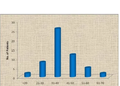
TABLE 2. AGE RATIO
Age No. of patients Percentage (%)
<20 2 3.6
21-30 8 14.5
31-40 26 47.3
41-50 12 21.8
51-60 5 9.1
61-70 2 3.6
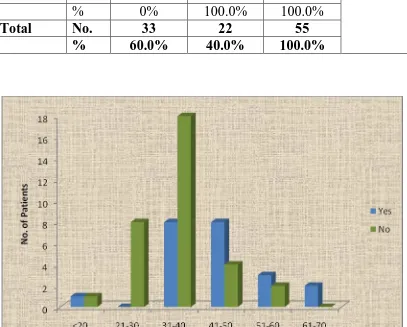
TABLE 3. AGE AND PRE-TRANSPLANT
Age Pre-transplant P value
No Yes Total
<20 No. 1 1 2
0.017
% 50.0% 50.0% 100.0%
21-30 No. 8 0 8
% 100.0% 0% 100.0%
31-40 No. 18 8 26
% 69.2% 30.8% 100.0%
41-50 No. 4 8 12
% 33.3% 66.7% 100.0%
51-60 No. 2 3 5
% 40.0% 60.0% 100.0%
61-70 No. 0 2 2
% 0% 100.0% 100.0%
Total No. 33 22 55
TABLE 4. SEX RATIO AND INCIDENCE
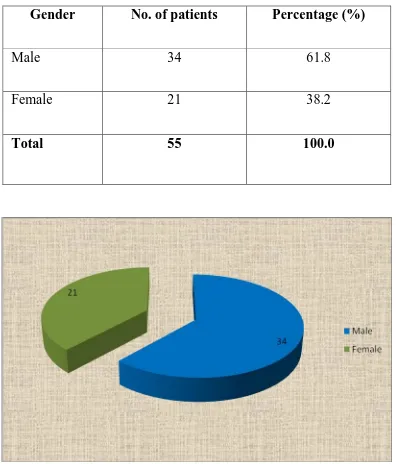
Gender No. of patients Percentage (%)
Male 34 61.8
Female 21 38.2
TABLE 5. GENDER AND PRE-TRANSPLANT
Gender Pre-transplant P value
No Yes Total
Male No. 20 14 34
0.524
% 58.8% 41.2% 100.0%
Female No. 13 8 21
% 61.9% 38.1% 100.0%
Total No. 33 22 55
TABLE .6. GENDER RELATION WITH PH
Gender No of patients Percentage (%)
Male
7 50.0
Female
7 50.0
Total
TABLE.7. RELATIONSHIP OF DONOR
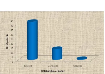
Relationship of donor No. of patients Percentage (%)
Related 42 76.4
Unrelated 12 21.8
Cadaver 1 1.8
TABLE 8. HYPERTENSION
Hypertension No. of patients Percentage (%)
No 34 61.8
Yes 21 38.2
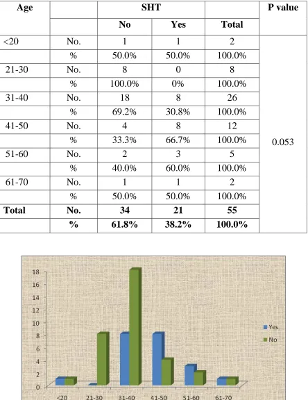
TABLE. 9. HYPERTENSION WITH AGE
Age SHT P value
No Yes Total
<20 No. 1 1 2
0.053
% 50.0% 50.0% 100.0%
21-30 No. 8 0 8
% 100.0% 0% 100.0%
31-40 No. 18 8 26
% 69.2% 30.8% 100.0%
41-50 No. 4 8 12
% 33.3% 66.7% 100.0%
51-60 No. 2 3 5
% 40.0% 60.0% 100.0%
61-70 No. 1 1 2
% 50.0% 50.0% 100.0%
Total No. 34 21 55
TABLE.10. HYPERTENSION ESRD WITH PH:
SHT No of patients Percentage (%)
No 8 57.1
Yes 5 42.9
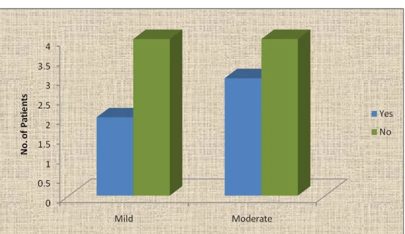
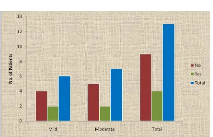
TABLE 11: SHT & PRE TRANSPLANT PH:
Mild Moderate Total P value
No No
4 4 8
0.471
% 50.0% 50.0% 100.0%
Yes No
2 3 5
% 33.3% 66.7% 100.0%
Total No 6 8 14
%
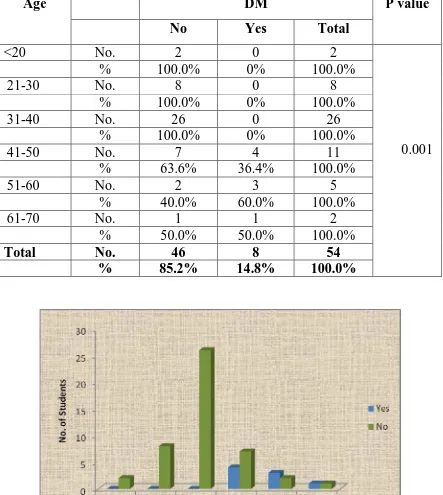
TABLE.12. DIABETES & ESRD IN RELATION WITH AGE PRE TRANSPLANT GROUP
Age DM P value
No Yes Total
<20 No. 2 0 2
0.001
% 100.0% 0% 100.0%
21-30 No. 8 0 8
% 100.0% 0% 100.0%
31-40 No. 26 0 26
% 100.0% 0% 100.0%
41-50 No. 7 4 11
% 63.6% 36.4% 100.0%
51-60 No. 2 3 5
% 40.0% 60.0% 100.0%
61-70 No. 1 1 2
% 50.0% 50.0% 100.0%
Total No. 46 8 54
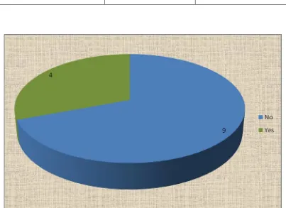
TABLE .13. DIABETIC ESRD WITH PULMONARY HYPERTENSION:
DM with ESRD No of patients Percentage (%)
No 9 64.3
Yes 4 35.7
TABLEBLE.14. DM & PRE TRANSPLANT
Mild Moderate Total P value
No No 4 5 9
0.657
% 44.4% 55.6% 100.0%
Yes No 2 2 4
% 40.0% 60.0% 100.0%
Total No 6 7 13
TABLE 15. CORONARY ARTERY DISESAE IN TRANSPLANT POPULATION
CAD No of patients Percentage (%)
No 43 78.2
Yes 12 21.8
TABLE.16. ESRD WITH CORONARY ARTERY DISEASE AND PH:
CAD No of patients Percentage (%)
No 11 84.7
Yes 2 15.3
Total 13 100.0
TABLE.17. CAD & PRE TRANSPLANT:
Mild Moderate Total P value
No No 6 5 11
0.154
% 54.5% 45.5% 100.0%
Yes No 0 2 2
% .0% 100.0% 100.0%
Total 6 7 13
TABLE. 18. PATIENTS WITH MULTIPLE RISK FACTORS AND PH:
Pt. with DM, SHT,CAD
No of patients Percentage (%)
No
11 78.6
Yes 3 21.4
TABLE.19.PATIENT WITH MULTIPLE RISK FACTORS & PRE TRANSPLANT:
Mild Moderate Total P value
No No 6 5 11
0.154
% 54.5% 45.5% 100.0%
Yes No 0 3 3
% .0% 100.0% 100.0%
Total No 6 8 14
TABLE .20. POST TRANSPLANT AFTER 3&6 MONTHS
Normal Mild Total P value
No No 11 0 11
0.003
% 100.0% .0% 100.0%
Yes No 0 3 3
% .0% 100.0% 100.0%
Total No 11 3 14
TABLE .21.OUTCOME OF PH IN POST TRANSPLANT 3 MONTHS:
Post transplant after 3 months
No of patients Percentage (%)
Normal
11 78.6
Mild
3 21.4
Total
TABLE .22.OUTCOME OF PH IN POST TRANSPLANT 6 MONTHS:
Post transplant after 6 months
No of patients Percentage (%)
Normal 11 78.6
Mild 3 21.4
DISCUSSION
It’s well known fact that in CKD G5D cardiovascular disease is most
common cause of mortality which is mostly manifested as LV dysfunction,
Ischemic heart disease, or acute Myocardial infarction. The other forms of
the cardiovascular disease, other rare manifestations may be pulmonary
hypertension37. But only limited data is available on pulmonary hypertension
in ESRD and Post transplant outcome.
We examined 75 patients out of which 55 cases underwent Renal
Transplant in our institute & also fulfilled the inclusion criteria was taken up
for the study, 41 patients were excluded by clinical history and examination,
laboratory investigation and exclusion criteria (5,7,15,21). Doppler
echocardiography was done in all 55 patients as a part of pre transplant work
up and in patient with PH post operatively 3 months and 6 months
echocardiography was done. Since it’s a non invasive parameter for
assessing pulmonary hypertension in this population during monthly OPD
Among the 55 patients who undergone renal transplantation 22
patients were found to have pulmonary hypertension in the population
(40%).
The Distribution age group and sex of the study population13 was
total of 55 patients out of which 31males(41.2%) and 21 females38.1%, and
only 2 were of 20 years, 26 were of 31-40 years, 12patients were 41-50
years, 5 patients were 51-60, and pre transplant pulmonary hypertension ‘p’
value of 0.017.
We compared gender ratio and found that pulmonary hypertension 7
male and 7 female, who fits in our inclusion criteria. As overall population
male are more common with 41.2%.and female of 38.1% with ‘p’ value of
0.524. in pulmonary hypertension group the ration is 1:1.
Most of our cases are live related donor with 76.2% unrelated are
21.8%, and deceased donor of only 1 patient.
We analysed systemic hypertension in our all population who
underwent renal transplant showed about 38.2%, with age related most
common is 3rd and 4th decade of life with 30.6% and 66.6% and ‘p’ value of
Out of 14 patients (42.9%) Hypertension ESRD, of which 2 had mild
pulmonary hypertension, 3 had moderate PH with ‘p’ value of 0.471.
Among the pre transplant group we found 8 cases (40%) having DM,
when compared to age relation compared 36.4% aged from 41-50, and only
3 were from 51-60. PH with ‘p’ value of 0.001.
In association with pulmonary hypertension and Diabetic ESRD we
found only 4 patients, 2 patients with mild PH, 2 with moderate PH.
calculating a ‘p’ value of 0.657.
In our study group we found 12cases to have coronary artery disease
and ESRD 21.8%outof 55 patients, with pulmonary artery hypertension 2
had moderate PH (45.4%) ‘ p’ value of 0.154.
We also analysed the data and found 3 patients (21.4%) had multiple
risk factors like SHT, DM.CAD& ESRD in the study group of 14 patients
(78.4%) ‘P’ value of 0.154.
In total of pre transplant workup with pulmonary Hypertension we
had 14 patients with mild and moderate PH, 8 cases Severe PH were not
Out of 55 patients who received renal transplant 22 had pulmonary
hypertension of which 14 patients were included for analysis.
Out of 14 patients 5had Hypertension and ESRD of which 2 had mild
PH and 3 had moderate PH, 4 patients had diabetes and ESRD of which 2
had mild PH, 2 had moderate PH, 2 patients had coronary artery disease and
ESRD both had moderate PH.
3 patients had SHT, DM, CAD & ESRD. All 3 had moderate PH.
Mild and moderate PH in Hypertensive ESRD, Diabetic ESRD,
Coronary artery disease & ESRD, became normal 3 and 6 month post
transplant.
The moderate PH in the hypertension, Diabetic, Coronary artery
disease & ESRD, group became mild PH in post Transplant 3and 6 months.
There was a significant favourable outcome in patients who
underwent Renal Transplant when followed up (with
echocardiography)(3,5,7) after first 3 months of transplant showed a ‘p’ value
of 0.002. And follow up after 6moths duration showed a good prognosis
concluded that patients have PH with ESRD has benefited by renal
transplantation.
Till date only one study on pulmonary hypertension in post renal
transplantation with Doppler echocardiography was done by Issa et al
reported pulmonary hypertension in ESRD group of patient’s Doppler
echocardiography was done as a part of workup. David et al., showed that
Non-invasive detection of pulmonary hypertension prior to renal
LIMITATIONS
The major limitations of this study are small sample size.
Observational study
Echocardiography which has
subjective variation, used as a parameter.
Cases of Severe Pulmonary hypertension are excluded because they
are not fit for renal transplant.
Right sided cardiac catheterization and Doppler echocardiography
SUMMARY AND CONCLUSION
14 patients with pulmonary hypertension and ESRD who has
undergone renal transplant were followed up in the post transplant
period. PH became normal in 11 patients during 3rd and 6th month. Of
these 11 patients 5 had Hypertension and ESRD, 4 had Diabetic and
ESRD, 2 had Coronary artery disease and ESRD.
In the remaining 3 patients moderate PH in the pre transplant period
regressed to mild PH on follow up. All the 3 co-morbid factors (DM,
SHT, CAD), were present in this sub- group which may be the reason
for incomplete resolution of PH.
Renal transplant offers a significant resolution of PH in all sub groups
BIBLOGRAPHY
1. Locatelli F, Marcelli D, Conte F, et al: Cardiovasculardisease in
chronic renal failure: thechallenge continues. Nephrol Dial
Transplant2000; 15: 69–80.
2. Simonneau, G, Robbins, IM, Beghetti, M, et al. Updated clinical
classification of pulmonary hypertension. J Am CollCardiol 2009;
54:S43.
3. Bossone, E, Bodini, BD, Mazza, A, Allegra, L. Pulmonary arterial
hypertension: the key role of echocardiography. Chest 2005;
127:1836
4. Mikami, T, Kudo, T, Sakurai, N, et al. Mechanisms for development
of functional tricuspid regurgitation determined by pulsed Doppler
and two-dimensional echocardiography. Am J Cardiol 1984; 53:160.
5. Yock, PG, Popp, RL. Noninvasive estimation of right ventricular
systolic pressure by Doppler ultrasound in patients with tricuspid
regurgitation. Circulation 1984; 70:657.
6. Task Force for Diagnosis and Treatment of Pulmonary Hypertension
of European Society of Cardiology (ESC), European Respiratory
Transplantation (ISHLT), et al. Guidelines for the diagnosis and
treatment of pulmonary hypertension. EurRespir J 2009; 34:1219.
7. Mathai S, Hassoun P. The role of echocardiography in the diagnosis
and assessment of pulmonary hypertension. Adv Pulm Hypertens.
2008;7:379–385.
8. Levey AS, Eckardt KU, Tsukamoto Y, et al. Definition and
classification of chronic kidney disease: a position statement from
Kidney Disease: Improving Global Outcomes (KDIGO). Kidney Int.
2005;67(6):2089-2100.
9. Winearls CG, Glassock RJ. Dissecting and refining the staging of
chronic kidney disease. Kidney Int. 2009;75(10):1009-1014.
10. Neugarten J, Kasiske B, Silbiger SR, et al. Effects of sex on renal
structure. Nephron. 2002;90:139-144.
11. Simonneau G, Robbins IM, Beghetti M, et al. Updated clinical
classification of pulmonary hypertension. J Am Coll Cardiol.
2009;54(1 suppl):S43-S54.
12. Runo JR, Loyd JE. Primary pulmonary hypertension. Lancet 2003;
361:1533.
13. Task Force for Diagnosis and Treatment of Pulmonary Hypertension
Society (ERS), International Society of Heart and Lung
Transplantation (ISHLT), et al. Guidelines for the diagnosis and
treatment of pulmonary hypertension. Eur Respir J 2009; 34:1219.
14. Simonneau G, Gatzoulis MA, Adatia I, et al. Updated clinical
classification of pulmonary hypertension. J Am Coll Cardiol 2013;
62:D34.
15. Updated clinical classification of pulmonary Hypertension Dana
Point, 2008.
16. Nakhoul F, Yigla M, Gilman R, Reisner SA, Abassi Z. The
pathogenesis of pulmonary hypertension in haemodialysis patients
via arterio-venous access. Nephrol Dial Transplant. 2005;20:1686–
1692.
17. Abdelwhab S, Elshinnawy S. Pulmonary hypertension in chronic
renal failure patients. Am J Nephrol. 2008;28: 990–997.
18. Bozbas SS, Akcay S, Altin C, et al. Pulmonary hypertension in
patients with end-stage renal disease undergoing renal
transplantation. Transplant Proc. 2009;41(7):2753–2756.
19. Kuhn, KP, Byrne, DW, Arbogast, PG, et al. Outcome in 91
consecutive patients with pulmonary arterial hypertension receiving
20. Raymond, RJ, Hinderliter, AL, Willis, PW, et al. Echocardiographic
predictors of adverse outcomes in primary pulmonary hypertension.
J Am CollCardiol 2002; 39:1214
21. Giaid A. Nitric oxide and endothelin-1 in pulmonary hypertension.
Chest. 1998;114:208S-212S.
22. Zoccali C. The endothelium as a target in renal diseases. J Nephrol.
2007; 20(12):39–44.
23. Arrigoni FI, Vallance P, Haworth SG, Leiper JM. Metabolism of
asymmetric dimethylarginines is regulated in the lung
developmentally and with pulmonary hypertension induced by
hypobaric hypoxia. Circulation. 2003;107:1195–1201.
24. Zoccali C, Bode-Böger S, Mallamaci F, et al. Plasma concentration
of asymmetrical dimethylarginine and mortality in patients with
end-stage renal disease: a prospective study. Lancet.
2001;358(9299):2113–2117.
25. Ahearn, GS, Tapson, VF, Rebeiz, A, Greenfield JC, Jr.
Electrocardiography to define clinical status in primary pulmonary
hypertension and pulmonary arterial hypertension secondary to
26. Bossone, E, Bodini, BD, Mazza, A, Allegra, L. Pulmonary arterial
hypertension: the key role of echocardiography. Chest 2005;
127:1836.
27. Mikami, T, Kudo, T, Sakurai, N, et al. Mechanisms for development
of functional tricuspid regurgitation determined by pulsed Doppler
and two-dimensional echocardiography. Am J Cardiol 1984; 53:160.
28. Yock, PG, Popp, RL. Noninvasive estimation of right ventricular
systolic pressure by Doppler ultrasound in patients with tricuspid
regurgitation. Circulation 1984; 70:657.
29. Fisher, MR, Forfia, PR, Chamera, E, et al. Accuracy of Doppler
echocardiography in the hemodynamic assessment of pulmonary
hypertension. Am J RespirCrit Care Med 2009; 179:615.
30. Berger, M, Haimowitz, A, Van Tosh, A, et al. Quantitative
assessment of pulmonary hypertension in patients with tricuspid
regurgitation using continuous wave Doppler ultrasound. J Am
CollCardiol 1985; 6:359.
31. Himelman, RB, Struve, SN, Brown, JK, et al. Improved recognition
of corpulmonale in patients with severe chronic obstructive
32. Astor BC, Matsushita K, Gansevoort RT, et al. Lower estimated
glomerular filtration rate and higher albuminuria are associated with
mortality andend-stage renal disease. A collaborative meta-analysis
of kidney disease population cohorts. Kidney Int.
2011;79(12):1331-1340.
33. Poggio ED, Rule AD, Tanchanco R, et al. Demographic and clinical
characteristics associated with glomerular filtration rates in living
ANNEXURE-I
LIST OF ABBREVIATIONS USED
PAH : Pulmonary Arteial Hypertension
PH : Pulmonary Hypertension
CKD : Chronic Kidney Disease
CAD : Coronary Artery Disease
DM : Diabetes Mellitus
SHT : Hypertension
CVD : Cardio Vascular Disease
ECG : Electro Cardiogram
CHD : Coronary Heart Disease
COPD : Chronic Obstructive Pulmonary Disease
HIV : Human Immunodeficiency Virus
WHO : World Health Organization
NO : Nitric Oxide
ANNEXURE-II
CASE PROFORMA
Name: Age: Sex:
IP No: OP No:
Diagnosis: Native Kidney Disease:
Causes of renal failure:
Duration of temp catheter:
Duration of AVF:
Risk Factors: DM / SHT / Smoker / Family History
General examination: BP: PR: SPO2: HR: CVS: RS: P /A: CNS:
Pre transplant work up & Echocardiography
Renal transplant date:
Pre transplant out come:
Echocardiography in first 3 months: