1
DISSERTATION ON
A COMPREHENSIVE STUDY ON INTESTINAL STOMAS
M.S. DEGREE EXAMINATION
BRANCH-I
GENERAL SURGERY
GOVERNMENT KILPAUK MEDICAL COLLEGE
THE TAMILNADU DR.MGR MEDICAL UNIVERSITY
CHENNAI
2
CERTIFICATE
This is to certify that this dissertation titled “A COMPREHENSIVE
STUDY ON INTESTINAL STOMAS” is the bonafide record work done by
Dr. UDAY PRASAD P.V,submitted as partial fulfillment for the requirements
of M.S. Degree Examinations Branch I, General Surgery, April 2014.
Prof. K. Kuberan, M.S Prof. P.N.ShanmugaSundaram, M.S
Dissertation Guide and Unit Chief Head of the Department,
Department of General Surgery General Surgery,
Government Royapettah Hospital Government Kilpauk Medical College
Kilpauk Medical College Chennai.
Chennai.
DEAN
Kilpauk Medical College
TN Dr MGR Medical University
3
DECLARATION
I, Dr. UDAY PRASAD P.V, solemnly declare that the dissertation submitted on the topic “A COMPREHENSIVE STUDY ON INTESTINAL STOMAS” is a bonafide work done by me from May 2011 to December 2013, towards partial fulfillment of the requirements of M.S Degree examinations, General Surgery, April 2014.
Chennai Dr. UDAY PRASAD P.V
4
ACKNOWLEDGEMENT
I sincerely thank Prof.P.Ramakrishnan MD,DLO, the Dean, Kilpauk Medical College for granting me permission to carry out and successfully complete my dissertation work.I consider it a privilege to have done this study under the supervision and guidance of Prof. K. Kuberan M.S, who was a constant source of inspiration and guidance.
5
ABSTRACT
BACKGROUND AND OBJECTIVES
Intestinal stomas are commonly constructed in an emergency as well as elective setting for a variety of indications. Historically associated with a high morbidity, evolution of skills on the part of the surgeon has lead to better understanding of the indications, technique of construction and management of a stoma. This study aims to evaluate the above mentioned parameters and hence improve the outcome of patients undergoing a stoma.
METHODS
50 patients admitted in Govt. Royapettah Hospital and later operated and managed with a stoma were closely followed up from the date of admission to the date of discharge and the various parameters were studied.
RESULTS
The indications, technique, complications and its management were studied in detail by following patients in person or through phone and the results were analyzed in detail.
INTERPRETATION AND CONCLUSION
6
loop colostomy was associated with no complications and was extremely well tolerated.
KEY WORDS
Intestinal stoma, complications, end colostomy,loop ileostomy, loop colostomy, Parastomal hernia, stomal prolapse, loop-end ileostomy.
7
S No Content Page No
1 Introduction 11
2 Aims and Objectives 13
3 Review of Literature 14
4 Materials and Methods 73
5 Observation and Results 75
6 Analysis and Discussion 91
7 Conclusion 97
8 Bibliography 98
9 Annexures
8
S No List of tables Page No
1 Age distribution of patients studied 75
2 Sex distribution of patients studied 76
3 Indications for surgery 77
4 Nature of the disease 79
5 Nature of presentation 80
6 Indication for stoma 81
7 Type of stoma 82
8 Nature of stoma 83
9 Complications of stoma 84
10 Complications associated with each type of stoma 85
11 Patient compliance to the procedure 87
12 Complication of stoma vs compliance 88
13 Type of stoma vs compliance 89
9
S No List of figures Page No
1 Classification of intestinal stomas 18
2 Classification of colostomy by anatomical location 19
3 The technique of constructing a “Blow- Hole” type stoma 25
4 The technique of constructing a tube type cecostomy 27
5 The technique of constructing a loop transverse colostomy 30
6 The technique of constructing an end colostomy 35
7 A rare occurrence of a stomal prolapse with a parastomal herniation
44
8 The technique of constructing an end ileostomy 49
9 The “Tripartite” fixation 50
10 The TURNBULL’S Technique Of Loop Ileostomy 52
11 The technique of closure of a loop ileostomy 52
12 The technique of construction of a loop- end stoma 55
13 Showing local sepsis following stoma and an ileostomy with prolapse
58
14 Construction of the nipple valve 65
15 The technique of constructing an ileal conduit 70
16 Age distribution of patients studied 75
10
18 Indications for surgery 78
19 Nature of the disease 79
20 Nature of presentation 80
21 Indication for stoma 81
22 Type of stoma 82
23 Nature of stoma 83
24 Complications of stoma 84
25 Complications associated with each type of stoma 86
26 Patient compliance to the procedure 87
27 Complication of stoma vs compliance 88
11
INTRODUCTION
Stomas are openings made on the surface of a part of a hollow viscus, usually a portion of the GIT in order to extrude its contents to the exterior. They can be made on a temporary or a permanent basis and can be constructed surgically on an emergency or elective basis. The various surgically constructed forms of stomas include gastrostomy, ileostomy and a colostomy.
12
Stoma is a life saving procedure and even though the first stoma was created more than 100 years ago, it continues as an important tool in the surgeons’ armamentarium. The incidence of permanent stomas like the end
13
AIMS AND OBJECTIVES
1. To study the various indications of intestinal stomas. 2. To study the techniques of intestinal stomas.
3. To study the complications of intestinal stomas and their management. 4. To study the overall compliance of patients in whom a stoma was
14
REVIEW OF LITERATURE
HISTORY
Intestinal stomas are amongst the most important developments in surgical specialties. The first stoma was said to have been constructed nearly 200 years ago and as of now there are an estimated 2 million people in the world living with a stoma. Management of a stoma was long seen as the most decisive factor which led to many people opting out of having one, but more recently this factor has been negated due to various advancements made in stoma care such that an entire specialty – ‘The EnterostomalTherapy unit’ was born to tackle the issues arising from post stomal care. As a result we now see many people, even sportsmen with a stoma leading a normal life. Many advances in stomasurgery, Enterostomal therapy, and ostomy management systems are responsible forthe full lives that these ostomates live and stomas are now a barely noticeable alternative to anal defecation.
15
often managed these stomas on their own with makeshift appliances, rarelywith the help of physicians. It was not until much later that physicians pondered thesurgical creation of an ostomy. Some of the fascinating historical events associated with a stoma arediscussed below.
In 1710 Alexis Littre suggested the creation of an abdominal stoma for the treatment of imperforate anus after observations made during the autopsy of a 6-day-old infant. This event was reported by Fontanel, the historian to the Royal Academy of Sciences in Paris. Littre’s idea remained untested for 66
years, until Pillore, a country surgeon from Rouen, France performed a cecostomy for the treatment of an obstructing rectal cancer.In 1757 Lorenz Heister firstrecommended the surgical creation of stomas for the treatment of abdominal trauma.Heister was resoundingly criticized by his colleagues based onthe inconvenience of exteriorized intestine. This was at atime when surgeons such as John Bell and Gene Palfin advocated closing the abdominal wound while leaving the injured intestines alone as the preferred treatmentfor penetrating intestinal trauma.
16
successful in relieving the obstructionbut not in curing the patient. This child died on the 10th day following surgery. Thecolostomy had its true beginning with the surgery of Duret, a naval surgeon at theMilitary and Marine Hospital at Brest. In 1793Duret performed the first successfulleft iliac colostomy in the treatment of imperforate anus in a 3-day-old infant.
In 1797 Professor Fine, surgeon-in-chief to the Hospital in Geneva, performed the firsttransverse loop colostomy in a 63-year-old woman suffering from rectal cancer1.Through a midline incision, he drew out an inflamed loop of bowel, passed a stitchthrough its mesentery and sewed it to the skin. The patient’s obstruction was relievedand she lived another 3 months. Fine believed
that he had created an artificial anusfrom the terminal ileum; however, autopsy revealed a successful transverse colostomy.With the advent of colostomies, it became necessary to create a means for thecollection of feces. The first mention of such a collecting device was reportedbyDaguesceau in 1795. He performed an inguinal colostomy in a farmer who impaledhimself on a cart stake while unloading wheat. Daguesceau also performed the first colostomy for the treatment of intractable perianalfistulas. Later SchitzingerandMadelung described a procedure of creating a proximal “singlebarreled” stoma while
17
Although the history of colostomy dates back to the early 1700s, the ileostomy was first created by Baum in 1879, which was a diverting ileostomy for an obstructing right sided colon cancer. The first successful creation of an elective ileostomy was by Maydin 1883, who did it along with a colonic resection. Finney described an ileostomy for an appendicular abscess but severe cutaneous reactions resultedowing to a naïve technique, and theprocedure never gained any popularity. These initial stomas were created within the confines of the laparotomy incision itself and it was Rankin who advocated creating a stoma in a separate incision in the right lower quadrant.
In the 1950’s Bryan Brooke of theUniversity of Birmingham in London
described the now famous Brooke ileostomy.In 1952 Brooke described the ileostomy that remains in use today. One sentence, “A moresimple device is to
evaginate the ileal end at the time of operation and suture themucosa to the skin; no complications have occurred from this”, accompanied by asingle illustration,
changed the ileostomy from a chronically inflamed and ulcerativestoma, frequently associated with dysfunction, to the functional “rosebud” we
18
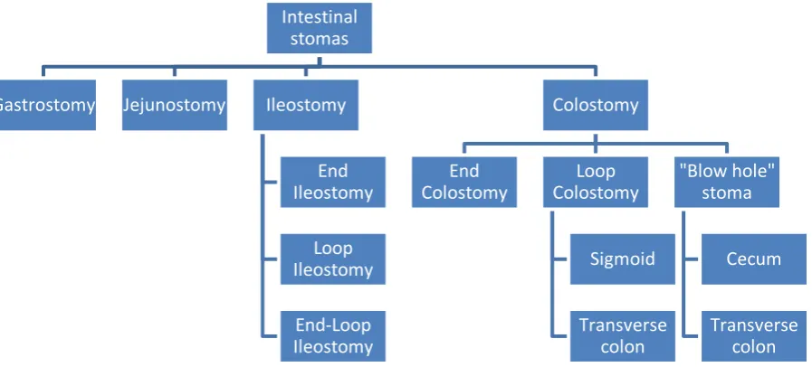
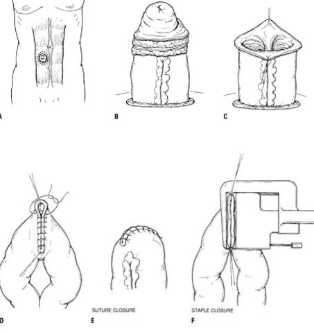
[image:18.595.81.530.162.365.2]CLASSIFICATION OF INTESTINAL STOMAS Figure 1: Classification of intestinal stomas
Gastrostomy: Exteriorizing a part of the stomach for feeding purposes in malignancies in esophagus.
Jejunostomy: Exteriorizing a part of the jejunum eg: in surgeries for perforation wherein primary repair is not feasible, and for nutritional purposes.
Ileostomy: Exteriorizing a part of the ileum eg: in surgeries for obstructed CA cecum or ascending colon where primary repair is not feasible, in perforations involving the ileum, in complicated ileocecal tuberculosis etc
Colostomy: Exteriorizing a part of the large bowel eg: in surgeries for malignancy, perforation, inflammatory bowel diseases etc.
Intestinal stomas
Gastrostomy Jejunostomy Ileostomy
End Ileostomy
Loop Ileostomy
End-Loop Ileostomy
Colostomy
End Colostomy
Loop Colostomy
Sigmoid
Transverse colon
"Blow hole" stoma
Cecum
19 COLOSTOMY
A colostomy is most commonly constructed for a rectal cancer and is usually placed in the anterior abdominal wall. Constructing a stoma in the perineum would be disastrous as evidenced by surgeons in the 19thcentury, as it has no sphincter control and an appliance of any sorts is difficult to place in the perineum without soiling the adjacent area. In fact, in an elderly patient with poor sphincter control, a distal colorectal anastomosis would serve as a “perineal colostomy”. Thus the construction of a stoma may be a better option
for a surgeon and the patient rather than restoring the intestinal continuity to an incontinent anus. Colostomies can be classified according to their anatomic location or by their function.
Classification by anatomical location
Figure 2:Classification of colostomy by anatomical location
End colostomies can be constructed in the descending or the sigmoid colon according to the viability of the inferior mesenteric artery on whose presence the sigmoid colon relies for vascularity. The left side of the colon
Proximal colon
• Cecostomy -Tube type
• Cecostomy -Blow Hole type
Mid colon
• Transverse
Colostomy - Blow Hole type
• Transverse
Colostomy - Loop type
Distal colon
• Sigmoid Colostomy - End and Loop type • Descending
20
merely serves as a conduit and has very few mass peristaltic movements per day whereas the more proximal colon is associated with absorption of water, electrolytes and has more regular and frequent peristaltic contractions. Thus a stoma constructed more proximally on the right side of the colon would have a liquid, foul- smelling high volume output and in essence it combines the worst of an ileostomy and a colostomy, thus should be avoided as against a stoma on the left colon which is more solid and has a less frequent, regulated output.
Transverse colostomies are usually constructed on a temporary basis for decompression of the large bowel in a case of a distally obstructing lesion, or for fecal diversion that is needed to protect a more distal anastomosis. Cecostomies, though rarely performed these days are used in emergency conditions for decompression of an obstructed proximal large bowel in an otherwise old, frail patient with multiple comorbid factors that prevent a major resection.
Classification by function
The intended function of a stoma is more important than the anatomical site wherein it is fashioned. The Colostomy is intended to serve either of the two purposes:
21
Hence, while a stoma is fashioned both its anatomical location and its intended purpose should be kept in mind and a prospective site and type of stoma is chosen by evaluating the patient meticulously.
Preoperative considerations:
The method of choosing the site to construct a stoma is common for all types of intestinal stomas and is mentioned here.Many patients are unsure as to what an ileostomy or colostomy is. Hence, imparting adequate knowledge and obtaining prior consent takes top priority. If an EnterostomalTherapist(ET) is available, then the patient must be counseled by the ET, who can provide specific information regarding the stomal appliances, dietary and clothing alterations and pouch management. Most importantly the ET will select the most appropriate site on the abdominal wall for stoma which will decrease post operative complications and improve the ostomates’ well being4
.
22
midline and the exact site of stoma is marked. The patient should sit up to ensure that the skin folds do not interfere with the stomal site and the patients’
belt line should be identified and avoided if possible as this decreases postoperative clothing restrictions. Despite any restrictions, the stoma must necessarily pass through the rectus to minimize the risk of post operative prolapsed or hernia.
In the distal colon, if an end colostomy (sigmoid or descending) or a loop colostomy (sigmoid) is contemplated, the most desirable position is usually in the left lower quadrant of abdomen. However, in obese patients, so as to not trap the stoma on the under-surface of a panniculus, it is desirable to site the colostomy in the left upper quadrant and hence making it more visible to the patient. For similar reasons, a distal transverse colostomy is fashioned more commonly over the left upper quadrant.Cecostomies are usually done in acute emergency settings and are usually placed on the skin right above the bowel wall. Ileostomies (end and loop ) are usually created in the right lower quadrant. Decompressing Colostomy
23
obstruction.Decompressing colostomy however does not necessarily divert the contents and as a result, it carries the risk of potentially fatal sepsis if there is distal perforation.
Types of Decompressing Colostomy They are of three types:
1. “Blow hole” decompressing stoma in cecum or transverse colon 2. Tube cecostomy
3. Transverse-loop colostomy
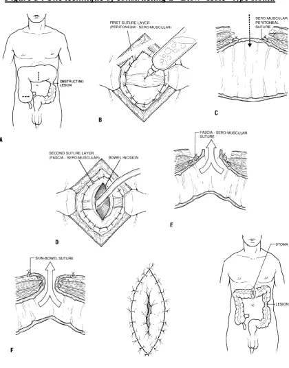
“Blow hole” - Cecostomy and Transverse Colostomy
Cecostomyis reserved for severely ill patients with massive distention and impending perforation of colon which is most often seen in malignant obstruction or Pseudo-obstruction syndromes in elderly and immuno-compromised patients5.The location of the stoma is usually right above the most distended part ofcecum which is to be decompressed.
Technique:
The technique of a “blow-hole” cecostomy and a transverse colostomy is essentially the same.
24
2. A first layer of interrupted, seromuscular, absorbable sutures is placed between the peritoneum and the seromuscular layer of the bowel to be decompressed. The skin incision made as above should be sufficient enough to allow a subsequent incision on the large bowel and suturing of the large bowel to the skin.
3. Needle decompression of the gas-distended viscus is performed to reduce the tension on bowel wall and subsequently a second layer of absorbable suture is placed between the seromuscular layer of the intestine and the fascia of the abdominal wall.
4. The colon is incised, usually with release of large amount of liquid and gas. The full thickness of intestine is then sutured to the full thickness of skin, again with absorbable sutures, and an appliance is placed over the stoma.
Disadvantages:
1. Since this is done through a small incision, one cannot evaluate other parts of the colon for potential ischemic necrosis.
2. Significant inflammation is usually noted in the abdominal wall around the stoma.
25
[image:25.595.80.501.223.759.2]These stomas are difficult to manage post-operatively and hence they must be rarely constructed and used only for short period of time with definitive resection performed as soon as possible.
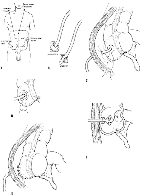
26 Tube cecostomy
A Tube Cecostomy is also a decompressing stoma whose indication and usage has decreased in the recent past due to its poor function and increased post-operative complications6.
Technique:
This is constructed by either approaching the cecum through a laparotomy incision or by making an incision in the abdominal wall over the distended cecum.A purse string suture is placed in cecal wall and a 1 cm incision is made over the dilated cecum and a large mushroom-tipped or Malecot catheter is introduced in the cecum. The purse string is then tightened to secure the catheter.A second purse string suture is placed and the tube is brought out through the skin incision in the right lower quadrant. TheCecum is sutured to peritoneum.
Advantages:
1. This can be performed quickly and hence useful in emergent settings. 2. Less chance of prolapse
Disadvantages:
27
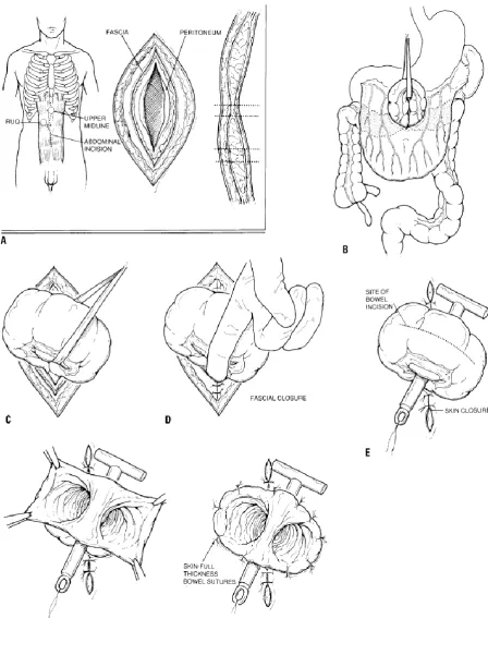
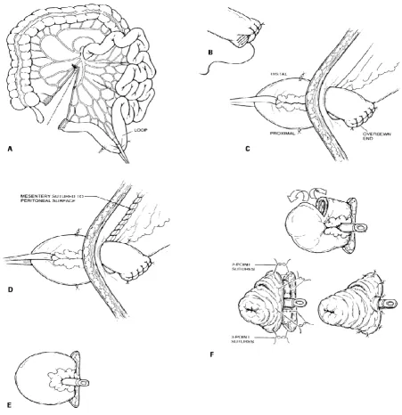
28 Transverse Loop Colostomy
These stomas are constructed to provide decompression of an obstructed colon and they also provide temporary diversion for protection of complicated distal anastomosis. When properly constructed, these can also serve as long term stomas. However in many instances an end loop stoma may serve to function better than a standard loop colostomy9. Prolapse and Para-stomal hernias do occur although their incidence is related to the technique of the surgeon.
Technique:
1. The site of the stoma is chosen and marked on the abdominal wall. In elective situation, the stoma can be placed through the rectus muscle either on the right or the left side or it can be brought out through the mid-line.
2. The mesentery and overlying omentumof the transverse colon is dissected free and a tracheostomy tape is placed around the colon at the site chosen for colostomy. The distal end of the bowel is marked with a suture which will prevent maturation of the incorrect segment and the tracheostomy tape and colon are mobilized and brought out through the laparotomy wound without twisting.
29
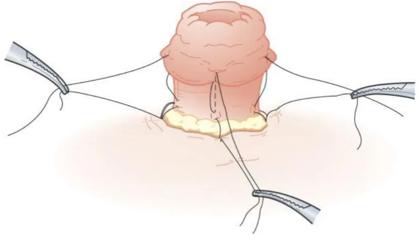
4. The protruding loop of colon is incised over its distal end comprising 80% of its circumference from mesentery to mesentery. The distal end is then “matured”. This is achieved by using the “tripartite” bites which
involve the full thickness edge of the bowel wall, the seromuscular layer at the fascial level and the dermis. The sutures are all held together with a pair of forceps and are tied together which will allow the stoma to evert nicely.
30
31 Closure of a temporary colostomy:
Before closure of a stoma, the following points must be considered and evaluated endoscopically and by contrast studies.
1. Is it safe to restore the intestinal continuity? 2. Is the integrity of distal bowel adequate? 3. Is the sphincter function distally adequate?
The adequacy of anal sphincter can be demonstrated by formal manometric and electro myographic studies or by simply giving the patient a 500 ml enema and asking him or her to hold until he or she can comfortably walk to the toilet and expel the enema.
Technique:
32 Diverting colostomy:
This is primarily constructed to provide diversion of intestinal content. It is performed when the distal segment of bowel has been completely resected, when there is a known or suspected perforation or obstruction of the distal bowel or when there is destruction or infection of the distal bowel. End colostomy and a loop transverse or sigmoid colostomy can act as a diverting colostomy.
Indications:
1. Abdomino-perenial resection 2. Diverticulitis
3. Anastomotic leakage 4. Trauma
5. Crohn’s disease
6. Complex anal sphincter reconstruction
33
sigmoid loop colostomy is employed for diversion, the distal bowel is still in partial continuity and hence when the stomal appliance is full, its contents could be forced into the distal bowel owing to pressure gradient. This phenomenon does not occur in an end colostomy, but it is critical in an end colostomy that the distal limb of the bowel should be vented to the atmosphere as a mucus fistula and not closed, whenever there is a distal obstructing lesion. If the distal limb is closed, there is a risk of closed loop obstruction and subsequent perforation. The decision on whether to close the distal stump or to fashion a mucus fistula depends on the length and the integrity of the distal segment. For example, in a patient undergoing sigmoid colectomy and colostomy for complicated diverticulitis, it is reasonable to close the rectal stump. However, in a patient undergoing abdominal colectomy and ileostomy for toxic colitis, it is preferable to bring the distal segment as a mucus fistula to avoid rectal stump blowout. Mucus fistula can be constructed through a separate opening or it can be fashioned in the same incision that is used to construct the proximal stoma. The construction of a loop sigmoid colostomy is done in the same manner as that of a loop transverse colostomy described above and hence the technique of constructing an end colostomy is alone mentioned below.
Technique:
34
2. An end colostomy requires mobilization of the entire left colon along with the splenic flexure. If there are concerns regarding viability of sigmoid colon, a descending colostomy is done.
3. An opening in the abdominal wall is made by excising a 3-5 cm disc of skin sparing the subcutaneous fat, as this aids in supporting the stoma in the postoperative period.
4. The fat is then separated with scissors and cautery to expose the anterior rectus sheath. The sheath is incised vertically for 3 to 4 cm. The incision can then be extended in a cruciate fashion laterally for upto 1cm if desired. Medial extension is avoided as it brings the stoma closer to the midline and would make closure of the midline wound more difficult. 5. The rectus abdominis is split in the direction of its fibers to expose the
posterior sheath. With the non dominant hand protecting the underlying viscera, the posterior sheath is bluntly opened with the scissors and the defect is enlarged to admit two fingers.
35
[image:35.595.92.492.229.625.2]7. The stoma is then matured by using “tripartite” bites as described above. Colostomies may be sutured without eversion also as distal colonic contents are not irritating to the surrounding skin. Skinis closed and the stoma appliance is fitted.
36
LONG-TERM COLOSTOMY MANAGEMENT 1. Enterostomal therapy(ET)
The enterostomal therapists contribute a lot to the overall success of a stoma in a patient. They not only provide preoperative counseling and post operative guidance, but also act as a long-term resource for individuals with stomas. They supply valuable information regarding appliance choices, suggest dietary or clothing modifications and aid in the management of complications such as skin necrosis, parastomal hernia, stomal prolapse etc. however if this support system is unavailable, then it is the surgeons’ responsibility to educate
the patient in the long term management of the stoma. A stomal appliance has a few components namely,
A skin barrier
An adhesive disk
A face plate
A drainable pouch
37
properly constructed stoma. A well-balanced diet, normal physical figure, ability to engage in normal recreational and sexual activity is all possible with a well-constructed colostomy.
The appliance must be emptied frequently to avoid overfilling and dislodgement of the pouch. This is usually determined by the location of the stoma and the patient’s natural bowel gas pattern. Colostomies usually empty only once or
twice a day or even once every other day. The entire appliance needs to be changed only every 4-7 days. The technique for changing an appliance is described below
1. The soiled pouch is removed by pushing down on skin while lifting up on pouch. Soiled pouch is discarded in an odor proof bag.
2. The stomal and peristomal skin is cleaned with a moist cloth and patted dry. In a cut to fit pouch, the stomal opening is cut to match the exact size of the stoma and the skin barrier paste is applied to the stoma and it is pressed into place.
38
carbohydrates. The amount of swallowed air can be minimized by avoiding the use of straws, excessive talking while eating, chewing gum, and smoking. Each individual can best identify which foods lead to gas production, but beans, broccoli, onions, Brussels sprouts, beer, and dairy products in lactose deficient individuals are common culprits. Avoiding these foods is a personal choice but will decrease the quantity and odor of stomal flatus. Yogurt, parsley, and orange juice have been associated with decreased odor. Odor-proof pouches, charcoal filters, and pouch deodorants (e.g., commercial deodorants, mouthwash, and perineal deodorants) may also help. Orally ingested deodorants are also available and include bismuth subgallate and chlorophyllin complex. However, the most important key to preventing odor is good peristomal hygiene and creating a leak-proof seal at the time of appliance change.
A period of adjustment occurs in all ostomates, but attention to detail at the time of appliance change combined with minor dietary and clothing modifications should make a stoma completely unnoticeable to all except the ostomate's closest acquaintances. In addition, abdominal stomas should not preclude participation in almost any physical activity.
Irrigation
39
improve the quality of life. Irrigation tends to clear the proximal bowel of its contents and temporarily eliminates the need for a stomal appliance, although most patients tend to keep an appliance fitted to permit flow of mucus and de odorized gas in between two bowel movements.
Principle :
The large bowel exhibits one or two mass movements per day and these can be stimulated by distention of the colon, which in turn is accomplished by irrigation. Hence irrigating the bowel tends to reduce the bowel movements to 1-2 per day. However in patients with irritable bowel syndrome this regulated bowel movement cannot be accomplished.
Technique :
The patient is instructed to feel for the stomal opening with a finger and advised to instill 500-1000 ml of water into the proximal loop. This would initiate mass peristalsis and tends to evacuate the bowel of its contents. The patient can then proceed with regular activity and once the bowel is cleared, the patient can even carry on without the need for a stomal appliance.
Advantages
1. Appliance need not be worn at all times. 2. Life style could be more regulated.
3. Passage of uncontrolled gas can be regulated. 4. less leakage of stool between irrigations
40 Disadvantages
1. it is a time-consuming ritual and some people feel discomfort when the bowel is distended during irrigation
2. Irrigation carries a minimal risk of perforation
3. Absorption of water during the irrigation process can be significant, and the patient with an irritable bowel syndrome will usually not achieve adequate control by irrigation and may be frustrated by attempting to do so
Complications of a colostomy
41
recurrence of the primary disease process. More rare causes would include stomal prolapse and parastomal hernia.
Stoma Stricture
Stomal stricture can be attributed to ischemia or serositis of the bowel wall. Ischemia often arises as a result of too much division of the mesentery, while serenities, not seen commonly these days was attributed to delayed opening of the colonic lumen. Both can lead to stricture and can be prevented by “maturing” the stoma, which essentially means suturing the full thickness of
the stoma to the skin. If, however a stricture has indeed developed, then it can be reversed by a simple procedure such as W- or Z- plastyunder local anesthesia. A larger stricture might however require a laparotomy. . Present incidence of stricture or stenosis has been reported to be around 10%10.
Parastomal hernia
42
such as obesity, advanced age and chronic obstructive pulmonary disease appear to increase the risk of parastomal herniation13. In contrast, technical issues such as lateral space closure, fascial fixation or stoma placement through the rectus muscle appear to have no effect on the incidence of these hernias. The use of prosthetic mesh prophylactically in the sublay position may reduce the risk of parastomal hernias14,15.
43 Parastomal prolapse
Prolapse of a stomal segment is most often seen with a transverse loop colostomy and the efferent limb is almost virtually the offending agent17. The reasons for the same are :
1. Long mesentery of the transverse colon which is not fixed retroperitoneally.
2. Large fascial defect to include the stoma.
3. Procedure done in a dilated bowel, which after decompression would broaden the actual fascial defect required to fashion the stoma and hence making the defect more lax and more predisposed to a prolapse.
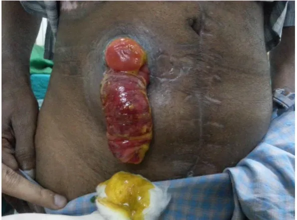
44
Figure 7 : A rare occurrence of a stomal prolapse with a parastomal herniation
Colostomy perforation
This occurs under rare conditions in which the colon is irrigated excessively with water or a contrast material is injected in excess. Treatment requires an immediate laparotomy and closure of the perforation with recreation of the stoma.
Stomalvarices
These varices develop as a result of abnormal Porto systemic anastomosis that develops between the portal venous system of the bowel and the cutaneous veins at the level of the muco cutaneous junction of the stoma. The typical “caput medusa” of the peristomal skin is indicative, especially in a patient with
[image:44.595.149.447.136.356.2]45
Patients with short life expectancies (e.g., extensive liver metastases) may be treated by mucocutaneous disconnection; the stoma is freed up to the level of fascia, thereby dividing the port systemic connections. Since these anastomoses typically reform within 1 year, more definitive solutions are required in most patients. More durable options include surgical shunts, transjugular intrahepatic port systemic shunts, or liver transplantation, based on life expectancy and the status of the associated liver disease
Ischemia
Edema and venous congestion are common after stoma creation owing to mechanical trauma and compression of the small mesenteric venules as they traverse the abdominal wall. This is typically self-limiting and requires no treatment18. However, ischemia may be related to tension on the mesentery or excessive mesenteric division, particularly in obese patients or those undergoing emergency surgery. A common error is dividing the sigmoidal vessels to obtain the length to allow a colostomy to reach the skin. In these cases, the inferior mesenteric vessels should instead be divided proximally and/or the splenic flexure mobilized, preserving the sigmoid arcades.
46
the stoma at fascial level, immediate laparotomy and stoma revision are required. Early ischemia is seen in 1% to 10% of colostomies.
ILEOSTOMY
An ileostomy refers to exteriorizing the ileum, more often distal than proximal, onto the abdominal wall. The stoma is constructed on a permanent basis for patients who require removal of the entire colon (and usually the rectum) and for inflammatory bowel disease. The use of a loop ileostomy is becoming more frequent because of the complex sphincter-preserving operations being performed for ulcerative colitis and familial polyposis. For these operations (restorative proctocolectomy), it is necessary to have complete diversion of intestinal flow while the pouches are allowed to heal and adapt. The loop ileostomy is also useful in cases where multiple and complex anastomoses must be performed distally, usually for Crohn's disease or rectal cancer. As sphincter-preserving operations are used more often, diminishing numbers of permanent ileostomies will be constructed, but equal number of temporary loop ileostomies will be constructed. The same principles used in constructing an ileostomy can be applied to the construction of a urinary conduit.
47
peristomal skin. Therefore, the stoma must be accurately located preoperatively, and it must have a spigot configuration to allow an appliance to seal effectively and precisely around the stoma.
Various types of ileostomies can be constructed. The most common has been the end ileostomy, using a technique popularized by Brooke and Turnbull. The loop ileostomy is used, as described, to protect diseased areas or surgical procedures distally. The end- loop ileostomy is a stoma that uses the principles of a loop ileostomy but is constructed as a permanent stoma when the mesentery and its blood supply need special protection. The continent ileostomy, a technique devised by the Swedish surgeon, Nils Kock, is an internal pouch that does not require the wearing of an external appliance. The urinary conduit is a stoma constructed of small intestine to provide a conduit to the outside for the urinary tract.
As in the case of a colostomy, choosing the exact site of an ileostomy is mandated pre-operatively and is done with the patient in sitting position and marking two lines one through the umbilicus vertically and the other through the inferior margin of the umbilicus, horizontally. The stoma must be placed such that it abuts on both lines at the right lower quadrant and doesn’t cross
48
umbilical fat fold and doesn’t point upward or downward. A majority of stoma
complications can be avoided by marking the site of a stoma precisely. The best incision for an end ileostomy is left paramedian, slanting to the midline fascia, which maintains opening the abdomen in the midline, yet places the skin incision away from the midline to aid in fixing a secure stomal appliance.
END ILEOSTOMY
49
Figure 8 : The technique of constructing an end ileostomy
50
thickness of the ileum, the seromuscular layer of ileum at the base of the stoma and then the dermis. Bites taken through the skin would result in stellate scarring of the stoma and hence should be avoided. Eight of these sutures should be taken, one in each quadrant and one in between each and they should be held in place and tied together so that the stoma everts nicely. This process known as “maturing the stoma” is important as it pours the ileal effluent directly
[image:50.595.90.504.453.687.2]into the stomal appliance and prevents soiling the skin around and hence prevents local sepsis and skin necrosis. A perfectly sized stomal appliance is fit around after cutting the skin barrier and it is held in place by using non irritant skin adhesives.
51 LOOP ILEOSTOMY
52
Figure 10: The TURNBULLS' Technique of Loop Ileostomy
Closure of loop ileostomy
53
54 LOOP – END ILEOSTOMY
55
Figure 12: The technique of construction of a loop- end stoma
Post-operative care and complications
56
liquid content will be passed everyday and an ideally constructed stoma should tolerate this effluent well and there should be no prolapse or retraction. A well-fixed appliance will not leak and damage the skin. Stoma-related complications may be classified as those that occur early (within 1 month of surgery) or late (>1 month postoperatively). The most common early complications are peristomal skin irritation, leakage, high output, and ischemia. The most commonly reported late complications include parastomal hernia, prolapse, obstruction, and stenosis. Parastomal hernia and prolapse have already been explained under the section- complications following colostomy.
57
colostomies. The most common complications were necrosis (22%), prolapse (22%), skin irritation (17%), and stenosis (17%). Risk factors for complications included inflammatory bowel disease, ischemic colitis, and increased body-mass index. As others have observed, obesity markedly increased the risk of skin irritation. Of particular note was the six fold decrease in stoma complications when an ET was involved in the patient's care.
Saghir et al. retrospectively reviewed 121 stoma patients and reported a 67.5% complication rate, 41% of which were considered minor, and 26% were considered major. Nine of the patients (7%) required revisional surgery. Complications were associated with older age, increased medical co morbidities, and an ostomy created by other than a colorectal surgeon.
Skin irritation and leakage :
58
which include antibiotics and topical application of zinc oxide. Changing the stomal appliance and refashioning the stoma could also help in many cases.
Figure 13 :Showing local sepsis following stoma and an ileostomy with prolapse
High output :
59
temporarily managed in an otherwise healthy adult with rehydrating solutions. However, patients who have lost considerable absorptive surface owing to previous bowel resection and/or those with recurrent/residual Crohn's disease are at particular risk. In addition to the loss of absorptive surface area, ileal resection also removes the fat or complex carbohydrate stimulation of the so-called ileal brake that slows gastric emptying and small bowel transit. Fluid and electrolyte maintenance in these patients may require a period of parenteral hydration and nutrition.
Ileostomy diarrhea may be treated in its milder forms with fiber supplements or cholestyramine, which can thicken secretions. Histamine H2
60
downstream limb until gastrointestinal continuity can be restored. This has led to weaning parenteral nutrition in a substantial number of patients.
A related problem in patients with an ileostomy is the development of urinary stones. The obligatory loss of fecal water, sodium, and bicarbonate reduces urinary pH and volume. Whereas approximately 4% of the general population develops urinary stones, the incidence in patients with an ileostomy is approximately twice that. Whereas uric acid stones comprise less than 10% of the calculi in the general population, they comprise 60% of stones in ileostomy patients. There is also an increase in the incidence of calcium oxalate stones.
Serositis
The majority of post-operative complications historically associated with an ileostomy were related to serositis which resulted in a partial obstruction at the stoma itself. These patients suffered massive fluid and electrolyte disturbance and the enormous sequestration of fluid secondary to bowel obstruction in such patients usually resulted in death. Historically termed “ileostomy dysfunction”, the phenomenon was anticipated after construction of
61
ileostomy, many patients may experience problems involving odor and gas. These can usually be managed by paying attention to food and medications ingested, by maintaining meticulous personal hygiene and by using various deodorant products.
An unusually long-term risk to the patient is dehydration which occurs in hot weather and during strenuous physical activity. This is exacerbated by a simple diarrhea so that many patients can go for dehydration before adequate control is achieved by medication. Hence it is of utmost importance for patients to maintain adequate intake of fluid and electrolytes.
Bowel obstruction
62
vomiting. These patients need to be admitted and started on intravenous fluid replacement. The stomal problem should be dealt by introducing a 24F Foley catheter in the stoma and by inflating the balloon with 3-5 ml of saline just beneath the fascia.
The stoma is irrigated with 50 ml of saline and the return of clear fluid or food particles is noted. A clear return suggests a more proximal obstruction and necessitates further evaluation using water-soluble contrast. Return of food particles indicates food blockage and a continuous irrigation would eventually result in clearing of all food debris and return of normal stomal function. This procedure often requires 12-24 hours of irrigation and intravenous fluid supplementation.
Rare complications include a para-ileostomy fistula which is seen in Crohn’s disease which is managed by modifying the appliance so that the fistula
63 CONTINENT ILEOSTOMY
The construction of an intestinal reservoir for feces was first described in 1967 by Nils Kock. His original description was based on the theory that interruption of coordinated peristalsis would enhance capacity. J- and S-shaped pouches have been used with similar results and an S-shaped pouch is described here.
The construction of a continent ileostomy, or Kock pouch, can be broken into four components:
The creation of a pouch
The creation of a nipple valve, which provides continence
The suspension of the pouch from the abdominal wall in such a way as to
prevent slippage of the nipple valve The creation of a stoma.
64
anastomosis, most often because of poor anal sphincter function, (c) the rare patients whose daily work takes them away from toilet facilities for long periods of time and who prefer a continent ileostomy to an ileo-anal anastomosis, and (d) patients with a failed ileo-anal anastomosis who desire to preserve continence and avoid an external appliance if the failure is unrelated to Crohn's disease or severe pouchitis.Use of the Kock pouch should be discouraged in (a) older patients who may be more prone to postoperative complications, including valve dysfunction, and may not tolerate reoperation, (b) patients with Crohn's disease, (c) obese patients, (d) critically ill patients such as those with toxic megacolon, (e) psychologically unfit patients who may not be able to intubate properly or tolerate complications and reoperations, and (f) patients in whom a significant amount of small intestine has already been removed.
The technique :
65
The valve is then fashioned. The serosal surface of the efferent limb of the ileum is scarified, with the electrocautery beginning at the pouch and extending for a distance of 10 cm toward the cut end. The peritoneum of that same segment is also stripped from the adjacent mesentery, which is also defatted. These maneuvers are designed to promote adherence of the ileum and its mesentery when the efferent limb is intussuscepted into the pouch to fashion the valve. The 10-cm efferent limb is intussuscepted into the pouch to form a nipple valve of approximately 5 cm in length. The intussusceptum is fixed in place with through-and-through sutures of 2-0 Vicryl, and three cartridges of stainless steel staples along both sides of the mesentery, care being taken to avoid injury to the vascular supply, and immediately opposite the mesentery using the GIA (U.S. Surgical Corp., Norwalk, CT) auto suture apparatus .
Figure 14 : Construction of the nipple valve
The placement of the staples and sutures is facilitated by stenting• the
66
of the pouch with interrupted 3-0 nonabsorbable sutures at the exit of the limb from the pouch to further anchor the intussusceptum in place. A circumferential defect is created through the abdominal wall just above the pubic hairline in the right lower quadrant, and the outflow tract is brought through the defect and amputated flush with the skin and matured into a stoma with interrupted 3-0 chromic catgut. The length of ileum between the pouch and the stoma should be kept short to avoid tortuosity and facilitate later intubation of the reservoir. This is aided by suturing the pouch to the undersurface of the anterior abdominal wall so that the nipple valve is perfectly aligned with the stoma .A No. 28 French catheter is passed through the stoma, efferent limb, and nipple valve, and its tip is positioned within the lumen of the pouch before the incision is closed. A suture of heavy silk is tied around the catheter at the level of the stoma so that the exact position of the catheter can easily be ascertained in the postoperative period. These precautions help prevent pouch perforation or tube slippage during postoperative recovery.
The advantages of continent ileostomy26,27 are
The patient need not wear an appliance The patient is continent between intubations
He or she may experience a better quality of life.
67 Not all patients are continent
It does require multiple intubations during the day
There can be difficulty in intubation and
The surgery is prolonged and carries a substantial risk of complications.
URINARY CONDUIT
68
The basic principles of construction of the conduit and stoma involve isolation of a segment of intestine, with maintenance of the mesenteric blood supply and enough mobility to allow the distal end to be used as a stoma and the proximal end to serve as the site for ureteral implantation. It is most important to maintain the isoperistaltic direction of the intestine, especially if the conduit is constructed of sigmoid colon. The conduit must not be made of irradiated bowel, even if this requires using either colonic or proximal small intestinal conduits. If the stoma is improperly constructed, there may be a stasis of urine, resulting in reflux and damage to the proximal tract.
69
70
71
LAPAROSCOPIC ILEOSTOMY AND COLOSTOMY
Laparoscopic creation of an ileostomy, whether alone or in conjunction with bowel resection can be created easily. As in the case of an open ileostomy, it is done to protect an anastomosis lower down or to provide diversion proximal to complex anovaginal fistula repair or anal canal reconstruction. Basic principles include patient and site selection are common to the traditional technique30.
At the time of trocar placement, the siting of the stoma should be considered. A trocar can be placed through the future stoma trephine, but sites adjacent to the trephine within the footprint of the stomal appliance must be avoided. The existing trocars placed for the bowel resection procedure can be used for the laparoscopic creation of the stoma also. If the ileostomy is created without any additional abdominal surgery, then only two ports are commonly necessary : one at the umbilicus for the camera and a second through the stoma site to manipulate the terminal ileum. Under either circumstance, the operative principles are similar.
72
created or in use. Ileal mobilization is rarely required. Extreme care should be taken ensure proper orientation of the bowel. The proper loop of bowel is grasped with a grasper through the stoma trephine and proximal and distal bowels carefully identified. If an additional port is available, the tip of a marking pen is grasped with a laparoscopic grasper and the distal end marked just beyond the grasper.
73
MATERIALS AND METHODS
Materials: All patients admitted to GRH and subsequently managed
with a stoma.
Methodology: All patients admitted in Govt. Royapettah Hospital and later operated and managed with a stoma were closely followed up from the date of admission to the date of discharge and the above perspectives were studied.
Type of Study:Descriptive Study Sample Size:100
Inclusion Criteria:All patients who were admitted in Govt. Royapettah Hospital, in the Department of General Surgery between May 2011 and November 2013 and managed with a stoma were taken for study.
Exclusion criteria:
1. Patients who were managed with a stoma done elsewhere and referred to our hospital for further care were not included in the study.
2. Pediatric cases were excluded from the study.
74
involving non-GIT sites viz. Urethrostomy were excluded from the study.
Data collection:The data of each patient was collected in a specially designed proforma which is enclosed.
ROUTINE INVESTIGATIONS - HB, TC, DC
- BT, CT, PT, APTT - LFT
- Serum electrolytes
- X-ray erect abdomen, X-ray chest P-A view - USG abdomen and pelvis
SPECIAL INVESTIGATIONS - CECT abdomen and pelvis - MRI pelvis
Ethical committee clearance was taken from the institution to conduct the study
75
[image:75.595.74.454.466.725.2]OBSERVATION AND RESULTS
Table 1: Age distribution of patients studied
Age groups (years) Frequency Percentage
15-25 10 20.0
26-35 5 10.0
36-45 13 26.0
46-55 8 16.0
56-65 13 26.0
>65 1 2.0
A total of 50 patients were included in the study. The maximum number of patients
were in the age group of 36-45 and 56-65 (n=13).
Figure 16: Age distribution of patients studied
10
5
13
8
13
1 20.0
10.0
26.0
16.0
26.0
2.0
0 5 10 15 20 25 30
15-25 26-35 36-45 46-55 56-65 >65
FREQUENCY
76 Table 2: Sex distribution of patients studied
Sex Frequency Percentage
Male 38 76%
Female 12 24%
Of the total 50 patients included in the study, 38 were male patients and 12 were female patients.
Figure 17: Sex distribution of patients studied
76% 24%
Male
77 Table 3: Indications for surgery
Diseases Frequency Percentage
Blunt abdominal trauma 2 4.0
CA Rectum 3 6.0
Diverticular disease
2 4.0
Hollow-viscus Perforation 17 34.0
Inflammatory bowel disease
2 4.0
Intestinal Obstruction – Benign
7 14.0
Intestinal Obstruction – Malignant 8 16.0
Penetrating Abdominal trauma 5 10.0
Peri-anal sepsis 2 4.0
Acute Mesenteric Ischemia 2 4.0
78 Figure 18 : Indications for surgery
2 3 2 17
2
7 8
5
2 2 4.0
6.0 4.0
34.0
4.0
14.0 16.0
10.0
4.0 4.0
0 5 10 15 20 25 30 35 40
FREQUENCY
79 Table 4: Nature of the disease
Nature of disease Frequency Percentage
Benign 39 78%
Malignant 11 22%
Of the 50 patients for whom a stoma was constructed, benign diseases accounted for 78%.
Figure 19: Nature of the disease
78% 22%
BENIGN
80 Table 5 :Nature of presentation
Nature of presentation Frequency Percentage
Elective 4 8%
Emergency 46 82%
82 % of t total patients presented as an acute emergency and only 4% patients had an elective indication for surgery.
Figure 20: Nature of presentation
8%
82%
ELECTIVE
81 Table 6 :Indication for stoma
Indication for stoma Frequency Percentage
Decompression 4 8%
Diversion 46 92%
Of the 50 patients, 92 % needed a stoma for diversion of the enteral contents and only 8% needed decompression as the principle behind a stoma.
Figure 21 :Indication for stoma
8% 82% 82% DECOMPRESSION
82 Table 7:Ttype of stoma
Type of stoma Frequency Percentage
End Colostomy 4 8.0
End Ileostomy 4 8.0
Loop Colostomy (Sigmoid) 2 4.0
Loop Ileostomy 28 56.0
Proximal Jejunostomy and End
Ileostomy 2 4.0
Transverse Loop Colostomy
10 20.0
Of the 50 patients, 56% patients underwent a loop ileostomy, which was the commonest procedure done (n=28) followed by a transverse loop colostomy.
Figure 22:Type of stoma
4 4 2
28 2
10
8.0 8.0 4.0
56.0 4.0
20.0
0 10 20 30 40 50 60
End Colostomy End Ileostomy Loop Colostomy
(Sigmoid) Loop Ileostomy
Proximal Jejunostomy and…
Transverse Loop Colostomy
PERCENTAGE
[image:82.595.75.492.412.689.2]83 Table 8 :Nature of stoma
Nature of stoma Frequency Percentage
Permanent 4 8%
Temporary 46 92%
Of the 50 patients, only 8% had a permanent stoma while 92 % had a temporary stoma which was eventually reversed.
Figure 23 :Nature of stoma
8%
92%
Permanent
84 Table 9: Complications of stoma
Complications Frequency Percentage
Nil 38 76
Hernia 2 4
Local Sepsis 5 10
Necrosis 3 6
Prolapse 1 2
Retraction 1 2
Local sepsis was the commonest complication associated with a stoma which was present in 10 % of the patients. However the majority of patients did not present with any complications (n=38)
Figure 24: Complications of stoma
38
2 5 3 1 1
76
4
10
6
2 2
0 10 20 30 40 50 60 70 80 90 100 110 120
Nil Hernia Local Sepsis Necrosis Prolapse Retraction
PERCENTAGE
[image:84.595.75.496.415.675.2]85
Table 10 :Complications associated with each type of stoma
COMPLICATION S
END COLOSTOM
Y
END ILEOSTOM
Y
LOOP COLOSTOM
Y (SIGMOID)
LOOP ILEOSTOMY
PROXIMAL JEJUNOSTOM
Y AND END ILEOSTOMY
TRANSVERS E LOOP COLOSTOM
Y
NIL 3 1 1 21 2 10
HERNIA 0 2 0 0 0 0
LOCAL SEPSIS 0 1 0 4 0 0
NECROSIS 0 0 0 3 0 0
PROLAPSE 1 0 0 0 0 0
RETRACTION
0 0 1 0 0 0
86
Figure 25 :Complications associated with each type of stoma
0% 20% 40% 60% 80% 100% End Colostomy
End Ileostomy Loop Colostomy
(Sigmoid) Loop Ileostomy Proximal Jejunostomy and End Ileostomy Transverse Loop Colostomy
87
Table 11 :Patient compliance to the procedure
Patient compliance Frequency Percentage
Good 37 74
Average 8 16
Poor 5 10
Of the 50 patients, most of them (74%) showed good compliance with the procedure.
Figure 26 :Patient compliance to the procedure
37
8
5 74.0
16.0
10.0
0 10 20 30 40 50 60 70 80
Good Average Poor
FREQUENCY
[image:87.595.73.456.345.614.2]88
Table 12 :Complicationof stoma vs compliance
Complications Good Average Poor
Nil 33 5 0
Hernia 1 1 0
Local sepsis 0 0 5
Necrosis 1 2 0
Prolapse 1 0 0
Retraction
1 0 0
Of the 50 patients, local sepsis as a complication resulted in a poor compliance in all patients while even certain high risk complications like prolapse and retraction were well tolerated.
Figure 27 :Complication of stomavs compliance
[image:88.595.72.487.420.718.2]89 Table 13 :Type of stoma vs compliance
Patient compliance Good Average Poor
End Colostomy 3 1 0
End Ileostomy 1 2 1
Loop Colostomy
(Sigmoid) 2 0 0
Loop Ileostomy 20 4 4
Proximal Jejunostomy
and End Ileostomy 1 1 0
Transverse Loop
Colostomy 10 0 0
90 Figure28 :Type of stoma vs compliance
Good Average
Poor 0
2 4 6 8 10 12 14 16 18 20
End Colostomy
Loop Colostomy
(Sigmoid) Proximal Jejunostomy and End Ileostomy 3
1 2
20
1
10
1 2
0
4
1
0
0 1
0
4
0
0
Good
Average
91
ANALYSIS AND DISCUSSION
Although the first stomas were described and constructed in the 19th century, they were associated with innumerable complications and hence did not establish themselves as a favorite amongst surgeons as well as patients. With the advent of better surgical techniques, asepsis and post-operative care, the traditional complications were associated with much lesser morbidity and the field of stoma saw a proportional increase in patient acceptance of the procedure and its aftermath. Nevertheless a stoma, though extremely beneficial and at times life saving, is a procedure which needs to be modified and its incidence minimized as the ultimate aim of a surgeon – “to perform a surgery which is without any morbidity and mortality and is well accepted by a patient”, will
never be fulfilled by constructing a stoma. We now stand in an era of greater scientific advancements, more so in the field of surgery wherein we try as much as possible to minimize the construction of a stoma. This study would throw more perspective on where we currently stand in terms of a stomal construction and management.
92
Most of the patients belonged to two age groups (36-45) and (56-65)
n=26 each. Only one patient presented above the age of 65. The high incidence of cases in the above age group could be attributed to the incidence of malignancies, inflammatory bowel disorders and intestinal perforations, all of which present most commonly in the above age groups.
76% of patients who were included in the study were males (n=38). Only
24% were females. None of the parameters that were studied showed a significant variation according to the sex of the patient. A high male incidence could also be attributed to the above mentioned conditions which also predominate amongst males.
A total of 17 patients, for whom the stoma was constructed, had hollow