A Dissertation on
A STUDY ON ROLE OF ANATOMICAL OBSTRUCTION
IN PATHOGENESIS OF CHRONIC SINUSITIS
Submitted to the
THE TAMILNADU DR. M.G.R. MEDICAL UNIVERSITY In partial fulfilment of the requirements
For the award of the degree of
M.S.BRANCH IV
(OTORHINOLARYNGOLOGY)
GOVERNMENT STANLEY MEDICAL COLLEGE & HOSPITAL
THE TAMILNADU DR. M.G.R. MEDICAL UNIVERSITY, CHENNAI, TAMILNADU
DECLARATION
I, DR. YAHYA ABDUL BASITH.M, Solemnly declare that the dissertation, titled “ A STUDY ON THE ROLE OF ANATOMICAL OBSTRUCTION IN THE PATHOGENESIS OF CRONIC SINUSITIS” is a bonafide work done by me during the period of MARCH 2012 to NOVEMBER 2013 at Government Stanley Medical College and Hospital, Chennai under the expert supervision of PROF.DR.T.BALASUBRAMANIAN, M.S., D.L.O., Professor and Head, Department Of Otorhinolaryngology , Government Stanley Medical College and hospitals, Chennai.
This dissertation is submitted to The Tamil Nadu Dr. M.G.R. Medical University in partial fulfilment of the rules and regulations for the M.S. degree examinations in Otorhinolaryngology to be held in April 2014.
Chennai-1 DR.YAHYA ABDUL BASITH.M
Date:
CERTIFICATE
This is to certify that this dissertation on “A STUDY ON ROLE OF ANATOMICAL OBSTRUCTION IN THE PATHOGENESIS OF CHRONIC SINUSITIS” presented here in by DR YAHYA ABDUL BASITH M, is the original work done in Department of Otorhinolaryngology, Government Stanley Medical College and Hospitals, Chennai in partial fulfillment of the regulations of the Tamilnadu DR.M.G.R Medical University, Chennai for the award of M.S (Otorhinolaryngology), under guidance and supervision during the academic year 2011 – 2014.
PROF. DR.T.BALASUBRAMANIAN, M.S., D.L.O.,
Professor and Head,
Department of Ent & Head and Neck Surgery, Stanley Medical College & Hospital,
Chennai.
PROF.DR.S.GEETHALAKSHMI M.D., Ph.D.,
Dean
Stanley Medical College & Hospital, Chennai.
I wish to express my sincere thanks to Prof. Dr.GEETHALAKSHMI, M.D., Phd, DEAN, Government Stanley Medical College and Hospital for having permitted me to utilize the facilities of the hospital for the conduct of the study.
My heartfelt gratitude to Prof. Dr. T.BALASUBRAMANIAN, M.S., D.L.O, Professor and Head, Department of Otorhinolaryngology, Government Stanley Medical College and Hospital for his motivation, valuable suggestions, expert supervision and for making all necessary arrangements for conducting this study.
I owe my sincere thanks to Prof. Dr. N.SEETHALAKSHMI M.S.,D.L.O., Professor of Otorhinolaryngology, Prof.Dr.RAMANIRAJ M.S, D.L.O Professor of Otorhinolaryngology, for supporting, guiding and encouraging me in this study.
I wish to thank my Assistant professors DR. ATHIYAMN M.S, DR.KARUPPASAMY M.S., D.L.O., DR.CHANDRAMOULI M.S., DR.NANMULLAI M.S., DR.BHARANIDHARAN D.L.O. for their valuable suggestion and help.
I also thank Mrs. Radhakalaiselvan, Audiologist and speech pathologist of ENT Department, Government Stanley hospital for her expert assistance.
I thank the staff nurses and theatre personnel, Government Stanley Hospital for their cooperation and assistance.
CONTENTS
S.NO TOPIC P.NO
01. ABSTRACT
02. INTRODUCTION 01
03. AIMS AND OBJECTIVES 03
04. REVIEW OF LITERATURE 04
05. MATERIALS AND METHODS 44
06. RESULTS AND OBSERVATIONS 58
07. DISCUSSION 71
08. CONCLUSION 75
09. BIBLIOGRAPHY 77
i ii iii iv v vi ANNEXURE PROFORMA
ETHICAL COMMITTEE APPROVAL LETTER PATIENT INFORMATION SHEET
INFORMED CONSENT FORM PLAGIARISM CHECK
A STUDY ON ROLE OF ANATOMICAL OBSTRUCTION IN PATHOGENESIS OF CHRONIC SINUSITIS
ABSTRACT
This is a study about the role of obstructive anatomic variations in the osteomeatal complex of the nasal cavity leading to the development of chronic sinusitis, conducted in a tertiary care centre between July2011 to November2013. About 50 patients were studied for the frequency of occurrence of main anatomical variants like nasal septum deviation, pneumatised middle turbinate, paradoxically curved middle turbinate and infraorbital ethmoid air cell(haller cell), which was compared with a control group of 50 patients on the basis of nasal symptoms of sinusitis and radiological imaging by CT scan of the paranasal sinuses. All the patients were evaluated for the effectiveness of endoscopic sinus surgery by evaluating for the improvement of symptoms and patency of the sinus drainage. Nasal septum deviation followed by pneumatised middle turbinate and paradoxical middle turbinate was the commonly observed anatomical variation at the osteomeatal complex and there was significantly good improvement of the disease symptoms and quality of life following endoscopic sinus surgery.
INTRODUCTION
Sinusitis is a commonly diagnosed condition in the general
population. As the inflammation nearly always involves the nasal
mucosa also, it is now more aptly called as “Rhinosinusitis”. It is a
Group of disorders characterised by inflammation of the mucosa of nose
and paranasal sinuses of at least 12 consecutive weeks duration. Patients
with chronic Rhinosinusitis may have acute flareups, in such conditions it
is called acute exacerbation of chronic sinusitis.
The Rhinosinusitis Task Force of the American Academy of
Otolaryngology-Head and Neck Surgery has classified the chronic
rhinosinusitis based on the duration of the symptoms. They are 1) Acute
(ARS) 7 days to <4 week, 2) sub acute 4 – 12 week, 3) chronic
(CRS) >12 week and 4) and acute exacerbation of chronic, if there is
acute worsening of chronic sinusitis with return to base line.
The etiological factors for chronic sinusitis can be broadly divided
into three overlapping groups: genetic or physiologic factors,
environmental factors and structural (obstructive) factors.
Here we assess the role of anatomical obstructions (structural
factors) in pathogenesis of chronic sinusitis, based on symptoms and
variations in the nose like nasal septal deviation, concha bullosa,
paradoxical middle turbinate and haller cell, leading to chronic sinusitis
have been analysed. Symptomatic patients with CT evidence of chronic
sinusitis will be subjected to undergo functional endoscopic sinus surgery
and correction of the anatomical obstruction, which is causing the
AIMS AND OBJECTIVES
AIMS OF STUDY
1) To study the role of major anatomical obstructions causing chronic
Sinusitis.
2) To find the prevalence of major anatomical variations in patients
with Chronic sinusitis.
3) To find out the effectiveness of surgical correction of anatomical
REVIEW OF LITERATURE
Chronic Rhinosinusitis (CRS) is a spectrum of diseases
characterised by Inflammation of mucosa of the nose and paranasal
sinuses of more than 12 consecutive weeks duration1,2.
Lanza and kennedy reported the now well accepted classification
of the Rhinosinusitis formed by the Rhinosinusitis Task force of the
American academy of Otolaryngology-Head and Neck surgery in 1997.3,4
This classification relies on two major criteria that help to identify
whether or not a patient has rhinosinusitis primarily on the basis of
symptoms and then to classify rhinosinusitis types based primarily on
temporal time frames from the onset of symptoms.
The symptoms of rhinosinusitis are divided into major and minor
categories. Patients with two major factors or one major and two minor
factors, are diagnosed to have the disease.4,6
Major Symptoms/Signs Facial pain/pressure
Facial congestion/fullness
Nasal obstruction/blockage
Nasal discharge/purulence, discolored posterior drainage
Hyposmia/anosmia
Purulent discharge on nasal examination
Minor Symptoms/Signs Headache
Fever (non acute rhinosinusitis)
Halitosis
Fatigue
Tooth pain
Cough
Ear pain/pressure/fullness
Many of the etiological factors have been postulated for the
Development of rhinosinusitis, which can be broadly classified in to
Genetic/physiological factors, environmental factors and structural
factors.5
Major structural factors (anatomical variants) in the nose leading to
sinus obstruction and inflammation are septal deviation, paradoxical
Anatomy of lateral wall of nose
Embryology:
The paranasal sinuses develop from the ridges formed in the lateral
nasal wall called ethmoturbinals8. During 8th week of development a
series of 5 to 6 ridges appear , but after regression and fusion only 3 to 4
ridges persist. The first ethmoturbinals regresses at the time of
development and its ascending part becomes the agger nasi and the
descending part form the uncinate process. The second ethmoturbinals
form the middle turbinate and the third ethmoturbinal form the superior
turbinate. The 4th and 5th ethmoturbinals join to form the supreme
turbinate.
There are primary furrows that lie between the ethmoturbinals, that
forms the various nasal meati and recesses. The first primary furrow lies
between the first and second ethmoturbinals. Its descending part forms
the ethmoid infundibulum, hiatus semilunaris, and middle meatus, and its
ascending part contribute to the formation of frontal recess. The
primordial maxillary sinus develops from the inferior aspect of ethmoid
infundibulum.9-11 The second and third primary furrows form the superior
The ethmoturbinals during their development from the lateral nasal
wall form bony structures that attach the ethmoid complex to lamina
papyracea and skull base. The furrows then develop in to numerous
prerecesses and recesses and contribute to the complex pneumatisation of
the ethmoid bone. As the development progresses, secondary
evaginations and invaginations form from the lateral nasal wall between
the maxilla and ethmoturbinals.12,13 The frontal sinus might have
developed from direct extension of frontal recess, one or more anterior
ethmoid cell or from the anterior aspect of the ethmoid infundibulum.14,15
Bighman et al highlighted the role of a cartilaginous nasal capsule
that surround the developing nasal cavity in addition to the ridges and
furrows.16 He observed that all three turbinates in the lateral nasal wall
develop from this cartilaginous nasal capsule.17 Wang et al also pointed
out the role of cartilaginous capsule. In their view they postulated that all
four pairs of paranasal sinuses develop from the cartilaginous capsule out
pouching of the nasal mucous membrane is only a secondary
phenomenon rather than the primary force in sinonasal development.
Sphenoid sinus develop as the nasal mucosa invaginates in to the
posterior portion of the cartilaginous nasal capsule during the third month
cavity referred as cartilaginous cupolar recess.18 In later months of fetal
life the surrounding wall of this cartilage get ossified and forms the so
called ossiculum bertini. In second and third years this ossiculum bertini
becomes attached to the body of sphenoid after its intervening cartilage is
resorbed.
Sinus pneumatisation is completed between 9 - 12 years in
majority of human subjects.19,20
The ethmoid sinus is commonly called ‘the labyrinth’ because of
its complexity and interpersonal variability. Many otorhinolaryngologists
and surgeons have decreased the complexity of the ethmoid sinus by
dividing it in to many series of lamellae on the basis of embryological
development. The first is the uncinate process, second is the ethmoid
bulla, third being the basal or ground lamella and fourth is that of the
superior turbinate.
The basal lamella of the middle turbinate is special, that it divides
the ethmoid cells in to two groups i.e anterior and posterior. The lamellae
are almost constant feature between the human subjects, and so their
intraoperative recognition is utmost important to help the surgeon
Anatomy of Lateral Wall of Nose
Anterior ethmoid
During anterior rhinoscopy a prominence can be noticed
immediately anterior to the middle turbinate’s attachment in to the lateral
wall. This is called agger nasi. In Latin language, agger means mound or
eminence, nasi means nose. This is a constant feature on nasal
examination and in most but not all cases, it gets pneumatised from an
anterior ethmoid cell called agger nasi cell. The agger nasi cell usually
takes its origin from the superior part of the ethmoid infundibulum or
are, anteriorly frontal process of maxilla, superiorly frontal recess,
anterolaterally nasal bones, and inferomedially lacrimal bone.
The agger nasi is closely related to the lacrimal bone, causing
epiphora in many patients with sinus disease. It is also important in
frontal sinus disease and its management. The pneumatisation of agger
nasi may extend inferomedially to pneumatise the uncinate process.24,25
Uncinate process
The uncinate process is sagittally oriented, almost paralleling the
ethmoid bulla. It is 3-4 mm wide and 1.5-2 cm in length. Its posterior
margin has no bony attachments and is free through most of its course.
The hiatus semilunaris lies directly behind its posterior margin.
Anteriorly and superiorly it attaches to the ethmoidal crest of the maxilla,
just inferior to anterior attachment of middle turbinate on to the lateral
wall of nose. Directly inferior to this it attaches with the posterior portion
Axial view of middle turbinate and uncinate with surrounding structures.
Posteroinferiorly the uncinate process fuses to the ethmoidal
process of inferior turbinate. The attachment to the inferior turbinate is
thick and often it splits or widen to attach with the stout inferior turbinate
bone., the uncinate gives off a small bony projection at its posterior and
superior end to attach to the lamina perpendicularis of the palatine
1. Uncinate attached to lamina papyracea
2. Uncinate attached to skull base
The uncinate does not have any bony attachments anterior and
posterior to its attachment to the inferior turbinate. At this site the lateral
wall of nose is devoid of bone and contain only mucosa of the middle
meatus, sinus mucosa and a thin connective tissue layer in between. They
are referred to as anterior and posterior fontanelles.
The posterior fontanelle on the lateral nasal wall is much larger and
distinct than the anterior fontanelle and also, it contains an opening in to
the maxillary sinus, the called the accessory ostium of the maxillary
sinus.. It can be mistaken for the natural ostium of the sinus and it is seen
in 20 – 25% of the population.27 The superior attachment of the uncinate
varies in different people. Most of the time it bends laterally to get
attached to the laminapapyracea of the orbit. It may also attach centrally
to the skull base or in the medial aspect to the superior part of the vertical
lamella of the middle turbinate.28 The uncinate forms the anterior and
medial boundary of the ethmoid infundibulum.
The uncinate may get displaced laterally against the orbit, as seen
in a case of hypoplasia of maxillary sinus, or it may be displaced
medially as which occur in cases of extensive polyposis within the
In rare cases the uncinate is so medialised that it recurves on itself
to give the appearance of a double middle turbinate. The uncinate process
can be pneumatised also In a small percentage of patients.24,25
Ethmoid Bulla
The ethmoid bulla is the largest and most constant of the anterior
ethmoid air cells. It is located within the middle meatus anterior to the
basal lamella of the middle turbinate and directly posterior to the uncinate
process. The cell has its base on the laminapapyracea and projects in to
the middle meatus medially. The cell has the appearance of a bulla, which
is a hollow, thin walled, rounded bony prominence.
The anterior wall of the ethmoid bulla can extend to the skull base
and form the posterior limit of frontal recess superiorly. The bulla can
blend with the ground lamella posteriorly. Anatomic variations of the
ethmoid bulla may occur. The ethmoid bulla can be one of the largest
ethmoid air cells and can lie in the lower aspect of the middle meatus
when highly pneumatised. In certain cases, a low lying bulla may
potentially narrow the infundibulum and thus, impair mucociliary
transport and ventilation. The ethmoid bulla is formed by pneumatisation
of, and behind, the second basal lamella or bulla lamella. When
and it is referred to as the torus lateralis8. This is estimated to occur in
approximately 8% of patients.
Hiatus Semilunaris
Hiatus semilunaris is a Latin word which can be translated as
hiatus means gap, cleft or passage way and semilunaris means crescent
shaped. It is the crescent shaped cleft in between the posterior free
margin of the uncinate process and the anterior wall of ethmoid bulla. It
is a two dimensional sagittally oriented cleft through which middle
meatus communicate with the ethmoid infundibulum. This cleft is further
defined as the hiatus semilunaris inferior by Grunwald to differentiate it
from smaller less defined hiatus semilunaris superior.8Hiatus semilunaris
superior is a cleft between posterior wall of the ethmoid bulla and ground
lamella of the middle turbinate through which the middle meatus
communicate with the lateral sinus.
Ethmoidal infundibulum
Strictly speaking, infundibulum means funnel shaped structure or
passage.29 It is a funnel - shaped passage through which the secretions
from various anterior ethmoid cells, maxillary sinus, and sometimes
anterior ethmoid region, the ethmoidal infundibulum is a three
dimensional space, bordered in medial aspect by uncinate process, by
laminapapyracea laterally, anterosuoeriorly by frontal process of maxilla
and superolaterally by lacrimal bone. In a sagittal perspective, the
ethmoid infundibulum is curved corresponding to the course of uncinate
and anterior wall of ethmoid bulla.
The ethmoid infundibulum and middle meatus communicate each
other through the hiatus semilunaris. Viewing in a coronal section
immediately above the level of maxillary ostia, the maxillary ostium
forms the inferior boundary, lateral boundary is the lamina papyracea and
medial boundary of the ethmoid infundibulum is formed by the uncinate
process.
Anterior wall of the ethmoid bulla forms the superior boundary,
and the superomedial boundary is formed by hiatus semilunaris.
The importance of the superior aspect of the ethmoid infundibulum
is its close relationship with the frontal recess. This relationship of the
infundibulum and frontal recess is to a large extent determined by the
uncinate process attachment superiorly. Most of the time the uncinate
recessus terminalis, which is the superior boundary of the ethmoidal
infundibulum.
The frontal recess may drain medial to uncinate process when the
uncinate attaches to the laminapapyracea laterally. The uncinate can
attach alternatively to the ethmoid roof or insert in to the middle
turbinate.
The frontal recess will be contiguous with the ethmoidal
infundibulum in these cases.
Several variations exist in the attachment of the uncinate and
relationship of the ethmoid infundibulum and frontal recess. The inferior
aspect of the infundibulum is also quite important due to its relationship
to the maxillary ostium. The natural ostium of the maxillary sinus is
commonly located in the posteroinferior one-third of the ethmoidal
infundibulum. The most inferoposterior portion of the infundibulum
terminates as it drains in to the middle meatus and blends with the
posterior fontanelle mucosa.
Sinus Lateralis (Suprabullar and Retrobullar Recesses)
The sinus lateralis is a variable air space, that lies behind and
suprabullar and retrobullar recesses. This space was first described by
Grunwald and may be highly developed, and in such cases is bordered by
the lamina papyracea laterally, superiorly the ethmoid roof, the basal
lamella of the middle turbinate posteriorly and the ethmoid bulla roof and
posterior wall inferiorly and anteriorly.
The sinus lateralis lies anterior to the middle turbinate basal
lamella, and thus it is anterior ethmoid in location. However it is not
considered an anterior ethmoid air cell as it does not have a single ostia
for ventilation and drainage. Instead, it is considered as a recess that
communicate with middle meatus via hiatus semilunaris superior.
Stammberger pointed out that if the ethmoid bulla do not extend to
the base of skull to form the posterior wall of the frontal recess, the sinus
lateralis can communicate with the frontal recess and the hiatus
semilunaris inferior.8,26 The ethmoid bulla often opens posteriorly, in to
the sinus lateralis.
Osteomeatal complex
The term osteomeatal complex has been widely described and
incorporated into our literature. It was first coined by Naumann.30 It
rather than an anatomic structure. It refers to the physiologic arrangement
of structures into which the frontal, maxillary, and anterior ethmoid
sinuses drain.
Anatomically, this corresponds to the area of the ethmoid
infundibulum and its surrounding structures.
It refers to the physiologic arrangement of structures into which the
frontal, maxillary, and anterior ethmoid sinuses drain. Anatomically, this
corresponds to the area of the ethmoid infundibulum and its surrounding
structures.
It is crucial to have a good patency of the sinuses for adequate
mucociliary function and thus subsequent drainage of the sinuses. The
sinonasal mucosa produces about 1 L of mucus per day, which is getting
cleared by the mucociliary transport. Obstruction of these ostia of sinuses
can lead to fluid accumulation and stagnation, thus creating a moist and
hypoxemic environment suitable for the pathogens to grow and flourish.
The Frontal Recess and Sinus
The frontal sinus drains in to the middle meatus and so to the nasal
cavity through a complex passage described as a ‘nasofrontal duct’.
Anatomic dissection reveals that, there is no existence of a true
duct. But in an attempt to redefine the nomenclature of this area and to
more accurately define the anatomy, the term frontal recess has
recommended.
Being the most anterosuperior aspect of the ethmoid sinus, the
frontal recess forms a connection with the frontal sinus.26 The frontal
recess boundaries are, laterally by the laminapapyracea, medially by the
middle turbinate, anteriorly by the posterior and superior wall of agger
nasi cell, the anterior wall of ethmoid bulla posteriorly(when present). If
frontal recess can communicate with the suprabullar recess and form a
complete posterior wall.
The frontal recess tapers towards internal os of the frontal sinus;
and above the os , as the anterior and posterior tables diverge to their
respective positions it get widened to form a hourglass-like appearance,
with the most narrowed portion being the frontal ostium. The complexity
of the anatomy of this region can be better understood when the effect of
the surrounding ethmoid cells such as the frontal cell, agger nasi cell, and
supraorbital ethmoid cells are taken in to consideration. There is an
intimate relationship existing between the frontal recess and agger nasi
cell.
Secretions from the frontal sinus used to follow a path through the
frontal recess and over the posteromedial surface of the agger nasi cell
and thus reaches the nasal cavity. If the frontal recess is relatively
narrowed and the agger nasi cell is extensively pneumatised, then the
patient may be predisposed to frontal sinusitis.
An extensively pneumatised agger nasi may be mistaken for the
frontal recess or frontal sinus during the course of the endoscopic
The residual posterosuperior wall of the agger nasi cell can be left
posteriorly to the ethmoid roof, and iatrogenic stenosis of the frontonasal
connection can occur, if a large agger nasi call is mistakenly opened for a
frontal sinus.31
In addition to the agger nasi cell, there are other ethmoid cells that
can have a close relationship with the frontal recess.
According to Van Alyea about 50% of cadaveric specimens
studied had anterior ethmoid cells that have encroached in to the frontal
sinus, and among these one-third were encroached in to the area of the
ostium of frontal sinus. .32He called them as ‘frontal cells’.
Schaeffer stated that the anterior ethmoid air cells can pneumatise
extending in to the frontal sinus and appear as the sinus has duplicated.
Stammberger stated that anterior ethmoid cells can develop from the
frontal recess, alongside the frontal sinus, in to the frontal bone. These
are called the bulla frontalis’ by Zuckerkandl.8
The supraorbital ethmoid cell is another anatomic variation in the
region of the frontal recess. Supraorbital ethmoid cells commonly occur
from pneumatisation of the orbital plate of the frontal bone by ethmoid
air cells. Kasper felt that these cells originated in the third and fourth
superiorly over the orbit in to the orbital plate of frontal bone.22
Pneumatisation of the orbital plate of bone can also occur from the frontal
sinus proper.
Maxillary sinus
The maxillary sinus is being a single chamber, is bounded
superiorly by the roof of orbit, inferiorly by the alveolus and dental
portion of the maxilla and hard palate, laterally by the zygomatic process,
posteriorly by a thin bony plate separating the cavity from the
pterygopalatine and infratemporal fossa and medially by the uncinate
process, fontanelles and inferior concha.
The maxillary sinus ostium is located within the most poster
inferior one-third of the infundibulum in 71.8%.15,27The most commonly
observed anatomical variation in the maxillary sinus region is the infra
orbital ethmoid cell or Haller’s cell. Haller’s cells are felt to arise from
the anterior ethmoid in 88% and the posterior ethmoid in 12%.33
Another anatomical variation is hypoplasia or atelectasis of the
maxillary sinus.34,35 In which the maxillary sinus is smaller, the
surrounding maxillary bone is thicker, and the uncinate process is
hypoplastic and lies against the inferomedial orbit, and hence the
infundibulum is atelectatic.
Uncinectomy will be more difficult in these patients due to the
lateral displacement of the structure and the risk of inadvertent orbital
entry due to this displacement against the orbit.
Middle Turbinate
The middle turbinate of the ethmoid bone, which develops from
the second ethmoturbinal is having many important features. Anteriorly
the turbinate attaches the agger nasi region at the crista ethmoidalis
(ethmoidal eminence) on the lateral wall. From here it traverses
superiorly and medially to attach vertically to the lateral aspect of
horizontally along the skull base and inferiorly to attach to the lamina
papyracea and medial wall of maxillary sinus.8 This segment is in a
coronal plane and it divides the ethmoidal labrynth in to anterior and
posterior groups and is referred to as ground lamella or basal lamella. The
posterior aspect of the middle turbinate is having its inferior attachment
to the lateral wall at the crista ethmoidalis of the perpendicular process of
palatine bone, which lies just anterior to the sphenopalatine foramen.
Posterior Ethmoid Sinus
The posterior ethmoid sinus is a group of 1 to 5 ethmoid air cells,
and as they are developmentally derived from the second and third
furrows, they drain in to the superior and supreme meati of the lateral
nasal wall. The posterior ethmoid sinus is bounded anteriorly by the
middle turbinate basal lamella, by the anterior wall of the sphenoid sinus
posteriorly, laterally by the laminapapyracea , by the vertical parts of the
superior and supreme turbinates and their corresponding meati medially,
and superiorly by the roof of ethmoid.
The posterior ethmoid cells, due to their proximity to the skull base
and optic nerve have special surgical significance. Anatomic variation in
the posterior ethmoid is therefore especially important for surgeons be
Onodi performed detailed investigations of the variability in
posterior ethmoid anatomy, and he specifically highlighted the
relationship that most posterior ethmoid cell could have with the optic
nerve.36 Onodi described 38 variations in the relationship of the most
posterior ethmoids to the optic nerve, arranging them in to 12 major
groups. He stressed that when the most posterior ethmoid cell was highly
pneumatised, it could stretch posteriorly along the lamina papyracea in to
the anterior wall of sphenoid sinus. When this occurred , the optic nerve
which usually considered to border the lateral and superior aspects of the
sphenoid sinus, would actually be adjacent to the posterior ethmoid cell.
if this anatomic variation is not appreciated , dissection in the posterior
ethmoid can result in trauma to the optic nerve and blindness.
Modern endoscopic sinus surgeons began to refer this anatomic
variation as Onodi cell.26 If the sphenoethmoidal cell is large, the carotid
canal can bulge in to the posterior ethmoid sinus as well. Onodi cautioned
the surgeons not to assume that, to reach the sphenoid sinus, one simply
extend the dissection through the limits of the posterior ethmoid. Surgical
dissection within the posterior ethmoid should always be oriented in an
inferior and medial direction, rather than a superolateral direction, to
Sphenoid Sinus
The sphenoid sinus is found embedded in to the clivus and is
limited posterosuperiorly by the sella turcica. Horizontally situated
septations are actually bony septations between the posterior ethmoid
sinus and sphenoid sinus and they do not represent septations in the
sphenoid sinus. The sphenoid sinus shows great variability in shape and
size.
It is of three types: type 1: Conchal sinus
It is the most primitive and with an incidence of 2-3%. Hence, the
sella turcica is totally surrounded by bone and remains totally unexposed
to the aerated sinus.
Type 2: Pre sellar pneumatisation
In this type, the posterior half of the sella turcica is surrounded by
bone, while the anterior part is pneumatised and has an incidence of
approximately 10-24%.
Type 3: Sellar pneumatisation.
It is the commonest type with an incidence of 86%
Physiology Of Mucociliary Clearance
The movement of the mucous blanket is referred to as mucociliary
clearance. Inflammatory sinus disease results primarily from the
interference of mucociliary clearance caused by the compromise of the
osteomeatal channels of the individual sinus cavities. The mucosa of the
paranasal sinuses and nasal fossae are lined by ciliated cuboidal
epithelium that secretes mucus. The cilia are in a constant motion and act
in concert to propel the mucus in each sinus toward the nasopharynx.
There are specific pattern of flow for each sinus and they persists even if
alternative openings are surgically created in the sinus.25
In the maxillary sinus, mucus flow is created centripetally toward
the primary ostium. Then the mucus is transported to the hiatus
semilunaris through the infundibulum, from where it passes in to the
middle meatus and then finally in to the nasopharynx. In the frontal sinus,
the mucus flows in to the primary ostium down through the frontal recess
and then in to the middle meatus, where it joins the flow from the
ipsilateral maxillary sinus.25
The sphenoid sinuses and posterior ethmoids clear their mucus in
to the spheno-ethmoidal recess. The mucus flow then enters the superior
CT Scan of Paranasal Sinuses
CT is currently the modality of choice in the evaluation of the
paranasal sinuses and adjacent structures. Its ability to optimally display
the bone, soft tissue and air provides an accurate picture of both the
anatomy and the extend of disease in and around the paranasal sinuses. In
contrast to standard radiographs, CT clearly shows the fine bony anatomy
of the osteomeatal channels.
Performing the initial ct scan after an adequate course of medical
therapy and pre-treatment with sympathomimetic nasal spray 15 minutes
prior to scanning in order to eliminate or diminish reversible mucosal
inflammation and reduce nasal congestion (mucosal edema), improve the
display of fine bony architecture and any irreversible mucosal disease.
Since the osteomeatal unit is best represented in the coronal
plane, it is the primary imaging orientation for evaluation of the sinonasal
tract. This can be achieved by direct coronal scanning or by reformatting
data acquired in the axial plane in to coronal plane images. Coronal study
is optimally performed with the patient in the prone position so that any
remaining sinus secretions do not obscure the osteomeatal unit. In
patients who cannot tolerate prone positioning (children, patients with
utilized. In this technique, the patient is placed in the supine position and
the neck is maximally extended. A pillow is placed under the patient’s
shoulders to facilitate positioning. The CT gantry is then angled to be
perpendicular to the hard palate. However it is not always possible to
obtain true direct coronal images with this technique. When direct
coronal study becomes difficult due to patient’s positioning, spiral
scanning or thin section, contiguous axial CT images with coronal
reconstructions are performed. Axial images complement the coronal
study, particularly when there is severe disease (opacification) of any of
the paranasal sinuses and surgical treatment is contemplated. The axial
studies are needed, as the posterior walls of the various sinuses are not
well seen, if at all, in the coronal plane. Axial images are particularly
important in visualizing the frontoethmoid junction and sphenoethmoid
recess. For axial scans which are 5mm thick, the orbitomeatal line is
taken as reference. The introduction of spiral CT and multidetector CT
scanners allowed even more refined reconstruction in planes other than
the primary scan plane.
Opacification of osteomeatal unit has been found to predispose
patients to the development of sinusitis. Opacification of osteomeatal unit
Diagnostic nasal endoscopy
The advent of nasal endoscopes has facilitated more accurate
diagnosis and proper treatment of sinonasal diseases. Diagnostic nasal
endoscopy has become a routine tool in evaluation of sinonasal diseases
nowadays.
For conducting a proper diagnostic nasal endoscopy, the nasal
mucosa is prepared by using mixture of local anesthetic and decongestant
to facilitate surface anesthesia and decongestion within a few minutes.
Ideally the nasal endoscopy is done using a 4mm, 30 degree endoscope,
and also using 2.7 mm scope in narrow passages. This angulation helps in
direct visualization of the nasal cavity and introduction along the axis,
hence inadvertent injury to the nasal mucosa is averted. It also helps in
adequate visualization of the nasal meatus and the entire nasopharynx.
The procedure is done in three steps as follows;
First pass: visualization of the nasal vestibule, nasopharynx, and inferior
meatus by passing the endoscope along the floor of the nasal cavity.
Second pass: evaluation of the sphenoethmoid recess and superior
meatus.
ANATOMIC VARIATIONS
Deviation of Nasal Septum:
Nasal septum deviation is one of the main causes of anatomical
obstruction at the level of middle turbinate in the osteomeatal complex.
Cottle classified nasal septal deviation in to three types.
1) Simple deviation: It is the most common type. Here there is only a
mild deviation with no obstruction.
2) Obstructive type: The deviated nasal septum touches the lateral
nasal wall or, but on decongestion with vasoconstrictors the
3) Impaction: There will be a septal spur with gross angulation of the
septum leading to severe obstruction, even after decongestion.
CT scan of a patient shows deviated nasal septum with spur
Concha bullosa:
Concha bullosa is nothing but pneumatisation of the turbinate,
usually the middle turbinate and is said to be one of the most common
anatomical variations in the nose. Based on the location, Bolger et al50
classified pneumatisation of the turbinate as lamellar concha bullosa
(pneumatisation of the vertical lamella of the turbinate only), bulbous
concha bullosa (pneumatisation of the whole part). There are many
studies in the literature suggesting the role of concha bullosa in the
etiology of sinus disease. If there is significant expansion of the concha, it
leads to compression of the uncinate process on to the lateral wall of nose
leading to obstruction of the osteo-meatal complex and ethmoid
infundibulum.
Coronal CT scan shows concha bullosa leading to maxillary sinusitis
There is disagreement in the literature regarding the origin of
middle turbinate pneumatisation. The proposed areas from which the
middle turbinate can get pneumatised are, anterior ethmoid, posterior
superior meatus is said to be responsible for the pneumatisation of the
vertical lamellar of the middle turbinate.
Endoscopic picture of Concha bullosa
Paradoxical middle turbinate:
Paradoxical turbinate is another anatomical variation of the nasal
cavity leading to obstruction and chronic sinusitis. Middle turbinate is
said to be paradoxical, if it is concave medially rather than laterally.
Usually paradoxical turbinate occurs where the mucosa is hyperplastic.
with the resultant curvature pointing to the septum. An excessively
curved paradoxical turbinate can compresses the uncinate process leading
to obstruction of the osteo-meatal complex.
Coronal CT scan showing paradoxical middle turbinates.
Haller’s cell:
Haller was an anatomist who described the “ethmoidal cell which
Excavates the os planum and os maxillare, outwardly continuing from the
ethmoid labyrinth capsule”.40 The cell is actually an ethmoid air cell that
pneumatises in to the floor of the orbit or roof of the maxillary sinus,
inferior and lateral to the ethmoidal bulla, closely related to the ethmoid
infundibulum and maxillary sinus ostium.
Haller’s cells are thought to originate from the anterior ethmoid in
88% and posterior ethmoid in 12%. A variety of terms have been used to
Refer to Haller’s cell, including the orbital cells, the
maxilla-ethmoidal Cell, and orbito-maxilla-ethmoidal cells.41, 42 Haller’s cells can obstruct
osteomeatal complex by their close proximity to the uncinate process.
Clark et al, 1989
He studied the incidence of concha bullosa in patients with chronic
Sinusitis and found that, 33% of patients with sinusitis had a concha
bullosa and 11% in the control group(p<0.001), in their radiological
study.
Bolger et al, 1991
He studied the sinus scans of the patients which was taken for inus
and non-sinus complaints, and he found no statistical difference in the
incidence of true concha bullosa and concha bullosa that involves the
vertical part only. But he noticed bulbous concha bullosa in 35.3% of the
scans of patients with rhinitis or sinusitis as compared to 13.9% in the
control group.
Yousem et al, 1991
He studied sinus scans of the patients with sinus and non- sinus
complaints and found that there was no higher risk of having sinusitis in
the presence of a concha bullosa. So he concluded that most of the
concha bullosa were small and did not cause significant narrowing and
Scribano et al, 1993
He observed in his study of CT scans that, maxillary sinusitis was
more in patients with a concha bullosa obstructing the osteomeatal
complex than in those, whose concha bullosa does not cause obstruction
at the osteomeatal complex.
Liu et al, 1998
Liu et al, in 1998 have demonstrated that when the size of the
anatomical variant is greater at the osteomeatal area, the higher the
frequency of association with alteration of the sinus mucosa and
sinusitis.
Kennedy et al, 1998
kennedy et al reported the incidence of infra orbital ethmoid cell
(Haller’s cell) as 10% after studying the coronal CT scan of the patients
with or without sinus problems.
Ameri et al, 2001
He carried out a case control study on 148 patients with chronic
and concha bullosa were the two anatomic variants significantly
associated with sinusitis in his study.
Talaiepour et al, 2004
He analyzed the CT scan of 143 patients presented to the hospital
with a clinical diagnosis of chronic sinusitis and found out the frequency
of anatomical variants among them. The frequency of major anatomic
variants were : Aggar nasi cell in 56.7%, haller cell in 3.5%, nasal septum
deviation 63%, and concha bullosa 35%.
M. Adeel, M. S. A. Rajput, S. Akhter, et al.,2011
They studied the CT scan of 87 patients presented to the hospital
with sinonasal symptoms and analyzed the presence of anatomical
variations among them. There were no anatomical variants in 37 scans.
16 scans showed single variation, while 18 showed two and 6 scans
showed three variations in an individual patient. Nasal septum deviation
was the most commonly observed variation followed by concha bullosa
and paradoxical middle turbinate.
Alkire BC, Bhattacharrya N et al
They studied the CT scan of 37 patients with recurrent acute
reasons. CT scans were analyzed for the presence of Haller cells, septal
spur and concha bullosa. There was more anatomical variations in the
study group, but was not statistically significant.
MATERIALS AND METHODS
Patients with chronic sinusitis having anatomical obstruction in
their coronal CT of paranasal sinuses, in the age group of 18 to 45 were
selected against a control group of population having anatomical
obstruction in their scans with no sinusitis. There was 50 patients in the
study group and 50 patients in the control group. Patients with CT scans
taken for other reasons (eg: faciomaxillary trauma) who were having any
of the anatomical variations in the nose were taken as the control group.
Patients in the study group were selected after proper history taking
and clinical examination. All the patients having symptoms suggestive of
chronic sinusitis were subjected to take coronal CT of the paranasal
sinuses. All of them further subjected to undergo a diagnostic nasal
endoscopic examination.
As septal deviation is a rule rather than an exception in the
population, only Cottle’s type 2 (obstruction) and type 3 (impaction)
were taken in to consideration while studying its prevalence.
All the patients in the study group underwent Functional
obstruction as the definitive treatment for their complaints. They were
followed up 1 week, 2 week, 1 month, 3 month and 6 months post
operative period by nasal endoscopic examination for disease clearance
and improvement of symptoms.
Study period
From March 2012 to November 2013 at Govt. Stanley medical college.
Inclusion criteria
1. Patients with symptoms of chronic sinusitis (major symptoms)
2. Symptoms present for more than 3 months
3. Age 18-45 years
4. Coronal CT scan of paranasal sinuses showing features of chronic sinusitis
5. Coronal CT scan of paranasal sinuses shows anatomical variation at OMC leading to obstruction and sinusitis.
6. No previous nasal surgeries in the past.
7. No evidence local sepsis from oral cavity and oropharynx.
Exclusion criteria
1. symptoms of chronic sinusitis for less than 3 months
2. Age <18 or >45 years
3. Symptoms of chronic sinusitis with no CT changes
4. Coronal CT scan not showing anatomical obstruction at OMC
5. History of previous nasal surgery
6. Evidence of focal sepsis from oral cavity or oropharynx
7. Patients with allergic rhinitis.
Method of study
Step 1: Patients with symptoms of chronic sinusitis were examined
clinically- anterior rhinoscopy.
Step 2: All these patients will be advised to take coronal CT scan of
paranasal sinuses
Step 3: Patients whose CT scan shows evidence of sinusitis and
anatomical obstruction at OMC were subjected to diagnostic
nasal endoscopy.
Step 4: Patients with chronic sinusitis due to anatomical obstruction
Sinus Surgery (FESS) along with correction of the
anatomical variation.
Step 5: Postoperatively all those patients were followed up at 1st
week, 2ndweek, 4thweek, 3rdmonth and 6thmonth with nasal
endoscopic examination by two criteria i.e
i. disease clearance
ii. improvement of symptoms
Surgical correction of anatomical obstruction
Patients with deviated nasal septum were corrected of their
obstruction either by septoplasty or submucous resection(SMR)
operation, according to the type of deviation i.e anterior or posterior. The
procedure was done under general anesthesia or local anesthesia.
First infiltration is given with a pre mixed solution of 2% lignocain
with adrenalin, 1: 100000 concentration on both sides of the cartilaginous
and bony septum. Then Freer’s hemitransfixation or Killian’s incision is
put according to the type of the deviation. In septoplasty
mucoperichondrial flap is elevated on one side and mucoperiosteal flap
Then deviated bony septum is removed and cartilage repositioned
after trimming the lower edge.
In submucous resection (SMR), mucoperichondrial and
mucoperiosteal flaps elevated on both sides of the nasal septum and as
much as the bony and cartilaginous septum is removed as is needed to
correct the deviation. Then flaps are repositioned and sutured.
Picture showing surgical correction of concha bullosa
Patients with concha bullosa, corrected with conchoplasty. A
concha bullosa is having a medial and lateral lamella. After packing the
nose with topical anesthetic and decongestant medication pre operatively,
turbinate as well as near the posterior end of the middle turbinate. Then
using sickle knife concha bullosa is incised over the anterior wall and the
lateral lamella is removed, after separating from the mucosa. This will
relieve the obstruction at OMC.
Endoscopic view of middle turbinate after correcting concha bullosa
A paradoxical middle turbinate causing severe obstruction to the
drainage of the sinuses was dealt with partial middle turbinectomy. After
preoperative anesthetic and decongestant packing infiltration is given
with premixed anesthetic solution over the middle turbinate. Then a cut is
made with turbinate scissors back for about 18mm from the anterior end
and partially freed piece of turbinate fractured downwards. It is then
Functional Endoscopic Sinus Surgery
The initial understanding that drainage of the diseased sinuses
alone by functional endoscopic sinus surgery, is sufficient for resolution
of the disease, has currently modified to some extend based on the new
understanding of the disease process. But the concept that surgery should
extend one stage beyond the diseased mucosa, detected at the time of
surgery or by CT scan, remains unchanged. So simply draining the
diseased sinuses will be insufficient in chronic diseases.
Post operative close endoscopic studies has revealed that most of
the time disease persists in localized areas and that it tends to recur at the
same site even if the diseased mucosa is removed. But it typically
resolves when the underlying diseased bone is removed. So it has been
postulated that the underlying bone may be an important factor in the
disease process, more so in case of chronic sinusitis. So wherever
possible the underlying osteitic bony septations have to be removed
Steps of the procedure
All cases of endoscopic sinus surgery were done under general
anesthesia with controlled hypotension after getting informed written
consent from the patients. The nose is packed with 4% lignocain with
adrenalin (1:100000) , 20 minutes prior to the surgery. The procedure
was done using 4mm, zero degree endoscope and camera connecting to
the monitor.
1. Uncinectomy:
The site of attachment of the uncinate on the lateral wall of nose
is identified. Then the sickle knife is used to remove the uncinate process
as close as possible to its attachment to the posterior edge of the lacrimal
duct ridge. The knife is moved parallel to the lateral nasal wall with
sawing action from anterosuperior to posteroinferior direction, taking
care not to injure the lamina papyracea. The uncinate is then removed
using a blakesley forceps. Uncinectomy can also be done using a back
biting forceps.
2. Middle meatal maxillary antrostomy:
The natural ostium of the maxillary sinus is best identified by
medially. The ostium can then be palpated using a ball-tipped probe. If
the ostium is patent and the disease is limited, then removal of some
additional uncinate only is required.
The maxillary sinus ostium is enlarged by extending the posterior
wall of the ostium to the posterior fontanelle. However tissue should not
be removed circumferentially to avoid stenosis of the ostium.
Tissue is also removed anteriorly using a back biting forceps or
punch, taking care not to injure the nasolacrimal duct, which lie anterior
to the ostium and uncinate process.
There are many theoretical consideration regarding the size of the
antrostomy to be performed. Experimental studies have demonstrated
that, too much exposure of the maxillary sinus mucosa to air flow
resulted in slowing of mucosal clearance. Hence ideally the ostium of
maxillary sinus and its mucosa should remain protected from airflow.
Hence there is a theoretical advantage to keep the surgically created
ostium small, except in few cases like diffuse chronic sinusitis of long
duration, or when there is evidence of osteitis in CT or during the time of
3. Frontal sinusotomy:
Frontal sinus is notorious for persistent and recurrent disease after
surgery. The two most common cause of disease in frontal sinus is, first
displacement of the uncinate process medially due to the presence of
infundibular disease, leading to obstruction of the frontal recess.
Secondly displacement of the frontal sinus ostium posteriorly due to an
enlarged agger nasi cell.
Frontal sinusitis caused by infundibular disease is treated by
opening the frontal recess after meticulous removal of the uncinate
process.
4. Ethmoidectomy:
Nasal obstruction, postnasal discharge and nasal congestion and
are the most common symptoms of ethmoid sinusitis. The ethmoidal
bulla is infractured in its medial aspect using a blakesley forceps and the
residual bony portions are removed with a forceps or a curette. The
medial orbital wall is identified from the starting itself to ensure lateral
positioning during the surgery. After removal of the bulla, the retrobullar
If the posterior ethmoid cells are to be opened, the basal lamella is
perforated just above its horizontal part. Then the remaining portions are
removed superiorly and laterally using a through cutting forceps. Then
the intercellular bony partitions are removed carefully between the
posterior ethmoid cells. The posterior most ethmoid cell is identified by
its pyramidal shape with its apex pointing posteriorly, laterally and
superiorly.
5. Sphenoidotomy:
The safest method of Sphenoidotomy from the ethmoid sinus is to
identify the superior meatus and superior turbinate by palpating medial to
the middle turbinate and superior turbinate. Then resect the most inferior
part of the superior turbinate and palpate the sphenoid ostium to enter the
sinus. The ostium is enlarged medially and inferiorly using kerrison’s
Post operative follow up:
All the patient who underwent the surgery was discharged on the
second post operative day. Post operative antibiotics were given for all
the patients for one week. They were followed up on the end of 1stweek,
2ndweek , 4th week, 3rdmonth and 6th month. The outcome of the surgery
was assessed by two factors i.e.
1) Improvement of the symptoms and
2) Patency of the drainage of the sinuses.
At the first post operative visit (1st week) , the nose is packed with
anesthetic decongestant pack and any blood clots and adhesions are
gently removed. After that all patients are instructed to irrigate the nose
with normal saline two times a day for 3 – 6 weeks. Mucosa would have
formed over the operative area by the end of 3rdweek and by the end of 6
weeks after surgery mucociliary flow is reestablished in most of the
Evaluation of the results of surgery:
Timing of evaluation: As it is always possible for sinus problems
to recur, the results of the surgery represent the success rate for a certain
time frame.
The mucosa is reestablished and mucociliary function starts by the
end of 3rd week, we start evaluating our patients by this time onwards.
Many of the patients who had no improvement by the end of 3rd week,
did well after 6 weeks or 1 month follow up time.
Method of evaluation:
We considered two main criteria for reporting the success rate of
surgery, that is improvement of the symptoms following the surgery and
patency of the sinuses on nasal endoscopic examination in each follow up
periods. There was contradiction between these two criteria during the
evaluation process i.e patients who had no improvement of their
symptoms, had their sinuses patent during nasal endoscopic examination.
So we considered symptomatic improvement as the main criteria to
rate the success of surgery than nasal endoscopic findings.
RESULTS AND OBSERVATIONS
The number of patients involved in the study group was 50, both
males and females and that in the control group was also 50. The study
results among them were as follows.
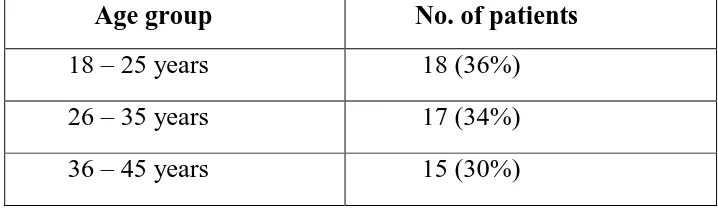
Age distribution of patients with chronic sinusitis
[image:67.612.136.495.291.395.2]Age group No. of patients 18 – 25 years 18 (36%) 26 – 35 years 17 (34%) 36 – 45 years 15 (30%)
Table 1: Shows the age distribution of patients with chronic sinusitis
Among the 50 patients studied, 18 (36%) patients were of the age
group 18 -25 years, 17 (34%) between 26 – 35 years and 15 (30%)
figure 1: shows the age distribution of cases.
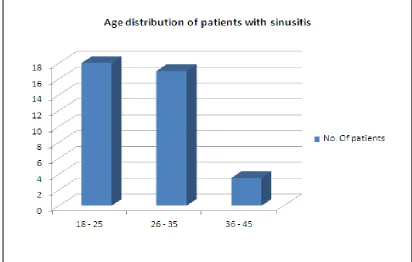
Sex distribution of patients with
Sex
MALE
FEMALE
Table 2
shows the age distribution of cases.
Sex distribution of patients with chronic sinusitis
No. of patients
25 (50%)
FEMALE 25 (50%)
[image:68.612.151.481.502.595.2]The incidence of chronic sinusitis was equal in both males and
females in our study.
[image:69.612.147.483.184.430.2]
Figure 2: showing the sex distribution of cases
The incidence of chronic sinusitis was equal in both males and
: showing the sex distribution of cases
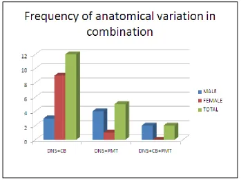
Frequency of anatomical variations.
Anatomical variation male female total
DNS 19 (38%) 18 (36%) 37 (74%)
CB 12 (24%) 10 (20%) 22 (44%)
PMT 8 (16%) 2 (4%) 10 (20%)
HALLER 2 (4%) 1 (2%) 3 (6%)
DNS + CB 3 (6%) 9 (18%) 12 (24%)
DNS + PMT 4 (8%) 1 (2%) 5 (10%)
[image:70.612.112.518.126.347.2]DNS + CB + PMT 2 (4%) 0 2 (4%)
Table 3:showing frequency of different anatomical variations
DNS =deviated nasal septum, CB =concha bullosa, PMT =paradoxical
middle turbinate
The frequency of occurrence of each anatomical variation was
analyzed among the patients with sinusitis. Among the 50 cases, 37
(74%) had nasal septal deviation, either alone or in combination with
other anatomical variation. Concha bullosa was seen in 22 (44%) cases, a
paradoxical middle turbinate in 10 (20%) cases and haller cell in 3 (6%)
There were patients with more than one anatomical variations i.e
12 (24%) cases had septal deviation and concha bullosa together, 5 (10%)
cases had septal deviation and paradoxical
patients had all the three occurring together.
[image:71.612.140.506.260.528.2]
Figure 3: showing frequency of major anatomical variations.
There were patients with more than one anatomical variations i.e
12 (24%) cases had septal deviation and concha bullosa together, 5 (10%)
septal deviation and paradoxical middle turbinate and
three occurring together.
showing frequency of major anatomical variations.
There were patients with more than one anatomical variations i.e
12 (24%) cases had septal deviation and concha bullosa together, 5 (10%)
and 2 (4%)
[image:72.612.151.500.91.352.2]
Figure 4: shows frequency of anatomical variation occurring in combination
Figure 5: frequency of anatomical variation occurring alone.
shows frequency of anatomical variation occurring in
of anatomical variation occurring alone.
shows frequency of anatomical variation occurring in
[image:72.612.184.450.452.646.2]Among the 50 cases studied, 17 (34%) had septal deviation alone
as the anatomical variation, 7 (14%) cases had concha bullosa alone, 3
(6%) cases were having
cases had only haller cell.
anatomical variations. (figure:5 & 6).
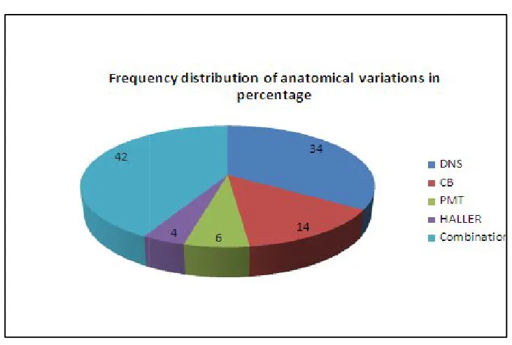
Figure 6: A Pie chart showing the isolated frequency of each
anatomical variation. DNS =deviated nasal bullosa, PMT =paradoxical middle turbinate.
Among the 50 cases studied, 17 (34%) had septal deviation alone
the anatomical variation, 7 (14%) cases had concha bullosa alone, 3
cases were having paradoxical middle turbinate alone and 2 (4%)
cases had only haller cell. Rest of the cases had combination of
(figure:5 & 6).
Pie chart showing the isolated frequency of each variation. DNS =deviated nasal septum, CB =concha bullosa, PMT =paradoxical middle turbinate.
Among the 50 cases studied, 17 (34%) had septal deviation alone
the anatomical variation, 7 (14%) cases had concha bullosa alone, 3
paradoxical middle turbinate alone and 2 (4%)
Rest of the cases had combination of
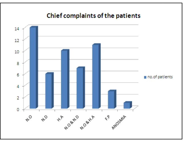
[image:73.612.135.501.262.509.2]Chief complaints of the patients with sinusitis complaints
Nasal obstruction Nasal discharge Headache
Nasal obstruction& headache Nasal obstruction& nasal discharge Facial pain
[image:74.612.124.516.140.323.2]Anosmia
Table 4: showing the frequency of chief complaints of the patients.
Figure 6: showing the frequency
N.O =nasal obstruction, N.D =nasal discharge, H.A =headache,
F.P =facial pain
Chief complaints of the patients with sinusitis
No. of patients 14 (28%) 6 (12%) 10 (20%) headache 11 (22%) Nasal obstruction& nasal discharge 7 (14%)
[image:74.612.172.492.371.618.2]3 (6%) 1 (2%)
Table 4: showing the frequency of chief complaints of the patients.
showing the frequency of presentation of chief complaints N.O =nasal obstruction, N.D =nasal discharge, H.A =headache, Table 4: showing the frequency of chief complaints of the patients.
When the presenting symptom or chief complaints of the patients
were analyzed, we found that 14 (28%) cases had nasal obstruction, 6
(12%) cases had nasal discharge, 10 (20%) had head ache as the main
complaint. 7 (14%) cases had both nasal obstruction and nasal discharge,
11 (22%) had both nasal obstruction and headache, 3 (6%) cases had
facial pain and one patient had anosmia as the chief complaint. (table:4,
figure:6).
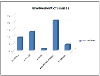
Involvement of sinuses in patients with sinusitis
Sinus involved No. of patients
Maxillary 9 (18%)
Ethmoid 13 (26%)
Frontal 1 (2%)
Maxillary & ethmoid 21 (42%)
All sinuses 4 (8%)
[image:75.612.132.501.336.495.2]