To Evaluate the Complications of TRUS guided Prostate Biopsy
A dissertation submitted to The Dr. M.G.R. Medical University, Tamilnadu, in partial
fulfillment of the requirements for M.Ch. Branch-IV (Genitourinary surgery)
examination to be held in August 2014
DEPARTMENT OF UROLOGY
BONAFIDE CERTIFICATE
This is to certify that the work presented in this dissertation titled “To Evaluate the complications of TRUS guided prostate biopsy” done towards fulfilment of the requirements of the Tamil Nadu Dr. M.G.R. Medical University, Chennai for the M.Ch (Branch– IV) (Urology) exams to be conducted in August 2014, is a bonafide work of the candidate Dr. Feroz Mohd Khan, Senior Post graduate student in the Department of Urology, Christian Medical College, Vellore under my guidance and
supervision. This dissertation has not been submitted, fully or in part to any other
board or University.
Guide Head of the Department Principal
Dr. Nitin S. Kekre Dr. Lionel Gnanaraj Dr. Alfred J Daniel Professor and Head, Professor and Head, Professor, Dept of Dept. of Urology Unit - 2, Dept. of Urology, Orthopaedics,
ACKNOWLEDGMENTS
My first and sincere appreciation goes to Prof Nitin S Kekre, my Guide for all I
have learned from him and for his continuous help and support in all stages of
this thesis. I would also like to thank him for being an open person to ideas,
and for encouraging and helping me to shape my interest and ideas.
I would like to express my deep gratitude and respect to Dr Nirmal T. J, my Co
guide, whose advices and insight was invaluable to me. And his overall
supervision provided the main foundation for this study.
I would like to thank Prof. Lionel Gnanaraj, Head of the Department of
Urology, Prof. Antony Devasia, Prof. Santosh Kumar and Prof. Chandrasingh,
faculty of the department of Urology for their expert advice and support.
My colleagues in the department of Urology merit special mention for their
cooperation Chandan Phulan, Vikas Rampal, Cornerstone Wann and Amit
Vijay Deshpandey. My sincere thanks to all other members of the department
of Urology, CMC, Vellore.
I would like to thank Ms Tunny Sebastian, statician at the Department of
Biostatics, CMC, Vellore for her help in doing the statistical analysis of the
results of the study.
I remain indebted to Dr. Mariyam Khan, my wife for her constant motivation
and encouragement during the period when I needed it most. I am profoundly
grateful towards her for her continuous and watchful guidance given to me all
throughout this work.
I would like to thank my parents, especially my mother and father for always
believing in me, for their continuous love and their supports in my decisions.
Without whom I could not have made it here.
CONTENTS
FRONT MATTER
PAGE NUMBER
Abbreviations
Introduction
1
Aim and objectives
3
Review of Literature
5
Materials and method
29
Results
35
Discussion
46
Conclusions
53
Bibliography
55
ANNEXURE
Patient Performa
Patient information
Consent form
ABBREVIATIONS
TRUS – Trans rectal Ultrasound
PSA – Prostate Specific Antigen
UTI – Urinary tract Infection
VAS – Visual Analogue Scale
LUTS - Lower urinary tract symptoms
PPNB – Peri prostatic nerve block
ASAP – Atypical small acinar proliferation
HGPIN – High grade prostatic intra epithelial neoplasia
FORMAT
TITLE OF THE ABSTRACT: To evaluate the Complications of TRUS guided Prostate Biopsy
DEPARTMENT
: UROLOGY
NAME OF THE CANDIDATE
: DR FEROZ MOHD KHAN
DEGREE AND SUBJECT
: MCh UROLOGY
NAME OF THE GUIDE
: DR NITIN SUHAKAR KEKRE
AIM / OBJECTIVES: Describe the objectives of your study (maximum 30 words)
The aim of the study was to assess prospectively, the complications following TRUS
guided prostate biopsy. The primary objective of the study was to assess urosepsis
requiring hospitalisation. The secondary objective was to assess the incidence of other
complications following TRUS guided prostate biopsy. These include; fever, hematuria,
hematochezia, urinary retention and pain or discomfort.
MATERIAL AND METHODS: Explain the clinical and statistical methods used (maximum 100
words)
All consecutive patients under evaluation of suspected carcinoma prostate were
included in the study. All patients underwent detailed history and physical examination.
Standard 12- Core prostate biopsy done. Inclusion Criteria: Raised prostate specific
antigen (PSA >4.0 ng/ml), abnormal digital rectal examination (DRE) or outside biopsy
report suggestive of prostate cancer but no slides/blocks available for review. Exclusion
Criteria: Patients refused to give consent, patients started on prophylactic antibiotics
two days prior to biopsy.
RESULTS: Summarise the findings and conclusions of your study (maximum 90 words)
A total of 89 patients underwent TRUS guided prostate biopsy. Fourteen patients were
regardless of their urine culture report. The remaining seventy five patients were included
in the study. The mean age of patients was 60.69 years. Diabetes and hypertension were
the most common associated co-morbid illnesses. Most common presentation was lower
urinary tract symptoms. Only five patients developed low grade fever. One patient
developed urosepsis and septic shock after prostate biopsy and was hospitalised.
Subgroup analysis showed that patients with positive urine cultures had more infection as
compared to the other group where urine culture was either sterile or contaminants.
Diabetic patients had more incidence of infection compared to non-diabetics. Ten patients
were on catheter prior to prostate biopsy. There was no infection noted in this group
following biopsy.
CONCLUSIONS:
Trans rectal ultrasound guided prostate needle biopsy is safe for diagnosing prostate
cancer. The most common complication was hematuria in 26.4% of cases, followed by
low grade fever. Incidence of sepsis requiring hospitalisation was very low in our study.
Increased incidence of infection in patients with positive urine culture suggests that
treatment of infection and documentation of negative urine culture before biopsy may be
wiser. Positive 7pre-biopsy urine culture and diabetes mellitus are risk factors which
should be looked into before planning prostate biopsy.
The most common non-cutaneous cancer in United States is prostate cancer, an
approximate 241,000 cases diagnosed in 2012. And also, the second most common cause of
cancer-related death (1). The recommended methods for screening of prostate cancer are
prostate specific antigen (PSA) test and rectal examination. But prostate needle biopsy is
necessary to make the diagnosis. Prostate biopsies were originally done with either digitally
guided or trans-perineal biopsy trucut. But with the development of trans rectal ultrasound,
all trucut biopsies have been replaced by trans rectal ultrasound (TRUS)-guided prostate
biopsy (2).
Majority of the prostate biopsy related complications are mild and self-limited but
sometimes it could be severe and life threatening. Infection, bleeding, and urinary retention
are the most common complications after prostate biopsy. Among Infectious complications,
mild fever, febrile UTI, and sepsis are common (3–6). Recently, there is an increase in
hospitalisation rate due to infectious complication following prostate biopsy (7). Multiple
factors could be responsible for recent trends. Bacterial resistance to fluoroquinolones and
the lack of standard regimen for antimicrobial prophylaxis before prostate biopsy
apparently the most common factors (8–11).
This is a prospective study, designed to evaluate the complications of TRUS guided prostate
AIMS:
The aim of the study was to assess prospectively, the complications following trans rectal
ultrasound (TRUS) guided prostate biopsy.
OBJECTIVES:
Primary objective of the study was to assess urosepsis requiring hospitalisation.
Secondary objective was to assess the occurrence of other complications following biopsy of
prostate. These
include- Fever
Hematuria
Hematochezia
Urinary retention
Historical Perspective:
Prostate biopsy was first described by Ferguson in 1930. He obtained cancer cells by
aspirating prostate tissue with 18G needle, trans-perineally (12). The first trans rectal core
needle prostate biopsy was performed by Astraldi in 1937 (13). There were various
instruments developed and modifications occurred since then for core needle biopsy. All
biopsies were done using finger guidance for palpable abnormality of prostate, either trans
perineal or trans rectal route.
Wild and Reid in 1955, first reported use of trans rectal ultrasound (TRUS) of prostate. It was
popularised by Watanabe et al. in 1970 (14,15). TRUS guided prostate biopsy was started in
late 1980s using 18 Gauge needle on a spring device (Biopty Gun). Since then it has become
a standard procedure. The “sextant biopsy” model was proposed by Hodge in 1989 (16).
Sextant biopsy was standard method for prostate biopsy until Stamey in 1995, suggested
taking samples from more lateral parts of the prostate to include peripheral zones. Studies
of radical prostatectomy specimen section analysis had shown that prostate cancer most
commonly arises from peripheral zones. Therefore laterally directed biopsies were advice to
increase the yield of cancer detection as well as reduce the missing out cancer foci (17).
Thereafter, technological advancement has improved the TRUS and its role in prostate
cancer detection. Recent advancement in prostate imaging includes Colour Doppler, MR
useful in cases where 1st biopsy came as negative but clinical suspicion for prostate carcinoma is high.
Epidemiology of carcinoma prostate:
Incidence of prostate cancer is commonest non skin cancer in U.S. population. The lifetime
risk of disease is estimated to be approximately 16%, with a 2.5% risk of death. Among U.S.
population, African-Americans have the highest incidence of carcinoma prostate.
Worldwide, it is the 5th most common cancer and the 2nd second most common among
men. Overall, incidence of carcinoma prostate was about 12%, 19% in developed countries
and about 5% in the developing countries. There is a wide variation of incidence among
countries and ethnicity. Asia has lowest incidence rates and highest among North America
and Scandinavian population (1,18). Introduction of PSA has led to much increase in
incidence of prostate cancer. Similar to prostate cancer incidence, there is wide variation in
mortality among countries. Highest rate of mortality in African countries southeast China,
Asia and North Africa has lowest mortality (18). The CONCORD study analysed cancer
survival in different countries and reported different survival rates for colorectal, breast and
prostate cancer. There was large variation in 5-year survival rates; it was highest in the
Canada, Australia, and United States and to lowest in Algeria, Poland and Denmark. The
accuracy of cancer registries, access to health care and quality of health care as well as PSA
screening affect prostate cancer reporting (19).
In recent period, there have been efforts to diagnose prostate cancer at earlier stage. This
described as small-volume, lower-grade prostate cancer. The 10-yr survival rate was similar
to general population (20,21).
Despite screening programme to detect early prostate cancer, a large number of significant
prostate cancers are still under diagnosed. Under-diagnosis was defined as inability to
diagnose high grade, high-risk, locally advanced or surgical margins positive, if resected,
prostate cancer (22).
Over-detection was defined as detection of small volume prostate cancer, localised with
Gleason score ≤4 or 5. Studies showed that about 1–7% of patients were over diagnosed
and the resulting into consequent overtreatment. Over-diagnosis of prostate cancer is a
major concerns in prostate oncology (23).
Role of Prostate Biopsy:
Initially prostate biopsy was used to diagnose prostate cancer. Now it has evolved from not
only to detect cancer but to assist in clinical management of patient. Prostate biopsy plays
an important role in active surveillance (AS) programme. Therefore it should be
reproducible. It has an important role in standardised staging and grading of prostate cancer
(20).
Historically, sextant biopsy was the standard to diagnose prostate cancer. Studies showed
that there was increased number of missing clinically significant prostate cancers due to
sampling error while doing sextant biopsy. This has resulted to the introduction of
extended biopsy in Gleason stage up gradation. It included 301 patients with low-risk
prostate, an extended (>or=10-core) prostate biopsy was done. 18 cores of biopsy were taken
in majority of patients. The up gradation was reported in 31.9% patients (28).
To detect missed cancer and to assess the tumor volume as well as prognosis, extended or
saturation prostate biopsies were advised (29).
Risk stratification of prostate cancer: (30)
There are three risk categories described for prostate cancer;
Low risk group:PSA < 10, Gleason score≤6 and stage T1 or T2a.
Intermediate risk group:PSA 10 - 20, or Gleason score = 7 or stage T2b or T2c.
High risk group:PSA > 20, or Gleason score ≥8 or stage T3, or two or more intermediate risk
factors.
Watanabe was the first to describe trans rectal ultrasonography (TRUS) of the prostate in
1968, later with technological advancement of ultrasound and use of trans rectal ultrasound
for prostate biopsy, it was included in routine clinical use (16). Increased numbers of
patients are undergoing TRUS guided prostate biopsy annually in the United. Prostate
cancer prevalence is high similarly large number of patients undergoing trans rectal prostate
biopsy under ultrasound guidance now a days.
Ultrasonography of the Prostate:
The prostate gland is located anterior to rectum between the bladder neck and the
urogenital diaphragm.
zones- Anterior fibro muscular stroma (AFS)
Central zone (CZ)
Transition zone (TZ)
Peripheral zone (PZ)
Periurethral zone
These zones cannot be differentiated on trans rectal ultrasonography (TRUS). Central and
peripheral zones of the prostate are located posteriorly and appear homogenous on TRUS.
Most of the adenocarcinoma prostate arises from these regions. Multiple, small and diffuse
calcifications of the prostate are a normal ultrasonographic finding. It is an age related and
not a pathological. Prostatic calculi are symptomatic if large, needs further evaluation and
[image:20.612.96.364.396.610.2]treatment.
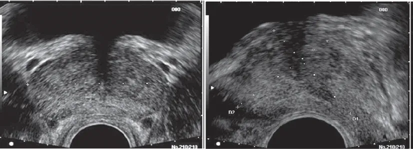
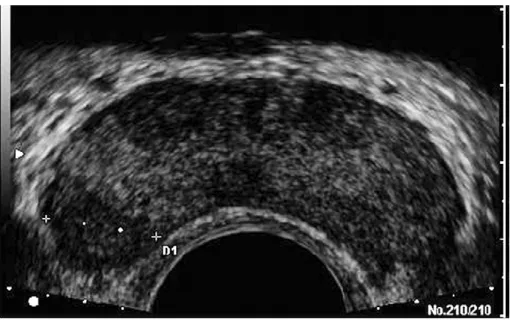
Figure 1
Normal Trans rectal ultrasound image.
A, Transverse view. B, Sagittal view
Gray-scale trans rectal ultrasonography (TRUS):
Currently, TRUS is the most common imaging for prostate evaluation. The most common
use of TRUS is for prostate cancer detection, there is other indications also esp. Infertility
where it can be useful. There are two types of endorectal probes, side-fire and end-fire
models with 6 to 10 MHz frequencies. The biplane probes give simultaneous transverse as
well as sagittal views. The resolution of images increases with increase in frequency of
ultrasound probe. High frequency probes have small focal length and image is very close to
the transducer. The 7-MHz transducer results in high-resolution image and is best for
peripheral zone of prostate. Lower frequency probes have large focal length but the
resolution of images will be low. They are accurate of volume measurements.
Techniques:
Prostate should be evaluated in both transverse as well as sagital planes. Prostate volume is
[image:21.612.98.511.67.216.2]calculated. Hypo-echoic lesions are looked in central and peripheral zones. Figure 2
Trans rectal ultrasound image of the prostate.
A, In the transverse
Patient positioning during TRUS:
Left lateral position is the most preferred position for prostate biopsy.
Prostate Volume Calculations:
Various formulas are described for calculation of prostate volume. Prostate measurements
are taken in three dimensions, axial plane, transverse and antero-posterior, to calculate the
volume. Most commonly used formula is
- Prolate spheroid (π/6 × transverse diameter2× antero-posterior diameter).
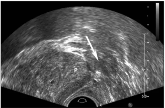
Hypo-echoic lesions within peripheral zones should be identified and biopsied. Majority of
suspected lesions for prostate cancer are hypo-echoic lesions. About 39% of malignancies
are iso-echoic and ~1% of tumors are hyper-echoic on TRUS. Although hypo-echoic lesions
are most commonly turn out to be prostate cancer, other disease processes like
granulomatous prostatitis, prostatic infarct and lymphoma can also produce hypo-echoic
lesions. About 17% to 57% of cases, a hypo-echoic lesion is malignant, required to biopsy
these lesions. Any contour abnormalities along the surface of the gland are suspected to be
prostate cancer. Extra capsular extension of prostate cancer is noted by loss of bright white
Prostate biopsy under image guidance:
Prostate gland imaging improves cancer detection by precisely visualising and characterising
the lesion as well as to guide the accurate and targeted biopsy. Currently, with the help of
newer imaging modalities such as MRI and contrast enhanced TRUS, a higher number of
image-targeted cancers in suspicious lesions have been found (32–34). These studies have
shown that targeted biopsies detect cancer of high grade and tumor volume than that of
conventional TRUS biopsy. Therefore targeted biopsy may be helpful in prostate cancer
grading, prognosis and deciding treatment. Recent studies had introduced modern imaging,
such as elastography. A prospective randomised study showed sensitivity and specificity of
real-time elastography as 61% and 68% respectively as compared to grey-scale ultrasound
which showed sensitivity and specificity of 15% and 92% respectively (35).
Routine prostate biopsy may be called as image-blinded prostate biopsy. Tumor lesions
[image:23.612.171.426.65.225.2]identified on image will result in better yield of malignant tissue than the image-blinded Figure 3: Hypo-echoic lesion noted on the
procedure. Image localisation of cancer provides precise identification of the cancer, which
would result in better lesion-targeted management. A systematic review on targeted biopsy
using image-guided prostate biopsy and MRI localised lesions had shown that cancer was
detected in 30% of MRI targeted cores versus 7% of systematic cores (36).
Another newer modality of prostate is MR/TRUS image fusion (37). However, Utility of
MRI/TRUS fusion imaging needs to be validated as the prostate is a deformable organ
changes its shape during TRUS but it remains the same on MRI (20).
Role of MRI/MR spectroscopy:
Recently, the role of MR spectroscopy and targeted biopsy was assessed. The high
diagnostic accuracy of magnetic resonance spectroscopic imaging and dynamic
contrast-enhanced imaging magnetic resonance (DCEMR) in the management of prostate cancer was
evaluated in one study. MRS detects metabolic activity of tissues and can deferntiate
normal from cancerous tissue based on ratios of creatine, choline and citrate production
and consumption (38). Cancerous tissue contains less citrate level and higher concentrations
of choline and creatine when compared with benign prostatic hypertrophy or normal
prostatic tissue. A randomised controlled trial, recently evaluated the role of MRS and
dynamic contrast enhanced MR (DCEMR) imaging in cases of prior negative biopsy but high
suspicion of prostate cancer. The patients with a prior negative prostate biopsy and
persistently elevated PSA levels, a combination of a standard 10-core biopsy scheme with an
oversampling strategy in sites targeted by combined MRSI/DCEMR indications resulted in
significantly higher cancer detection rates. At the second biopsy, a prostate cancer detection
0.01). The prostate cancer detected with the help of MRS/DCEMR were high grade (Gleason
score ≥7 (4+3) 61.6%) (39).
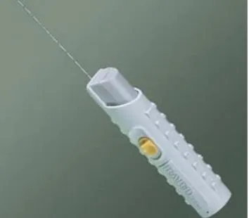
Prostate Biopsy Technique:
18 Gauge needle loaded on a spring action device is used for prostate biopsy. When trigger
button of device is pushed, the inner needle advances 23 mm followed by outer hollow core
needle. The prostate tissue is caught between inner needle and outer sheath and therefore
disengaged from the prostate gland. The device is design to obtain 15 mm to 17 mm length
of the tissue during biopsy.
Techniques of Extended Core Biopsy:
The sextant biopsy had a low yield of cancer detection therefore sampling of more tissue
from laterally focused cores are introduced. Various studies have shown improved detection
rate of cancer by laterally directed biopsy cores in addition to the standard systematic
sextant technique. Extended core biopsy includes 8 to 13 cores (24,40,41). 6 cores biopsy is
not adequate for prostate cancer detection.
Transition zone as well as seminal vesicles is not routinely sampled because of low cancer
detection rate at initial biopsy. Transition zone biopsy may help in case of large gland (more
Trans rectal ultrasound guided prostate needle biopsy is the gold standard for diagnosing
prostate cancer. PSA based screening programs has resulted in detection of early prostate
cancer. There is a significant increase incidence of organ confined disease and potentially
curable due to PSA screening (42). Most accepted PSA threshold above which biopsy
advised is >4.0 ng/ml, although optimal PSA threshold is not yet defined in asymptomatic
men. As the PSA threshold was lowered to >2.5 ng/ml, it has resulted in increased detection
of organ confined prostate cancer at the time of radical prostatectomy.
This observation has led to recommendation of prostate biopsy in younger men (below 60
years if PSA rises above 2.5ng/ml (43). There is no PSA level that can exclude prostate
cancer in age range as shown in Prostate Cancer Prevention (PCPT) Trial. Thompson and
colleague in their data showed that men with PSA less than 4.0 ng/ml, large numbers of
patients were diagnosed as prostate cancer. Prostate cancer was diagnosed in 15% of
patient with PSA level < 4.0, and there will be 15% of patient having a Gleason score of ≥ 7
[image:26.612.97.508.80.219.2](22).
Figure 4
A, Sextant biopsy B, 10-core biopsyC, 12-core biopsy
To reduce the chances of under-diagnosis of high risk disease, prostate biopsy was
extensively performed as well as PSA threshold for performing biopsy was also reduced in
the last decade. This has resulted to increased number of “insignificant cancer” being
diagnosed. Similarly, there is an increased number of biopsy cores to diagnosed prostate
cancer.
Urinary tract infection (UTI) or prostatitis may cause false elevation of PSA. Most of the
cases, PSA value ranges between 2.5–10.0. Guidelines recommend repeating such abnormal
PSA value before deciding upon performing prostate biopsy (20).
Number of prostate biopsy cores:
Increased numbers of cores and proper localisation of these cores were considered optimal
to sample entire prostate. Haas et al. did biopsy on prostates of 164 autopsy patients.
18-core biopsy was performed in all patients. Analysis showed that 12-18-core specimen detected
most of cancers which is clinically significant with 80% sensitivity (44). It was found that
cancer detection was related more to location of samples rather number of biopsy cores
and samples containing lateral and apical cores were representing the peripheral zone (PZ)
tissue where prostate cancer mostly occurs.
Initial biopsy:
Currently the 12-core biopsy is widely accepted method of biopsy. Some studies did show
that taking biopsy cores from apical region on each side improves the yield of detecting
prostate cancer. Cancer is missed mostly at the apical location during initial biopsy (45).
volume of prostate. Extended biopsy has a significant superiority of cancer detection rate
when compared with sextant biopsy (46). Studies have shown that extended biopsy cores
>12–14 as initial prostate biopsy scheme has no advantage over standard 12-core biopsy
(47,48).
Repeat biopsy:
Indications:
Inadequate prostatic tissue to diagnose or exclude prostate cancer.
Previous negative biopsy but persistent clinical or biochemical suspicion for
carcinoma prostate (e.g. abnormal DRE, persistently raised or rising PSA).
Previous biopsy of multifocal High Grade PIN and/or Previous Suspicious
Appearances (ASAP)
Systematic prostate biopsy has the potential to miss small volume prostate cancer in some
patients. Therefore these patients undergo repeat biopsy. About 30% to 50% of patients
detected to have cancer on repeat biopsy, mostly with extended biopsy scheme (20).
Scattoni et al. proposed a model for repeat prostate biopsy considering the clinical
characteristics of the patients. Patients with ASAP, a 14-core biopsy without TZ sampling
were done. A 14-core biopsy with four TZ cores if there is no ASAP but %free PSA was ≤10%.
If there is no ASAP and %free PSA >10%, a 20-core biopsy including four TZ cores was found
to be useful (49).
Ideally, samples should be located at different sites after negative biopsy to identify tumours which
were not sampled in the previous biopsy. In case of multifocal HGPIN or ASAP, repeat biopsy to an
location. There is a risk of carcinoma in the entire gland in case of HGPIN (50,51). The natural
history of atypical small acinar proliferation (ASAP) is not well defined as compared to
HGPIN, if ASAP is seen in the biopsy, there is a significant chance of diagnosing prostate
cancer on repeat biopsy. Current recommendations are to repeat biopsy within 3 to 6
months in case of ASAP or HGPIN on initial biopsy. The most common areas missed during
initial biopsy are apices and anterior prostate.
Saturation Biopsy:
Saturation biopsy has been recommended by some investigators to maximise the cancer
detection rate in patients where the clinical criteria put them at high risk for prostate cancer
despite a previous benign biopsy. It has been performed with peri prostatic nerve block,
under sedation or under general or spinal anaesthesia (26,52). The office-based trans rectal
saturation biopsy technique with biopsy cores of ≥20 has increased the prostate cancer
diagnosis by 30%. However, complication rate was similar to that of standard biopsy (53).
Cancer detection rate by saturation biopsy protocol is similar to extended core (10-12 Core)
biopsy, which is less morbid procedure. Therefore role of saturation biopsy has decreased
with time.
When serum PSA level is in the range of 4.0 to 10.0 ng/ml, % free PSA less than 25% helps in
95% chances of detection of prostate cancer and avoids 20% prostate biopsies, and the risk
of cancer increased further decline of free PSA (42). Similarly, a PSA velocity >0.75
ng/ml/year has increased suspicion of prostate cancer and suggests biopsy (54). The clinical
Before deciding on a repeat prostate biopsy to detect suspected prostate cancer, an
elevated PSAD and PSAD-TZ suggests repeat biopsy to diagnosed prostate cancer.
Prostate Biopsy contraindications:
(55)
Significant coagulopathy
Painful condition
Immunosuppression
Acute prostatitis
Prophylactic Antibiotics:
There are various prophylactic antibiotic regimens described. American Urological
Association recommends antibiotic prophylaxis before trans rectal prostate biopsy (56).
Controversy exits regarding the duration of antibiotics after biopsy. Studies have shown that
fluoroquinolone prophylactic regimens, single-dose oral is equally effective than 3-day
regimens to prevent infections (56,57).
Rectal Cleansing Enema prior to Biopsy:
Various strategies reducing infectious complications have been explored. One study by
Gil-Vernet used povidone-iodine for rectal cleansing prior to prostate biopsy and reported 0.2%
incidence of E. coli epididymitis. Another study using the same protocol also showed similar
result (58,59). Contrary to these studies, Zaytoun et al. could not find any difference in
complications using enema (60). Cochrane review has concluded that risk of bacteraemia
was reduced with enema plus antibiotics in comparison to antibiotics only but risk of fever
To reduce the infectious complications, several studies have assessed the role of expanding
the antimicrobials, using different techniques for biopsy and rectal swab cultures. Adding
ciprofloxacin to amoxicillin-clavulanate resulted decreased of infections(61). Adibi et al.
evaluated the addition of gentamicin to trimethoprim-sulfamethoxazole or ciprofloxacin and
compared it with the group where gentamicin was not added and noticed decreased rate of
hospitalization (62). Another study have reported favourable results with addition of
amikacin (63). Ceftriaxone was added to lidocaine during peri-prostatic nerve block in one
study, resulted in decreased incidence of sepsis (64). Drawbacks of broad spectrum
prophylaxis are increased adverse effects, potential increase in antimicrobial resistance and
cost. However, the expenses of hospitalisation increases significantly if admission is required
due to post biopsy infection rather using more intensive prophylaxis prior to biopsy and it is
more cost effective. There is another concept of targeted prophylaxis under investigation. A
culture swab from rectum is taken and is plated on agar. Patients’ ciprofloxacin sensitive
rectal swab cultures can receive the same as prophylaxis; others to get alternate antibiotics.
Positive swab culture from rectum is considered as risk factor for prostate biopsy related
infection but it does not always lead to clinical infection. Several prevalence studies have
shown that approximately 25% of rectal swab cultures contain fluoroquinolone-resistant
organisms but actual clinical infection occurs in a very small number of these patients (11).
There are no randomized control trials to show that targeted prophylaxis decreases the
chances infection and expenditure when compared with routine prophylaxis.
There are studies showed that technical modifications had influenced the incidence of
infection. Trans perineal approach has been suggested as an alternative technique for
prostate biopsy as it bypasses the rectum which is the source of bacterial contamination and
or cleaning the needle with iodine solution is not associated with infectious risk. There are
conflicting reports regarding infectious risk while using same needle in subsequent patients
or new needle each time for biopsy. However, adequate reprocessing/disinfection of biopsy
probes and needle guide are of paramount importance. General recommendations for
assessment of patient prior to prostate biopsy include a complete evaluation including
history and examination and risk factor assessment for bacterial resistant and infectious
complications (66).
Risk factors for infectious complications:(66)
Patient-related:
Co morbidities; Diabetes, COPD
Heart valve
Benign prostate enlargement
Recent UTI
Recent antibiotics usage, particularly fluoroquinolone
Hospitalisation in the recent past
Indwelling urethral catheter
Pre biopsy urine culture positivity
Procedure-related:
Increase number of biopsy cores
Repeat biopsy
Contaminated ultrasound gel
Infection following TRUS biopsy:
Infection following TRUS guided prostate biopsy is a well-established risk, therefore
evidence supporting antimicrobial prophylaxis. Regarding use of prophylactic antibiotics, a
Cochrane review has reported significant reduction of bacteriuria, bacteraemia, incidence of
fever, urinary tract infection (UTI), and rate of hospitalisation (6). American Urological
centres use prophylactic antibiotic prior to prostate biopsy however, there was a wide
variation among studies regarding duration of antimicrobial use, many of these studies did
not show significant benefit if used for >24 hours period (67–70).
Incidence of infection and related complications:
Infection rate varied in different studies, with reported rate of hospitalization range from 0–
6.3% (71,72). One study on infection have reported, incidence of UTI in about 3.5% patients
and 3% required hospitalisation after biopsy. Simsir et al showed similar incidence of
septicaemia (73,74). Contrary to them, other studies had shown decreased incidence of
sepsis (0.6% to 1.7%) following prostate biopsy (60). Recently, there is an increase incidence
of antimicrobial resistance particularly fluoroquinolone. Data from US SEER–Medicare
showed a 2 fold increase in hospitalization rate for infectious complication as compared to
controls (7). The risk of sepsis is similar between the first and subsequent biopsies (74).
There was an increased incidence of infectious complications following TRUS-Biopsy
between 1996 and 2005. There was an increased hospital admission from 1.0% to 4% in this
duration and about 70% were related to sepsis (75). There was a significant increase in
hospital admission following prostate biopsy during 1993 to 2010. Majority of infection
related complications resulted from E coli, with increased resistance to fluoroquinolone,
ampicillin and sulfamethoxazole- trimethoprim (76).
Bleeding following Prostate biopsy:
Bleeding following TRUS-Biopsy is one of the most common complication which bothers
bleeding. Factors responsible for these complications are size of prostate gland,
anticoagulation drugs, and number of biopsy cores.
Hematuria:
Hematuria is common after prostate biopsy, with incidence of 10–84% (71,75,77). This wide
range is because of different definitions for hematuria (blood seen in urine, requirement of
catheterisation or need for hospitalisation) and duration. One study has reported incidence
of hematuria as 65.8% base on questionnaires, in majority of patients it did not bother to
them (only about 6% patient considered it as a major problem) (77). Hematuria for >3days
days was reported in about 23% of patients. Incidence was more in case of large prostate
size (78)(60). Increased risk of bleeding as the number of cores increase during prostate
biopsy is controversial. Ghani and colleague performed biopsy in 760 men and found no
difference in the prevalence of hematuria and number of biopsy cores (79). Another study
showed increased chances of bleeding as the number of biopsy core increased (80). Prostate
biopsy needle size does not affect bleeding rates. Hospital admission following prostate
biopsy was reported as 1.4% within 30 d, out of which 20% were related to bleeding (75).
Gross hematuria requiring catheterization following biopsy was noted in 0.4% of patients,
bleeding requiring admission in 0.14% patient (81). Mild hematuria is commonly noticed
after biopsy but it is very rare to have significant bleeding which require hospitalisation
Hematochezia or Rectal bleeding:
Incidence of hematochezia is ranging from 1.3% and 45% (71). The various studies have
reported increasing rate of bleeding with extended prostate biopsy and patients on
anticoagulation but it was not related to size of prostate biopsy needle (79,82). In majority
of cases, rectal bleeding was common (36.8%), but it was a major problem in only 2.5%
cases (77). Patients who are counselled properly regarding rectal bleeding and hematuria, it
is of little consequence. It is very rare to notice massive rectal bleeding following prostate
biopsy and it could be fatal. There are various treatment alternatives available to control
rectal bleeding like balloon tamponade, injection of adrenaline endoscopically,
sclerotherapy or endoscopic direct vessel clipping (83–86).
Hematospermia:
Varied incidence of hematospermia reported in literature (1.1–93%) (87). This could be
related cultural issues, varied perceptions or social stigma. One study reported almost all
patient had hematospermia (92.6%). This is an alarming 25% of men (77). Post prostate
biopsy, there was a gradual decline in hematospermia over time. There was an anxiety and
decreased sexual activity with hematospermia which improved subsiquently. ERSPC study
showed hematospermia in about 50% patient and it was found that incidence of
hematospermia is related to age, prostate volume, and history of TUR of prostate (78). As
the number of biopsy core increases the incidence of hematospermia also increases.
Anticoagulation:
Discontinuation of anticoagulation prior to biopsy is a very critical issue and it has to be
looked at thoughtfully. There is cardiovascular risk when anticoagulation is stopped while a
factors which modify the balance of risks and benefits. Giannarini and colleague assessed
the role of aspirin continuation before prostate biopsy and evaluated 196 men, divided
them into three groups, aspirin group, aspirin replaced with LMWH or no aspirin group. All
three groups did not show any difference in bleeding rate (p = 0.26). However, there was
prolonged duration of bleeding for men on anticoagulation. This study showed that aspirin
prolongs duration of bleeding but it did not increase bleeding risk (88). It is safe to perform
prostate biopsy without discontinuing aspirin as the risk of bleeding is very low; however,
same conclusion can be drawn for warfarin and clopidogrel as there are very few studies
had looked into it.
Pain after TRUS biopsy of Prostate:
For TRUS biopsy prostate, analgesia was not always routinely used. However, it causes
significant pain and discomfort as well as anxiety. Increased pain was reported as reluctance
to second biopsy, if required (77).
Measures of pain:
Visual analogue scale (VAS; 0 = none and 10 = worst pain) is the most commonly used
measure to assessed pain. Other method is a five-point scale . When using VAS to assess the
pain, it is considered clinically meaningful if VAS >2 points.(66)
Pain management related to biopsy of Prostate:
There are numerous factors which can contribute to pain during biopsy; anxiety is the main
concern, which may be greater in young patients. Size of the needle does not affect the
rectum, volume of prostate and biopsy cores (88). Left lateral decubitus position during
biopsy was found to have slightly less pain, although it did not reach statistical significance.
Sedo analgesia prior to prostate biopsy has been described. Although very effective, it is
difficult for use as outpatient, and it also need monitoring of patient post biopsy with
significant increase of expenditure. However, it remains a viable option for select patient.
Peri prostatic nerve blockade (PPNB) is a safe procedure, and 20 cc of Lignocain significantly
reduces pain. Various techniques have been described for PPNB; infiltration to the apex,
infiltrating the basal region and combined techniques. One study has assessed peri
operative difficulty if any in those patients who received nerve block during biopsy and do
not found any significant difference in operability in those in whom PPNB was given (89).
Intra rectal creams, gels, and lidocaine suppositories are described to reduce the pain during
biopsy. These agents found to be more effective when compared with placebo but most
[image:37.612.165.448.425.610.2]studies found them to be inferior compared to PPNB (90).
LUTS and retention of urine after biopsy:
Risk of urinary retention is very low following prostate biopsy. The incidence reported in
literature is 0.2% to 1.7% (60,81,3). Number of cores taken during biopsy has no correlation
with incidence of retention of urine (3). Raaijmakers and colleague have noted certain
factors which are directly linked to retention of urine, includes volume of prostate, ratio of
transition zone to total prostate volume and a higher IPSS score (60,78).Affect of α-blockers
on the incidence of urinary retention following prostate biopsy have been studied. 66
patients were randomized to Tamsulosin versus no Tamsulosin in a prospective study. There
was an increased flow rate & significant improvement in IPSS noted in the Tamsulosin
group. Overall risk of retention is low (<2%). About >25% patients had deterioration of lower
urinary tract symptoms after biopsy for a brief period, even though it is not always
recommended to use alpha blocker for majority of patients (91).
Mortality following Prostate biopsy:
Overall, there is a very low risk of mortality following prostate biopsy. A Canadian
population based study (N=75,190) showed 0.09% mortality following prostate biopsy (75).
An analysis of SEER data (N=17,472) suggested mortality rate as 0.31% (7). Hospital
admission due to infectious complication following prostate biopsy play a significant role
Materials
and
Design and duration of study:
A prospective observational study design carried out at our institute from September 2013
to March 2014.
Approval of Institutional Review Board and Ethics Committee was obtained. The IRB no.8452
Inclusion Criteria:
Raised serum prostate specific antigen (PSA >4.0 ng/ml) or
Abnormal digital rectal examination (DRE) or
Both
Outside biopsy report suggestive of prostate cancer but no slides/blocks available for
review
Exclusion Criteria:
Patients refused to give consent
Patients started on prophylactic antibiotics two days prior to biopsy
All consecutive patients under evaluation of suspected carcinoma prostate were included in
the study. All patients underwent detailed history and physical examination. Co-morbidities
were assessed if any, especially diabetes mellitus. Detailed medication history including
steroid, insulin, anti-coagulation medication, anti platelet drugs was taken. If patient was on
Warfarin, it was decided to change to either unfractionated heparin (UFH) or low molecular
weight heparin (LMWH) with an appropriate time of bridge therapy before proceeding for
prostate biopsy. Similarly, if patient was taking antiplatelet drug like Clopidogrel, it was
All patients were asked to give urine sample for culture & sensitivity on prior visit. If urine
culture was negative, single dose of Inj. Amikacin 15mg/Kg IV was given just before doing
biopsy. But if culture was positive, patients received antibiotics for 3days prior to biopsy and
were continued for total of 7 days according to sensitivity.
An information sheet was provided to all patients and those who consented to take part,
were included in the study.
Peri prostatic nerve block was given to all patients before TRUS guided prostate biopsy. 5ml
of Injection Lignocain 2% (v/v) mixed with 5ml of normal saline and injected 5ml each on
both sides. 22 gauge 17cm long spinal needle was used for this purpose.
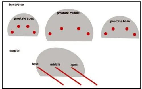
Standard 12- Core prostate biopsy done in each patient and samples were labelled properly
[image:41.612.180.430.396.553.2]according to site and side of the prostate and sent for pathology.
Figure 6: Biopsy locations in a 12-core biopsy
Ultrasound machine (B K Medical system) model Flexi focus 200 was utilised for prostate
imaging and to guide prostate biopsy. The ultrasound probe (rectal) model 8808e was a
7.5MHz biplane probe with slot for TRUS biopsy needle.
[image:42.612.102.510.118.439.2]
Figure 7: Ultrasonography Machine (Courtesy- B K Medical system) model Flexi focus 200and Trans rectal probe
[image:42.612.219.396.508.663.2]Outcomes:
Primary outcome was to assess urosepsis requiring hospitalisation.
Secondary outcome was to assess the incidence of other complications following TRUS
guided prostate biopsy. These
include- Fever
Hematuria
Hematochezia
Urinary retention
Pain or discomfort
All patients’ data including follow up, up to 30 days, were prospectively recorded, including
duration of hospitalisation including intensive care unit (ICU), if required.
Outcome Definitions:
Sepsis: SIRS with documented or clinically high suspicion of infection (SIRS includes -
temperature ≥ 38 C˚ or < 36 C˚; heart rate > 90 beats/minute; respiratory rate > 20
breaths/minute or respiratory alkalosis; WBC > 12,000 or < 4000 or immature forms > 10%
in case of normal range of total WBC) (6)
Infection – Any fever post biopsy more than 37.5 C˚
Gross hematuria – Visible blood in urine
Hematochezia – Blood noticed in stool
Urinary retention – Unable to pass urine after biopsy
Sample size calculation:
A target sample size of 95 was calculated using a precision of 4% and 90% desired confidence
level for the infection related complications. This was carried out assuming an average incidence
of infection related complication of up to 6%. This incidence was arrived at after a
comprehensive literature review for this statistics.
n= 4 p x q
d
2
p = 0.06
q= 1-p = 0.94
d= 0.004
Statistical analysis:
Statistical analysis was performed using Statistical Package for Social Sciences (SPSS®)
version 18 (IBM Corporation, USA).
Frequencies and percentages are used represent the categorical variables. eg; age, prostate
volume etc.
Descriptive statistics are used to represent continuous variables, eg; mean standard
deviation.
Chi square test and Fisher’s exact test were used to find the relationship between two
variables.
pvalue < 0.05 was considered as statistically significant.
A total of 89 patients underwent TRUS guided prostate biopsy for suspected carcinoma
prostate on the basis of raised PSA or abnormal rectal examination during study period.
Fourteen patients were excluded from the study as they received oral prophylaxis before
prostate biopsy regardless of their urine culture report. The remaining seventy five patients
were included in the study.
The demographic characteristics of the patients are given in Table 1. The mean age of
patients was 60.69 years. Diabetes and hypertension were the most common associated
co-morbid illnesses, being seen in over 58% of the patients. Some of these patients had both
[image:46.612.89.524.324.637.2]diabetes mellitus as well as hypertension as co-morbidities.
Table 1 Demographic characteristic (n=75)
Age (yrs)(SD) 60.69 (9.57)
Prostate size (cc) (SD) 24.07 (13.29)
PSA (ng/ml) 96.75
Co- morbidity
DM
HT
CAD
COPD
Post CVA
CKD
18
26
01
02
02
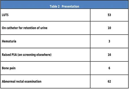
Clinical Presentation:
Most common presentation of these patients was lower urinary tract symptoms. During
evaluation, they were suspected to have prostate cancer either an abnormal rectal finding
or raised PSA. At our institution, we do not do screening for prostate cancer. Therefore the
number biopsies performed during the study period was quit small as compared to any
other studies where they do routine screening for prostate cancer. Some patients have
more than one clinical presentations eg; having LUTS and came with a report of raised PSA
(done elsewhere). Similarly, bone pain with LUTS was another common presentation.
[image:47.612.87.535.360.672.2]Details of clinical presentation are given in Table 2
Table 2 Presentation
LUTS 53
On catheter for retention of urine 10
Hematuria 3
Raised PSA (on screening elsewhere) 16
Bone pain 6
Pathological Features of Prostate Biopsy:
Among all the patients who underwent TRUS guided prostate biopsy, only 38 patients were
diagnosed as adenocarcinoma prostate rest of them did not show evidence of malignancy
on biopsy tissue. Majority of non-malignant pathology specimen were reported as focal mild
inflammation or chronic prostatitis. One patient’s biopsy was reported as granulomatous
prostatitis suggestive of Tuberculosis. He had other features suggestive of urinary
Tuberculosis.
Majority of cases had higher Gleason’s score on biopsy. 63% of patients had Gleason’s score
>7 and peri-neural invasion was seen in more than 81% of patients. Table 3 shows details of
[image:48.612.87.528.380.694.2]pathological features of prostate biopsy.
Table 3 Pathological features of Prostate biopsy
Adenocarcinoma prostate 39 (52%)
Gleason score
>7
7
3+4=7
4+3=7
<7
24 (61%)
7 (18%)
6 (15%)
2 (5%)
Peri-neural invasion 31 (81.58%)
Tumor volume (mean) 56.45%
Patients who were diagnosed as adenocarcinoma prostate on TRUS biopsy or presented
with bony pain underwent bone scan. Total of 38 patients had undergone bone scan, out of
which only seventeen patients did show evidence of osseous metastasis.
Pain during Prostate Biopsy:
Pain during TRUS biopsy prostate was minimal in majority of patients. On visual analogue
[image:49.612.87.525.291.398.2]scale (VAS), it was 2 or less in 84% patients as shown in Table 4
Table 4 Visual analogue scale VAS (n=75)
2 or less 63 (84%)
more than 2 12 (16%)
Complications following Prostate Biopsy:
Only five patients developed low grade fever after biopsy which subsided with antipyretics.
None of them required hospital admission. Out of these 5 patients, two patient’s pre biopsy
urine culture was positive, two had sterile culture and one had contaminants in urine
sample. Those two patients in whom urine samples were positive, they received appropriate
duration of antibiotics before and after procedure according to culture sensitivity.
Another 71 years of age gentleman with multiple co-morbidities, developed urosepsis and
septic shock after prostate biopsy. His pre-biopsy urine sample grew two organisms for
patient developed features suggestive of sepsis and was hospitalised. The antibiotic was
upgraded as the patient’s condition deteriorating. He was kept in ICU for 2 days. The urine
and blood culture sent before upgrading the antibiotic did not grow any organism. Total
duration of hospitalization was 12 days.
Twenty patients noticed mild hematuria post biopsy which settled in one or two days. Only
two patients had several episodes of gross hematuria lasted for more than 2 days which
ultimately resolved on its own. None of them required catheterization or bladder wash.
Two patients were catheterised after prostate biopsy as they were having overflow
incontinence. One patient was catheterised post biopsy due to sepsis. None of the patient
developed urinary retention following prostate biopsy.
Six patients noticed blood in stool after biopsy which settled on its own.
Incidence of hematospermia after prostate biopsy was not assessed in this study
[image:50.612.87.537.471.720.2]population. Table 5 summarizes the complications of Prostate biopsy.
Table 5 Complications following prostate biopsy (n= 34)
Minor Complications
Low grade fever
Hematuria < 2 days
Hematochezia
5 (6.7%)
20 (26.4%)
6 (8%)
Major Complications
Sepsis
Hematuria > 2 days
Urinary retention
1 (1.3%)
2 (2.7%)
Among all the patients, 20 of them had positive urine culture.
urethral catheter. E. coli was the most common organism found. Most of these urine
samples grew single organism. Five urine samples grew 2 organisms and one sample grew
three organisms. Most of these multiple organism urine samples were from patients who
were on per urethral catheter.
were shown in Figure 9
0 2 4 6 8 10 12 14 16 18 20
Figure 9: Showing complications following Prostate biopsy
Among all the patients, 20 of them had positive urine culture. Five of them were on per
E. coli was the most common organism found. Most of these urine
samples grew single organism. Five urine samples grew 2 organisms and one sample grew
three organisms. Most of these multiple organism urine samples were from patients who
catheter. Pattern of micro-organisms seen on urine culture samples
no. of patients
Figure 9: Showing complications following Prostate biopsy
Five of them were on per
E. coli was the most common organism found. Most of these urine
samples grew single organism. Five urine samples grew 2 organisms and one sample grew
three organisms. Most of these multiple organism urine samples were from patients who
organisms seen on urine culture samples
Antimicrobial resistance pattern among positive urine culture samples:
There were twenty patients in whom pre-biopsy urine culture was positive. Urinary
organisms were found resistant to one or more of the eight most common antimicrobial
agents used. Organisms were most frequently resistant to Cefpodoxime in our study. Seven
patients’ cultures were resistant to Co-trimoxazole and Nitrofurantoin. Only three urine
culture samples were resistant to Ciprofloxacin. Reason for apparently low resistance to
Ciprofloxacin was because sensitivity for this antimicrobial was not checked in 70% of urine
samples. See figure 10 for detail below.
0 2 4 6 8 10 12
Organisms
[image:52.612.96.548.64.354.2]Staph aureus NF GNB Morganella Klebsiella Enterobacter Enterococcus Pseudomonas E coli
Subgroup Analysis:
Relationship of Urine Culture with
Urine culture
Positive
No growth or contaminants
0 2
Amox/clavu Amikacin Cefoperazone sulb Nitorfurantoin Co-trimoxazole Gentamicin Cefpodoxime Ciprofloxacin
[image:53.612.90.506.66.305.2]Figure 11: Antimicrobial r
Table 6: Relationship of urine culture with infection
ulture with Infection:
Rate of infection (%)
4/20 (20.0%)
2/55 (3.6%)
4 6 8 10
: Antimicrobial resistance patterns of pre biopsy urine cultures
: Relationship of urine culture with infection
p Value
12
esistance patterns of pre biopsy urine cultures
[image:53.612.88.522.466.585.2]As shown in Table 6, patients with positive urine cultures had more infection as compared
to the other group where urine culture was either no growth or contaminants. There was a
statistically significant association of positive urine culture with rate of infection.
Relationship of Urine Culture with Hematuria:
Urine Culture Incidence of hematuria p Value
Positive 6/20 (30.0%)
0.939 No growth or contaminants 16/55 (29.1%)
In both groups almost equal number of patients had hematuria. There was no association
found between urine culture and incidence of hematuria. See Table 7
Risk of infection in diabetic patients:
Diabetes mellitus Incidence of infection (%) p Value
Present 2/18 (11.1%)
0.626
Absent 4/57 (7.0%)
Table 7: Relationship of Urine culture with hematuria
In our study, it was found that diabetic patients had more incidence of infection compared
to non-diabetics. But the association of diabetes and incidence of infection did not reach to
a statistical significance as shown in Table 8.
Association of Infection with indwelling urethral catheter:
Per urethral catheter Rate of Infection (%) p value
Catheter 0/10 (0%)
1.000
No catheter 6/65 (9.2%)
In our study, ten patients were on catheter prior to prostate biopsy. There was no infection
noted in this group following biopsy. Six patients in non catheter group had infection (9.2%).
This increase incidence of infection in no catheter group did not reach statistical significance
(p= 1.000). Table 9
Trans rectal ultrasound guided prostate biopsy has remained the gold standard to diagnose
prostate cancer. Biopsy related complications are not uncommon. Majority of the
complications are mild and self-limited but sometimes it could be severe and life
threatening. Infection and bleeding from urethra and rectum are the most common
complications following prostate biopsy. Some recent studies have reported increasing
trends of hospitalisation following prostate biopsy due to infection related complications.
To reduce the incidence of infection following biopsy, there were many prophylactic
regimens including oral as well as intravenous antibiotics recommended by various studies
(56,57). In our study, one dose of intravenous Injection Amikacin 15mg/kg was given just
before TRUS biopsy in those in whom urine culture showed no growth or contaminants.
Otherwise patients received 3 days of antibiotic course before biopsy and continued for
total of seven days according to culture and sensitivity.
Rectal cleansing with povidone-iodine and enema plus antibiotics has been explored by
various studies with controversial conclusions. Some studies say that it reduces infectious
complications but few of them conclude no difference in complications (58–60). However,
Cochrane review has concluded that risk of bacteraemia was reduced with enema plus
antibiotics when compared with antibiotics alone but risk of fever or infection was similar in
both groups(6). We have not used any kind of rectal cleansing or enema before prostate
biopsy.
Loeb S et al have identified various risk factors for infectious complications. These include;
Co-morbidities like Diabetes, COPD, Heart valve, benign prostate enlargement , recent
biopsy urine culture etc. (66) In our study , the risk factors identified were Diabetes mellitus,
COPD, per urethral catheter and positive urine cultures.
Prophylactic antibiotic before prostate biopsy is being used by all centres but the particular
antibiotic, dose and duration varies widely among centres. However, most studies showed
no significant benefit if duration is more than 24hours (67,69,70). At our centre, we utilised
single dose of intravenous Injection Amikacin just before biopsy.
Febrile UTI following prostate biopsy is common. Reported incidence rate of infection in
different studies is around 3% (73,74). In our study, only one patient had febrile UTI which
progressed to sepsis and required hospitalisation despite of being on antibiotics prior to
procedure.
Incidence of fever reported in literature is about 3% to 3.5% (78,92). Our study had reported
fever in 6% of patient, higher than reported in previous studies.
Hospitalisation rate in our study was 1.3% similar to other studies in which it was 0.6% to
1.7% (60). But other studies have reported incidence of hospitalisation of 3.1% to 3.06% (73)
Another study reported increase in hospitalisation rate from 1% to 4.1% from 1996 to 2005
(75)
Hematuria is very common complaint following TRUS biopsy of prostate. Its incidence varies
in literature from 10-84% (71,75,77). There are various definitions of hematuria in different
studies (visible blood, need for catheterisation or hospitalisation, also duration of
hematuria). In a cohort study, incidence of hematuria was reported as 65.8%, but
bothersome hematuria was only 6.2% (77). Our study reported hematuria in 26.4% patients.
But bothersome hematuria which lasted for more than 2 days was noted in only 2.7%
large prospective study on prostate cancer screening had reported prolonged hematuria (>3
days) in 22.6% and it was correlated with prostate volume (78). We could not assess this
association in our study population as the number patient in our study was quit small as
compared to ERSPC study where approximately 6000 persons participated. There are
studies which had evaluated relationship of hematuria with number of biopsy cores, size of
the biopsy needle etc. and showed conflicting results. We did not evaluate these factors in
present study.
Incidence of hematochezia ranged from 1.3% and 45%. Studies had shown that incidence of
bleeding increases with increased number of prostate biopsy cores and anticoagulative
drugs (79).Our study did show incidence of rectal bleeding around 8%. A very low incidence
rate as compared to previously reported in various studies. This could be due to smaller
needle size used for biopsy and proper patient evaluation before biopsy. In most of the
cases of rectal bleeding, if patients are counselled properly regarding rectal bleeding and
hematuria, it is of little consequence. It is very rare to see massive rectal bleeding and could
be fatal. No such massive hematochezia noted in our study. Rectal bleeding was noted in
few patients in our study but it was self limiting.
Hematospermia was noted in almost all studies following prostate biopsy. Its incidence
varied from as low as 1% to as high as 93% (87). Gradually it declined over several weeks.
Studies have reported anxiety and reduced sexual activity associated with hematospermia
which resolved after about eight ejaculations. ERSPC study showed hematospermia in 50.4%
(78). Our study population included majority of patients above 60 years of age. And almost
all of them did not have intercourse for a long time, either because of decreased libido or
TRUS guided prostate biopsy causes significant amount of pain. Therefore some form of
analgesia is mandatory now. One study noted that TRUS biopsy prostate was associated
with significant pain and discomfort as well as anxiety. This has resulted in reluctance to
subsequent biopsy, among those it was required (77). There are other factors affecting pain
during biopsy like rectal compliance, volume of prostate and number of prostate biopsy
cores. Left lateral decubitus position during biopsy was found to have slightly less pain,
although the difference may not be clinically significant. Various types of
anaesthesia/analgesia were described for prostate biopsy. Among them, peri prostatic
nerve block (PPNB) is safe and effective procedure. We performed prostate biopsy in left
lateral decubitus position and injected 2% Lignocain 5ml diluted with 5ml of normal saline as
PPNB. It was very effective in reducing pain during biopsy and majority of our patients (84%)
did not have clinically significant pain (based on VAS ≤2).
Risk of urinary retention following TRUS-Biopsy prostate is very small (0.2% to 1.7%).
Number of cores taken during biopsy has no correlation with incidence of retention of urine.
One study had assessed factors directly linked to retention of urine, includes volume of
prostate, ratio of transition zone to total prostate volume and a higher IPSS score (78,81,3).
In our study, no patient had retention following prostate biopsy. There were two patients
with overflow incontinence and therefore were catheterised after prostate biopsy.
Overall, the risk of mortality is very low following prostate biopsy. Some studies have
The subgroup analysis of our study showed that patients with positive urine cultures had
increased rate of infection compared to other group with sterile urine culture or
contaminants. It was