“
A CLINICAL STUDY ON VENTRAL HERNIAS
”
Dissertation submitted to
THE TAMILNADU DR. M.G.R MEDICAL UNIVERSITY
CHENNAI
In partial fulfilment of regulations
For award of the degree of
M.S (GENERAL SURGERY)
BRANCH
–
1
KILPAUK MEDICAL COLLEGE
CHENNAI-600 010
BONAFIDE CERTIFICATE
This is to certify that the work entitled “A CLINICAL STUDY ON VENTRAL HERNIAS” is a bonafide work performed by Dr.VINODH.D, post
graduate student, Department of General Surgery, Kilpauk Medical College, Chennai-10, under guidance and supervision in fulfilment of regulations of the Tamilnadu Dr.M.G.R Medical University for award of M.S. Degree Branch I (General Surgery) during the academic period from May 2012 to April 2015.
Prof. N. Gunasekaran., M.D., D.T.C.D., The DEAN
Government Kilpauk Medical College Chennai - 600 010.
Prof. P.N.Shanmugasundaram, M.S. Prof. R.Kannan, M.S. Professor and Head Professor and Head
CERTIFICATE
Certified that Dr. VINODH. D has worked on the dissertation
“A CLINICAL STUDY ON VENTRAL HERNIAS” under my guidance and
Supervision. The consolidated report presented here is based on bonafide cases Treated in Govt. Royapettah Hospital & Kilpauk Medical College Hospital. The Observations and conclusions made by the candidate are his own and have been Verified by me.
Dr. R. KANNAN, M.S., (GUIDE)
Professor and Head
Department of General Surgery
DECLARATION
I solemnly declare that this dissertation “A CLINICAL STUDY ON VENTRAL HERNIAS” was prepared by me at Government Royapettah hospital and Kilpauk Medical College and Hospital, Chennai, under the guidance and supervision of Prof P. N. Shanmugasundaram M.S, Professor and Head of
Department of General Surgery, KMCH and Prof. R. Kannan, M.S., Professor
and Unit Chief, Government Royapettah Hospital, Chennai.
This dissertation is submitted to The Tamil Nadu Dr. M.G.R. Medical University, Chennai in partial fulfilment of the University regulations for the
award of the degree of M.S. Branch I (General Surgery).
Kilpauk Medical College & Hospital Chennai.
ACKNOWLEDGEMENT
At the outset, I would like to thank my beloved Dean,
Kilpauk Medical College Prof. N.Gunasekaran M.D., D.T.C.D., for his kind
Permission to conduct this study in Govt. Royapettah Hospital & Kilpauk Medical College and hospital.
I would like to express my special thanks to Professor and Head, Department of General Surgery, Prof. P.N.Shanmugasundharam M.S,
Kilpauk Medical College and Hospital for permitting me to conduct this study.
I would like to thank wholeheartedly, Prof. R.Kannan M.S.,
Professor and Head, Department of General Surgery, Govt. Royapettah Hospital for their encouragement and guidance during the study.
I also express my special thanks to my Assistant Professor of Surgery Dr. Rosy Adhalene Selvi, M.S and Dr. B.N. Kalaiselvan, D.N.B for
their assistance and guidance.
I also thank all my postgraduate colleagues who have been a source of constant help and encouragement during my study.
Finally, I wholeheartedly thank all my patients for their active
co-operation in this study, without which this would not have become a reality.
CONTENTS
S.No Topic Page No
1 LIST OF FIGURE 8
2 INTRODUCTION 10
3 AIM OF THE STUDY 12
4 REVIEW OF LITERATURE 13 5 MATERIALS AND METHODS 96 6 OBSERVATION AND RESULTS 97
7 DISCUSSION 105
8 SUMMARY 111
9 CONCLUSION 113
10 BIBLIOGRAPHY 114
LIST OF FIGURES
Fig. No FIGURE Page No
1 INNERVATION & ARTERIAL SUPPLY OF ANTERIOR ABDOMINAL WALL 20
2 MUSCLES OF ANTERIOR ABDOMINAL WALL 23
3 CROSS SECTION OF ANTERIOR ABDOMINAL WALL 24
4 MUSCLES OF THE ANTERIOR
ABDOMINAL WALL 25
5 ANTERIOR ABDOMINAL WALL TRANSVERSE SECTION OF 25
6 ANTERIOR ABDOMINAL WALL DEEP ARTERIES & VEINS OF 28
7 FUNCTIONS OF THE ANTERIOR ABDOMINAL MUSCLES 30
8 ABDOMINIS MUSCLE & RECTUS SHEATH ANTERIOR VIEW 33
9 ABDOMINAL INCISIONS 35
Fig. No FIGURE Page No
11 PARAUMBILICAL HERNIA 55
12 PARAUMBILICAL HERNIAL SAC CONTAINING OMENTUM 57
13 INCISIONAL HERNIA 2LSCS – PREVIOUS 75
14 INCISIONAL HERNIA MESH REPAIR 76
15
NON-ABSORBABLE SUTURE MATERIAL & POLYPROPYLENE
MESH
84
16 INCISIONAL HERNIA SAC 90
17 ANATOMICAL REPAIR OF HERNIAL SAC 90
18 RIVES-STOPPA MESH REPAIR 91
INTRODUCTION
DEFINITION:
Hernia:
A hernia is defined as an area of weakness or complete disruption of the fibro muscular tissues of the body wall. Structures arising from the cavity contained by the body wall can pass through, or herniate, through such a defect. While the definition is straightforward, the terminology is often misrepresented. It should be clear that hernia refers to the actual anatomic weakness or defect, and hernia contents describe those structures that pass through the defect. These hernias are basically classified into two types, depending upon their visibility.
a) External hernias are those which are visible from outside, like inguinal, incisional, femoral, epigastric.
Ventral Hernia:
Are those hernias, which occur through the anterior abdominal wall. The anterior abdominal wall is the site of a variety of hernias due to man’s erect posture which renders the anterior abdominal wall weak. Almost all these hernias protrude through the abdominal wall to form palpable swellings.
AIMS OF THE STUDY
1. To study the various etiologies of ventral hernias.
2. To determine the age distribution, sex ratio & clinical presentation of individual hernias.
REVIEW OF LITERATURE
The word Hernia is derived from the Greek word (Hernias, bud) meaning an offshoot, a budding or bulge. The Latin word Hernia means rupture or tear. Hernia was recognized about 1000 years ago. Probably the reason for this is the upright position which man has assumed during the revolutionary process. Hernia was treated by several ways with the available simple measures like bandages, ointment, poultices and localized concoctions. Cutting and countering operations were common in India, China and Japan long before Hippocrates.
Omphalocele was well known to Ambrose pare who described in his book ‘The
Works published in 1634. Astly Cooper (1804) was the first person to report the successful treatment of exompholos and he was the originator of one stage repair of small omphalocele.
Astley Cooper discovered the Transversalis Fascia and pointed out that this layer was the main barrier to herniation.
Lucas Championnere apparently was one of the first to use the overlapping fascia technique in 1891.
latter resulted in a recurrence rate of 11% while after using a tension free mesh repair is amounted to only 1%.
INCISIONAL HERNIA:
Witzel (1900), Goepel (1900), Barlett (1903) and McGavin (1909) advocated the use of Silver wire filigree.
Koontz and Throckmorton (1948) used Tantalum Gauze.
Fascia Lata grafts used in the form of strips of sheets have been reported.
Shortly the advent of synthetic Plastic sheets and the polyvinyl alcohol sponge were used.
HISTORY OF SURGICAL MESHES:
Artificial material was introduced in 1889 by Witzel who used a mesh of silver wire for abdominal wall hernias.
In 1959, Usher et al. reported the successful implantation of surgical meshes at first in 13 dogs and afterwards inpatients with abdominal wall hernias.
Busse in 1901 even used meshes made of gold wire.
In 1940, Ogilvie published the use of cloth mesheds to treat contaminated gun shot wounds with defects of the abdominal wall.
HISTORIC OVERVIEW OF MESH REPAIR
No. Event Introduction
1 Polyester mesh Wolsten Holme Arch Surg., 1956, 73, 1004 2 Polypropylene mesh Usher Arch. Surg. 1962;84;325
3 GPRVS Stoppa et al., 1973 (72) 4 Trans-inguinal preperational prostheses
Prostheses
Rives et al., chirurgie, 1973; 99:564.
5 Subfascial prosthesis to Lichtenstein Lichtenstein and Schulman, 1986(44)
6 Preperitoneal prosthesis by Extraperitoneal access
extraperitoneal access
Nyhus et al., An. Surg., 1988; 208:733.
Wantz,Surg.,1989;169:408
Wantz, Surg., 1989;169:408 7 Mesh plug Rutkow/Robbins Surgery, 1993; 114:3. 8 Plug Laparoscopy Shultz et al., clin. Laser Mon., 1990;8:103
9
Intraperitoneal onlay mesh
prosthesis (IPOM)
Transabdominal preperitoneal prosthesis (TAPP)
Shultz et al., clin. Laser Mon., 1990;8:103
Corbitt, Surg. Laparos Endose, 1991; 1:23.
10 TEPP
Ferzil etal.,laparosendcsc,Surg.,
1992;2:281
PARAUMBILICAL HERNIA:
Celsus in the first century A.D used an elastic suture in the treatment of umbilical hernias.
Willian J Mayo, on Aug 4th 1898 delivered his classical paper, remarks on a radical cure of hernia. He instituted the new classical technique of overlapping fascia for repair of umbilical hernia.
In 1979 Usher described a technique of repair using Marlex Mesh.
EPIGASTRIC HERNIA:
Epigastric hernias were first described in 1285.The term epigastric hernia was introduced by Leveille in 1812.
The first successful operation on this hernia was reported by Maunnior in 1802.
Ulrike Muschaweck in 2003 concludes using a Mesh plug in an epigastric hernia has advantages over the commonly used methods.
SPIGELIAN HERNIA:
EMBRYOLOGY
The abdominal wall begins to develop quite early in the embryo, but it does not achieve its definitive structure until the umbilical cord separates from fetus at birth. Most of the abdominal wall forms during closure of the midgut and reduction in relative size of the body stalk.
The primitive wall is somatopleure (ectoderm and mesoderm without muscle, blood vessels, or nerves). The somatopleure of the abdomen is secondarily invaded by mesoderm from the myotomes that developed on either side of the vertebral column. This mesodermal mass (hypomere) migrates ventrally and laterally as a sheet, and the edges differentiate while still widely separated from each other into the right and left rectus abdominis muscles. The final opposition of these muscles in the anterior midline closes the body wall.
Approximation of the two rectus abdominis muscles in the midline proceeds from both caudal and cranial ends and is complete by the 12th week, except at the umbilicus. The final closure of the umbilical ring awaits the separation of the cord at birth, but the ring may remain open in which case an umbilical hernia is present. Most such hernias gradually close spontaneously.
ANATOMY OF THE ANTERIOR ABDOMINAL WALL
ANTERIOR ABDOMINAL WALL:
The anterolateral abdominal wall is a complex musculoaponeurotic structure.it is bounded by the flare of coastal margins and xiphoid process of sternum above and by the iliac crests, inguinal ligaments and pubis below. The structures that comprise the anterior abdominal wall are skin, subcutaneous tissue, superficial fascia, antero-lateral muscles of the abdomen, together with their enveloping fascial sheaths and aponeurosis, transversalis fascia, extra peritoneal adipose and areolar tissue and parietal peritoneum.
I. SUPERFICIAL FASCIA:
The fascia contains fat, cutaneous nerves, cutaneous vessels and superficial lymphatics.
Below the level of umbilicus fascia is divided into a superficial fatty layer (fascia of camper) and a deep membranous layer (fascia of scarpa). Most part of the fascia is a single layer that contains variable amount of fat.
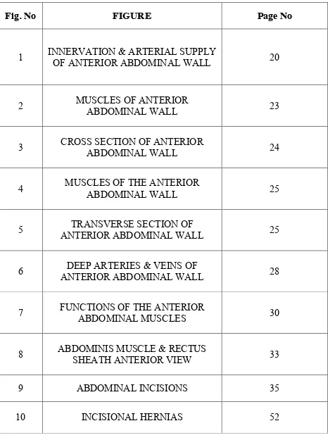
II. CUTANEOUS NERVES
Skin of anterior abdominal wall is supplied by the lower six thoracic nerves and by the first lumbar nerve.
III..CUTANEOUS ARTERIES AND VEINS
The venous drainage corresponds to arteries
FIGURE NO.1 1
SEGMENTAL INNERVATION OF THE ANTERIOR ABDOMINAL WALL AND ARTERIAL SUPPLY TO THE ANTERIOR ABDOMINAL
IV.SUPERFICIAL LYMPHATICS
Above the level of the umbilicus, the lymphatics run upwards to drain
into the axillary lymphnodes. Below the level of umbilicus they run
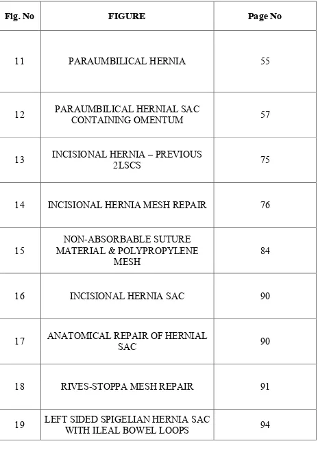
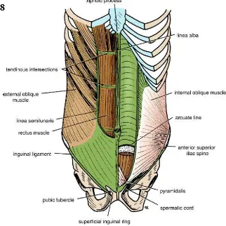
[image:22.595.141.457.138.415.2]ANTERIOR ABDOMINAL WALL MUSCLES:
1. THE EXTERNAL OBLIQUE MUSCLE:
This muscle is largest and thickest of the flat abdominal muscles. Its broad origin includes the last seven ribs, the thoracolumbar fascia, the external lip of iliac crest and the inguinal ligament that inserts into pubic tubercle. The muscle belly gives way to a flat, strong aponeurosis at about the midclavicular line, and it inserts medially into the linea alba. The aponeurosis passes anterior to sheath of rectus abdominis and with care, it can be dissected from it. In general the fascicles pass from the superolateral to inferomedial. Thus the direction of force generated by contraction is superolateral. The nerve supply of this is from ventral rami of lower six thoracic spinal nerves.
2. THE INTERNAL OBLIQUE MUSCLE:
Inguinal ligament pursue a downward course and insert into os pubis between symphysis and the tubercle. Some of the lower fibres are pulled into the scrotum by the testis as it passes through the abdominal wall and called the cremastric muscles of the spermatic cord. The Nerve supply of this is from Ventral rami of lower six thoracic and first lumbar spinal nerves.
3. THE TRANSVERSUS ABDOMINIS MUSCLE:
It is the smallest of the three flat muscles and originate from lower five ribs, the Thoracolumbar fascia, the internal lip of iliac crest, and the lateral third of the inguinal ligament. The direction of its fibers is transverse and they give way to a flat aponeurosis that inserts into the linea alba. The aponeurosis passes behind the rectus sheath in its upper two-third. The fibers that originate from inguinal ligament pass downward to insert into os pubis, as do the fibers of the internal oblique. Occasionally, the lower fibers of both muscles inserts by means of a common tendon called conjoint tendon. The nerve supply is from the Ventral rami of lower six thoracic and first lumbar spinal nerves.
NOTE: The neurovascular plane of the abdominal wall lies between the internal oblique and transverses abdominis.
MUSCLES OF THE ANTERIOR ABDOMINALWALL
MUSCLES OF ANTERIOR ABDOMINAL WALL(CROSS-SECTION)
MUSCLES OF THE ANTERIOR ABDOMINAL WALL FIGURE NO 4
4. THE RECTUS ABDOMINIS MUSCLE:
It is a long strap like muscle which arises from two tendinous heads. The lateral head arises from the lateral part of pubic crest, the medial head from the anterior pubic ligament. The fibers run vertically upwards and get inserted into xiphoid process, seventh, sixth and fifth costal cartilages.
The nerve supply is from the Ventral rami of lower six or seven thoracic spinal nerves.
5. THE CREMASTER MUSCLE:
The muscle is fully developed only in the male. In female it is represented by a few fibers only. Along with the intervening connective tissue, the muscle loops form a sac like cremastric fascia around spermatic cord deep to external spermatic fascia.
6. THE PYRIMIDALIS MUSCLE.
It is a rudimentary muscle in human beings. This is a small triangular muscle arising from anterior surface of body of pubis. Fibers pass upwards and medially to be inserted into linea alba.
The nerve supply is from the Subcostal nerve which is the ventral ramus of the twelfth thoracic spinal nerve.
7. DEEP ARTERIES AND VEINS OF ANTERIOR ABDOMINAL WALL
FIGURE NO 6
DEEP ARTERIES AND VEINS OF ANTERIOR ABDOMINAL WALL
8. DEEP NERVES OF THE ANTERIOR ABDOMINAL WALL
9. FUNCTIONS OF ANTERIOR ABDOMINAL WALL MUSCLES:
The abdominal muscles provide a firm but elastic support for the abdominal viscera against gravity. This is chiefly due to the tone of the oblique muscles, especially the internal oblique. They are also accessory muscles of respiration.
The oblique muscles assisted by the transversus, can compress the abdominal viscera and thus help in all expulsive acts, like micturition, defecation, parturition, vomiting.
The external oblique can markedly depress and compress the lower part of the thorax producing forceful expiration, as in coughing, sneezing, blowing, shouting.
Flexion of the lumbar spine is brought about mainly by the rectus abdominis.
Lateral flexion of the trunk is done by one sided contraction of the oblique muscles.
FIGURE NO 7: PICTURE SHOWING FUNCTIONS OF ABDOMINAL WALL MUSCLES
10. RECTUS SHEATH:
posterior lamina of the aponeurosis of the internal oblique and aponeurosis of the transverse muscle. Below the arcuate line anterior wall is formed by aponeurosis of all the three flat muscles. The aponeurosis of the transverses and internal oblique are fused, but the external oblique aponeurosis remains separate. There is deficiency of posterior wall.
CONTENTS:
The rectus abdominis muscle The pyramidalis muscle External oblique
Transverses muscle muscle
Internal oblique muscle rectus muscle The superior epigastric artery and veins The inferior epigastric artery and veins
The terminal parts of lower six thoracic spinal nerves
The aponeurosis of the transverses and internal oblique are fused.
11. LINEA ALBA:
The linea alba is a tendinous raphe formed by interlacing fibers of the three aponeurosis forming the rectus sheath. It extends from the xiphoid process to the pubic symphysis
. Above the umbilicus it is about 1 cm wide, but below the umbilicus it is narrow and difficult to define. It is so called because it is a white line.
12. FASCIA TRANSVERSALIS
This fascia lines the inner surface of the transversus abdominis muscle. It is a continuous lining of the abdominal cavity and is considered to be the strongest layer of the abdominal wall.
Deep inguinal ring is an oval opening in the fascia transversalis.
ANTERIOR VIEW SECTION OF THE ABDOMINIS MUSCLE AND THE RECTUS SHEATH.
13. CONJOINT TENDON
It is formed from lower fibres of internal oblique and lower part of aponeurosis of transverse abdominis. It is attached to pubic crest and pectineal line. It descends behind the superficial inguinal ring and acts to strengthen the medial portion of the posterior wall of the inguinal canal.
14. INGUINAL LIGAMENT
[image:35.595.154.476.392.714.2]It is the thick, in rolled lower border of the aponeurosis of external oblique and stretches from anterior superior iliac spine to the pubic tubercle. Its grooved abdominal surface forms the floor of the inguinal canal.
FIGURE NO 8
15.EXTRAPERITONEAL ADIPOSE AND CONNECTIVE TISSUE
LAYER
It contains adipose tissue, inferior epigastric artery and vein and fetal structures, medial umbilical ligaments (obliterated umbilical artery), obliterated urachus (median umbilical ligament), ligamentum teres (obliterated umbilical vein).
16. PARIETAL PERITONEUM
It is the inner most layer of the abdominal wall. It is a thin layer of dense irregular connective tissue and is covered on the inside by a layer of simple squamous mesothelium. The peritoneal membrane is innervated from above downward in a sequential manner by spinal nerves T7-L1. The peritoneum provides little strength in wound closure, but it affords remarkable protection from infection if it remains unviolated.
ANATOMY 0F
FIGURE NO 9
THE ANATOMY OF ABDOMINAL INCISIONS
Incisions through the abdominal wall are based on certain anatomical principles. The intra-abdominal pressure is considerable and the surgeon aims at leaving the abdominal wall as strong as possible after operation, Otherwise there exists a real fear that a portion of abdominal contents may leave the abdominal cavity through the weak area which is caused by a badly placed incision resulting in hernia. The principles governing abdominal incisions are;
1. The incision must give ready access to the part to be investigated and should admit extension if required.
3. The incision must be transverse, as the scar left in the peritoneum is best protected by muscle.
4. The rectus muscle may be cut transversely without seriously weakening the abdominal wall, as such a cut passes between two adjacent nerves without injuring them. The rectus has a segmental nerve supply so that there is no risk of a transverse incision cutting of the distal part of the muscle from its nerve supply, as would obtain if a muscle was divided which depend on a single nerve.
5. The incision must divide no nerves.
CLASSIFICATION OF VENTRAL HERNIA
A. Congenital – Present at birth 1. Omphalocele
2. Gastroschisis 3. Umbilical: infant B. Acquired
1. Midline
Diastasis recti Epigastric
Umbilical: Adult, Acquired, Paraumbilical 2. Median
Supravesical 3. Paramedian
Spigelian Interparietal C. Incisional
It depends on previous operative incision D. Traumatic
INCIDENCE
Epigastric Hernia:-
They are not very common. The incidence varies from about 0.8% to as high as 7-8% of all hernias operated upon. More common in men than in children and rare in women. Mostly seen in early adulthood, middle age and also seen in multipara. Up to 20% may be multiple, but usually one is dominant Strangulation is very unusual.
Umbilical Hernia:-
Adult umbilical and Para umbilical hernias are more common in women and in obese persons. Estimates of the incidence of umbilical hernia at birth vary greatly. In Caucasian infants, they range between 10-30%. In children
of African descent, it may be several times greater. Children with raised
intrabdominal pressure owing to ascites, COPD, or ventriculoperitoneal shunt,
also tend to develop an umbilical hernia. The incidence of Para umbilical
In his study Ghori, Jain et. al. reports the maximum incidence (41%) in female at .the age of 40 years. The adult umbilical hernia may undergo strangulation at any time. Strangulation is more common in women than in men, occurring between 40-50 years and very small number between 50-70 years. They account for 0.03% of the total hernias operated upon.
Incisional Hernia:
The frequency with which hernia results in the scar of abdominal
SPIGELIAN HERNIA:
There are about 1000 case reports described about spigelian hernia. They are common in fifth or sixth decade. Both sex are affected equally. Strangulation is common. In one review of the subject, in 1984 the mean age was 50 years and the ration of women to men was 1.4: The ratio of hernias on
right side to hernias on the left side was 1.6:1. The hernia was bilateral in 24 of
744 patients. In ten cases there were more than one hernia on the same. Side.
Most of the hernias were located below the level of the umbilicus; only 28
were above this level. The youngest recorded patient was six days old, and the
oldest was 94 years of age. Incarceration at the time of the operation occurred
in 69 to 325 patients (21.2%). The hernial sac was situated subcutaneous in
only 15 cases while in most cases the hernia was located between the
musculoaponeurotic layers of the anterior abdominal wall.
AETIO PATHO-GENESIS
The main causes for production of ventral hernia can be classified into congenital and acquired causes.
1). Congenital Causes:-
Congenital sac, apertures in the linea alba and aponeurosis or in linea semi lunaris.
The umbilicus is sometimes imperfectly developed at birth permitting the viscera to protrude through the umbilical cord.
Congenital muscle defects. 2) Acquired Causes:-
The hernia may result from any condition which tends to weaken the abdominal wall or tends to increase the intra-abdominal pressure. Post-operative Incisional hernias may result from imperfect closure of peritoneum and anterior abdominal wall following laparotomy.
Chronic strain (e.g. whooping cough in children, chronic Bronchitis, constipation, urinary out flow obstruction in adults).
Stretching and relaxation of abdominal musculature because of increase in size of contents e.g. Obesity, Pregnancy.
Obesity - Fat acts like a pile driver as it separates muscle bundles and layers, weakening aponeurosis and favours the outcome of hernia.
EPIGASTRIC HERNIA:
This is a small protrusion, usually composed of pre peritoneal adipose tissue occurring in the linea alba between the xiphoid process and umbilicus. The hernia varies considerably in size from pea nut size to a tennis ball size. It is possibly owing to lack of fibres at midline decussation which allows
preperitoneal fat to be herniated between the gaps. It starts as protrusion of a lobule of fat through an abnormally wide opening for blood vessels or through a congenital defect in the linea alba and posterior rectus sheath. The fact that it is common between 20 and 50 years of age probably reflects a balance between
a congenital defect and a rise of intra-abdominal pressure, adiposity, and
weakening of the muscles in adults. It is more frequent in people with a wide
linea alba.
Epigastric hernia is generally considered an acquired lesion, probably related to excessive strain on the anterior abdominal wall aponeurosis.
Moschowitz emphasized the importance of blood vessels perforating the linea alba and prolongation of the transversalis fascia at this point.
Askar’s studies also demonstrated that fibers originating from the
the linea alba at a site midway between the xiphoid and the umbilicus. Uncoordinated vigorous, synchronous contraction of the diaphragm and upper abdomen may occur during straining and coughing. The force caused by upward traction on the diaphragm and lateral traction on the tendinous intersection would be maximal at this point of attachment midway between the xiphoid and the umbilicus, the most common site of Epigastric Hernia.
Omentum is not uncommonly found in the epigastric hernia, but stomach, colon and small intestines are rarely found in them. At the earlier stage the hernia consists of only fat and known as fatty hernia of linea alba.
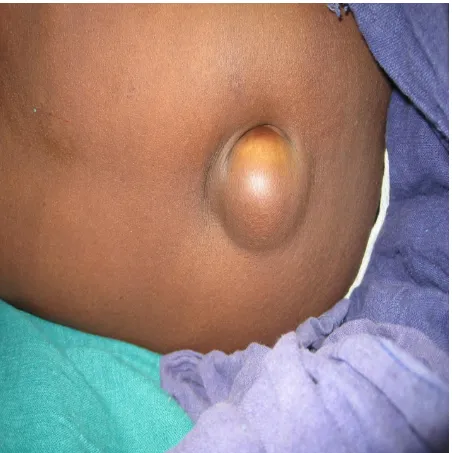
UMBILICAL HERNIA:-
Hernias occurring at or around the umbilicus.
Infantile umbilical hernia
If the scar formation is abnormal or ring is large, herniation of the intra-abdominal contents will occur through this defect. Because of these factors there is increased incidence in premature babies of umbilical hernia formation.
Para umbilical hernia
Etiological factors can be divided into congenital and acquired factors.
Congenital:
Due to anatomical weakness, maldevelopment of abdominal wall few
variations in their attachment and arrangement of abdominal muscle.
A positive relation between the pattern of aponeurotic decussating and herniation has been demonstrated by study conducted by Askar with a single midline decussating a midline hernial defect is seen. Congenital widening of the umbilical orifice predisposing factor.
Acquired:
a)Predisposing factors
1) Faulty ligation of umbilical cord
Umbilical cord ligation more 4-5cm from the abdominal wall may give rise to development of hernia.
2) Umbilical sepis- weakness umbilical area.
3) Increased intrabdominal pressure, due to chronic cough, constipation, Straining while passing urine, ascites.
b)Contributing factors 1. Low birth weight
2. Race
3. Sex: Female: Male=3:1 4. Family history:
Familial history contributes but no generic pattern of inheritance has been seen.
5. Age: more common in children< 2yrs and elderly people.
It occurs through a weak spot in the linea alba either above or below the umbilicus. In this type the umbilicus is normal. The exact etiology is obscure.
The most reasonable hypothesis seems to be the one given by Mayo. He considered para umbilical hernias to be caused by downward traction of the abdominal wall bearing on a fixed point at the umbilicus.
The site of attachment of lower tendinous insertion of rectus abdominis to the lateral border of linea alba seems to be the critical spot for the
development of para umbilical hernia. The hernia progresses to attain enormous
size. The content may be from omentum to small bowel and large bowel. Because the fibers break unevenly, many locules develop. Because the neck is small, complications are common, because of subclinical infections adhesions within the sac is very common. Coverings of the sac are peritoneum, fibrous linea alba and skin.
6. Multiparty due to stretching and weakening of the anterior abdominal wall
musculoaponeurotic layer.
7. Associated conditions-some congenital condition like mongolism cretinism, meningomyelocele, hurler’s syndrome, and amourotic family idiocy may be
SPIGELIAN
A spigelian hernia is one that protrudes through the linea semilunaris at any point in its extent. The most common site is at the junction of linea semilunaris with the linea semi circularis of Douglas.
The spigelian hernias has also been called “Masked Hernia” because in
some cases, the hernia protrudes deep to the aponeurosis of the external oblique and may be difficult to identify. Factors such as age, obesity, multiple pregnancies, straining increases intra-abdominal pressure, and paralysis have been cited as predisposing causes. Incarceration of the spigelian hernia is a frequent phenomenon, because the hernial neck, in addition to being narrow, most often is firm and fibrous.
Ageing and weight loss are generally regarded as important causative factors.
Spigelian hernia is associated with a high rate of intestinal obstruction, which can probably be explained by the combination of a small hernial opening with rigid edges and the fact that the hernia is often only diagnosed when symptoms consistent with intestinal obstruction are apparent.
INCISIONAL HERNIA
The post-operative ventral abdominal hernia or Incisional hernia is due to failure of lines of closure of abdominal wall following laparotomy. The approximated tissue separate and abdominal organs bulge through the gap. It is covered from inside out with peritoneum, scar tissue and skin. The hernias grow to reach enormous size and truly large hernias may contain most of the abdominal contents.
Etiology:-
There are many factors which causes failure of wound healing. The two main causes are poor surgical technique and sepsis. There are two types of Incisional hernias early and late type.
Early Hernia:-
It occurs soon after the original laparotomy closure, often involving the whole length of wound, grows rapidly. The main causes are
I. POOR SURGICAL TECHNIQUE:
1. Execution of Non Anatomical Incisions 2. Poor Wound Closure Technique
II. WOUND SEPSIS
III. USAGE OF DRAINAGE TUBES
IV. GENERAL CONDITION:-
Obesity, Old age, generalized weakness, hypoproteinaemia, anemia, diabetes mellitus, chronic liver failure, ascites, prolonged steroid therapy, immuno suppressive therapy, smoking and any other factor which persistently rise the intra-abdominal pressure or factors which influence the rate of Incisional hernia occurrence.
V. POST OPERATIVE COMPLICATION:-
Chronic coughs, distention of abdomen, benign prostatic hyperplasia, Stricture urethra, constipation are all factors favourable for development of Incisional hernia.
VI. TYPE OF OPERATION:-
VII. POST OPERATIVE WOUND DEHISCENCE:-
Burst abdomen whether covered by skin or frank evisceration is often followed by Incisional hernia, whether re-sutured or treated by open method. This is not surprising since practically all the conditions mentioned previously are also the causal factors in burst abdomen as reported by Efron in 1965.
Late Hernias:-
CLINICAL MANIFESTATIONS
INCISIONAL HERNIA
The patient’s complain of an unsightly bulge in the operation scar as well as of
PARAUMBILICAL HERNIA
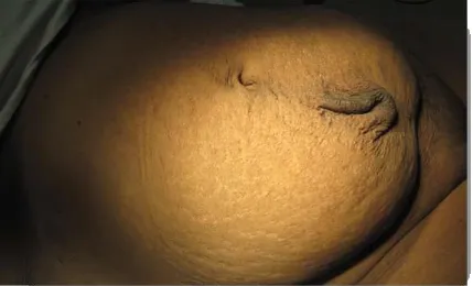
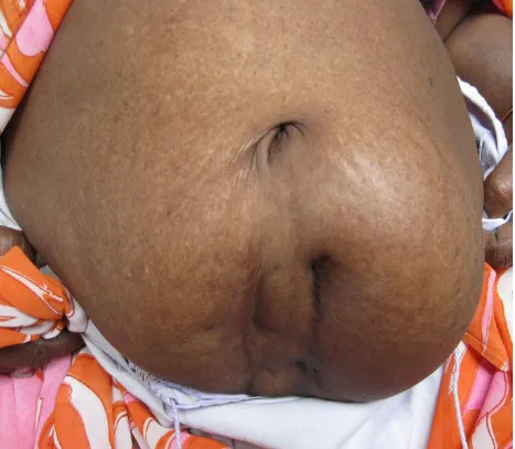
It usually develops in middle and old age and it is commonly found in case of obese females. The other symptoms are swelling and pain. This hernia soon becomes irreducible because of omental adhesions with in the sac. Gastrointestinal symptoms are common due to traction on stomach or transverse colon. It is a protrusion through the linea alba just above or just below the umbilicus and it is rounded or oval in shape, the edges are well made out, and surface is smooth. The consistency is soft when it contains intestine and firm when it contains omentum and has most expansible cough impulse.
Radiological imaging is not normally required to assist in the management of hernias. Clinical examination usually allows an accurate diagnosis. However, herniography, USG, CT and MRI scan are all established and accepted investigations for imaging hernias in cases of diagnostic uncertainty.
Most of the frequent complication of umbilical hernias is irreducibility. This is due to loculation with in the sac and omental adhesions with sac.
The most frequent complication of umbilical hernia is incarceration with or without strangulation; both are extremely rare in infants and children The incidence of obstruction and strangulation in adults is 10%. Previously
reducible or partially reducible can go for obstruction and strangulation. Strangulation is a frequent complication of a large Para umbilical hernia in
EPIGASTRIC HERNIA
The usual epigastric hernia is symptom less. The patient may complain of mild or even severe pain in the mass and of exquisite tenderness to tough. The pain is exacerbated by exertion and relieved by rest in the supine position. The smaller hernias may become painful because of strangulation of the preperitoneal fat nipped by the sharp facial edges of the opening. Omentum in the sac may strangulate in which case the hernia may become swollen, painful and tender, and the overlying skin may redden. Larger hernias containing bowel may also strangulate, but this is rare.
It usually present with a small round swelling in the midline between xiphisternum and umbilicus. They are often irreducible, sometimes multiple. In obese patients the typical smooth, rounded, slightly tender lump may be lost in the depths of subcutaneous fat.
DIVARICATION OF RECTI
SPIGELIAN HERNIA
Patients complain of pain or a lump or both at the site of herniation. The pain is sharp and constant or intermittent, or there is a dragging, uncomfortable feeling. If strangulation of the hernial contents is present, the pain will be severe or constant and associated with symptoms and signs of complete or partial (Richter) intestinal obstruction, going on to gangrene and peritonitis. Localized perforation into the sac may cause an abdominal wall abscess and even fistula.
MANAGEMENT
INVESTIGATIONS
As all patients of ventral hernia need surgical intervention they are evaluated medically by assessing their general condition for which routine blood investigations, urine examinations, chest radiographs (where needed) are done.
The assessment of abdominal wall hernias has long been a clinical skill that only occasionally required the supplementary radiological assistance of herniography. In almost all cases the correct diagnosis can be reached on the basis of the patients history symptoms and clinical examination
INDICATIONS FOR INVESTIGATIONS:
1. Patients who are obese
2. patients with palpable masses within deep layers of the abdomen
3. Patients with pain and complaints within the abdominal wall but without causative clinical findings.
But in some patients certain investigations are of benefit like: 1. Ultrasound of the abdomen
2. Herniography
Ultrasound abdomen, Herniography Computed Tomography, Magnetic Resonance Imaging are all established and accepted investigations for imaging hernias in cases of diagnostic uncertainty.
I. SONOGRAPHY
Sonography is indicated primarily in patients with palpable masses within deep layers of the abdominal wall, especially in obsess patients and in patients with pain located within the abdominal wall without any causative findings. In patients with hernia, a measurement of the defect can be done.
Sonographic Criteria for Hernias
Incisional Hernia
Sonography shows the typical hernial pattern with a fascial gap and protruding hernial sac. After mesh repair for hernia, a recurrence can occur at the edge of the mesh which can be seen sonographically.
Most hernias noted are incidental findings. The accurate demonstration of size, site, and contents of sac is useful in assessing the potential risk of strangulation or the likely success of hernia repair. Imaging is also useful when early dehiscence of the muscle layer in an anterior abdominal wall closure occurs without disruption of the overlying skin.
In comparison with CT or herniography, the ultrasonography is time as well as cost saving and not burdened with risks such as contrast allergy.
Epigastric Hernia
The hernia is visualized by a characteristic midline fascial defect.
Predictive Value
Divarication of Rectus Abdominis
Can be clearly visualized by sonography and the resulting herniation in abdominal wall.
Spigelian Hernia
A defect in the outline and an anterior bulging of the rectus margin confirm the hernia.
II. COMPUTED TOMOGRAPHY
Computed tomography is an excellent method of evaluating the abdominal wall and its relations to the abdominal viscerae. Lesions can be easily identified, owing to their different density.
There are several reports in the literature concerning the primary diagnosis of spigelian hernia by CT which can elegantly demonstrate.
CT allows exact evaluation of the volume and content of giant hernias. CT is also used to differentiate postoperative findings such as haematoma, abscess, or recurrence of hernia after laparoscopic repair of ventral hernia.
III. MAGNETIC RESONANCE IMAGING
Compared to CT, MRI offers the advantage of direct multiplane imaging without ionizing radiation and the use of contrast agents. A relative merit of MRI is the excellent demonstration of abdominal wall layers.
In conclusion, CT and MRI are not the first method of choice in the diagnosis of abdominal wall hernias. However these methods are useful in distinguishing hernias from benign, malignant or inflammatory lesions of the abdominal wall and their correlation to the intra-abdominal cavity, if clinical examination and sonography fails. In cases of abdominal wall relaxation, MRI allows direct comparison of the affected and the unaffected sides. The disadvantages include higher cost, limited availability and potential allergic reaction to contrast medium.
IV. HERNIOGRAPHY
Herniography has a low complication rate, relating mainly to accidental colonic puncture, of less than 1%, contrast allergy, and irradiation to pelvic region. It is invasive and is likely to be replaced by cross sectional imaging.
OPERATIVE MANAGEMENT
PRE-OPERATIVE PREPARATION
1. Optimal skin hygiene.
2. Weight reduction for obese patient. 3. To stop smoking.
4. The repair of a large postoperative ventral hernia should be delayed for atleast
one year after the operation that caused the hernia or after a previous attempt at repair.
5. Wait for atleast one year after all infection and sinuses have healed.
6. Associated cardiovascular, respiratory, renal conditions, Diabetes Mellitus,
hypertension and other general illness must be diagnosed, assessed, and treated. The operation is usually elective and must be delayed until the patient is in an optimal state.
7. Perioperative antibiotics are used more liberally.
8. The patient is investigated for coexisting abdominal pathology so that it can be
dealt with at the same operation.
9. The repair of a large postoperative ventral hernia should be delayed for atleast
one year after the operation that caused the hernia or after a previous attempt at repair.
11.Associated cardiovascular, respiratory, renal conditions, Diabetes Mellitus, hypertension and other general illness must be diagnosed, assessed, and treated. The operation is usually elective and must be delayed until the patient is in an optimal state.
12.Perioperative antibiotics are used more liberally.
13.The patient is investigated for coexisting abdominal pathology so that it can be
dealt with at the same operation.
INDICATIONS
1. Pain and discomfort.
2. Large hernias with small openings.
3. A history of recurrent attacks of sub-acute obstruction, incarceration,
irreducibility and strangulation,
GENERAL PRINCIPLES IN REPAIR OF VENTRAL HERNIAS
1. Spinal and epidural anaesthesia gives excellent relaxation with minimal
respiratory depression.
2. Hemostasis should be as careful and as effective as possible.
3. Permanent suture material should be used for the repair.
4. The choice of incision is governed by the orientation of the defect.
5. Healthy fascia must be isolated.
6. Closure of the sac is done in one layer, incorporating both fascia and
peritoneum after opening the sac, freeing all adhesions, reducing the viscera and exploring the abdomen.
The main danger in all forms of hernia is that of strangulation. Hernia left alone has got the tendency to increase in size and land up in complication one day or other. So there is hardly any reason for not operating on all hernias as soon as they are diagnosed.
EPIGASTRIC HERNIA:-
Simple Closure:-
It is employed for small hernias. The herniated fat is exposed through small transverse incision and is excised. The fascial opening in linea alba and some centimeters of fascia around it are cleared of fat. If the small sac is empty it is pushed into peritoneal cavity. If the sac has irreducible contents, the sac is opened and contents are pushed into abdominal cavity and sac closed. The opening in the linea alba is closed transversely with continuous or interrupted sutures of fine mono-filament polypropylene or polyamide, by taking generous bites of the edges.
Reconstructions of Linea Alba:-
When several such epigastric hernia occurs nearby it probably reflects a generalized weakness of the midline fascia, complete linea alba from sternum to umbilicus is repaired.
a) Modified Shoelace Technique.:-
b) Vertical Mass Closure Technique:-
Repair is done by taking bites that include not only linea alba but also part of anterior and posterior rectus sheath.
c) Vertical Overlap Technique:-
Vertical incision is made along the anterior rectus sheath to create a flap. The flaps are double breasted and repaired.
Repair of Large Epigastric Hernias:-
The larger hernial sac is opened, contents are replaced into abdomen, and sac is excised. Upper and lower edges of the fascial openings are approximated by continuous mass closure. The repair is reinforced by onlay nylon darn. Still larger hernias are treated by use of synthetic mesh.
Koontz's Operation:-
Ker's Operation:-
The sac is excised and peritoneum closed. Longitudinal incision is made in the posterior rectus sheath on both sides. The sheath flap is dissected free of rectus muscle. The gap in the posterior rectus sheath is reinforced with mesh. The rectus muscles are approximated. The flaps are double breasted anterior to the rectus muscles.
ADULT UMBILICAL HERNIA: -
INCISIONAL HERNIA
The main steps of surgery are as follows. Elliptical skin incision is made around the previous scar. Incision is extended above and below previous incision so that the healthy area can be identified and used to enter into peritoneal cavity without blindly disturbing the sac with adherent contents. After freeing the adhesions and dissecting out the sac, the sac is excised. Linea alba/Rectus sheath is strengthened. Layers of abdominal wall are closed.
There are various methods of repair of Incisional hernia.
I. Repair of Abdominal Wall:-
1. Anatomical layer by layer reconstruction. 2. Layered reconstruction - Cattell's operation.
II. Overlap Methods:-
1. Transverse overlap –Mayo’s.
2. Vertical overlap - Rutherford Morris. 3. Lanenskiold's Ribbon overlap procedure. 4. Chaimoff - Dintsman fascial flap method. 5. Muscle flap procedure.
6. Raviteh's operation.
III. Latice or Darn Repair:-
1. Burton's fingered fascia lata graft repair. 2. Nylon Darn - Huntor.
4. Stainless steel darning - Abel.
5. Skin /Duramater /fascia / tendon darning. 6. Shoelace Darn Repair.
IV. Extensive Tissue rearrangement technique:-
Nuttell's operation.
V. Repair by Implants:-
Various materials were used for this technique 1. Stainless steel plates, gauze.
2. Tantalum gauze. 3. Silver filigree. 4. Pliable plastic sheet.
5. Whole thickness skin graft. 6. Poly vinyl alcohol sponge. 7. Nylon Tricot.
8. Polypropylene mesh
9. Polytetra fluoroethylene (PTFE).
Rodney maingot advises 3 basic methods for repair of these hernias 1. Resuture
2. Shoelace darn repair
PROCEDURES:
I. REPAIR OF ABDOMINAL WALL:
1. Anatomical Layer by Layer Reconstruction:-
It is ideal for small hernias. The sac is excised and closed with chromic catgut. Anterior and posterior rectus sheaths are dissected out and closed with non-absorbable suture materials as one would do at the end of a laparotomy.
2. Cattell's Operation:-
Hernial sac is opened. Neck of the sac sutured in first layer, excess sac is cut off. Cut edge of the sac is sutured in second layer. Posterior rectus sheath is sutured in third layer. Muscles are sutured in fourth layer. Anterior rectus sheath is sutured in fifth layer.
II. OVERLAP METHODS:
Mayo's operation:- After excising and suturing the sac, rectus sheaths are dissected free of fat and overlapped transversely over each other and sutured in place.
Langenskiold's Operation:-
operative margin of the hernial orifice and tightened. These strips are either folded back and fastened with sutures or tied together in pairs. Over which skin is closed.
Chaimoff - Dintsman's Fascial Flap methods:- A vertical cut is made laterally
over the anterior rectus sheath and the fascia is raised and separated from the muscle. The raised flap is overlapped and sewed together.
Muscle Flap Procedures:- In upper abdominal hernias pectoralis major pedicle
flaps were used by Kenneth Me Kenzie in lower abdominal hernias tensor fascia femoris flap were used by O.H. Wangen Sten and Me Kenzie.
Raviteh's Operation:- Used for larger suprapubic hernias. Anterior rectus
sheath is dissected and overlapped and sutured. Inferior border of flaps are sutured to connective tissue over symphysis pubis.
III. LATICE OR DARN REPAIR:
Burton's Fingered Fasica lata Graft:- The hernial sac is dissected and
Several parallel lateral incisions of 2cm width are made on either side of the excess part of the graft. Graft is now placed under rectus sheath. Same numbers of slots are made about 2cms from the margin of hernial orifice on either side. The fascia lata strips are drawn through the slots. The strips are folded back, tightened, twisted in pairs with the opposite strips and held in place with sutures.
Maingot's Keel Operation:- Used for larger hernias. The sac is dissected but
not opened. Fibrofatty tissues at the margins of the ring are removed to expose healthy aponeurosis all around the sac. The loose peritoneal sac is inverted into abdomen by layers of sutures. The sac now resembles a keel of a ship dipping into peritoneal cavity. When strong aponeurosis margin of the hernia is reached they are sutured together with series of closely applied mattress sutures or continuous right angled Cushing's stitch
Shoelace Darn Repair:- Skin and fat are dissected out of hernia, as well as
by a continuous over and over suture of mono filament nylon. This creates a new linea alba. The sac remains unopened throughout the operation. The gap between the anterior rectus sheath is closed by the second suture with 6 m length of heavy monofilament nylon each starting at one end of the incision in the rectus sheath, and meeting in the middle, of the line of repair, where they are tied to one another. Skin closes over this with a drain
IV. EXTENSIVE TISSUE REARRANGEMENT TECHNIQUE:
Nuttell's Operation:-It is a type of repair in which extensive mobilization and
rearrangement of abdominal muscles were carried out. It was used for sub umbilical massive incisional hernias.
V. IMPLANTS:
Repair of Incisional hernia is one of the few instances in surgery in which implants of foreign materials were used to bridge the gaps, before the use of natural tissue. The modern era of prosthetic hernia repair began in 1958 when Usher reported his experience with polypropylene mesh. Later polyamide mesh and recently PTFE mesh were introduced. With the development of modern synthetic non-absorbable suture materials, three basic methods have emerged for repair. Resuture, shoelace darn technique and synthetic non-absorbable mesh closure. Resuture is used for small hernias.
TYPES OF PROSTHETIC MESH REPAIR:
Many variations and combinations of mesh repair have been described. They are as follows,
Underlay Graft:- A mesh may be sutured in place deep to peritoneum. Inlay Graft:- Mesh is placed between peritoneum and abdominal wall and sutured to edge of the defect.
Overlay/onlay Graft:- Larger mesh is placed over the defect and sutured. Both Inlay and Overlay:- are used in combination,
CHOICE OF MATERIAL
The ideal mesh is one that is cheap and universally available, is easily cut to the required shape, is flexible, slightly elastic and pleasant to handle. It should be practically indestructible and capable of being rapidly fixed and incorporated by human tissues. It must be inert and elicit little tissue reaction. It must be sterilisable and non-carcinogenic.
Polypropylene mesh meets the requirements of the ideal prosthesis and is
today the most commonly used material for repair of all types of hernia.
a)POLYPROPYLENE MESH (MARLEX,PROLENE)
This is currently most widely used prosthetic material in hernial repair. It is formed of knitted monofilament plastic fibers and has minimal elasticity or stretch capacity. Prolene elicits an intense desmoplastic reaction in tissue, accompanied initially by serous exudation and resulting eventually in the formation of a sheet of scar that uses the mesh as a scafford for its formation. The mesh thus becomes densely incorporated in the scar. In 1963, Usher introduced knitted monofilament polypropylene mesh into clinical practice. The disadvantages are visceral adhesions, erosion into the bowel/skin causing enterocutaneous fistula/ sinus formation, erosion of mesh into urinary bladder.
Sterilization: gamma radiation; after removal from its package, the mesh
FIGURE NO 15 : NON-ABSORBABLE SUTURE MATERIAL
POLYPROPYLENE MESH
It is supplied as a felted sheet in which fibers randomly interlace. It is used for vascular prosthesis. It is strong, pliable, soft, smooth and slippery to touch, biologically inert and causes little tissue reaction. It is costly.
c) POLYESTER MESH (DACRON) MERSILENE
It is multifilament knitted mesh. It is cheap, freely available, light, and supple, has a pleasant, soft feel and is strong and elastic. It excites greater tissue inflammatory reaction than prolene. It tears easily.
d) FASCIA LATA
It is harvested from lateral aspect of the thigh. It is strong and flexible although minimally elastic. The use has been abandoned.
INDICATIONS FOR MESH REPAIR
The indications are:
Repair of recurrent incisional hernias: successful repair of recurrent hernias in patients, whose musculature is of poor quality and weak and flabby, fascial coverings are thin and weak, requires prosthestic material.
In primary repair of massive hernia in which tissues are deficient and repair without tension cannot be accomplished readily by conventional techniques of direct suturing. The employment of a bridging prosthesis in a massive incisional hernia will enable the surgeon to avoid excessive tension in wound closure and the hazards of increased intra-abdominal pressure.
When continued presence of forces tending to disrupt in the future are reasonably predicable. There are certain conditions which present a relatively high risk of recurrence unless prosthetic materials are used. They are chronic cough, increased intra-abdominal pressure from obesity and massive incisional hernias.
Hesselink et al. have shown that any ventral/incisional hernia greater than 4 cm and recurrent hernial have a high rate of recurrences if not repaired with mesh.
Both large underlay and large overlay graft can be used together for very weak abdominal wall.
Reinforcing strips - Onlay and Underlay strips can be used. Wrap Around - Reinforcement of wound edges with mesh.
Two sheets of mesh sutured to abdominal wall then sutured to each other to draw together to the edge of the wound.
Onlay Technique
In this technique usually a polypropylene mesh is sutured to the anterior rectus sheath after the fascial defect has been closed primarily.
Procedure
After managing hernial sac and its contents as described in Mayo’s repair,
aponeurosis is approximated using polypropylene suture and prosthetic mesh is placed over the aponeurosis and fixed with polypropylene suture material. Suction drain placed subcutaneous tissue and skin sutured.
The potential advantage of this repair keeps the mesh separated from the abdominal contents by full abdominal muscle fascial wall thickness.
48
when the surgical wound becomes infected. No studies available to accurately state recurrence rates with this repair.
Inlay Mesh Repair
After reducing the sac and its contents, peritoneum is closed using chromic catgut and mesh fixed with polypropylene suture material. Rectus sheath is closed over the mesh. Suction drain kept and wound closed in layers. The potential advantage of this repair keeps the mesh separated from the abdominal contents by full abdominal muscle fascial wall thickness.
Disadvantages of this repair include, a repair under tension, large subcutaneous dissection that allows for seroma formation, and mesh infection when the surgical wound becomes infected. No studies available to accurately state recurrence rates with this repair.
.
Intraperitoneal Underlay Mesh Repair
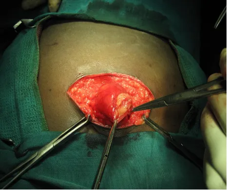
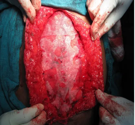
FIGURE NO 16: INCISIONAL HERNIA SAC
[image:91.595.99.498.489.746.2]RIVES-STOPPA TECHNIQUE (RETRORECTUS MESH REPAIR)
Another promising technique is the Rives-stoppa procedure developed for the repair of incisional hernias. Prosthetic material is used to close the defect in a so called sublay technique. The prosthesis is placed between the rectus abdominis muscle and posterior sheath. Above the umbilicus, dissection is performed above the posterior rectus fascia and underneath the rectus muscle. Below the umbilicus, the lack of a posterior rectus fascia necessitates dissection in the preperitoneal space. A large piece of polypropylene mesh is placed in the space created, and fixed to muscle layer above with full or partial thickness suture. The recurrence rate with this repair have been stated to be less than 10%.
LAPAROSCOPIC REPAIR OF INCISIONAL HERNIA
hernia repair with mesh is reasonable alternative to conventional repair for defects that will require mesh. Although this technique can probably not be justified for typical umbilical defects that would otherwise be closed primarily, further studies looking specifically at issues of cost, return to work, and long-term durability may establish the laparoscopic technique as the preferred mesh repair for large ventral hernias.
SPIGELIAN HERNIA
FIGURE NO 19: PICTURES SHOWING HERNIAL SAC AND CONTENTS ILEAL
BOWEL WITH MESENTRY IN LEFT SIDED SPIGELIAN HERNIA
[image:95.595.77.434.70.307.2]COMPLICATIONS AND PROGNOSIS OF VENTRAL HERNIA
MATERIALS & METHODS
Place of Study : Department of General Surgery,
Govt Royapettah hospital& Govt Kilpauk Medical College
Hospital.
Type of Study : Prospective & Observational study
Sample Size : 76
Inclusion criteria:
1. Adult patients above the age of 12 years.
2. Patients presenting with clinically apparent ventral hernia in outpatient
Department or in emergency who underwent surgery for the same.
Exclusion criteria:
1. Pediatric cases below the age of 12 years
2. Patient with ventral hernia who were unfit for surgery or refused
Surgery.
Type of analysis: Clinical data analysis
Data collection:
1. Patients were evaluated with a standardized questionnaire
2. Patients were subjected to thorough physical examination and
relevant investigations.
3. Patients were followed up for approximately 2 months after
PERCENTAGE OF DIFFERENT TYPES OF
VENTRAL HERNIAS
Among the 76 cases of ventral hernias studied 42 (55.26%) were incisional; 7 (9.21%) were epigastric; 17 (22.36%) were umbilical; and 9 (11.84%) were paraumblical and 1(1.31%) was Spigelian hernia. Three patients (one was a c/o cirrhosis with PHT with umbilical hernia and another a c/o coronary artery disease with incisional hernia) and another c/o coronary artery disease with COPD, who were unfit for surgery, were not included in the study.
Incisional Hernia
Paraumblical hernia
Epigastric hernia
Umblical Hernia
Spigelian hernia
SEX DISTRIBUTION OF VENTRAL HERNIAS
Ventral Hernia
Male
Female
Incisional Hernia
Male
AGE DISTRIBUTION
The youngest patient with a ventral hernia was a female patient with an epigastric hernia aged 24 years and oldest was a male patient aged 72years with an umbilical hernia. The highest incidence of Ventral hernia was noted in the
4th decade that is 27 cases which amounted to 35.6 % and the lowest incidence was in the 8th decade that is one case which is 1.3%. Among Incisional hernia
more cases were found in the 3rd decade (9 cases), 4th decade (14 cases), 5th decade (10 cases) which amounted to 78.57 % of all incisional hernia. In
epigastric hernia 2 cases each were found in 3rd, 4th, 5th decade and 1 case in
6th decade. The incidence in umbilical hernia was highest in 5th decade that is
CLINICAL FEATURES
All cases of ventral hernias presented with a swelling (100%). 30 cases presented with pain (39.47 %), 6 cases presented with features of obstruction (7.9 %). No case of incisional hernia presented with strangulation. 30 cases of ventral hernias were obese (cases) (39.47%), 3 cases had chronic cough (3.94%), secondary to chronic obstructive pulmonary disease. 3 cases were multiparous (5.35%). 13 cases had anemia (17.10%) and 3 cases had hypothyroidism (3.94%).
12
5
4
9
1 3
1 2
0 2 4 6 8 10 12 14
Incisional Hernia Paraumblical hernia Epigastric hernia Umblical Hernia Spigelian hernia
Clinical features
PREVIOUS SURGERY IN INCISIONAL HERNIA
OPERATIVE PROCEDURE
Ventral Hernia
Emergency
Elective
Ventral Hernia
Anatomical repair
Out of 76 ventral hernias 6 cases (7. 9%) were operated as emergency. 3 out of 6 Ventral Hernias—2 Umbilical and 1 Paraumbilical were anatomically
repaired. Out of 42 Incisional hernias 3 cases (7.14%) were operated as
Emergency, 2 were caused by previous hysterectomy and 1 by previous PS
Which presented with Features of obstruction. The other 39 cases (92.85%)
DISCUSSION
This present study has been compared to other series of similar nature, 76 cases of ventral hernia were taken up for this study which was done between April 2014 to September 2014.
Distribution of ventral hernia
This present study of 76 cases of ventral hernia had 42/76 cases (55.26%) of Incisional hernia, 7/76 cases (9.21%) of epigastric hernia and 17/76 (22.36%) of umbilical hernia, 9/76(11.84%) paraumbilical hernia. There was 1(1.31%) case of spigelian hernia. In S.M.Bose series (1999) of 175 cases 110 were incisional hernia(62.86%) 44 were umbilical hernias (25.13%), 21 cases were epigastric hernia (37.13%) 100% of all cases presented with swelling in the anterior abdominal wall, 18.26% presented with pain, 8.7% presented with features of obstruction with strangulation. This compares well with the S.M.Bose series (1999).
Epigastric hernia
SERIES PERCENTAGE 1 S.M.BOSE (1999) 12%
2 M.MOHAN RAO (1986) 9.86% 3 PRESENT SERIES 9.21%
The incidence of epigastric hernia in the present series is comparable with that of the M.MOHAN RAO series (36) and is slightly lower than S.M.BOSE series In this study of 7 cases of epigastric hernia 5 cases were male (71.4%). This agrees well with the Ponka series (38) which states that epigastric hernia is rarely seen in infants and children and is commonly seen in Males.
Clinical features
Pain was a presenting complaint in 57.14% of cases, swelling was a presenting Complaint in 100% of cases. There were no features of strangulation or
obstruction and