INFERIOR BORDER FIXATION FOR DISPLACED
MANDIBULAR ANGLE FRACTURES
A Dissertation s ubmitted in
Partial fulfillment of the requirements
for the degree of
MASTER OF DENTAL SURGERY
BRANCH – III
ORAL AND MAXILLOFACIAL SURGERY
THE TAMIL NADU DR. M.G.R. MEDICAL UNIVE RSITY
CHENNAI – 600 032
`
This i s to cer tify that Dr. S ATI SH V ASANT H.D, Pos t Gr adua te
studen t (2012 –201 5) in the Dep art men t of Oral and max illof acial
Sur ger y, Ta mi l Nad u Go ve rn me nt De ntal Co lle ge and Hospi tal,
Chenna i – 600 003 has done this dissertation titled
“COMPARISON OF SUPERIOR BORDER AND INFERIOR
BORDE R FI XATI ON F OR DI S PLACE D MA NDIB ULAR
AN GLE FRA CTUR ES ” u nder my di re ct guid ance a nd su per vision
in parti al fulf ill men t of th e re gu latio n s laid dow n by T he Ta mi l
Na du Dr. M. G.R. Me dica l U niver sit y, Chenn ai – 600 032 for
M.D.S., (Branch – III) Oral a nd Ma xi ll ofacial S urger y de gree
exa min atio n.
D r. D. DURAI RAJ, M DS., Dr. B . SARAVANAN, M DS., PhD Prof esso r and Guid e Prof ess or and H OD
Depa rt ment o f O ra l and M axi llo fa cia l Surg e ry , Tami l Nadu Gove rn ment Denta l Co lleg e and Hospital ,
Chennai – 600 003.
D r. S. PREM K UM AR, M DS., Pri ncipal in cha rge
I, DR.D.SATISH VASANTH, do hereby declare that the dissertation titled “C OM PAR ISON OF SU PE RIO R B ORD ER AN D IN FE RIO R
B ORDER FIX AT ION FOR D ISPLAC ED M ANDIB U LAR A NGL E FRA CTU RES” was done in the Department of Oral and Maxillofacial Surgery, Tamil Nadu Government Dental College & Hospital, Chennai-600 003. I have
utilized the facilities provided in the Government dental college for the study in
partial fulfillment of the requirements for the degree of Master of Dental Surgery in
the speciality of Oral and Maxillofacial Surgery (Branch III) during the course period
2010-2013 under the conceptualisation and guidance of my dissertation guide, Prof. Dr.D.Durairaj, M.D.S. I declare that no part of the dissertation will be utilized for gaining financial assistance for research or other promotions without obtaining prior
permission from the Tamil Government Dental College & Hospital. I also declare that
no part of this work will be published either in the print or electronic media except
with those who have been actively involved in this dissertation work and I firmly
affirm that the right to preserve or publish this work rests solely with the prior
permission of the Principal, Tamil Nadu Government Dental College & Hospital,
Chennai 600 003, but with the vested right that I shall be cited as the author(s).
Signature of the PG student Signature of Guide
This agreement herein after t he “Agreement” is entered into on this day ………. between the Tamil Nadu Government Dental
Co lleg e a nd Ho sp it a l repre se nt ed b y it s Prin cipa l ha ving addre s s at Ta mil N adu Go ver nme nt De nt a l Co l lege a nd Ho sp it a l, C he nna i -
600003 , (here a ft er re ferred t o as, ‘t he co lle ge ’)
And
D r.D.DURA IRA J M DS., aged 54 year s wo rk ing a s Pro fe s so r in Depart me nt o f Ora l a nd Ma x illo fac ia l S urger y at t he co lleg e, ha ving
res ide nce addre s s At Sr i Ku mara n I lla m, 3/19, Fir st St reet , Ragha va
Nagar, Mo o vara sa mp et , Che nna i-60 0091 (here a ft er re ferred t o as t he
'Pr inc ipa l I nve st ig at o r')
And
D r.D.SAT ISH VAS AN TH, age d 28 year s curre nt ly st ud ying a s fina l
ye ar Po st G raduate student in t he Depart me nt o f Ora l a nd
Ma xillo fa c ia l surger y, Ta mil N adu Go vernme nt De nt a l Co lle ge a nd
Ho sp it a l, C he nna i -3 r esid ing at P lo t No 7, Do o r No 1, G -4 ,
Pa la niap pa Nag ar, Va la sara vakka m, C he nna i. ( here a ft er re ferred t o as
the ‘PG and co - invest igator’)
Wherea s t he ‘P G st ude nt as part o f his curr icu lu m u nd ert akes t o
resear c h o n “COMPARISON OF SUPERIOR BORDER AND INFERIOR
BORDER FIXATION FOR DISPLACED MAN DIBULAR ANGLE FRACTURES”
fo r w hic h purpo se t he Gu ide s ha ll act as Pr inc ip a l inve st ig at o r and t he
Co lleg e s ha ll pro vide t he requ is it e in fra s t ruct ure bas ed o n a va ila bilit y
a nd a lso pro vide fa c ilit y t o t he PG st udent as t o t he e xt e nt po ss ible a s a
co nfide nt ia lit y is su es t hat ar ise in t his r eg ard
No w t his agr ee me nt w it ne ssed a s fo llo ws :
1. The part ie s agree t hat a ll t he Re sear c h mat er ia l a nd o w ner s hip
t here in s ha ll be co me t he ve st ed r ig ht o f t he co lle ge, inc lud ing in
part icu lar a ll t he co p yr ig ht in t he lit erat ure inc lud ing t he st ud y,
resear c h a nd a ll o t her re lat ed papers.
2. To t he e xt ent t hat t he co lle ge ha s le ga l r ight t o do go , sha ll gr a nt
to lic e ns e o r a ss ig n t he co p yr ig ht so ve s t ed w it h it fo r med ic a l
a nd/o r co mmerc ia l u sage o f int erest ed p erso ns/e nt it ies su b ject t o
a reaso na ble t er ms/co nd it io ns inc lud ing r o ya lt y as de e med by t he
co llege.
3. The Ro ya lt y so rece ived by t he co lleg e s ha ll be s hared equa lly
by a ll t he t hree part ies.
4. The PG/ Re searc h st ude nt and PG/Pr inc ip a l I nvest igat o r sha ll
und er no c ircu mst a nce s d ea l w it h t he co p yr ig ht , Co nfide nt ia l
in fo r mat io n a nd k no w - ho w-ge nerat ed dur ing t he co urse o f
resear c h/ st ud y in a ny ma nner w hat so ever , while s ha ll so le we st
w it h t he co lle ge.
5. The PG st ude nt and Pr inc ipa l I nve st ig at or undert ake no t to
d ivu lge (o r) cau se t o be d ivu lged a ny o f t he co nfide nt ia l
in fo r mat io n o r, kno w -ho w t o anyo ne in a ny ma nner w hat so ever
a nd fo r a n y purpo se w it ho ut t he e xpr es s wr it t en co nse nt o f t he
co llege.
6. All e xpe nse s pert a ining t o t he rese arc h s ha ll be dec id ed upo n by
t he pr inc ipa l in ve st igat o r/Co -invest ig at or o r bo rne so le by t he
PG st udent .(co -inve st igat o r)
7. The co lle ge s ha ll pro v ide a ll infr ast ruct ure a nd ac ce ss fac ilit ies
w it hin a nd in o t her inst it ut es t o t he e xt ent po ss ib le. T his
inc lude s pat ie nt int eract io ns, int ro ducto ry let t ers,
it s co mp let io n. Ho we ver t he se le ct io n a nd co nduct o f re se arc h,
to pic a nd are a res earc h by t he St ude nt Re s earc her u nd er gu ida nce
fro m t he Pr inc ip a l I nve st igat o r sha ll be su bje ct to t he pr io r
appro va l, r eco mme ndat io ns a nd co mme nt s o f t he Et hic a l
Co mmit t ee o f t he Co lleg e co nst it ut ed fo r this purpo se.
9. It is agreed t hat as regard s o t her asp ect s no t co vered u nder t his
agree me nt , but whic h pert a in t o t he rese arc h u ndert ake n b y t he
PG st udent , und er gu ida nce fro m t he Pr inc ipa l I nve st ig at o r, t he
dec is io n o f t he Co lle ge sha ll be bind ing a nd fina l.
10. I f a ny d isput e ar is e s a s t o t he mat t ers re la t ed o r co nnect ed t o t his
agree me nt her e in, it s ha ll be re ferred t o arbit rat io n in ac co rdance
w it h t he pro vis io ns o f t he Ar bit r at io n and Co nc iliat io n Act ,
1996.
In w it nes s w hereo f t he p art ie s here ina b o ve me nt io ned ha ve o n
t his t he d a y mo nt h a nd ye ar here in a bo ve me nt io ned set t he ir ha nd s t o
t his agre e me nt in t he pre se nce o f t he fo llo w ing t wo w it ne s se s.
Co lleg e repres e nt ed b y it s Principa l
Guide
PG Student
Wit ne ss es
1.
2.
“Gratitude can transform common days into thanksgivings, turn routine jobs into joy, and turn ordinary opportunities into blessings.”
- William Arthur Ward
-Let me begin this acknowledgement by thanking the Almighty for showering His
bountiful blessings on me and giving me this opportunity.
I would be failing in my duties if I do not thank my parents and my sister who have
been the backbone of my life and who have sacrificed so much so that I can fulfill my
dreams.
My guide, Prof. Dr. D. Durairaj- I do not have words to thank him. His words of
wisdom and guidance during my study have helped me complete it in a successful
manner. He will be an unforgettable person in my life.
I owe a ton of gratitude to the Head of my Department, my mentor, Prof. Dr. B.
Saravanan. Without his blessings, none of this would have been possible. His
constant encouragement and support has motivated me constantly during the duration
of my study. I am privileged to be his student.
I would also like to thank Prof. Dr. Prem kumar, Principal Incharge, Tamil Nadu
her share of patients for the study and her constant encouragement and words of
wisdom.
My acknowledgement would be incomplete if I fail to mention my co guide, Dr.
Davidson Rajiah, Assistant Professor. His words of encouragement and urgency
have helped me complete my study.
I am indebted to Dr. G. Suresh Kumar , Dr. Arun Kumar, Dr. J.Balaji, Associate
Professors, who with their timely advice and words of encouragement have helped me
during the course of my PG study.
I am thankful to Dr. S.B. Sethurajan, Dr .D. Karthikeyan, Dr. T. Rohini, Dr.K.
Kamalakannan, Dr.M.Arulmozhi , Assistant Professors for their continued support
and inspiration to guide me through the study.
I would like to thank Dr. Junaid who, with his timely help and experience helped me
in my statistical analysis.
I would like to thank the other staff members of the Department of Oral and
maxillofacial surgery , Department of Oral Medicine & Radiology , TNGDC and
Rajiv Gandhi Government General Hospital, Chennai for all the help provided to me
during the duration of my post graduation.
Lastly, I would like to thank all my friends & colleagues who were the silver lining
and the pillar of strength when the days were dark and gloomy for their unwilling
BACKGROUND:
The purpose of this prospective study is to compare the efficacy of superior border
fixation and inferior border fixation in displaced mandibular angle fractures.
MATERIALS AND METHODS:
The total study sample was fourteen and was divided into two groups. Group
A(Intra oral) was treated with open reduction and internal fixation by means of 2 × 4 hole
titanium miniplate with 2 × 8mm screws over the superior border of angle of mandible in
accordance with champy’s principle. Group B (Extra oral) was treated with 2 × 4 hole
titanium miniplate with 2 × 10mm screws over the inferior border of angle of mandible.
The efficacy and complications associated with both techniques were evaluated.
RESULTS:
There were no significant differences in terms of complications, the clinical union of
fracture , radiographic assessment of fracture after surgery, occlusal harmony between the
two groups although the parameters like patient satisfaction, mouth opening at the end of
3 months follow up, duration of intra operative time, ease of surgery were in favor of
extra oral group. Also the incidence of usual demerits associated with the extra oral
approach such as facial nerve paresis and scarring were less compared to other studies.
CONCLUSION:
Although Champy’s miniplate fixation through intra oral approach is followed
worldwide for fixation of angle fracture, the displaced angle fractures are better reduced
and fixed with inferior border plating which provides easy access, adaptation of plates,
short surgical time with minimal complications.
M - Male
F - Female
OPG - Orthopantomogram
CT - Computed Tomography
ORIF -Open reduction and Internal Fixation
SD -Standard Deviation
IMF- Intermaxillary Fixation
IO - Intra Oral
EO- Extra Oral
3M- Third Molar
IMFS- Intermaxillary Fixation screws
S.No
Title
Page No
1.
Introduction
1-3
2.
Aim and Objectives
4
3.
Review of Literature
5-26
4.
Surgical Anatomy
27-28
5.
Materials and Methods
29-31
6.
Surgical Procedure
32-40
7.
Case Reports
41-45
8.
Observation and Results
46-48
9.
Discussion
49-57
10.
Summary and Conclusion
58-59
11.
Bibliography
“The face is a picture of the mind with the eyes as its interpreter.”
― Marcus Tullius Cicero
Facial fractures are one of the most common presenting injuries to the
hospitals worldwide. It is usually associated with head injuries thereby increasing
the morbidity of these patients. There must be proper understanding of the
principles of evaluation and surgical treatment for adequate management and
rehabilitation of these patients1. The treatment of these fractures have gained
valuable significance since it deals with the aesthetics and facial contour and any
failure to treat, will lead to a life with secondary deformity and that of poor
quality. In the maxillofacial region, mandibular fractures causes significant facial
deformity as well as loss of masticatory efficiency and speech. The fracture of
condyle is the most common of the facial fractures. Next in line is the mandibular
angle fracture which constitutes about 23-42% of all mandibular fractures2. The
cause, patterns and fruequency of these fractures are diverse varying on the mode
of injury.
The distinct anatomical shape of the mandible plays a role in the fracture
pattern involving the mandible. It is a tubular bone which is bent into a blunt v
shape. Its strength depends in its dense cortical plates. The cortical bone is very
thick anteriorly and at lower border of mandible, while posteriorly the lower
border is relatively thin. This scenario makes mandible strongest anteriorly in the
midline with progressively less strength towards condyles. This basic shape is
bone. Masseter muscle and medial pterygoid muscle is attached to the angle while
temporalis muscle relates to the coronoid process. The form of alveolar process
relies upon the presence of teeth and force transmitted through it. Also it is said
that mandible is not a smooth curve of uniform cross section but a v shaped
specimen. Because of this there are parts of mandible that has greater force per
unit area and so tensile strain develops in these locations.3
The angle of mandible is not a precise anatomical site but a region. The
clinical angle is the junction between alveolar bone and ramus of mandible at the
origin of internal oblique line. The junction between the mandibular body and the
ramus at origin of external oblique line is called is called surgical angle. The place
where the lower border meets the posterior border of the ramus is designated as
anatomical angle or gonion.
The most persistent cause of facial fractures include Road traffic Accidents
and assaults. The other causes comprise sport injuries and falls4. The primary goal
of mandibular fracture treatment is the restoration of anatomical form and proper
function with priority to achieve adequate occlusion. Of all the mandibular
fractures the mandibular angle fractures is associated with highest post surgical
complications5.
The management of these fractures has gone through various stages of
evolution ranging from closed to open treatment. The appropriate option depends
on the nature of the fracture, age of the patient and also the medical and
Conservative management is achieved by the reduction of fracture segments with
adequate occlusion by using direct or indirect interdental wiring to accomplish
immobilisation in that position by means of maxillo mandibular fixation. The
possible demerits include inconvenience to the patient and obstruction of airway.
The open treatment by means of internal fixation provides proper
maintenance of aligned fracture fragments and obviates the post operative MMF
period7. The advantage of this method is convenience to the patient by avoiding
complications of immobilisation8. Various options available are single plate at
superior border or inferior border, two plates, lag screws, 3d plates or
bioresorbable plates. Although undisplaced fractures can be managed by intra oral
means, displaced fractures can be treated by various means9.The intra oral method
provides good access to superior oblique ridge but it doesn’t provide any access to
the inferior border to reduce the segments accurately10.The extra oral method
provides direct visualisation and fixation of the displaced fracture segments but
there is possibility of scar and facial nerve injury.11
AIM
The aim of this prospective study was to compare the Open Reduction and
Internal Fixation (ORIF) of the fracture via an intraoral approach with a single
monocortical titanium miniplate at the superior border versus extraoral approach
with a single titanium miniplate with bicortical screws at the inferior border for
displaced mandibular angle fractures.
OBJECTIVES
The following parameters were compared with the two surgical
approaches 1) superior border fixation by transoral technique 2) inferior border
fixation by submandibular approach for management of displaced mandibular
angle fractures.
1. Surgical ease and exposure.
2. Intraoperative time of surgery.
3. Patient satisfaction.
4. Preoperative and post operative mouth opening.
5. Preoperative and post operative occlusion.
6. Wound infection & dehiscence.
7. Facial nerve paresis.
8. Clinical union.
9. Scar assessment.
The examination , diagnosis and treatment of the mandibular fractures
and other surgical treatments began as early as 1650 B.C as described by an
Egyptian papyrus. Hippocrates (460 to 375 B.C) "Father of medicine" was the
first one to describe the basic principles of modern fracture repair, reduction and
stabilisation. He used circumdental wiring for the reapproximation of fracture
segments. He also advocated the use of adjacent teeth and external bandaging of
the face to make the fracture immobile. He insisted that reapproximation and
immobilisation are paramount for treatment of mandibular fractures. Salerno
from Italy has written a textbook describing the importance of establishing proper
occlusion. In 1492,”Cyrugia” authored by Guglielmo Salicetti spoke about the
theory of maxillomandibular fixation by stating that ‘tie the teeth of uninjured jaw
to the teeth of injured jaw’. In the year 1795, Chopart and Desault decribed the
effects of elevator muscles and depressor muscles on mandibular fragments 12.
In the nineteenth century, Buck, used an iron loop and kinlock using a
silver wire to treat mandibular fractures with an open reduction. In the year 1881,
Gilmer described the use of 2 heavy rods placed on either side of the fracture line
that were wired together4.
In 1886, Hansmann from Hamburg used the corrosion free metal plates for
mandibular fracture fixation13.
Schede (1888), is credited with the first use of true bone plate made up of
steel and secured with four screws. In 1960s, Luhr developed the vitallium
In the early 1970s AO/ASIF developed the concept of dynamic
compression plating for mandible using the eccentrically placed screws to
generate compression. In 1970s, Michelet introduced the concept of non
compression plating which was later popularised by Champy and co workers and
now being used widely.14
INCIDENCE:
There are many possible causes for mandibular fractures but vehicular
accidents(43%) and assaults (34%) are one of the most common causes of
mandibular fractures 4.
Ellis (1985) in his study has stated that angle of mandible is the second
most common site for fractures caused by alleged assaults and the third most
fractured region in the event of falls.15
Mohammed Hosein (2003) in his study of assessment of maxillofacial
fractures had suggested that assault is the most common cause of maxillofacial
fracture in developed countries and traffic accidents remain the most frequent
cause of fractures in the developing countries.16
Also it is said that the most common site of mandibular fractures in cases
of assault is mandibular body while in fractures due to fall, fracture is most likely
to happen in condyles. In cases where trauma occurs due to road traffic accident
The age of the patient also plays a significant role in influencing the site of
fracture and it is said that the condylar fractures are more common in children16.
According to Alleyson (2008), Angle fractures are the most common
fractures accounting for 30% of all the mandibular fractures17.
Considering the gender difference, fractures occur more commonly in
male population and are often associated with alcohol consumption.
Pathologic fractures can result from conditions like osteo-radionecrosis,
bisphosphonates-related osteonecrosis, and benign or malignant tumours or cysts
that weaken the structure of the angle to the point where a fracture occurs from
minimal or no trauma18.
CLASSIFICATION:
The mandibular angle can be best described as an anatomic region rather
than a precise anatomic location. This area is designated as a triangular area with
the superior edge being the junction of the horizontal body and the vertical ramus,
usually where the third molar is or was located. The anterior border of the
masseter muscle forms the anterior limiting border and the posterior border of the
triangle is formed by an oblique line which extends from the third molar region to
the posterior superior attachment of the masseter muscle 18.
Fractures through the mandibular angle can be classified in different ways.
never communicates to the outside environment; whereas an open fracture is
partially or completely exposed intraorally or extraorally. Extraoral open fractures
occurs rarely and happens only in high-velocity or penetrating injuries.Intraoral
open fractures occurs due to tearing of the gingiva overlying the angle at its
superior border. Communication of the fracture to the mouth through the
periodontal ligament also creates an open fracture.18
Angle fractures can also be classified as simple or communited. Simple
fractures involve only a single break through the bone whereas the communited
fractures display multiple breaks. The communited fractures are more often
caused by high impact trauma such as gunshot wounds and high velocity motor
vehicles accidents.18
The degree of fracture separation can be another basis for classification
and is classified as complete or greenstick fractures. Complete fractures occur
when there is disruption of both the medial and lateral cortices. Greenstick
fractures, which are usually rare, occurs with disturbance of only one cortex .18
Mandibular angle fractures can also be classified as favorable or
unfavorable. A favourable fracture occurs when the masseter and medial
pterygoid muscle act on the proximal and distal segments of the fracture and help
to reduce it. The more common unfavorable fracture is the one that involves
separation of the proximal and distal segments due to muscle pull. An unfavorable
fracture is further classified as horizontally or vertically unfavorable. In the case
pterygoid muscles distracts the proximal segment superiorly and the suprahyoid
muscles act to distract the distal segment inferiorly. A vertically unfavorable
fracture is the one when this fracture pattern allows for the distal segment to be
pulled medially by the medial pterygoid muscle.18
Mandibular angle fractures may also occur in combination with many
other facial or mandibular fractures. When angle fractures occur in combination
with any other mandibular fractures, the most common secondary fracture site will
be at the contralateral parasymphysis.19
The presence of bilateral mandibular angle fractures is quite rare but, when
present, it requires special attention because of the possibility that the dentate
segment can become displaced posteriorly, resulting in airway compromise. Close
observation of patients with these types of fractures is a must to prevent airway
collapse.18
DIAGNOSIS:
Because of the routine use of CT scans in emergency departments, the
importance of the physical examination is often overlooked. The extraoral
examination should begin first with a visual inspection. Swelling, ecchymosis,
step deformity and tenderness on palpation at the inferior border may be a sign of
an angle fracture .A thorough cranial nerve examination should be a routine
practice in any physical examination, with special attention regarding potential
changes in the third division of the fifth cranial nerve. Mandibular angle fractures,
hypoesthesia, anesthesia, or dysesthesia of inferior alveolar nerve. Facial nerve
(cranial nerve VII) injury is rare with angle fractures, but this can occur with
penetrating trauma. It is imperative to document those findings in the preoperative
evaluation as a baseline for postoperative monitoring of the patient.18
Intraoral examination of the patient can reveal ecchymosis, gingival
lacerations, and bleeding in the posterior buccal and lingual vestibules. Evaluation
of the occlusion may reveal a malocclusion, with premature tooth contact on the
fractured side and an open bite on the contralateral side. In a case of bilateral
mandibular angle fractures, an anterior open bite and posterior displacement of the
tooth-bearing segment can occur.18
When using plain films, at least 2 views of the mandible should be
obtained. The radiographs should be perpendicular from each other to ensure
proper evaluation of fracture. The use of plain films has fallen out of favour due to
the availability of CT scans in most hospital emergency departments. Axial CT
scans along with sagittal and coronal reconstructions provide excellent
visualization of all dimensions of the fracture and are the gold standard in
diagnosis. In the clinic and as an initial screening tool, a panoramic radiograph is
still a valuable tool, especially when considering the ease of obtaining them, the
TREATMENT:
There has been considerable change in the treatment of mandibular angle
fractures over the past 3 decades. There is still wide acceptance of the usage of
closed and open treatment of these fractures. The alternatives are dictated by the
nature of the fracture, age of the patient and medical and the psychological status,
cost, and occasionally surgeon preference and training.18
Closed reduction treatment for mandibular angle fractures can only be
used with favourable fracture patterns. In favourable fractures, the elevator
muscles of mandible are less probable to cause the rotation of proximal segment
superiorly and anteriorly when the segment is not securely fixed to dentate part of
the mandible. In such circumstances, closed reduction is usually achieved with
fixation screws. The use of arch bars gives no added stability of the proximal
segment of the angle because, unlike in the dentate portions of the mandible, an
arch bar will not be able to provide a superior tension band at the angle of
mandible.18
After the closed reduction of fracture, an immediate postoperative
panoramic radiograph should be obtained to confirm the proper reduction of the
fracture segments. Maxillo mandibular fixation can also be used alone or be used
in combination with external pin fixation devices when there is a comminuted
fracture with several small bony fragments that cannot be stabilized using
Pranav D. Ingole (2014) using a total of 50 patients with minimally
displaced mandibular fractures established the supremacy of InterMaxillary
Fixation Screws (IMFS) when comparing with the eyelet interdental wiring. IMFS
is a safe as well as time saving technique. It is a cost-effective, straightforward,
and a viable alternative to the cumbersome eyelet interdental and other wiring
techniques that are usually used for providing IMF, in the presence of satisfactory
occlusion during closed reduction or intraoperative open reduction and internal
fixation of fractures. In addition to that, oral hygiene can be maintained, and the
chance of glove perforation rate was very low using IMFS.20
Moshood F. Adeyemi (2012) compared the healing outcome of a short
period of (2weeks) intermaxillary fixation (IMF) with the conventional (4-6
weeks) IMF in the management of fractures of the mandibular tooth-bearing
segment. The healing outcome was quite comparable in both the groups. But, the
healing time was significantly longer in the group with the short IMF period. Also
the recovery of maximal mouth opening, oral hygiene status, and loss of body
weight in the study group were significantly better than those that of control
group. This study suggested that a short period (2 weeks) of IMF in the
management of minimally displaced mandibular fractures of the tooth-bearing
area in the young adults is a suitable alternative to the usual method in terms of
the healing outcome.21
Anshul Rai (2012) compared the efficacy of eyelet wiring and direct
interdental (Gilmer) wiring to achieve intermaxillary fixation (IMF).He states that
complications, and requires a shorter operating time with minimally displaced
fractures.22
Kyle Tracy (2012) compared the outcomes of mandible fractures treated
by open reduction and internal fixation and adjunctive intermaxillary fixation
(IMF) using 2 different techniques, the embrasure wires vs arch bars. Patients
treated with embrasure wire IMF had slightly better clinical outcomes in
comparison to arch bar IMF. But, there is a significant cost reduction for patients
treated with embrasure wire for IMF.23
Griffin Harold West (2014) assessed whether simple mandibular
fractures could be treated successfully in an open or closed method by using
maxillomandibular fixation (MMF) screws .Uncomplicated mandibular fractures
were successfully treated by using MMF screws in open and closed methods. But,
the utility in closed treatment was low because of the significant screw failure and
patient’s noncompliance. There was a minimal long-term damage to the
periodontium and the dental roots. The cost of screws was negligible compared to
time savings.24
G.C.S. Cousin (2009) did a study consisting of a total of 150 successive
patients treated with wire-free fixation of 146 mandibular and 5 maxillary
fractures. He had stated that IMF using wire has certain disadvantages. IMF
application increases the operating time and also the costs of fracture
management. Eyelets, particularly the arch bars, usually do compromise gingival
surgeons are at great risk of needle stick injuries, and of acquiring blood-borne
virus infections and so the avoidance of jaw wiring has its advantages.25
SURGERY:
Aleysson O. Paza, 2008 did a retrospective study from April 1999 until
July 2004.114 patients were treated for 115 fractures of the angle of the mandible.
The results showed that the use of either an extraoral ORIF with the AO/ASIF
reconstruction plate, or intraoral ORIF ,using a single miniplate, is associated with
the fewest complications, ranging from 0% to 7.5% .Severity of the trauma and
the social risk, including alcohol abuse, smoking, intravenous and non intravenous
drug abuse, were the factors that contributed to the development of postoperative
infection.26
R. Bryan Bell 2008 conducted a retrospective cohort study of 162
patients. The purpose of the study was to analyze the complications associated
with a series of mandibular angle fractures which were treated by ORIF and to
find if the method of intraoperative maxillomandibular fixation (MMF) affected
the patient’s outcome. It was concluded that the use of intraoperative interdental
wire fixation (arch bars or “Stout wires”) used as an aid to the open reduction,
stabilization, and fixation was not always necessary for successful clinical
outcomes in selected patients. The clinician should in a position to select the
appropriate technique based on the patient’s injury pattern, expected compliance
and also treating surgeon’s experience and available resources. Larger and more
well-powered studies are needed to determine equivalency between the treatment
Paolo Scolozzi 2008 evaluated prospectively the accuracy and also the
reliability of a specific ad hoc reduction compression forceps used in the intraoral
open reduction of transverse and well displaced mandibular angle fractures and
established that it results in a high rate of success.28
Alparslan Esen 2008 performed an experimental in vitro study to
compare the stability of titanium and absorbable plate and screw fixation systems
for mandibular angle fractures. 21 sheep hemi -mandibles were used to evaluate 3
different plating techniques .The study demonstrated that the system of titanium
plate and screw fixation had greater resistance to occlusal loads when compared
to absorbable plate and screw systems. Also, a second absorbable plate fixation
provides a more favorable biomechanical behavior in comparison to a single
absorbable plate placement.29
Peter Bui, 2009 conducted a study to determine the rate of postoperative
infection and the efficacy of removing tooth in the line of mandibular angle
fractures treated with 2.0-mm 8-hole titanium curved strut plates. The use of this
plate is said to be associated with a low infection rate (8.2%). He further added
that the infection rate for those mandibular angle fractures with teeth in the line of
fracture retained was about 14% compared with 5.6% in those fractures with the
teeth in the line of fracture extracted.30
Burak Bayram, 2009 compared the fixation reliability and stability of
eleven sheep hemimandibles. The stability of mandibular angle fractures fixed
with titanium miniplates under simulated chewing forces was significantly higher
than that of the resorbable system. Metallic and resorbable fixation systems
therefore cannot be used interchangeably to treat the mandibular angle fractures
under similar loading conditions.31
A. W. Sugar, 2009 compared the fixation of simple mandibular angle
fractures using a single miniplate either placed from a combined transbuccal and
intraoral approach, or intra-orally alone in 140 consecutive patients .He
demonstrated that the combined transbuccal and oral procedure was, on the basis
of the principal outcome measure (probability of plate removal and infection
requiring further surgery), safer and more effective than the standard intra-oral
technique. The combined approach was overwhelmingly preferred by the surgeons
who carried out both procedures.32
Edward Ellis 2010 worked about to evaluate treatment outcomes
prospectively when isolated mandibular angle fractures are treated by 1) nonrigid
fixation that includes five to six weeks of maxillomandibular fixation, 2) nonrigid
but functionally stable fixation by using a single miniplate, and 3) rigid fixation
using 2 miniplates. It was concluded that the use of a single miniplate was
associated with fewer complications when compared to use of 2 plates or if an
interosseous wire and MMF were employed. It was also found to be the least
Krushna Bhatt 2010 did a study using bioresorbable fixation versus
titanium for equivalence in terms of clinical union and complications by using the
American Association of Oral and Maxillofacial Surgeons parameters of care .The
small sample size of the study did not allow any meaningful conclusion to be
extracted from the present study in terms of the primary question of achieving
union. Both groups matched in outcomes when evaluated only on a clinical basis
of patients. The avoidance of repeat surgery for hardware removal is a definite
advantage of using resorbable plates. But, the results are still inconclusive in
favor of any particular plating system.34
Ribeiro-Junior 2010 performed a in vitro study to evaluate the influence
of the type of miniplate and the number of screws installed in the proximal and
also the distal segments on the stability and resistance of Champy’s osteosynthesis
lines in mandibular angle fractures. The results demonstrated that locking
miniplates offer more resistance in comparison to conventional miniplates and that
long locking miniplates provide greater stability compared to short locking
miniplates.35
Edward A. Longwe 2010 did a retrospective study of 337 fractures of the
angle, body, and parasymphyseal regions of the mandible in the period from 2001
to 2006. The study advocates the use of a 2.0-mm miniplate adapted along
Champy’s line of ideal osteosynthesis using an intraoral, transmucosal approach
and fixation with monocortical (where feasible, bicortical) screws plus two weeks
body, and symphyseal mandibular fractures and it results in a low complication
rate. 36
L. A. Bregagnolo 2010 orchestrated a in vitro study to compare, by
mechanical in vitro testing, a 2.0-mm system made with polyL-DL-lactide acid
with an analogue titanium based system. He postulated that despite more failures,
the polyL-DL-lactic acid-based system was found to be effective.37
Mohammad Bayat, 2010 evaluated the treatment of mandibular angle
fractures using a single biodegradable plate and addressed the possible
complications such as malocclusion, infection, wound dehiscence, and non-union
in these patients. Based on this limited series of patients, it was formulated that the
use of a single biodegradable plate for unilateral mandibular angle fractures is a
reliable fixation scheme with minor complications.38
Anil Kumar Danda, 2010 compared the postoperative complications that
occur after the fixation of mandibular angle fractures with two non compression
miniplates, in which a single plate is fixed on the superior border of the mandible
and the other plate is fixed to the lateral aspect of the mandible, with the standard
technique of a single noncompression miniplate fixed on the superior border of the
mandible in 54 patients .Results of the study showed that the use of non
compression miniplates used for treating noncomminuted fractures of the
mandibular angle does not seem to have any advantage over the use of a single
Heidrun Schaaf , 2011 did a retrospective investigation comparing the
patients treated with miniplates and with lag screws. The major parameters for the
outcome analysis were fracture gaps at four defined measuring points on
postoperative radiography. This study demonstrated a smaller fracture gap when
using the lag screw fixation. This fixation method using 2 miniplates showed
wider fracture gaps in comparison with 1 miniplate. The main advantage of the lag
screw lies in providing compression to the fracture fragments so that primary bone
healing can be obtained. The lag screw offers the advantages of a minimally
invasive technique, short surgery time, no need of plate contouring, and less
osteosynthesis material, and hence minimal cost. An intraoral approach is possible
for lag-screw fixation, with a minimal transbuccal approach for correct screw
angulation alone.40
Alparslan Esen orchestrated an in vitro experimental study to test the
reliability of a single malleable titanium miniplate for fixing fractures of the
mandibular angle. 18 sheep hemimandibles were used to evaluate the 2 plating
techniques. The groups were tested with a single non compression titanium
miniplate or a single malleable titanium miniplate. Their results clearly show that
malleable plates alone had insufficient stability to support fractures of the
mandibular angle . From a clinical point, we think that intermaxillary fixation may
be needed to support the malleable miniplate fixation during the early
postoperative period after a fracture of the mandibular angle. 41
Eduardo Hochuli-Vieira 2011 performed a study to evaluate the clinical
and a rectangular grid miniplate with 4 holes and stabilisation with monocortical
screws. The rectangular grid miniplate that was used in this study was stable for
the treatment of simple mandibular angle fractures through intraoral access had
lower complication rates, easy handling and adjustment, and a low cost. As for
other methods, the use of a smaller sized plate with less rigidity in the presence of
other existing mandibular fractures may increase the rate of complications. It is
therefore important to emphasize that the use of the rectangular grid miniplate
should be indicated mandatorily in fractures with sufficient interfragmentary
contact.42
Z. O. Pektas 2012 conducted a study to evaluate the effects of
horizontally favourable and unfavourable mandibular fracture patterns on the
basis of fixation stability of titanium plates and screws by simulating chewing
forces. Favourable and unfavourable mandibular fractures on twenty two sheep
hemimandibles were fixed with 4-hole straight titanium plates and 2.0 mm
titanium screws according to the Champy’s principle. It was found that there was
no evidence for the need to apply different treatment modalities for mandibular
fractures regardless of whether the factures are favourable or not.43
S. Laverick 2012 designed a study to investigate the null hypothesis that
there is no difference in the incidence of post operative removal of an infected
plate. Miniplates placed on the mandibular external oblique ridge and that placed
on the buccal surface of the mandible by a transbuccal approach to treat the
fracture of the angle of the mandible were compared. They found that the
comparison to ridge plating in the treatment of angle fractures. Transbuccal
plating is not more time consuming than ridge plating and there is no significant
scarring or facial nerve damage in association with this approach. If mandibular
fractures are being treated according to the Champy’s principles it is therefore
recommended that angle fractures should be treated with a monocortical
osteosynthesis plate that can be placed against the buccal side of the mandible
using transbuccal method.44
David R. Kang, 2013 did a retrospective evaluation of 10 patients over a
2-year period using a 7-hole angle plate for stabilisation for their angle fracture.
The patients were evaluated for post-operative complications including pain,
malocclusion, and infection .The 7-hole angle plate was found to be a good first
option when more rigid or semi rigid fixation is required and the best alternative
when the Champy technique was found to be ineffective.45
B.T.Suer, 2014 did a in vitro experimental study to test the stability and
resistance to mechanical force of a new titanium miniplate design. 30 fresh sheep
hemimandibles, sectioned at the angle region, were used to evaluate the two
plating techniques. The results of this in vitro study cannot be actually compared
to a actual patient care but the findings demonstrate that this new design miniplate
offers more resistance and stability to the lateral displacing forces occurring at
the fracture site than conventional single miniplates. Also this new design titanium
miniplate could be useful in the treatment of non-comminuted, non-complicated,
Julie Kimsal ,2014 did a finite element analysis to biomechanically
evaluate the different fixation methods used to fixate mandibular angle fractures.
3 fixation scenarios were considered: a single tension band onto the superior
mandibular border, a single bicortical angle compression plate placed at the
inferior border and the tension band with bicortical plate used together. A single
tension band placed on the superior border provided more angle fracture stability
in comparison to a single bicortical plate which was placed inferiorly and
provided comparable stability to the combination plate fixation scheme. High
stress over the single tension band configuration may explain the clinical
observations of plate failure.47
F.B.Trivellato 2014 designed a in vitro study to determine the mechanical
resistance of a 2.0-mm titanium system applied to the mandibular angle, in cases
with or without continuity of the inferior border of the mandible. He found better
results in the group with continuity of the inferior border of the mandible in
comparison to the subgroup without continuity. He further added that
discontinuity of the inferior border of the mandible did not decrease the
mechanical resistance of the fixation .48
Joseph E. Cillo Jr, 2014 did a study to determine the incidence, etiology,
and the outcomes of bilateral mandibular angle fractures treated with the transoral
method of rigid fixation on one side and non rigid fixation on the other side. The
fixation method used in their study was the angle fracture treated with rigid
fixation (2 plates) and the other group was treated with non-rigid fixation (single
for comparison, the results of the present study show that this same principle holds
when treating bilateral fractures of the angle. 49
THIRD MOLAR AND ANGLE FRACTURE:
D. H. Duan, 2008 did a retrospective study from January 1991 to April
2005, and totally 902 patients were treated for mandible fractures at Peking
University. The incidence of fractures was compared in 700 patients with and
without impacted mandibular 3rd molars(M3s). The results showed that the
patients with impacted M3s had a significantly lower risk of condylar fracture but
a higher risk of angle fracture than those without impacted M3s when injured by a
moderate trauma force.50
Krishnaraj Subhashraj, 2009 evaluated the relationship between the
status and position of mandibular third molars and the angle fractures of mandible.
The study confirmed an increase in risk of angle fractures in the presence of a
lower third molar, and also as a variable risk for angle fracture, depending on the
position of third molar.51
A. Thangavelu 2010 did a retrospective cohort designed for patients
attending the Division of Oral and Maxillofacial Surgery from January 2001 till
October 2008. The primary predictor variable was patients with impacted
mandibular 3rd molarss were 3 times more likely to develop angle fractures and
impacted M3s. This study provides significant clinical evidence to suggest that the
removal of unerupted mandibular third molars predisposes the mandible to
increased chances of condyle fractures.52
Pavan M. Patil 2012, designed a study to assess the influence of the
presence and status of impaction of mandibular third molars on the incidence of
fractures of the angle and condyle of mandible. He concluded that the condylar
fractures were significantly more common among patients presenting with erupted
or absent third molars, while there were significantly more angle fractures in those
with incompletely erupted third molars.53
A.N.Bobrowski, 2013 did a study and concluded that when proper
surgical techniques and guidelines recommended in the literature are observed
and when adequate principles of functionally stable fixation are used and when
socioeconomic conditions, nutrition, proper oral hygiene, bad habits, and the
acceptance of postoperative orientations are individualized, the possibility of
eventual postoperative infectious complications will decrease.54
Saba Naghipur, 2014 designed a study to determine whether any
relationship exists between the presence of mandibular third molars (M3s) and
fractures of mandibular angle and condyle and whether the risk of these fractures
varies with the M3 position. He came to a conclusion that the presence of
impacted M3s increased the risk of angle fracture and simultaneously decreases
the risk of condylar fracture. However there appears to be no relation between M3
COMPLICATIONS
Rudolf Seemann, 2010 published a retrospective study and the
complication rates of mandibular angle fractures treated by open reduction were
assessed. Here the rate of revision surgery (6.31%) was slightly increased in
comparison with other studies. Wound-healing disturbances accounted for most of
the complications, followed by infections .No significant differences were found
between the angle fractures treated with 1 miniplate or using 2 miniplates.56
Lipa Bodner 2011 did a review of 189 documented cases of iatrogenic
fractures of the mandible (IFM) associated with the teeth removal. The reasons for
its occurrence are thought to be multi-factorial and it includes age, sex, degree of
tooth impaction, relative volume of the tooth in the jaws, pre-existing infection or
any bony lesions, failure to maintain a soft diet in the early postoperative period,
and the surgical technique used.57
INTRA ORAL VS EXTRA ORAL:
Vincent thoma, 2002 did a retrospective study to evaluate results and
complications associated with transoral and extraoral approaches for open
reduction and internal fixation of mandibular body, angle, and ramus fractures.
They concluded that the decisions regarding treatment approaches for open
reduction and fixation of mandible fractures often relate to surgeon’s experience
and training, modifying factors that can affect uncomplicated healing such as
fracture locations and its displacement, comminution of the fracture, any
cases, the choice of approach is affected by availability of equipment and
experience of operating room personnel .It is said that more difficult cases
involving an edentulous, atrophic mandible or comminution should be considered
for an extraoral exposure.58
Pushkar Mehra, 2008 compared the treatment outcomes between the
rigid extra-oral fixation and semi-rigid intra oral fixation for the management of
isolated angle fractures of mandible and to develop a protocol for successfully
managing those fractures in an indigent population. Isolated mandibular angle
fractures can be effectively treated in an indigent population with intraoral
monocortical fixation or an extraoral bicortical fixation techniques. Use of a
standard protocol involving early surgical management with limitation of
periosteal reflection, concomitant removal of third molars associated with
fractures, and short-term maxilla mandibular fixation ensures a predictable success
with a low incidence of complications.59
Marginal Mandibular Branch of the Facial Nerve:
After the facial nerve divides into temporofacial and cervicofacial
divisions, the marginal mandibular branch takes origin and extends anteriorly and
inferiorly within the substance of the parotid gland. The marginal mandibular
branch or branches, which supply motor fibers to the facial muscles in the lower
lip and chin, represent the most important anatomic hazard when performing the
submandibular approach to the mandible. Studies have shown that the nerve
passes below the inferior border of the mandible in a significant minority of cases.
In Dingman and Grabb's classic dissection of 100 facial halves, the marginal
mandibular branch was as much as 1 cm below the inferior border in 19% of
cases. Anterior to the point where the nerve crossed the facial artery, all
dissections displayed the nerve above the inferior border of the mandible.60
Ziarah and Atkinson found an even higher number of cases in which the
marginal mandibular branch passed below the inferior border. In 53% of 76 facial
halves, they found the marginal mandibular branch below the inferior border
reaching the facial vessels, and in 6%, the nerve continued for a farther distance of
as much as 1,5 cm before turning upward and crossing the mandible. The farthest
distance between a marginal mandibular branch and the inferior border of the
mandible was 1.2 cm. In view of these findings, most surgeons recommend that
the incision and deeper dissection be at least 1.5 cm below the inferior border of
the mandible. Another important finding in the study by Dingman and Grabb was
that only 21% of cases had a single marginal mandibular branch between the
angle of the mandible and the facial vessels; 67% had two branches , 9% had three
Facial Artery
After its origin from the external carotid, the facial artery follows a
cervical course, during which it is carried upward medial to the mandible and in
fairly close contact with the pharynx. It runs superiorly, deep to the posterior belly
of the digastric and stylohyoid muscles, and then crosses above them to descend
on the medial surface of the mandible, grooving or passing through the
submandibular salivary gland as it rounds the lower border of the mandible. It
appears on the external surface of the mandible around the anterior border of the
masseter muscle. Above the inferior border of the mandible, it lies anterior to the
facial vein and is tortuous.60
Facial Vein
The facial (anterior facial) vein is the primary venous outlet of the face. It
begins as the angular vein, in the angle between the nose and eye. It generally
courses with the facial artery above the level of the inferior mandibular border, but
it is posterior to the artery.Unlike the facial artery, the facial vein runs across the
surface of the submandibular gland to end inthe internal jugular vein. 60
Fig 1-Cadaveric dissection showing facial artery, facial vein and facial nerve
PATIENTS :
The patients with angle fractures who reported to the Department of Oral
and Maxillofacial Surgery, Tamil Nadu Government Dental College and Hospital,
Chennai were selected for the study using the criteria as discussed below.
METHODOLOGY :
The patients who reported with angle fractures were categorised as
displaced , the if displacement of Inferior Alveolar Nerve( IAN) canal was greater
than 2mm and then were randomly assigned into two groups Group A and Group
B. The Group A patients were treated with superior border fixation by transoral
approach and Group B patients were treated with inferior border fixation by
submandibular approach.
INCLUSION CRITERIA
1. Displaced Angle fractures (displacement of inferior alveolar canal greater
than 2mm).
2. All healthy Individuals between 18- 55 yrs of age, of both sexes will be
included.
3. Patient who were willing for follow up of 3 months.
EXCLUSION CRITERIA
1. Severely Comminuted fractures.
2. Medically compromised patients.
STUDY DESIGN: Prospective Study
SAMPLE SIZE: 14
The patients who reported with angle fractures to our department were
carefully examined after proper recording of the mode of injury, any relevant past
medical / surgical history, history of drug allergies if any. After that these patients
were sent for routine blood investigations and digital Orthopantomogram (OPG)
to assess whether the fracture falls into our criteria of displaced fracture ( >2mm
of displacement of IAN canal).If it does, the patients were randomly assigned into
two groups- group A (intra oral) & group B ( extra oral). The surgical procedures
were explained to the patient clearly and informed consent was obtained. All the
other required investigations were obtained and surgery was carried out under
local / general anaesthesia after getting assessment from the concerned
anaesthetist. The following parameters were assessed in these patients
A)INTRA –OP ASSESSMENT:
1. Surgical ease and exposure.
2. Intraoperative time
B) POST – OPERATIVE ASSESSMENT :
1. Patient satisfaction
3. Occlusion
4. Wound infection & dehiscence
5. Facial nerve paresis
6. Clinical union
7. Scar assessment.
8. Fracture reduction after 3 months with radiographs
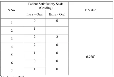
PATIENT SATISFACTION
The satisfaction of the patient was assessed by the patients in scale of 0 to 2
0 - Very satisfied
1 - Satisfied
2 - Not satisfied
FACIAL NERVE PARESIS:
The facial nerve paresis is assessed in the scale of 0 to 2
0 - No facial nerve paresis ;
1 - Temporary facial nerve paresis
2 - Permanent facial nerve paresis
SCAR ASSESMENT
The scar was assessed with the values ranging from 0 to 2
0 - Unnoticeable (invisible)
1 - Barely noticeable (barely visible)
SUBMANDIBULAR APPROACH
Step 1. Preparation and Draping
Pertinent landmarks useful during dissection should be exposed throughout
the procedure. For operations involving the mandibular ramus/angle, the corner of
the mouth and lower lip was exposed within the surgical field anteriorly and the
ear, or at least the ear lobe, posteriorly. These landmarks helps the surgeon to
mentally visualize the course of the facial nerve and to see whether the lip moves
if stimulated.
Step 2. Marking the Incision and Vasoconstriction
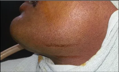
The skin was marked before injection of a vasoconstrictor. The incision is
1.5 to 2 cm inferior to the mandible. Some surgeons prefer to parallel the inferior
border of the mandible; others place the incision in or parallel to a neck crease.
Incisions made parallel to the inferior border of the mandible may be unobtrusive
in some patient; however, extensions of this incision may be noticeable unless
hidden in the submandibular shadow. A less conspicuous scar result when the
incision is made in or parallel to a skin crease. It should be noted that skin creases
below the mandible do not parallel the inferior border of the mandible but run
obliquely, posterosuperiorly to anteroinferiorly. Thus, the further anterior the
surgeon makes an incision in or parallel to a skin crease, the greater the distance to
dissect to reach the inferior border of the mandible. Both incisions can be
Mandibular fractures that shorten the vertical height of the ramus by their
displacement (i.e., condylar fractures in patients without posterior teeth or those
not placed intomaxillomandibular fixation) will cause the angle of the mandible to
be more superior than it would be following reduction and fixation. Therefore, the
incision should be 1.5 to 2 cm inferior to the anticipated location of the inferior
border. The incision is located along a suitable skin crease in whatever
anteroposterior position needed for mandibular exposure. For a fracture that
extends toward the gonial angle, the incision should begin behind and above the
gonial angle, extending downward and forward until it is in front of the gonial
angle. For fractures located more anterior than the gonial angle, the incision does
not have to extend behind and/or above the gonial angle, but may extend farther
anteriorly.
Vasoconstrictors with local anesthesia injected subcutaneously to aid in
hemostasis should not be placed deep to the platysma muscle because the
marginal mandibular branch of the facial nerve may be rendered nonconductive,
making electrical testing impossible. Alternatively, a vasoconstrictor without local
anesthesia can be used both superficially and deeply to promote hemostasis.
Step 3. Skin Incision
The initial incision is carried through skin and subcutaneous tissues to the
level of the platysma muscle. The skin was undermined with scissor dissection in
all directions to facilitate closure. The superior portion of the incision was
undermined approximately 1 cm; the inferior portion was undermined
extensively to allow retraction of the skin anteriorly or posteriorly to increase the
amount of mandibular exposure. In this manner, a shorter skin incision can
provide a great amount of exposure. Hemostasis can then be achieved with
electrocoagulation of bleeding subdermal vessels. See fig 4.
Step 4. Incising the Platysma Muscle
Retraction of the skin edges reveals the underlying platysma muscle, the
fibres of which run superoinferiorly. Division of the fibres can be performed
sharply, although a more controlled method is to dissect through the platysma
muscle at one end of the skin incision with the tip of a hemostat or Metzenbaum
scissor. After undermining the platysma muscle over the white superficiallayer of
deep cervical fascia, the tips of the instrument were pushed back through the
platysma muscle at the other end of the incision. With the instrument deep to the
platysma muscle, a scalpel is used to incise the muscle from one end of the skin
incision to the other. The anterior and posterior skin edges can be retracted
sequentially to allow a greater length of platysma muscle division than the length
of the skin incision.
The platysma muscle passively contracts once it is divided, exposing the
underlying superficial layer of deep cervical fascia. The submandibular salivary
gland can also be visualized through the fascia, which helps form its capsule.
Step 5. Dissection to the Pterygomasseteric Muscular Sling
Dissection through the superficial layer of deep cervical fascia is the
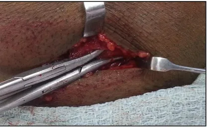
associated. The facial vein and artery are usually encountered when approaching
the area of the premasseteric notch of the mandible, as may the marginal
mandibular branch of the facial nerve. The facial vessels can be isolated, clamped,
and ligated if they are in the way of the area of interest. See fig.6. When
approaching the mandible posterior to the premasseteric notch, these vascular
structures generally are not encountered; if they are easily retracted anteriorly. The
marginal mandibular branch, however, occasionally is inferior to the mandible
posterior to the premasseteric notch, so care must be taken.
Dissection through the superficial layer of deep cervical fascia was
accomplished by nicking it with a scalpel and bluntly undermining with a
hemostat or Metzenbaum scissors. The level of the incision and undermining of
the fascia should be at least 1.5 cm inferior to the mandible to help protect the
marginal mandibular branch of the facial nerve. Thus, dissection through the
fascia at the level of the initial skin incision was performed, followed by
dissection superiorly to the level of the periosteum of the mandible. See fig 5.The
capsule of the submandibular salivary gland if often entered during this dissection,
and the gland is retracted inferiorly. A consistent submandibular lymph node
(node of Stahr) is usually encountered in the area of the premasseteric notch and
can be retracted superiorly or inferiorly. Its presence should alert the surgeon to
the facial artery just anterior to the node, deep to the superficial layer of the deep
cervical fascia. If encountered it was ligated as in fig 6.The marginal mandibular
branch of the facial nerve may be located close by, within or just deep to the
superficial layer of deep cervical fascia, passing superficial to the