JOURNAL OFVIROLOGY, Dec. 2008, p. 12221–12231 Vol. 82, No. 24 0022-538X/08/$08.00⫹0 doi:10.1128/JVI.01557-08
Copyright © 2008, American Society for Microbiology. All Rights Reserved.
RNA Interference Inhibits Respiratory Syncytial Virus Replication and
Disease Pathogenesis without Inhibiting Priming of the Memory
Immune Response
䌤
Wenliang Zhang and Ralph A. Tripp*
Department of Infectious Diseases, Center for Disease Intervention, University of Georgia, Athens, Georgia 30602
Received 23 July 2008/Accepted 19 September 2008
Respiratory syncytial virus (RSV) is a major cause of morbidity in infants, young children, and the elderly worldwide. Currently, there is no effective vaccine, and antiviral drugs to control infection are limited. RNA interference is a powerful tool amenable to development of antiviral drugs. Using small interfering RNA (siRNA) targeting the RSV P gene (siRNA-P), RSV replication can be silenced both in vitro and in a BALB/c model of RSV infection. In this study, we examine the effect of siRNA prophylaxis on the primary and memory immune response to RSV infection in mice. We show that mice prophylactically treated with siRNA-P to decrease but not eliminate RSV replication exhibit reduced pulmonary inflammation and lung pathogenesis and produce a robust anti-RSV memory response when subsequently challenged with RSV. The pulmonary T-cell memory response was characterized by high numbers of CD44hi
CD62Llo
CD4ⴙand CD8ⴙT cells, M2 peptide tetramerⴙCD8ⴙT cells expressing gamma interferon, and an RSV-specific antibody response. The results support the hypothesis that siRNAs can be developed as effective antiviral drugs that can be used to reduce the viral load and parameters of pathogenesis without limiting the induction of the memory immune response.
Respiratory syncytial virus (RSV) is an enveloped, negative-strand RNA virus belonging to the Paramyxoviridae family. RSV is a primary cause of morbidity and some mortality in infants, young children, the elderly, and the immune sup-pressed, causing bronchiolitis and pneumonia-related illness (20, 23, 68). In addition, RSV infection has also been linked to the development or exacerbation of airway hyper-responsive-ness in children (5, 18, 44); thus, there is a substantial need for effective vaccines and antiviral drugs. To date, all RSV vacci-nation strategies have proven to be ineffective (23, 47). Com-pounding issues, natural RSV infection does not lead to sus-tained protective immunity since individuals may be repeatedly infected with the same or different strains (7, 52). The features that contribute to the lack of durable immunity are not fully understood, but antigenic variation as a mechanism is unlikely. Seasonal epidemics linked to RSV infections do not appear to have a major role in susceptibility to reinfection since the two major viral surface proteins that are the targets of neutralizing antibody, i.e., attachment (G) and fusion (F) proteins, do not exhibit dramatic seasonal variation such as observed for influ-enza virus (10, 29). However, these proteins have been shown to modify aspects of the immune response, particularly the G protein, which has been shown to inhibit fractalkine-mediated responses, alter trafficking of CX3CR1⫹ cells immune cells, modify the magnitude and cadence of cytokine and chemokine expression, affect TCR Vusage by CD4⫹T cells, and affect the interface with the neuroimmune system through induction
of the proinflammatory tachykinin, substance P (31, 59–66). It is likely that these and other mechanisms contribute to im-mune dysregulation that may facilitate virus replication and/or contribute to persistence of RSV infection (5, 72).
Numerous studies in BALB/c mice have shown that cell-mediated immunity is important in the resolution of RSV infection, a feature mediated by both CD4⫹and CD8⫹T cells (49). The RSV M2 protein has been identified as a major
H-2d-restricted cytotoxic-T-lymphocyte target (15, 35, 36).
Consistent with the lack of durable immunologic memory gen-erated after RSV infection, M2-specific effector CD8⫹T cells isolated from the lungs of infected mice have been shown to have a reduced capacity to express gamma interferon (IFN-␥), whereas similar effector CD8⫹T cells in the spleen have no deficit in IFN-␥expression (8). Intriguingly, a rapid loss in the frequency of RSV-specific memory CD8⫹T cells occurs in the lungs of the infected mice during the resolution phase of in-fection, a finding consistent with the notion that natural RSV infection does not lead to sustained protective immunity (12, 13, 55). Thus, converging lines of evidence suggest that RSV protein expression contributes to the dysregulation of immune function, a feature that negatively affects the development of immune memory.
Since safe and effective RSV vaccines are not available, disease management has focused on passive immunotherapy for high-risk patients. Palivizumab, a humanized immunoglob-ulin G (IgG) monoclonal antibody targeting the RSV F protein is currently used as a passive immunoprophylaxis (14, 24). However, this treatment has modest prophylactic efficacy, highlighting the need for an actual antiviral that could be used as a treatment. RNA interference (RNAi) is a mechanism that inhibits gene expression at the stage of translation by hindering the transcription of specific genes (30, 43, 45). The process is * Corresponding author. Mailing address: Department of Infectious
Diseases, Center for Disease Intervention, University of Georgia, 501 DW Brooks Dr., Rm. 347, Athens, GA 30602. Phone: (706) 542-1557. Fax: (706) 542-5771. E-mail: rtripp@vet.uga.edu.
䌤Published ahead of print on 25 September 2008.
12221
on November 8, 2019 by guest
http://jvi.asm.org/
mediated by small interfering RNA (siRNA) that has comple-mentary nucleotide sequences to the targeted RNA strand. The siRNAs are guided to their cognate targets by components of the RNA-induced silencing complex, where they may cleave the target to prevent translation into protein (58). RNAi is a compelling tool for rationalizing drug design and is being tested as a prophylactic and therapeutic antiviral agent for a range of viruses, including human immunodeficiency virus and hepatitis virus, as well as RSV (3, 4, 32, 42, 56). Given the pace of RNAi applications and demonstrated efficacy, it is likely that RNAi-based therapeutics will evolve to be a major therapeutic modality for antiviral treatment of numerous viruses.
RSV has been successfully targeted by siRNA (2–4, 6). In-tranasal (i.n.) delivery of an in vitro-active siRNA directed at the P gene of RSV significantly inhibits RSV replication (6). In these studies, siRNA prophylactically delivered to mice 4 h before RSV infection reduced lung virus titers and prevented pulmonary pathology. When RSV-infected mice were treated therapeutically with the drug, the level of antiviral efficacy was diminished, but lung virus titers were still reduced. Since RSV replicates almost exclusively in the respiratory epithelium of humans (71), siRNA antiviral drugs can be directly adminis-tered using topical or aerosol delivery methods. Although the preliminary evidence suggests siRNA drugs targeting RSV may be beneficial, unfortunately nothing is known about the effects of siRNA treatment on the memory response to viral challenge.
In the present study, we examined the effect of siRNA pro-phylaxis on the primary and memory immune response to RSV infection in BALB/c mice. Mice were treated prophylactically with siRNA targeting the RSV P gene to reduce but not elim-inate RSV replication so that the relationship between virus load, disease pathogenesis, and immunity could be evaluated. We show that siRNA drugs that reduce RSV replication ef-fectively prevent lung disease pathogenesis and allow for ro-bust anti-RSV memory immune responses.
MATERIALS AND METHODS
siRNA.The wild-type (WT) siRNA corresponding to the sequence AAGCC CTATAACATCAAATTCAA of the P gene mRNA (6) of RSV strain A2 and a mismatch (MM) control siRNA of similar content were used. Each strand of the siRNA was 21 nucleotides long and contained 3⬘-terminal dTdT extensions. All siRNAs were commercially synthesized (Dharmacon ThermoFisher). The WT or MM siRNAs were diluted in phosphate-buffered saline (PBS; 100 nM) and i.n. instilled in mice.
Virus infection.Vero cells (African green monkey kidney cells) were grown in Dulbecco modified Eagle medium (DMEM) containing 5% fetal bovine sera (DMEM-5%; HyClone). Respiratory syncytial virus strain A2 (RSV) was prop-agated in Vero cells as previously described (64). Briefly, semiconfluent Vero cells were prepared and washed with PBS. RSV was diluted in DMEM, and the cells were infected at a multiplicity of infection of 1. The virus was allowed to adsorb for 2 h at 37°C, after which DMEM-5% was added, and the cells were incubated at 37°C for 4 days. At day 4 postinfection (p.i.), the virus was recovered by removing the cell culture supernatant, freeze-thawing the infected cells, and centrifuging the cell lysate to remove debris and recover the virus from the cell lysate supernatant.
Mice, treatment, and infections.Four-to-six-week-old specific-pathogen-free female BALB/c mice were purchased from Charles River Laboratories, housed in microisolator cages, and fed sterilized water and food ad libitum. The studies were reviewed and approved by the university institutional review committee. Mice were prophylactically treated for 12 h by topical instillation of either WT or MM siRNA (2 mg/kg) or PBS prior to infection. In primary immune response studies, the treated mice were challenged with 106
PFU of RSV strain A2 (RSV/A2) diluted in PBS (Gibco-BRL), and 10 mice/group/day were harvested
at day 0, 2, 4, 6, or 8 p.i. A portion of these mice were rested for 3 weeks p.i. and, for memory immune response studies, were i.n. challenged with 106
PFU of RSV strain A/Long (10 mice/group/day) and harvested at day 0, 2, 4, 6, 8, or 10 p.i. The organs were collected from primary and memory immune mice after anestheti-zation, and exsanguinations were mediated by severing the right caudal artery. The blood was collected for sera, and the bronchoalveolar lavage (BAL) fluid collected by lavaging the lungs of the mice three times with 1 ml of PBS. The lungs from three mice/group/day not collected for BAL were harvested for histopathology or determination of the virus titer determined by immunostaining plaque assay on Vero cells as previously described (64).
Flow cytometry.The percent positive B220, CD3, CD4, CD8, CD11b, CD44, CD62L, DX5, and RB6-8C5 cell subsets were determined for BAL cells by flow cytometry. Cells were blocked with 10% normal mouse sera (Jackson Labora-tories) in flow buffer (PBS plus 1% bovine serum albumin) and then stained with the appropriate combinations of fluorescein isothiocyanate- or phycoerythrin-labeled anti-B-cell (RA3-6B2), anti-CD3 (2C11), anti-CD4 (RM4-5), anti-CD8 (53-6.7), anti-CD11b (M1/70), anti-CD44 (KM114), anti-CD62L (DREG-56), anti-pan NK cell (DX5), anti-pan neutrophil (RB6-8C5), or mouse isotype an-tibody control (all from BD Pharmingen, San Diego, CA) as previously described (64). For intracellular cytokine staining, BAL cells were incubated for 3 h in the presence of 10g of brefeldin A (BD Pharmingen)/ml. After incubation, the cells were washed with flow buffer and blocked with 10% normal mouse sera (Jackson Laboratories) in flow buffer. After blocking for 15 min, the cells were washed with flow buffer and incubated with optimal concentrations of anti-CD4 PerCP-Cy5.5 (BD Pharmingen) or CD8 Pe-Cy5.5 (BD Pharmingen) for 30 min at 4°C. Cells were then washed with flow buffer and fixed with 4% paraformal-dehyde. Cells were then washed in permeabilization buffer (BD Pharmingen) and stained with an optimal concentration of anti-IFN-␥-phycoerythrin (clone XMG1.2; BD Pharmingen) or anti-interleukin-6 (anti-IL-6; clone; BD Pharmin-gen) antibody diluted in permeabilization buffer. Cells were washed and analyzed on a BD LSRII flow cytometer using FACSDiva software (Becton Dickinson, Mountain View, CA) from⬎10,000 lymphocyte gated events.
RSV-specific antibody response.The antibody titer and isotypes of the sera samples were determined as previously published (57). Briefly, 96-well high binding enzyme-linked immunosorbent assay (ELISA) plates (Corning Costar, Corning, NY) were coated with 1g of RSV/A2 (106
PFU/ml) or 1g of uninfected Vero cell lysate and blocked with blocking buffer containing PBS, 0.3% Tween 20 (Sigma), and 0.01 M EDTA buffer (Sigma) (pH 7.0). Dilutions of the sera were made in blocking buffer, added to the wells, and incubated for 1 h at 37°C. After five washes in PBS containing 0.1% Tween 20, biotinylated goat anti-mouse IgG (BD Pharmingen) was added to the wells, and the plates were incubated for 1 h at 37°C. After washing, an appropriate dilution of strepta-vidin conjugated to horseradish peroxidase (Zymed Laboratories, Inc., San Fran-cisco, CA) was added to the wells, and the plates were incubated for 30 min at room temperature. After removal of the unbound streptavidin by washing, per-oxidase substrate (2,2⬘-azino-di[3-ethyl-benzthiazoline sulfonate]; Kirkegaard and Perry Laboratories, Gaithersburg, MD) was added to the wells for 20 min. The reaction was stopped with 1% sodium dodecyl sulfate. The determination of anti-RSV titers was determined by optical densities at 410 nm (OD410; reference
value of 630 nm) on an ELISA plate reader (Tecan, Research Triangle Park, NC). The data are presented as the endpoint titer calculated as the reciprocal of the geometric mean of the dilution that resulted in an OD410reading of 0.03.
Isotype determination kits (ThermoScientific, Rockford, IL) were used for an-tibody isotyping as described by the manufacturer.
RSV M2 tetramer staining.BAL cell suspensions were blocked with purified anti-Fc␥RII/III monoclonal antibody (BD Pharmingen) for 30 min. Cells were washed with flow buffer and incubated with optimal concentrations of anti-CD8 FITC antibody (BD Pharmingen). Cells were washed and incubated with optimal concentrations of M282-90-specific H-2k
d
allophycocyanin-conjugated tetramers (obtained from the Emory University Tetramer Core Facility, Atlanta, GA) for 45 min at 4°C. After tetramer staining, cells were washed twice with flow buffer and analyzed on a BD LSRII flow cytometer using FACSDiva software from
⬎10,000 lymphocyte gated events.
Histopathology.Histopathological examination was performed for lungs from siRNA-treated and untreated mice infected with RSV. Tissues were fixed in 10% buffered formalin, embedded in paraffin, sectioned, and stained with hematoxylin and eosin prior to light microscopy observation. Multiple sections from each tissue block were analyzed under light microscopy.
Statistics.For statistical evaluation between two treatments, attest for un-paired samples was used to compare the responses between WT and MM siRNA-treated mice.Pvalues of⬍0.05 or⬍0.01 were considered significant. The Kruskal-Wallis test (Prism Graph Pad, La Jolla, CA) was used to compare the medians between the three treatment groups; the significance was set atP⬍0.05.
on November 8, 2019 by guest
http://jvi.asm.org/
RESULTS
Prophylactic siRNA treatment reduces virus lung titers and lung pathogenesis.Previous prophylactic and therapeutic stud-ies have shown that siRNAs targeting the P gene can reduce RSV replication (6). Using the same siRNAs targeting the P gene (WT), a scrambled MM siRNA control, or PBS, the level of lung virus replication was determined. Mice prophylactically treated by i.n. inoculation with 200, 100, or 50 nM siRNA for 12 h prior to RSV infection had reduced lung virus titers at day 4 p.i., an effect that was dose dependent, in contrast to MM siRNA- or PBS-treated mice, where no effect on virus titer was observed (Fig. 1A).
[image:3.585.136.454.66.472.2]To evaluate in vivo efficacy of siRNA prophylaxis, mice were i.n. treated with 100 nM (2 mg/kg) WT or MM siRNA or PBS vehicle and 12 h posttreatment were i.n. infected with 106PFU of RSV/A2 (Fig. 1B). Prophylactic treatment with WT siRNA reduced virus titers at all time points examined compared to MM siRNA- or PBS-treated mice, and at days 4 and 6 p.i. the WT siRNA-treated mice had significantly (P⬍0.05) reduced virus titers compared to MM siRNA- or PBS-treated mice. No substantial lung pathogenesis was detected WT siRNA-treated mice at any time point examined. MM siRNA- and PBS-treated mice did not show substantial histopathology until day 6 p.i. For comparison, the histopathology at day 6 p.i. is shown FIG. 1. Prophylactic siRNA treatment reduces virus lung titers. (A) Mice were prophylactically treated i.n. with 4, 2, or 1 mg/kg of WT siRNA (WT200, WT100, or WT50, respectively) or MM siRNA (MM200, MM100, or MM50, respectively) or with PBS for 12 h prior to RSV/A2 infection (106PFU). Lungs were harvested at day 4 p.i., and virus titers were determined by immunostaining plaque assay with anti-F protein monoclonal
antibody (clone 131-2A) as previously described (64). The data are presented as the mean log10PFU/g titer⫾the standard error (SE;n⫽five
mice/treatment/time point) in three separate experiments. The limit of virus detection is between 5 and 10 PFU/g of lung tissue. (B) To evaluate the kinetics of siRNA efficacy, mice were i.n. treated with 2 mg of WT or MM siRNA/kg or PBS vehicle and at 12 h posttreatment i.n. infected with 106PFU of RSV/A2. Lungs were collected at day 2, 4, 6, or 8 p.i. and assayed by immunostaining plaque assay using Vero cells as previously
described (64). (C) Lung histological pathogenesis was evaluated in WT siRNA-treated (i), PBS-treated (ii), and MM siRNA-treated (iii) mice at day 6 p.i. as previously described (26). Asterisks indicate a significant (P⬍0.01) difference from the control.
VOL. 82, 2008 RNAi OF RSV REPLICATION AND THE IMMUNE RESPONSE 12223
on November 8, 2019 by guest
http://jvi.asm.org/
for WT siRNA-treated mice (Fig.1Ci), PBS-treated mice (Fig.1Cii), and MM siRNA-treated (Fig.1Ciii) mice. The MM siRNA- and PBS-treated mice showed increased peribronchio-lar, perivascuperibronchio-lar, and interstitial lymphocytic infiltrates typical of RSV-mediated pathogenesis (26). These results suggest that the extent of lung pathogenesis following RSV infection ap-pears associated with the virus load.
BAL response in siRNA-treated mice.To determine whether the reduction in virus titer associated with WT siRNA treat-ment affected the primary immune response, mice were pro-phylactically treated with 100 nM (2 mg/kg) WT or MM siRNA or treated with PBS vehicle. At 12 h posttreatment, the mice were i.n. infected with 106PFU of RSV/A2, and the BAL and sera were collected at days 0, 2, 4, 6, or 8 p.i. The magnitude of the primary BAL response to RSV infection was highest for mice treated with MM siRNA or PBS compared to WT siRNA-treated mice (Table 1). Consistent with peak virus ti-ters (Fig. 1B), the highest number of BAL cells infiltrating the lung occurred at day 6 p.i. for all treated mice, and at day 8 p.i., all treated mice had similar BAL cell numbers indicating equil-ibration of BAL cell numbers among the groups of mice.
The BAL cell types were determined during the primary immune response to RSV infection of WT and MM siRNA-and PBS-treated mice (Table 2). Peak CD4⫹and CD8⫹T-cell numbers were detected at day 8 p.i. with no significant differ-ences (P ⬍ 0.0.5) in total cell numbers between siRNA- or PBS-treated mice. A minimal B220⫹ cell response was ob-served at all time points for all treatments. Interestingly, higher numbers of DX5⫹cells occurred at day 6 p.i. in WT siRNA- or MM siRNA-treated mice compared to PBS-treated mice, and MM siRNA-treated mice had significantly (P⬍ 0.05) higher DX5⫹cell numbers at day 8 p.i. compared to WT siRNA- or PBS-treated mice. These data suggest that siRNA treatment affects the DX5⫹cell response at late time points. It is likely that DX5⫹ cells are being recruited by cytokines or chemo-kines produced in the lung microenvironment at these time points; however, the mechanism is not known. It may be pos-sible that off-target siRNA activities contribute to these find-ings, although similar experiments have shown that treatment with similar siRNAs alone do not induce detectable type I IFN levels (6). It is also unlikely that the DX5⫹cells are responding directly to the siRNAs because they are rapidly degraded in vivo (38). Since this effect occurs late in the response, i.e., day 6 p.i., and only DX5⫹cells appear affected by siRNA treat-ment, there is no evidence that siRNA treatment has a general proinflammatory effect in the lung. A higher number of RB6-8C5⫹ cells, confirmed by hematoxylin and eosin staining as polymorphonuclear leukocytes, and CD11b⫹ cells were
[image:4.585.43.542.81.144.2]de-tected at day 2 p.i. in PBS- and MM siRNA-treated mice compared to WT siRNA-treated mice, but no other substantial differences were observed at the other time points examined. The differences in the recruitment of these innate immune cells between groups of treated mice are unclear, but it is possible that this may be linked to a reduced host cell response TABLE 1. Total BAL cell numbers during primary RSV infectiona
Treatment group
Total BAL cell count (SE)
Day 0 Day 2 Day 4 Day 6 Day 8
PBS 2.9⫻104(0.5) 6.0⫻105(1.5)† 6.5⫻105(1.5)† 1.2⫻106(3.1)† 7.0⫻105(1.2)†
MM 3.1⫻104(0.8) 5.8⫻105(2.1)† 7.9⫻105(2.8)† 1.4⫻106(30.5)† 8.0⫻105(1.5)†
WT 3.0⫻104(0.5) 3.9⫻105(0.5)*† 3.8⫻105(0.2)*† 0.5⫻106(3.5)*† 6.5⫻105(1.1)† aMice were i.n. treated with PBS, 100 nM (2 mg/kg) MM siRNA (MM), or 100 nM (2 mg/kg) WT siRNA (WT) specific for the RSV P gene prior to RSV/A2 infection
(106PFU). The total cell numbers were determined from three to five mice/treatment in three separate experiments. *, Statistically different (P⬍0.05) from MM- and
PBS-treated mice; †, statistically different (P⬍0.05) from day 0 response.
TABLE 2. Total number of cell types in the BAL during the primary response to RSV infectiona
Cell type and day p.i.
Total no. of cells (SE)
WT MM PBS
CD4⫹
2 1.5⫻103(3.0) 3.1⫻103(2.2) 2.5⫻103(1.5)
4 16.5⫻103(3.1) 18.5⫻103(2.5) 14.4⫻103(2.8)
6 16.2⫻103(2.1) 16.0⫻103(2.4) 12.2⫻103(1.6)
8 33.5⫻103(5.8) 29.5⫻103(6.0) 31.0⫻103(5.2)
CD8⫹
2 2.8⫻103(1.1) 5.8⫻103(2.2) 5.1⫻103(1.5)
4 18.5⫻103(1.8) 17.9⫻103(2.8) 18.9⫻103(2.8)
6 18.4⫻103(2.5) 21.0⫻103(2.1) 19.4⫻103(2.6)
8 48.6⫻103(5.5) 39.5⫻103(5.7) 48.0⫻103(5.5)
B220⫹
2 0.09⫻103(0.1) 0.1⫻103(0.1) 0.1⫻103(0.1)
4 0.5⫻103(0.1) 0.2⫻103(0.1) 0.8⫻103(0.1)
6 0.5⫻103(0.5) 0.6⫻103(0.2) 0.5⫻103(0.1)
8 0.3⫻103(0.1) 0.1⫻103(0.1) 0.6⫻103(0.2)
DX5⫹
2 2.5⫻103(0.5)† 5.0⫻103(1.1) 4.8⫻103(1.2)
4 5.0⫻103(0.5) 8.0⫻103(1.2) 6.5⫻103(1.3)
6 7.5⫻103(1.2)†‡ 14.4⫻103(2.8)‡ 1.3⫻103(1.1)†
8 1.9⫻103(1.1)† 7.0⫻103(1.2) 0.5⫻103(0.5)†
RB6-8C5⫹
2 3.8⫻103(0.8)†‡ 11.8⫻103(1.8)* 10.0⫻103(1.5)*
4 1.9⫻103(0.8) 2.5⫻103(1.8) 3.3⫻103(2.5)
6 5.8⫻103(2.4) 6.0⫻103(1.8) 7.4⫻103(2.0)
8 8.8⫻103(1.4) 8.0⫻103(2.7) 9.8⫻103(0.4)
CD11b⫹
2 3.1⫻103(0.5)†‡ 5.9⫻103(0.5) 5.1⫻103(0.5)
4 10.0⫻103(2.8) 10.8⫻103(2.8) 11.0⫻103(2.2)
6 5.0⫻103(1.2) 4.1⫻103(1.5) 5.9⫻103(1.5)
8 5.5⫻103(1.8)† 9.0⫻103(1.1) 7.5⫻103(1.5) aMice were i.n. treated with 100 nM (2 mg/kg) WT siRNA specific for the
RSV P gene, 100 nM (2 mg/kg) MM siRNA, or PBS prior to RSV/A2 infection (106PFU). The total cell numbers were determined from four to five mice/
treatment in three separate experiments using appropriate monoclonal antibod-ies and flow cytometry. *, Statistically different (P⬍0.05) from WT-treated mice; †, statistically different (P⬍0.05) from MM-treated mice; ‡, statistically different (P⬍0.05) from PBS-treated mice.
on November 8, 2019 by guest
http://jvi.asm.org/
[image:4.585.301.539.317.669.2]to infection associated with a decreased virus load in WT-treated mice (Fig. 1B).
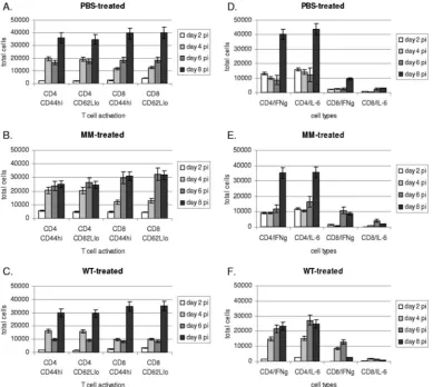
T-cell activation and intracellular cytokine expression. To determine the level of T-cell activation in the BAL, the levels of CD44hiand CD62Lloexpression by CD4⫹and CD8⫹T cells
were determined at various time points after RSV infection (Fig. 2A to C). CD4⫹and CD8⫹T cells from the BAL of mice prophylactically treated with WT or MM siRNA or PBS for 12 h prior to infection, i.e., day 0, had very low levels of CD44hi or CD62Lloexpression, e.g., the expression levels were below those observed for RSV-infected mice at day 2 p.i. (Fig. 2A to C). Both CD4⫹and CD8⫹T cells had increased CD44hiand CD62Llo expression at day 4 p.i., and the peak numbers of activated CD4⫹and CD8⫹CD44hiand CD62LloT cells oc-curred between days 4 and 6 p.i. for MM siRNA-treated mice (Fig. 2B) and at day 8 p.i. for PBS (Fig. 2A)- and WT siRNA (Fig. 2C)-treated mice. MM siRNA- and PBS-treated mice had significantly (P⬍0.05) higher numbers of activated CD4⫹T cells between days 4 and 6 p.i. compared to WT siRNA-treated mice and significantly (P⬍0.05) higher numbers of activated
CD8⫹T cells at 6 p.i. than WT siRNA-treated mice, suggesting that increased T-cell activation may be linked to a higher virus load (Fig. 1B).
[image:5.585.99.488.68.416.2]To determine the pattern of cytokines expressed by T cells in the BAL, intracellular IFN-␥(Th1-type) and IL-6 (Th2-type) expression was determined for CD4⫹and CD8⫹T cells at days 0 to 8 p.i. (Fig. 2D to F). No significant cytokine response or difference (P⬍0.05) in the total number of CD4⫹or CD8⫹T cells expressing IFN-␥or IL-6 was observed at day 0 (prior to infection). CD4⫹T cells in the BAL of any treatment group expressed significantly (P⬍0.01) higher levels of IFN-␥and IL-6 at day 8 p.i. compared to CD8⫹T cells and, at other time points, also expressed higher IFN-␥and IL-6 levels than CD8⫹ T cells. It is interesting that the magnitude of IFN-␥and IL-6 expression by CD4⫹T cells from WT siRNA-treated mice was significantly (P⬍0.05) higher at days 4 to 6 p.i. compared to MM siRNA- or PBS-treated mice. Overall, the pattern of CD4⫹T-cell cytokine expression suggested a mixed Th1/Th2-type cytokine response. However, the pattern of CD8⫹T-cell cytokine expression in all treated groups, although of lower FIG. 2. CD44hiand CD62Lloexpression by CD4⫹and CD8⫹T cells in the BAL. The levels of CD44hiand CD62Lloexpression by CD4⫹and CD8⫹T cells were determined at days 2 to 8 after RSV infection of PBS-treated (A), MM siRNA-treated (B), and WT siRNA-treated (C) mice. To address the association between CD44hiand CD62LloT-cell expression and Th1/Th2 cytokine expression, the levels of intracellular IFN-␥
(Th1-type cytokine) and IL-6 (Th2-type cytokine) were determined for CD4⫹and CD8⫹T cells at days 2 to 8 p.i. of PBS-treated (D), MM siRNA-treated (E), and WT siRNA-treated (F) mice. The data are presented as the mean total cells⫾the SE fromn⫽five mice/treatment/time point in three separate experiments.
VOL. 82, 2008 RNAi OF RSV REPLICATION AND THE IMMUNE RESPONSE 12225
on November 8, 2019 by guest
http://jvi.asm.org/
magnitude compared to CD4⫹ T cells, was suggestive of a Th1-type response. CD8⫹T-cell IFN-␥levels were significantly (P⬍0.05) higher than IL-6 at day 8 p.i. in PBS-treated mice, higher at days 6 to 8 p.i. in MM siRNA-treated mice, and higher at days 4 to 6 p.i. in WT siRNA-treated mice (Fig. 2D to F). These results suggest that the pattern and magnitude of IFN-␥and IL-6 expression is not directly linked to the level T-cell activation (Fig. 2A to C) and so not likely to be linked to the virus load.
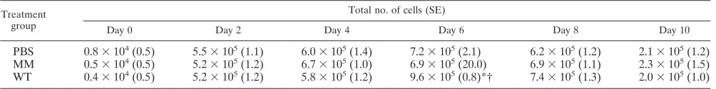
BAL cell response, virus clearance, and lung pathology dur-ing the memory response.To determine how a reduction in virus titer associated with prophylactic WT siRNA treatment affects the memory response to challenge, mice prophylacti-cally treated with 100 nM (2 mg/kg) WT or MM siRNA or treated with PBS and previously infected with RSV were i.n. challenged 3 weeks later with 106PFU of RSV/A Long and the BAL, lungs, and sera collected at days 0, 2, 4, 6, 8, or 10 p.i. BAL cell numbers were similar among the treated mice except at day 6 p.i., where WT siRNA-treated mice had significantly (P⬍ 0.05) higher numbers of total BAL cells (Table 3). As expected, the total number of BAL cells for any treatment group rapidly increased versus the unchallenged but RSV-immune mice.
The total number of CD4⫹ or CD8⫹ T cells expressing CD44hi or CD62Llo was determined in the BAL of mice treated with PBS, WT siRNA, or MM siRNA at days 0 to 10 p.i. No significant difference (P⬍0.05) in the total numbers of CD4⫹or CD8⫹CD44hiand CD62LloT cells were deter-mined between the treated mice at days 0 to 2 p.i. (range, 500 to 5,500). However, between days 4 and 6 p.i. in WT siRNA-treated mice, the total number of CD8⫹CD44hiT cells in the BAL (range, 11,000 to 39,500) was significantly (P ⬍ 0.05) higher compared to MM siRNA- or PBS-treated mice (range, 3,200 to 6,500), and CD8⫹CD44hiT-cell numbers peaked at day 6 p.i. (range, 21,500 to 39,500). In contrast, there was no significant difference in the total number of CD4⫹CD44hiT cells at any time point between days 0 and 6 p.i. (range, 500 to 5,500) in PBS-, WT siRNA-, or MM siRNA-treated mice. Likewise, the total number of CD8⫹CD62LloT cells (range, 28,000 to 41,000) was significantly (P⬍0.05) higher between days 4 and 6 p.i. in the BAL of WT siRNA-treated mice compared to PBS- or MM siRNA-treated mice, and the CD8⫹ CD62LloT-cell numbers peaked at day 6 p.i. (range, 29,500 to 39,000). There was no significant difference (P⬍0.05) in the total number of CD4⫹ CD62Llo T cells at any time point between days 0 and 6 p.i. (range, 1,000 to 5,500) in PBS-, WT siRNA-, or MM siRNA-treated mice. Between days 8 and 10 p.i., the total numbers of CD8⫹CD44hior CD62LloT cells
in the BAL of WT siRNA-treated mice were similar (range, 20,500 to 27,500) and remained significantly higher (P⬍0.05) compared to CD4⫹CD44hior CD62LloT cells in the BAL of MM siRNA- or PBS-treated mice (range, 2,200 to 4,500). These results suggest that CD8⫹CD44hiand CD8⫹CD62Llo are a principal memory T-cell population in the RSV memory response in WT siRNA-treated mice and that memory CD8⫹ T cells are more predominant than CD4⫹T cells in the BAL. In addition to investigating the T-cell types, the numbers of innate immune cell types in the BAL was determined. No significant difference (P⬍0.05) in the total number of DX5⫹ or RB6-8C5⫹cells was detected at any time point examined in PBS-, MM siRNA-, or WT siRNA-treated mice. However, there were higher numbers of DX5⫹ cells (range, 17,000 to 24,000) in WT siRNA-treated mice between days 4 and 6 p.i. compared to PBS- or MM siRNA-treated mice (range, 14,000 to 21,500), and the total number of RB6-8C5⫹ cells were higher in the BAL of WT siRNA-treated mice at days 4 to 6 p.i. (range, 16,000 to 22,000) compared to PBS- or MM siRNA-treated mice (range, 13,500 to 16,000).
[image:6.585.43.545.81.144.2]The lung virus titers in the treated mice were determined. The highest lung virus titers measured occurred at day 2 post-challenge in all treated groups; however, virus titers were sig-nificantly lower (P ⬍ 0.05) at day 4 postchallenge in WT siRNA-treated mice compared to MM siRNA- or PBS-treated mice (Fig. 3A). No virus was detectable in the lungs of WT siRNA-treated mice at day 4 postchallenge; however, similar levels of virus (1.8 to 2.6 log10PFU g/lung tissue) was detected in the lungs of MM siRNA- and PBS-treated mice. All treated groups of mice cleared the virus by day 6 p.i. No lung histo-pathology was evident at day 0 prior to challenge for any treated group, and no substantial histopathology was detected in WT siRNA-treated animals at days 2, 4, and 6 p.i. (Fig.3Bi, Biv, and Bvi, respectively). However, PBS- and MM siRNA-treated mice showed similar levels of peribronchiolar, perivas-cular, and interstitial lymphocytic infiltrates typical of RSV-mediated pathogenesis (26) at day 2 p.i. (Fig. 3Bii and Biii, respectively), day 4 p.i. (Fig. 3Bv and Bvi, respectively), and day 6 p.i. (Fig. 3Bvii and Bix, respectively). These results show that prophylactic WT siRNA treatment, despite decreasing the virus load during the primary immune response, sufficiently primes for a robust memory immune response to RSV chal-lenge that is not linked with substantial pulmonary disease pathogenesis compared to MM siRNA- or PBS-treated mice. Anti-RSV antibody responses.The evidence that mice pro-phylactically treated with WT siRNA recovered from virus challenge faster (days 2 to 4 p.i.) than MM siRNA- or PBS-treated mice (days 4 to 6 p.i.; Fig. 3A) suggested a difference in TABLE 3. Total BAL cell numbers after RSV challenge of immune micea
Treatment group
Total no. of cells (SE)
Day 0 Day 2 Day 4 Day 6 Day 8 Day 10
PBS 0.8⫻104(0.5) 5.5⫻105(1.1) 6.0⫻105(1.4) 7.2⫻105(2.1) 6.2⫻105(1.2) 2.1⫻105(1.2)
MM 0.5⫻104(0.5) 5.2⫻105(1.2) 6.7⫻105(1.0) 6.9⫻105(20.0) 6.9⫻105(1.1) 2.3⫻105(1.5)
WT 0.4⫻104(0.5) 5.2⫻105(1.2) 5.8⫻105(1.2) 9.6⫻105(0.8)*† 7.4⫻105(1.3) 2.0⫻105(1.0) aMice prophylactically treated with WT or MM siRNA or PBS and i.n. infected with 106PFU RSV/A2 were rested for 3 weeks p.i. and then i.n. challenged with
106PFU of RSV/A Long. BAL cells were collected at the days indicated postchallenge. The total cell numbers were determined from four to five mice/treatment in
three separate experiments. *, Statistically different (P⬍0.05) from MM-treated mice; †, statistically different (P⬍0.05) from PBS-treated mice.
on November 8, 2019 by guest
http://jvi.asm.org/
the quality of the memory response related to treatment. To determine whether the differences were in part related to the antibody response, the IgG1 (Th2-type) and IgG2a (Th1-type) anti-RSV antibody responses were examined in sera collected from the treated mice (Fig. 4). The antibody response was Th1/Th2 mixed, with no significant differences in IgG1 or IgG2a responses between the treated groups of mice. WT siRNA-treated mice had slightly higher IgG1 and IgG2a anti-body responses at day 10 p.i. compared to MM siRNA- or PBS-treated mice, but this difference likely does not explain the more rapid viral clearance by this group. Neutralizing an-tibody titers were also evaluated in the sera from WT and MM siRNA-treated and PBS-treated mice at day 6 p.i; however, no statistical differences (P⬍ 0.05) in efficacy were determined when the neutralizing anti-RSV serum titers ranged from 6.0 to 6.9 reciprocal mean log2between the treated mice.
Memory T-cell responses.The higher numbers of CD44hiand CD62LloCD8⫹T cells in the BAL of WT siRNA-treated
mem-ory mice compared to MM siRNA- or PBS-treated mice indi-cated that the memory CD8⫹T-cell response may be associated with enhanced virus clearance. To address this possibility, the memory CD8⫹T-cell response to the immunodominant RSV M2 peptide (M282-90) (36) was determined at days 0 to 6 p.i. using M2-Kdtetramer staining (Fig. 5). The results show that signifi-cantly (P⬍0.05) higher total numbers of M2-specific CD8⫹T cells trafficked to the lung between days 4 and 6 p.i. in WT siRNA-treated mice compared to MM siRNA- or PBS-treated mice (Fig. 5A), a finding consistent with the results showing faster virus clearance in these treated mice compared to MM siRNA- or PBS-treated mice (Fig. 3A). Notably, there was a significantly (P ⬍ 0.01) higher total number of M2-specific CD8⫹ T cells expressing intracellular IFN-␥between days 4 and 6 p.i. in WT siRNA-treated mice compared to MM siRNA- or PBS-treated mice (Fig. 5B). These data suggest that CD8⫹memory T-cell response in WT siRNA-treated mice likely contributes to the enhanced rate of virus clearance.
FIG. 3. Lung virus titers in the memory response to RSV challenge. (A) To determine the effect of treatment on lung virus titers in the memory response to RSV challenge, mice prophylactically treated with WT siRNA (WT), MM siRNA (MM), or PBS were challenged with 106PFU of
RSV/A Long. The lungs were harvested at days 2, 4, and 6 postchallenge to determine virus titers as previously described (64). The data are presented as mean log10PFU/g titer⫾the SE fromn⫽5 mice/treatment/time point in three separate experiments. (B) The lung histopathology
was evaluated in the treated mice challenged with RSV. No lung histopathology was evident at day 0 prior to challenge for any treated group (data not shown). Histopathology was evaluated for WT siRNA-treated mice at day 2, 4, or 6 p.i. (panels i, iv, and vi, respectively); for PBS-treated mice at day 2, 4, or 6 p.i. (panels ii, v, and vii, respectively); and for MM siRNA-treated mice at day 2, 4, or 6 p.i. (panels iii, vi, and ix, respectively) as previously described (26). Asterisks indicate a significant (P⬍0.01) difference from the control.
VOL. 82, 2008 RNAi OF RSV REPLICATION AND THE IMMUNE RESPONSE 12227
on November 8, 2019 by guest
http://jvi.asm.org/
To determine the pattern of cytokines expressed by memory T cells in the BAL, intracellular IFN-␥(Th1-type) and IL-6 (Th2-type) expression were determined for CD4⫹and CD8⫹T cells at days 0 to 6 p.i. The total number of CD4⫹ T cells expressing IFN-␥was similar between days 4 and 6 p.i. for MM siRNA- or PBS-treated mice (range, 20,000 to 27,500). In contrast, the total number of CD4⫹T cells expressing IFN-␥ from WT siRNA-treated mice was significantly (P ⬍ 0.05) higher (range, 38,500 to 42,500). No significant difference (P⬍
0.05) in the total number of CD4⫹T cells expressing IL-6 was observed at any time point among the groups of treated mice (range, 3,500 to 10,500). A similar trend for IFN-␥and IL-6 expression was observed for CD8⫹T cells from MM siRNA-or PBS-treated mice, where the total number of CD8⫹T cells expressing IFN-␥between days 4 and 6 p.i. ranged from 28,000 to 40,000 and, in contrast, the total number of CD8⫹T cells expressing IFN-␥from WT siRNA-treated mice was signifi-cantly (P⬍0.01) higher (range, 59,000 to 72,000). No substan-tial difference in IL-6 expression was observed (range, 2,500 to 4,500) among the groups of treated mice. The higher total number of CD8⫹T cells expressing IFN-␥from WT siRNA-treated mice is consistent with the finding of higher total num-bers of M2-specific CD8⫹T cells in WT siRNA-treated mice compared to MM siRNA- or PBS-treated mice (Fig. 5A)— features that may be linked to more rapid virus clearance in memory mice (Fig. 3A).
DISCUSSION
The most successful approach to control RSV infection to date has been prevention or treatment with anti-RSV antibod-ies. RSV-immune globulin (RespiGam) was the forerunner for use in children less than 2 years of age at high-risk of RSV
infection (70). This treatment was basically supplanted with palivizumab, an IgG1 humanized monoclonal antibody that selectively binds the RSV F protein and is neutralizing (27, 70). Palivizumab has been shown to reduce the rate of hospitaliza-tion and is the current primary means of RSV prevenhospitaliza-tion since no safe and efficacious vaccine is available (24). Despite the utility of anti-RSV antibodies to control infection, new antivi-ral drug approaches are being sought that have potentially broader application and efficacy.
The use of RNAi drugs to target viruses as a disease inter-vention strategy is an approach that continues to grow both in academia and in industry since for many important human viruses, e.g., RSV, there is a lack of efficacious vaccines and antiviral drugs. RNAi drugs whose active components are siRNAs appear to be ideal for inhibiting respiratory viruses such as RSV because there are multiple siRNA targets in conserved viral genes and because siRNAs can be targeted to sites of infection, i.e., the respiratory epithelia. It is also important to consider that RNAi drug efficacy is independent of immune status; thus, these drugs may be an effective treatment strategy in the very young, the elderly, or immunocompromised indi-viduals who are often susceptible to severe disease, particularly following RSV infection (1, 23, 34).
[image:8.585.314.527.66.257.2]RNAi of RSV replication was demonstrated using a 21-nucleotide long double-stranded interfering RNA targeting the RSV P gene (6). Using this approach, it was previously shown that RSV silencing by RNAi is highly efficient in that nanomolar siRNA concentrations led to RSV silencing both in vitro and in vivo (4, 6), and the effect was highly specific in that siRNAs targeted only the gene of interest and silencing was not related to induction of an IFN response. Although the efficacy of siRNA treatment to reduce virus replication was clear, it remains unclear how siRNA treatment affects the host immune FIG. 4. RSV-specific antibody responses in treated mice.
RSV-spe-cific IgG1 or IgG2a antibody responses in siRNA- or PBS-treated mice were determined at days 2, 6, 8, and 10 p.i. in mice challenged with 106
PFU of RSV/A Long. The data are presented as the mean ELISA OD⫾the SE fromn⫽five mice/treatment/time point in three sep-arate experiments.
FIG. 5. CD8⫹T-cell memory response to the RSV M2 peptide. The CD8⫹T-cell memory response to the immunodominant RSV M2 peptide82-90 (36) was determined at days 0 to 6 p.i. using M2-Kd
tetramer staining. (A) The total number of M2 tetramer⫹cells was determined at days 0, 2, 4, and 6 p.i. The data are presented as mean total cells⫾the SE fromn⫽five mice/treatment/time point in three separate experiments. (B) The total number of M2 tetramer⫹ cells expressing intracellular IFN-␥was determined at days 0, 2, 4, and 6 p.i. The data are presented as mean total cells⫾the SE fromn⫽five mice/treatment/time point in three separate experiments.
on November 8, 2019 by guest
http://jvi.asm.org/
[image:8.585.42.288.69.285.2]response to infection or subsequent challenge. This issue is not trivial and is particularly important for RSV, where natural infection does not seem to induce durable immunity and indi-viduals may be repeatedly infected with the same and different strains of RSV (9, 40, 50, 59).
In the present study, we examined the effect of prophylactic siRNA treatment targeting the RSV P gene on the primary and memory immune response to RSV infection or challenge. We address here questions related to the pathogenic threshold associated with virus load and the quality of the innate and adaptive immune response to RSV infection and challenge. This relationship is important because RSV disease severity is thought to be principally due to the host immune response (19, 46, 66), and the history of attenuated RSV vaccine studies has often revealed inadequate immunogenicity (16, 22, 41, 67), suggesting the virus load is relevant in the outcome of the memory response. To determine whether some of the immune responses associated with WT siRNA prophylaxis could be emulated by reducing the titers of RSV/A2 during primary infection, we also performed a series of experiments in which mice were i.n. inoculated with 106, 105, 104, or 103 PFU of RSV/A2. The results suggested that the quality of the primary immune response was different at the two time points exam-ined, i.e., days 4 and 6 p.i. compared to WT siRNA-treated mice, particularly in the total number and type of BAL cells recruited to the lung, as well as in the pattern of intracellular cytokine expression. These results were not unexpected be-cause Toll-like receptors are affected by RSV F and G proteins (21, 28, 37, 53), and decreasing the virus concentration effec-tively decreases the amount of these viral surface proteins for interaction with Toll-like receptors, which impacts the induc-tion of innate and adaptive immune responses. Also, mice prophylactically treated with WT siRNA receive a virus inoc-ulum of 106PFU, but the level of virus replication is limited by RNAi-mediated P gene silencing following virus infection and not because of the virus titer during infection. Thus, this aspect of the study was confounding and not pursued further. The significance of an appropriate host response to RSV disease severity is evident in formalin-inactivated RSV vaccine studies wherein disease enhancement has been associated with Th2-type cytokine shifts and remarkable changes in pulmonary cell trafficking, particularly the development of pulmonary eosino-philia (11). These features are linked to quantitative differ-ences in the pattern, type, and magnitude of the host response to RSV and RSV proteins. We show that a reduction in the lung virus load-mediated WT siRNA is associated with re-duced pulmonary cell infiltration and largely rere-duced levels of cell activation, cytokine expression, and pathogenesis during the primary response to infection. These findings are consis-tent with a reduced inflammatory response and the hypo-thesis that the virus load contributes to disease severity in part due to the host immune response. However, the reduced in-flammatory response associated with WT siRNA treatment prophylaxis did not detrimentally affect the memory T-cell or antibody response to RSV challenge. Indeed, the memory T-cell response was robust in WT siRNA-treated mice. It has been shown that CD4⫹ and CD8⫹ T cells make significant independent contributions to the restriction of RSV replica-tion in the mouse model (17, 25, 33, 51, 54), and a dominant role for CD4⫹ T cells is helpful for effective CD8⫹ T-cell
priming (69). Since the CD4⫹T-cell response was similar be-tween WT and MM siRNA-treated mice during the primary immune response, but the CD8⫹T-cell memory response was by several parameters more dynamic for WT siRNA-treated mice, the results suggest that CD4⫹priming of CD8⫹T cells is effective under conditions of reduced virus load associated with WT siRNA treatment. In addition, since CD8⫹ T cells (in particular RSV M2-specific CD8⫹T cells) have been shown to have an important role in the regulation of Th2 CD4⫹T cells responding to RSV infection, as well as in regulating lung pathogenesis (25, 33, 48, 54), the results suggest that CD8⫹T cells were appropriately primed in WT siRNA-treated mice since the memory response was associated with mixed Th1/ Th2-type cytokine and antibody responses and limited lung pathology was observed.
As indicated in the present study, one of the features asso-ciated with the vigorous RSV-specific memory response in WT siRNA-treated mice was the development of high numbers of RSV M2 tetramer-specific, IFN-␥expressing CD8⫹ T cells. RSV M2-specific CD8⫹T cells have been shown to regulate and reduce Th2-mediated pathology in an IFN␥-independent manner (48), a finding that may be consistent with a limited level of lung histopathology observed in WT siRNA-treated mice compared to MM siRNA- or PBS-treated mice. It is not clear what accounts for the differences in RSV M2 tetramer-specific CD8⫹T-cell numbers between treated mice; however, it is possible that the pattern of IL-6 expression may affect the regulation of this response. IL-6 was expressed earlier and to generally higher levels in WT siRNA-treated mice than in MM siRNA- or PBS-treated mice. IL-6 has been shown to have an important role in the development of T-cell memory to influ-enza virus and, specifically, that its ability to potentially sup-press CD4⫹CD25⫹regulatory T cells (39).
The implications of the present study are that RNAi drugs can be developed to target conserved RSV genes, e.g., the P gene, and can be used in vivo to reduce virus replication and diminish the parameters of disease pathogenesis without impairing priming of the memory response. The findings presented here suggest that virus load may be linked with disease pathogenesis in part due to the magnitude of the host immune response. However, patho-genesis does not appear to be directly linked to the RSV-specific CD8⫹ T-cell response because WT siRNA-treated mice have higher numbers of RSV M2-specific CD8⫹T cells and have low levels of pulmonary pathogenesis compared to MM siRNA- or PBS-treated mice. It is possible that the reduced virus load related to WT siRNA treatment limits the threshold required for some RSV genes to modify aspects of RSV immunity, leading to a more appropriate antiviral response. For example, RSV G protein ex-pression has been linked to modified cytokine and chemokine response by BAL cells, altered trafficking and responses by CX3CR1⫹T cells, and molecular mimicry of fractalkine-medi-ated responses (31, 41, 59, 63, 64, 66). The findings from these studies provide a foundation for new RSV disease intervention studies that use RNAi technologies and may offer a new direction in treatments for RSV.
ACKNOWLEDGMENTS
We thank Jackelyn Crabtree, Jamie Barber, and Les B. N. Jones for assistance in maintaining cell cultures, flow cytometry, and helpful discussions.
VOL. 82, 2008 RNAi OF RSV REPLICATION AND THE IMMUNE RESPONSE 12229
on November 8, 2019 by guest
http://jvi.asm.org/
This study was supported in part by the Georgia Research Alliance.
REFERENCES
1.Anonymous.2007. Brief report: respiratory syncytial virus activity–United States, July 2006-November 2007. MMWR Morb. Mortal. Wkly. Rep.56: 1263–1265.
2.Barik, S.2004. Control of nonsegmented negative-strand RNA virus repli-cation by siRNA. Virus Res.102:27–35.
3.Barik, S.2004. Development of gene-specific double-stranded RNA drugs. Ann. Med.36:540–551.
4.Barik, S., and V. Bitko.2006. Prospects of RNA interference therapy in respiratory viral diseases: update 2006. Expert Opin. Biol. Ther.6:1151– 1160.
5.Becker, Y.2006. Respiratory syncytial virus (RSV) evades the human adap-tive immune system by skewing the Th1/Th2 cytokine balance toward in-creased levels of Th2 cytokines and IgE, markers of allergy: a review. Virus Genes33:235–252.
6.Bitko, V., A. Musiyenko, O. Shulyayeva, and S. Barik.2005. Inhibition of respiratory viruses by nasally administered siRNA. Nat. Med.11:50–55. 7.Black, C. P.2003. Systematic review of the biology and medical management
of respiratory syncytial virus infection. Respir. Care48:209–233.
8.Braciale, T. J.2005. Respiratory syncytial virus and T cells: interplay be-tween the virus and the host adaptive immune system. Proc. Am. Thorac. Soc.2:141–146.
9.Broor, S., S. Parveen, P. Bharaj, V. S. Prasad, K. N. Srinivasulu, K. M. Sumanth, S. K. Kapoor, K. Fowler, and W. M. Sullender.2007. A prospec-tive three-year cohort study of the epidemiology and virology of acute res-piratory infections of children in rural India. PLoS ONE2:e491. 10.Cane, P. A., and C. R. Pringle.1995. Molecular epidemiology of respiratory
syncytial virus: a review of the use of reverse transcription-polymerase chain reaction in the analysis of genetic variability. Electrophoresis16:329–333. 11.Castilow, E. M., M. R. Olson, and S. M. Varga.2007. Understanding
respi-ratory syncytial virus (RSV) vaccine-enhanced disease. Immunol. Res.39: 225–239.
12.Chang, J., and T. J. Braciale.2002. Respiratory syncytial virus infection suppresses lung CD8⫹T-cell effector activity and peripheral CD8⫹T-cell memory in the respiratory tract. Nat. Med.8:54–60.
13.Chang, J., A. Srikiatkhachorn, and T. J. Braciale.2001. Visualization and characterization of respiratory syncytial virus F-specific CD8⫹T cells during experimental virus infection. J. Immunol.167:4254–4260.
14.Chavez-Bueno, S., A. Mejias, R. A. Merryman, N. Ahmad, H. S. Jafri, and O. Ramilo.2007. Intravenous palivizumab and ribavirin combination for respi-ratory syncytial virus disease in high-risk pediatric patients. Pediatr. Infect. Dis. J.26:1089–1093.
15.Connors, M., A. B. Kulkarni, P. L. Collins, C. Y. Firestone, K. L. Holmes, H. C. Morse III, and B. R. Murphy.1992. Resistance to respiratory syncytial virus (RSV) challenge induced by infection with a vaccinia virus recombinant expressing the RSV M2 protein (Vac-M2) is mediated by CD8⫹T cells, while that induced by Vac-F or Vac-G recombinants is mediated by anti-bodies. J. Virol.66:1277–1281.
16.Crowe, J. E., Jr.1998. Immune responses of infants to infection with respi-ratory viruses and live attenuated respirespi-ratory virus candidate vaccines. Vac-cine16:1423–1432.
17.Crowe, J. E., Jr., C. Y. Firestone, and B. R. Murphy.2001. Passively acquired antibodies suppress humoral but not cell-mediated immunity in mice immu-nized with live attenuated respiratory syncytial virus vaccines. J. Immunol. 167:3910–3918.
18.Dakhama, A., Y. M. Lee, and E. W. Gelfand.2005. Virus-induced airway dysfunction: pathogenesis and biomechanisms. Pediatr. Infect. Dis. J.24: S159–S169.
19.DeVincenzo, J. P.2005. Factors predicting childhood respiratory syncytial virus severity: what they indicate about pathogenesis. Pediatr. Infect. Dis. J. 24:S177–S183.
20.Ebbert, J. O., and A. H. Limper.2005. Respiratory syncytial virus pneumo-nitis in immunocompromised adults: clinical features and outcome. Respi-ration72:263–269.
21.Ehl, S., R. Bischoff, T. Ostler, S. Vallbracht, J. Schulte-Monting, A. Poltorak, and M. Freudenberg.2004. The role of Toll-like receptor 4 versus interleu-kin-12 in immunity to respiratory syncytial virus. Eur. J. Immunol.34:1146– 1153.
22.Englund, J. A.1999. Prevention strategies for respiratory syncytial virus: passive and active immunization. J. Pediatr.135:38–44.
23.Falsey, A. R.2007. Respiratory syncytial virus infection in adults. Semin. Respir. Crit. Care Med.28:171–181.
24.Feltes, T. F., and H. M. Sondheimer.2007. Palivizumab and the prevention of respiratory syncytial virus illness in pediatric patients with congenital heart disease. Expert Opin. Biol. Ther.7:1471–1480.
25.Graham, B. S., L. A. Bunton, P. F. Wright, and D. T. Karzon.1991. Role of T lymphocyte subsets in the pathogenesis of primary infection and rechal-lenge with respiratory syncytial virus in mice. J. Clin. Investig.88:1026–1033. 26.Graham, B. S., M. D. Perkins, P. F. Wright, and D. T. Karzon.1988. Primary
respiratory syncytial virus infection in mice. J. Med. Virol.26:153–162.
27.Greenough, A.2002. Respiratory syncytial virus infection: clinical features, management, and prophylaxis. Curr. Opin. Pulm. Med.8:214–217. 28.Groskreutz, D. J., M. M. Monick, L. S. Powers, T. O. Yarovinsky, D. C. Look,
and G. W. Hunninghake.2006. Respiratory syncytial virus induces TLR3 protein and protein kinase R, leading to increased double-stranded RNA responsiveness in airway epithelial cells. J. Immunol.176:1733–1740. 29.Hacking, D., and J. Hull.2002. Respiratory syncytial virus: viral biology and
the host response. J. Infect.45:18–24.
30.Hannon, G. J.2002. RNA interference. Nature418:244–251.
31.Harcourt, J., R. Alvarez, L. P. Jones, C. Henderson, L. J. Anderson, and R. A. Tripp.2006. Respiratory syncytial virus G protein and G protein CX3C motif adversely affect CX3CR1⫹T-cell responses. J. Immunol.176:1600– 1608.
32.Huang, D. D.2008. The potential of RNA interference-based therapies for viral infections. Curr. HIV/AIDS Rep.5:33–39.
33.Hussell, T., C. J. Baldwin, A. O’Garra, and P. J. Openshaw.1997. CD8⫹T cells control Th2-driven pathology during pulmonary respiratory syncytial virus infection. Eur. J. Immunol.27:3341–3349.
34.Khanna, N., A. F. Widmer, M. Decker, I. Steffen, J. Halter, D. Heim, M. Weisser, A. Gratwohl, U. Fluckiger, and H. H. Hirsch.2008. Respiratory syncytial virus infection in patients with hematological diseases: single-center study and review of the literature. Clin. Infect. Dis.46:402–412.
35.Kulkarni, A. B., M. Connors, C. Y. Firestone, H. C. Morse III, and B. R. Murphy.1993. The cytolytic activity of pulmonary CD8⫹lymphocytes, in-duced by infection with a vaccinia virus recombinant expressing the M2 protein of respiratory syncytial virus (RSV), correlates with resistance to RSV infection in mice. J. Virol.67:1044–1049.
36.Kulkarni, A. B., H. C. Morse III, J. R. Bennink, J. W. Yewdell, and B. R. Murphy.1993. Immunization of mice with vaccinia virus-M2 recombinant induces epitope-specific and cross-reactive Kd-restricted CD8⫹cytotoxic T cells. J. Virol.67:4086–4092.
37.Kurt-Jones, E. A., L. Popova, L. Kwinn, L. M. Haynes, L. P. Jones, R. A. Tripp, E. E. Walsh, M. W. Freeman, D. T. Golenbock, L. J. Anderson, and R. W. Finberg.2000. Pattern recognition receptors TLR4 and CD14 mediate response to respiratory syncytial virus. Nat. Immunol.1:398–401. 38.Layzer, J. M., A. P. McCaffrey, A. K. Tanner, Z. Huang, M. A. Kay, and B. A.
Sullenger.2004. In vivo activity of nuclease-resistant siRNAs. RNA10:766– 771.
39.Longhi, M. P., K. Wright, S. N. Lauder, M. A. Nowell, G. W. Jones, A. J. Godkin, S. A. Jones, and A. M. Gallimore.2008. Interleukin-6 is crucial for recall of influenza-specific memory CD4 T cells. PLoS Pathog.4:e1000006. 40.Madhi, S. A., L. Kuwanda, C. Cutland, and K. P. Klugman.2006. Five-year cohort study of hospitalization for respiratory syncytial virus associated lower respiratory tract infection in African children. J. Clin. Virol.36:215–221. 41.Mahalingam, S., J. Schwarze, A. Zaid, M. Nissen, T. Sloots, S. Tauro, J.
Storer, R. Alvarez, and R. A. Tripp.2006. Perspective on the host response to human metapneumovirus infection: what can we learn from respiratory syncytial virus infections? Microbes Infect.8:285–293.
42.Manjunath, N., P. Kumar, S. K. Lee, and P. Shankar.2006. Interfering antiviral immunity: application, subversion, hope? Trends Immunol.27:328– 335.
43.Meister, G., and T. Tuschl.2004. Mechanisms of gene silencing by double-stranded RNA. Nature431:343–349.
44.Mejias, A., S. Chavez-Bueno, H. S. Jafri, and O. Ramilo.2005. Respiratory syncytial virus infections: old challenges and new opportunities. Pediatr. Infect. Dis. J.24:S189–S197.
45.Mello, C. C., and D. Conte, Jr.2004. Revealing the world of RNA interfer-ence. Nature431:338–342.
46.Moore, M. L., and R. S. Peebles, Jr.2006. Respiratory syncytial virus disease mechanisms implicated by human, animal model, and in vitro data facilitate vaccine strategies and new therapeutics. Pharmacol. Ther.112:405–424. 47.Murata, Y., and A. R. Falsey.2007. Respiratory syncytial virus infection in
adults. Antivir. Ther.12:659–670.
48.Olson, M. R., and S. M. Varga.2007. CD8 T cells inhibit respiratory syncytial virus (RSV) vaccine-enhanced disease. J. Immunol.179:5415–5424. 49.Openshaw, P. J.1995. Immunity and immunopathology to respiratory
syn-cytial virus. The mouse model. Am. J. Respir. Crit. Care Med.152:S59–S62. 50.Parveen, S., S. Broor, S. K. Kapoor, K. Fowler, and W. M. Sullender.2006. Genetic diversity among respiratory syncytial viruses that have caused re-peated infections in children from rural India. J. Med. Virol.78:659–665. 51.Plotnicky-Gilquin, H., A. Robert, L. Chevalet, J. F. Haeuw, A. Beck, J. Y.
Bonnefoy, C. Brandt, C. A. Siegrist, T. N. Nguyen, and U. F. Power.2000. CD4⫹T-cell-mediated antiviral protection of the upper respiratory tract in BALB/c mice following parenteral immunization with a recombinant respi-ratory syncytial virus G protein fragment. J. Virol.74:3455–3463. 52.Power, U. F.2008. Respiratory syncytial virus (RSV) vaccines–two steps back
for one leap forward. J. Clin. Virol.41:38–44.
53.Shingai, M., M. Azuma, T. Ebihara, M. Sasai, K. Funami, M. Ayata, H. Ogura, H. Tsutsumi, M. Matsumoto, and T. Seya.2008. Soluble G protein of respiratory syncytial virus inhibits Toll-like receptor 3/4-mediated IFN-
induction. Int. Immunol.20:1169–1180.
54.Srikiatkhachorn, A., and T. J. Braciale.1997. Virus-specific CD8⫹T
on November 8, 2019 by guest
http://jvi.asm.org/
phocytes downregulate T helper cell type 2 cytokine secretion and pulmo-nary eosinophilia during experimental murine respiratory syncytial virus infection. J. Exp. Med.186:421–432.
55.Srikiatkhachorn, A., and T. J. Braciale.1997. Virus-specific memory and effector T lymphocytes exhibit different cytokine responses to antigens dur-ing experimental murine respiratory syncytial virus infection. J. Virol.71: 678–685.
56.Stram, Y., and L. Kuzntzova.2006. Inhibition of viruses by RNA interfer-ence. Virus Genes32:299–306.
57.Tebbey, P. W., C. A. Scheuer, J. A. Peek, D. Zhu, N. A. LaPierre, B. A. Green, E. D. Phillips, A. R. Ibraghimov, J. H. Eldridge, and G. E. Hancock.2000. Effective mucosal immunization against respiratory syncytial virus using pu-rified F protein and a genetically detoxified cholera holotoxin, CT-E29H. Vaccine18:2723–2734.
58.Tijsterman, M., and R. H. Plasterk.2004. Dicers at RISC: the mechanism of RNAi. Cell117:1–3.
59.Tripp, R. A.2004. Pathogenesis of respiratory syncytial virus infection. Viral Immunol.17:165–181.
60.Tripp, R. A., A. Barskey, L. Goss, and L. J. Anderson.2002. Substance P receptor expression on lymphocytes is associated with the immune response to respiratory syncytial virus infection. J. Neuroimmunol.129:141–153. 61.Tripp, R. A., A. Dakhama, L. P. Jones, A. Barskey, E. W. Gelfand, and L. J.
Anderson.2003. The G glycoprotein of respiratory syncytial virus depresses respiratory rates through the CX3C motif and substance P. J. Virol.77:6580– 6584.
62.Tripp, R. A., L. Jones, and L. J. Anderson.2000. Respiratory syncytial virus G and/or SH glycoproteins modify CC and CXC chemokine mRNA expres-sion in the BALB/c mouse. J. Virol.74:6227–6229.
63.Tripp, R. A., L. P. Jones, L. M. Haynes, H. Zheng, P. M. Murphy, and L. J.
Anderson.2001. CX3C chemokine mimicry by respiratory syncytial virus G glycoprotein. Nat. Immunol.2:732–738.
64.Tripp, R. A., D. Moore, L. Jones, W. Sullender, J. Winter, and L. J. Ander-son.1999. Respiratory syncytial virus G and/or SH protein alters Th1 cyto-kines, natural killer cells, and neutrophils responding to pulmonary infection in BALB/c mice. J. Virol.73:7099–7107.
65.Tripp, R. A., D. Moore, J. Winter, and L. J. Anderson.2000. Respiratory syncytial virus infection and G and/or SH protein expression contribute to substance P, which mediates inflammation and enhanced pulmonary disease in BALB/c mice. J. Virol.74:1614–1622.
66.Tripp, R. A., C. Oshansky, and R. Alvarez.2005. Cytokines and respiratory syncytial virus infection. Proc. Am. Thorac. Soc.2:147–149.
67.van Drunen Littel-van den Hurk, S., J. W. Mapletoft, N. Arsic, and J. Kovacs-Nolan.2007. Immunopathology of RSV infection: prospects for de-veloping vaccines without this complication. Rev. Med. Virol.17:5–34. 68.Welliver, R. C.2003. Respiratory syncytial virus and other respiratory
vi-ruses. Pediatr. Infect. Dis. J.22:S6–S12.
69.Williams, M. A., B. J. Holmes, J. C. Sun, and M. J. Bevan.2006. Developing and maintaining protective CD8⫹memory T cells. Immunol. Rev.211:146– 153.
70.Wu, H., D. S. Pfarr, G. A. Losonsky, and P. A. Kiener.2008. Immunopro-phylaxis of RSV infection: advancing from RSV-IGIV to palivizumab and motavizumab. Curr. Top. Microbiol. Immunol.317:103–123.
71.Zhang, L., M. E. Peeples, R. C. Boucher, P. L. Collins, and R. J. Pickles. 2002. Respiratory syncytial virus infection of human airway epithelial cells is polarized, specific to ciliated cells, and without obvious cytopathology. J. Vi-rol.76:5654–5666.
72.Zhao, D. C., T. Yan, L. Li, S. You, and C. Zhang.2007. Respiratory syncytial virus inhibits interferon-alpha-inducible signaling in macrophage-like U937 cells. J. Infect.54:393–398.
VOL. 82, 2008 RNAi OF RSV REPLICATION AND THE IMMUNE RESPONSE 12231