0022-538X/10/$12.00 doi:10.1128/JVI.02690-09
Copyright © 2010, American Society for Microbiology. All Rights Reserved.
Viral Nucleic Acids in Live-Attenuated Vaccines: Detection of
Minority Variants and an Adventitious Virus
䌤
†
Joseph G. Victoria,
1,2Chunlin Wang,
3Morris S. Jones,
4Crystal Jaing,
5Kevin McLoughlin,
5Shea Gardner,
5and Eric L. Delwart
1,2*
Blood Systems Research Institute, San Francisco, California 941181; Dept. of Laboratory Medicine, University of
California, San Francisco, California 941182; Stanford Genome Technology Center, Stanford, California 943043;
Clinical Investigation Facility, David Grant USAF Medical Center, Travis AFB, California 945354; and
Lawrence Livermore National Laboratory, Livermore, California 945515
Received 22 December 2009/Accepted 25 March 2010
Metagenomics and a panmicrobial microarray were used to examine eight live-attenuated viral vaccines. Viral nucleic acids in trivalent oral poliovirus (OPV), rubella, measles, yellow fever, varicella-zoster, multiva-lent measles/mumps/rubella, and two rotavirus live vaccines were partially purified, randomly amplified, and pyrosequenced. Over half a million sequence reads were generated covering from 20 to 99% of the attenuated viral genomes at depths reaching up to 8,000 reads per nucleotides. Mutations and minority variants, relative to vaccine strains, not known to affect attenuation were detected in OPV, mumps virus, and varicella-zoster virus. The anticipated detection of endogenous retroviral sequences from the producer avian and primate cells was confirmed. Avian leukosis virus (ALV), previously shown to be noninfectious for humans, was present as RNA in viral particles, while simian retrovirus (SRV) was present as genetically defective DNA. Rotarix, an orally administered rotavirus vaccine, contained porcine circovirus-1 (PCV1), a highly prevalent nonpatho-genic pig virus, which has not been shown to be infectious in humans. Hybridization of vaccine nucleic acids to a panmicrobial microarray confirmed the presence of endogenous retroviral and PCV1 nucleic acids. Deep sequencing and microarrays can therefore detect attenuated virus sequence changes, minority variants, and adventitious viruses and help maintain the current safety record of live-attenuated viral vaccines.
Highly effective, safe, and relatively inexpensive, live-atten-uated viruses protect against numerous human and animal viral infections. Attenuation is achieved by genetically adapting viruses for replication in a different host species or under nonphysiological conditions, such that viruses lose their patho-genic potential in their original host species while remaining sufficiently antigenic to induce lasting protective immunity. Live-attenuated vaccines are highly efficacious due to the phys-iologic presentation of native antigen to the host’s immune system and include the earliest human vaccine developed by serial passages of rabies virus in rabbits. In very rare instances, one attenuated viral vaccine, the oral poliovirus vaccine (OPV), can accumulate mutations as well as recombine with other coinfecting enteroviruses and revert to a pathogenic state (18, 24). Attenuated live vaccines also carry a potential risk of contamination with adventitious viruses introduced during the attenuation process, from the cell lines used, and/or from the animal sera or other biologics often used in cell cultures. Very early Theiler’s yellow fever attenuated virus was once “stabi-lized” with human plasma thought to contain hepatitis B virus, resulting in many cases of hepatitis (5, 28). Some early Sabin poliovirus vaccines were contaminated with the simian virus 40 (SV40) polyomavirus from the monkey cells used to amplify polioviruses. While carcinogenic in rodents, SV40 has no
epi-demiologic association with human cancers (10). Avian leuko-sis virus (ALV) and endogenous avian virus (AEV) have been reported in attenuated vaccines grown in chicken embryo fi-broblasts (CEF), but extensive testing has also ruled out hu-man infections (14, 15). Vaccine-associated ALV and AEV are thought to originate from endogenous retroviruses in the chicken germ line (14, 15, 17).
Because the chemical inactivation used in the manufacture of killed-virus vaccines is also likely to inactivate adventitious viruses, we focused on eight live-attenuated viruses, OPV (Biopolio), rubella (Meruvax-II), measles (Attenuvax), yellow fever (YF-Vax), human herpesvirus 3 (HHV-3) (Varivax), rotavirus (Rotarix and Rotateq), and multivalent measles/ mumps/rubella (MMR-II), to resequence the attenuated vi-ruses and test for the presence of adventitious vivi-ruses after viral particle purification, massively parallel pyrosequencing, and viral sequence similarity searches. Vaccine nucleic acids were also analyzed using a panmicrobial microarray.
MATERIALS AND METHODS
Viral particle purification.Lyophilized live-attenuated vaccines were resus-pended in 200l of manufacturer-appropriate sterile diluent (Merck; lot no. 4089) or sodium chloride solution (Sanofi Pasteur; lot no. UF198AB). Rotarix (GlaxoSmithKline [GSK]; lot no. A41XA799A) and Rotateq (Merck; lot no. 1724X) were resuspended in a 1-dose volume of accompanying oral diluent. A total of 200 l of Biopolio (Bharat Biotech; lot no. 63CM9004), Meruvax (Merck; lot no. 1198X), Attenuvax (Merck, lot no. 1440X), YF-VAX (Sanofi Pasteur; lot no. UF430AA), MMR-II (Merck; lot no. 1732X), Rotarix, Rotateq, or Varivax (Merck; lot no. 1526X) was filtered through a 0.45M filter (Millipore). Filtrate containing viral particles was treated with a mixture of DNase and RNase to remove exogenous, unprotected nucleic acids as described previously (36). Viral * Corresponding author. Mailing address: BSRI, 270 Masonic Ave.,
San Francisco, CA 94118. Phone: (415) 923-5763. Fax: (415) 567-5899. E-mail: delwarte@medicine.ucsf.edu.
䌤Published ahead of print on 7 April 2010.
† The authors have paid a fee to allow immediate free access to this article.
6033
on November 8, 2019 by guest
http://jvi.asm.org/
nucleic acids were then extracted using a QIAamp viral RNA extraction kit (Qiagen).
Sequence-independent amplification of viral nucleic acids.Viral cDNA syn-thesis and random PCR amplification were performed as previously described (36). Briefly, 100 pmol of primer consisting of an arbitrarily designed 20-base oligonucleotide followed by a randomized octamer (8N) sequence at the 3⬘end was used in a reverse transcription (RT) reaction (Superscript III; Invitrogen). For each live-attenuated vaccine, two distinct primers containing different 20-base fixed sequences were used in separate RT reactions. A single round of DNA synthesis was then performed using Klenow fragment polymerase (New England Biolabs [NEB]), and then PCR amplification of double-stranded DNA using a primer consisting of only the 20 fixed bases was performed. Independent dupli-cate PCRs were performed for each random primer, generating a total of 4 separate reactions for each vaccine. Random PCR DNA products were sepa-rated on an agarose gel, and fragments from 500 bp to 1,000 bp were excised and extracted. DNA was pooled and sequenced on a single pyrosequencing gasket using GS FLX Titanium reagents.
Microarray analysis. Nucleic acid samples were reverse transcribed to cDNA using tagged random primers as described above (36), except that primer A2 (5⬘-GATGAGGGAAGATGGGGNNNNNNNNN-3⬘) was used (39). The cDNA was synthesized into double-stranded cDNA and then am-plified by random PCR using primer B2 (5⬘ -GATGAGGGAAGATGGGG-3⬘). The PCR product was purified using the QiaQuick PCR cleanup kit (Qiagen) and labeled with Cy-3-labeled nonamers from Trilink Biotechnol-ogies. Two micrograms of labeled sample was hybridized to the microbial detection array (MDA) for 16 h, washed, and scanned using the Axon 4000B fluorescent scanner as previously described (16).
Scan data were analyzed as described previously (11), except that additional stringency criteria were applied to the results. Probes were classified as detected or undetected according to whether their intensity exceeded a threshold, equal to the 99th percentile of intensities for negative-control probes. Log-odds scores for presence of targets in a database of 43,705 viral genome sequences were com-puted using a greedy forward selection method, and targets with scores less than 5 were excluded. The remaining candidate targets were filtered further according to the fraction of expected probes detected for each target. The expectation value of the total target-specific detected probe count was calculated as the sum of the predicted detection probabilities for all probes with BLAST hits to the target sequence with BLAST scores less than 42. The detected probe expected count is the sum of detection probabilities for probes with hits to the target that had intensities above the detection threshold. Candidates for which the detected probe expected count was less than 20% of the total expected count were excluded from the final results. When significant log-odds scores were obtained for a group of closely related targets, scores and probe counts were reported for the highest-scoring target in the group.
Sequence read classification.The 454 sequence reads were sorted into their vaccine samples of origin according to the unique sequence tag (20 bases of the unique random PCR primer). The sequence reads from each vaccine were then assembled using the program Sequencher (Genecodes), with an overlap set as ⱖ95% over 35 bp to merge two fragments. The assembled sequences and the singlets greater than 100 bp were compared to the NCBI nonredundant nucle-otide and protein databases using BLASTn and BLASTx, respectively. Se-quences were classified as viruses, bacteria, and eukaryota based on the taxo-nomic origin of the best-hit sequence. Sequences whose best alignment E value was⬎10e⫺5were grouped as unclassifiable.
Virus-specific PCR for ALV, SRV, and PCV1.RT-PCR conditions for ALV and simian retrovirus 1 (SRV1) included generating cDNA with Maloney murine leukemia virus (M-MLV) reverse transcriptase (Promega) using a random hex-amer, per the manufacturer’s protocol. PCR conditions for ALV, SRV, and porcine circovirus-1 (PCV1) each contained a 0.25 mM final concentration of dNTPs, 1⫻NEB buffer, 0.2 mM each appropriate primer, and 5 UTaq poly-merase (NEB). For ALV and SRV, reverse transcriptase reactions were per-formed on either untreated samples or samples treated for 30 min with 2 U of rDNase I (Ambion) and then subjected to DNase inactivation according to the manufacturer’s instructions (Ambion). All PCR cycles included an initial 95°C denaturation for 5 min and a postcycling final extension at 72°C for 7 min. All first-round PCRs included 35 cycles at 95°C for 30 s, 56°C for 30 s, 72°C for 45 s (ALV); 95°C for 30 s, 50°C for 30 s, 72°C for 45 s (SRV1); or 95°C for 30 s, 55°C for 30 s, 72°C for 30 s (PCV1). A total of 1l of PCR product was used in the second round of PCR, which consisted of 30 cycles with cycling conditions identical to those of round 1 for ALV and SRV but consisting of 95°C for 20s, 55°C for 20s, and 72°C for 20s for PCV1. First-round primer sequences were as follows: ALV-F1 (5⬘-CTAGCCTGTTGGTCCGTTAAA-3⬘), ALV-R1 (5⬘-CCT ACAAGCCTTTTGCAACTTC-3⬘), SRV-F1 (5⬘-GAATCTGTAGCGGAYAA
TTGGCTT-3⬘), SRV-R1 (5⬘-GGGCGRATKGCTGCYTGACA-3⬘), PCV-F1 (5⬘-TTGGTGTGGGTATTTAAATGGA-3⬘), and PCV-R1 (5⬘-GCAGCCATC TTGGAAACAT-3⬘). Second round primers sequences were as follows: ALV-F2 (5⬘-GGAACATGTCAATAAGATCGGC-3⬘), ALV-R2 (5⬘-ATTCCGTGTGAT AGCTGATTGA-3⬘), SRV-F2 (5⬘ -ACTTGTTAGGGCAGTCCTYTCWGG-3⬘), SRV-R2 (5⬘-ACAGGCTGGRTTAGCRTTTTCATA-3⬘), PCV-F2 (5⬘-TAT AGGGGTCATAGGCCAAGTT-3⬘), and PCV-R2 (5⬘-CCCTACCTTTCCAAT ACTACCG-3⬘).
Sequencing of SRV DNA in Rotateq.A dose of Rotateq vaccine was resus-pended in sterile diluent and extracted using the QIAamp viral RNA kit (Qiagen). SRV-1 (GenBank accession no. U85506) (35) and African green monkey (trace ID AC216616) sequences were used to design primers span-ning the length of the genome. Genome sequences were obtained in both the forward and reverse directions.
Nucleotide sequence accession numbers.GenBank accession numbers for the sequence data reported in this paper are in the short-read archive under accession number SRA012283. The PCV1 and SRV sequences have been deposited under GenBank accession numbers HM143844 and HM143845.
RESULTS
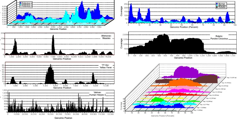
Deep sampling of the attenuated viruses.Viral particles in eight live-attenuated vaccines were purified through a 450-nm filter to eliminate bacterium-sized particles, digested with DNase and RNase enzymes to remove nonparticle protected “naked” nucleic acids, amplified by random PCR, and sub-jected to metagenomic pyrosequencing (4, 7). A total of 501,753 reads above a 50-bp length (average length 311 bp) were generated. Sequence reads were aligned against the ref-erence genome(s) of the expected attenuated virus(es). Viral regions sequenced and the depth at which different positions were covered are shown in Fig. 1, while Table 1 reports the percentage of viral reads and genome coverage for each vac-cine. Remaining sequence reads were assembled to generate contigs, and both singlet sequences and contigs were then classified using BLAST with the GenBank nonredundant da-tabase. Sequences were classified as viral, eukaryotic, or bac-terial/unclassifiable based on the best BLASTx match with an expectation value (E value) of 10e⫺5 or lower (Fig. 2).
Se-quences that exhibited a best match with an E value greater than 10e⫺5were deemed unclassifiable and typically were short
reads (less than 150 bp) with a weak match to a bacterial sequence. The amplification of bacterial and unclassifiable se-quences, as well as viral (bacteriophage and prophages) and eukaryotic (human and plants), when pure water alone was used as the nucleic acid input indicated that low-level contam-ination of reagents and enzymes may account for some of the nonvaccine sequences detected in vaccines (Fig. 2).
Adventitious viruses in vaccines.In addition to the attenu-ated virus, we also found other eukaryotic virus sequences. Attenuvax generated 4 reads covering 700 nucleotides of the ALV genome. Rotateq generated a single 276-nucleotide read with 96% identity to the SRV genome. Rotarix contained 6,344 reads with 98% identity to PCV1, covering the complete cir-cular genome (Fig. 1).
Nature of nonvaccine viral sequences.To confirm the pres-ence of ALV, SRV, and PCV1, total nucleic acids were directly extracted from all vaccines (without prior filtration and nucle-ase treatment), and both single-round and nested PCR (nPCR) was used to test for the presence of each virus (Table 1). ALV was detected in all three vaccines propagated in chicken embryo fibroblasts, although at lower levels detectable only by nPCR in MMRII and YF-Vax (Table 1, ⫹). The
on November 8, 2019 by guest
http://jvi.asm.org/
presence of SRV in the Vero cell-derived Rotateq vaccine was confirmed with a single round of PCR (Table 1,⫹⫹). SRV was also found by nPCR in the other Vero cell-derived Rotarix vaccine. However, SRV DNA was not detected in the Vero cell-derived OPV. Nucleic acids from all vaccines were tested for PCV1 using nPCR; only Rotarix was PCV1 positive. Sec-ond lots of both Rotarix (lot no. A41FA902A) and Rotateq (lot no. 0288Y) were also tested for the presence of SRV and PCV1. Both were positive for SRV by nested PCR. The second lot of Rotarix was also positive in a single-round PCR for PCV1, while Rotateq remained negative. To ascertain whether PCV1 was found in other GlaxoSmithKline (GSK) vaccines, we obtained a single dose of Pediarix containing noninfectious proteins from diphtheria, tetanus, pertussis bacteria, hepatitis
B, and killed polioviruses. Pediarix was negative for PCV-1 DNA using nPCR (data not shown). End-point dilution nested PCR was then performed to determine the approximate amount of PCV1 in both Rotarix lots (no. A41XA799A and no. A41FA902A) and was determined to be ⱖ175,000 and
[image:3.585.46.542.70.318.2]ⱖ250,000 PCV1 DNA copies per vaccine dose, respectively. In order to determine if the retroviral ALV and SRV se-quences originated from viral particles, the Rotateq and At-tenuvax preparations were spun to pellet viral particles that were then resuspended and extracted. An aliquot of each ex-traction was also treated with DNase. The DNase-treated and untreated extracts were then amplified by single-round or nested PCR, with or without cDNA synthesis using reverse transcriptase. ALV was still detectable after DNase treatment
FIG. 1. Viral genome sequencing coverage. Depth of sequence coverage at each genomic position for OPV, Attenuvax, YF-Vax, Varivax, MMR-II, Rotarix (PCV1), and Rotateq. For vaccines containing multiple genomes (MMRII) or multiple segments (Rotateq) of variable lengths, coverage is normalized to genetic position as percentage of fragment length. Depth of coverage for rotavirus and rubella virus from Rotarix and Meruvax-II, respectively, are not shown due to low coverage.
TABLE 1. Pyrosequencing coverage of viral genomes and PCR detection of endogenous retroviruses and PCV1
Vaccine Virus Total no. of reads
Viral sequences (%)
Genomic coverage (%)
Avg coverage (fold)
Nonvaccine viruses
PCR RT-PCRa
ALV SRV PCV1 ALV SRV
Biopolio Polio 1 150,899 40.25 99.5 2,009 None ⫺ ⫺ ⫺ ⫺ ⫺
Polio 2 150,899 14.08 99.6 639
Polio 3 150,899 39.72 97.0 2,425
Meruvax-II Rubella 14,389 0.97 41.8 ⬍1 None ⫺ ⫺ ⫺ ⫺ ⫺
Attenuvax Measles 9,863 11.96 95.9 21 ALV ⫹⫹ ⫺ ⫺ ⫹⫹ ⫺
YF-Vax Yellow fever 30,770 1.43 78.3 11 None ⫹ ⫺ ⫺ ⫹ ⫺
Varivax Human herpes 3 116,905 89.00 99.0 287 None ⫺ ⫺ ⫺ ⫺ ⫺
MMR-II Measles 39,646 1.19 95.8 9 None ⫹ ⫺ ⫺ ⫹ ⫺
Mumps 39,646 6.95 99.9 55
Rubella 39,646 ⬎0.01 28.4 ⬍1
Rotarix Rotavirus 15,249 ⬎0.01 20.6 ⬍1 PCV-1 ⫺ ⫹ ⫹ ⫺ ⫺
Rotateq Rotavirus 18,671 2.45 92.6 9 SRV-1 ⫺ ⫹⫹ ⫺ ⫺ ⫺
a
Samples were treated with DNase prior to reverse transcription to eliminate proviral DNA.⫺, nPCR negative;⫹, nPCR positive;⫹⫹, 1st-round PCR positive.
on November 8, 2019 by guest
http://jvi.asm.org/
[image:3.585.44.541.570.715.2]and required cDNA synthesis for its detection, indicating that it was in the form of encapsulated RNA. SRV did not require cDNA synthesis for its PCR detection and was no longer de-tectable after DNase treatment, indicating that it was likely present in the form of noninfectious host cell DNA released from Vero cells (Table 1). The presence of endogenous SRV provirus in Vero cells was confirmed by single-round PCR
using DNA extracted from uninfected Vero cells freshly ob-tained from ATCC (ATCC CCL-81), the parental cell line of the Vero E6 clone used to generate the vaccine (ATCC CRL-1586). As 41% of the 13,520 pyrosequence reads from the Rotateq were of primate origin, it is likely that the SRV se-quence detected was the result of leakage of host cell DNA, including proviral DNA. To further analyze the Rotateq SRV DNA, the entire coding region was sequenced, revealing 94% nucleotide identity to SRV-1 (GenBank accession no. U85506) (35). The inactivating mutations of proviral SRV-1 genomes in baboons (35) were not observed in the Rotateq-associated SRV genome. However, a frame-shifting single-nucleotide in-sertion in the polymerase gene was identified at genomic po-sition 3726, providing evidence that genetically defective SRV proviral DNA is present in Vero cells and in vaccines derived from these cells.
Microarray analysis of vaccine samples.The microbial de-tection array (MDA) developed at Lawrence Livermore Na-tional Laboratory contains 388,000 probes designed from all sequenced viral and bacterial organisms (11). The expected attenuated viruses were detected in all vaccines tested (Table 2). Hybridization was also seen to both PCV1 and PCV2 probes. Cross hybridization of labeled PCV1 DNA to PCV2 probes was due to the high sequence similarity of these two viruses. OPV was not tested. Two separate total nucleic acid preparations of the MMR-II vaccine yielded different results, identifying measles and mumps viruses in one preparation and rubella in the other. In vaccines grown in chicken embryo fibroblast (CEF), signals to avian endogenous retrovirus (AEV) were detected. The anticipated detection of AEV re-flects the presence of multiple loci of these endogenous pro-viruses in the chicken germ line (8, 9), release of AEV viral particles into CEF-derived vaccines (15, 34, 38), as well as possible cross-hybridization with ALV sequences, since the more conserved region of related viral groups are used in the microarray. The detection of human endogenous retrovirus K (HERVK) in Varivax, MMR-II, and Meruvax was the ex-pected consequence of their manufacture using human cell lines (Table 2). The origin of the baboon endogenous retrovi-rus signal for Rotateq is assumed to be related to the African green monkey-derived Vero cell used in its manufacture and cross-hybridization of its endogenous retroviruses to the ba-boon endogenous retrovirus probes.
Both nucleic acid extracted directly from the vaccines as well as nucleic acids extracted from vaccines that had first been filtered and nuclease digested (to enrich for viral particle-associated nucleic acids) were used as input to generate the DNA hybridized to the microarrays (Table 2, TOT and VP). The viral particle nucleic acid enrichment step (Table 2, VP) reduced the number of probes yielding signal from producer cell-derived endogenous retroviruses to undetectable levels for Attenuvax, Meruvax, and Rotateq, while YF-Vax exhibited
⬃50% reduction compared to total nucleic acid extraction. No difference was observed between the two extraction methods in the detection of any vaccine virus, with the exception of Ro-tarix, for which the VP purification procedure resulted in de-tection of only 7 of the 11 rotavirus genome segments, yielding a low overall log-odds score for the entire genome.
No pathogenic revertant sequences in the oral poliovirus vaccine.Since the year 2000, 426 cases of acute flaccid
pa-FIG. 2. Taxonomic distribution of sequences in 8 live-attenuated viral vaccines (a to h) and water (i). Reads were classified using BLASTx as either viral (V), eukaryotic (E), or bacterial/unclassifiable (E/U) (see Materials and Methods). Vaccine viral input (PFU) is listed based on manufacturers’ reported titers as either the 50% tissue cul-ture infectious dose (TCID50), PFU, or infectious units (IU).
on November 8, 2019 by guest
http://jvi.asm.org/
[image:4.585.57.268.57.562.2]ralysis in 11 countries have been attributed to 9 outbreaks of OPV revertants, resulting in pathogenic vaccine-derived polio-virus (VDPV) (http://www.cdc.gov/mmwr/preview/mmwrhtml /mm5836a3.htm). Recombination of attenuated poliovirus with other human enterovirus C viruses, together with several re-version mutations to wild-type poliovirus sequences, are the most common route in the generation of VDPV (http://www .polioeradication.org/content/fixed/VDPV_background.asp). In-depth sequencing of OPV allowed for detection of low-level polymorphisms generated byin vitroculture of the three vaccine serotypes. No mutations associated with reversion to poliovirus pathogenicity were observed as minority variants (Table 3). Mu-tations at levels lower than 2%, a conservative cutoff exceeding published rates of pyrosequencing error (22, 37), were observed at sites known to be polymorphic (26). For example, PV3 exhib-ited a C-to-T change at position 2493 in 0.5% (38/8,250) of se-quence reads. The detection of high-frequency frameshifting mu-tations also provided evidence of a mixture of both attenuated and defective PV2 (6%) and PV3 (30%) genomes (Table 4). Coverage depths for human herpesvirus 3 (HHV3) Varivax and
mumps virus in the MMR-II vaccine were also sufficient to ex-amine variation from reported vaccine strains and the presence of nucleotide polymorphisms. The percentage of nucleotide posi-tions for HHV3 in Varivax differing from those reported for the Oka vaccine strain (31) was less than 0.1%, with 110 mixed bases at both previously described and novel polymorphic positions (31). No mutation reached 100% of the HHV3 population in Varivax. Mumps virus in MMRII exhibited only a 0.30% muta-tion frequency from a published vaccine strain (accession number FJ211586) (32) with 4 nucleotide substitutions and 31 mixed-base polymorphisms.
DISCUSSION
[image:5.585.44.540.79.445.2]Using viral metagenomics, we identified nucleic acids from endogenous retroviruses and one adventitious virus in attenu-ated viral vaccines. The detection of ALV and EAV particles in live vaccines derived from chicken embryo fibroblast has been reported previously and investigated extensively (14, 15, 17). Seroconversion to ALV and EAV was not detected among
TABLE 2. Detection of viral sequences using panmicrobial microarray
Vaccine and cell line (origin)c Processinga Virus Log-odds
score Probe hybridization
b
Attenuvax from CEF TOT Measles virus 679.9 107/155
Avian leukosis virus 492.8 103/169
Avian endogenous retrovirus 276.2 43/59
VP Measles 673.6 105/154
YF-Vax from CEF TOT Yellow fever virus 621.4 88/120
Avian endogenous retrovirus 437.6 57/60
VP Yellow fever virus 628.3 89/120
Avian endogenous retrovirus 111.7 26/59
Varivax from MRC-5 (human) TOT Human herpes 3 608.6 103/130
Human endogenous retrovirus K 230.5 26/28
VP Human herpes 3 614.7 104/130
Human endogenous retrovirus K 199.7 26/28
Rotarix from Vero E6 (AGM) TOT Rotavirus A 482.3 232/802
Porcine circovirus 1 248.8 39/40
Porcine circovirus 2 533.7 105/120
VP Rotavirus A 2.4 203/802
Porcine circovirus 1 232.7 39/40
Porcine circovirus 2 505.2 96/103
Rotateq from Vero (AGM) TOT Rotavirus A 3559.8 490/802
Baboon endogenous retrovirus 256.5 44/82
VP Rotavirus A 2954.7 489/802
MMR-II from CEF and WI-38 (human) TOT (1st prep) Measles virus 688.8 104/154
Mumps virus 555.7 75/85
Human endogenous retrovirus K 222.3 22/28 Avian endogenous retrovirus 159.7 31/60
TOT (2nd prep) Rubella virus 58.1 26/74
Human endogenous retrovirus K 229.6 26/28
VP Not done NA
Meruvax-II from WI-38 (human) TOT Rubella virus 260.4 49/74
Human endogenous retrovirus K 247.3 26/28
VP Rubella virus 57.8 28/74
a
TOT refers to total nucleic acids directly extracted from the vaccine suspensions. VP refers to nucleic acids extracted from the vaccine suspensions following viral particle-associated nucleic acid purification using filtration and nuclease treatment.
b
NA, not applicable.
c
AGM, African green monkey.
on November 8, 2019 by guest
http://jvi.asm.org/
vaccinated individuals, indicating that these viruses were non-infectious to humans (14, 15).In vitroinoculation studies of a variety of human cell lines and peripheral blood mononuclear cells have also failed to show replication of CEF-derived ret-roviral particles, and there was no evidence of ALV pret-roviral integration (19, 27, 29). ALV and EAV in attenuated viral vaccines are therefore not of concern as infectious agents.
The detection of SRV DNA in Vero cells reflected the presence of endogenous SRV DNA in the germ line of African green monkeys, from which this cell line was derived. SRV is thought to be a highly prevalent infection of old-world mon-keys and endogenized at multiple copies in the genomes of many nonhuman primate species (23, 30, 35). The removal of SRV nucleic acid from the Rotateq vaccine by DNase indi-cated that it was present as naked DNA released from Vero cells. The detection of a retroviral inactivating mutation in the Rotateq SRV DNApolgene also indicated a defective nature for this endogenous retrovirus. A prior study showed two cases of SRV seroconversion in people with extensive blood and saliva contacts with nonhuman primates presumably following infection with replication-competent SRV (20). Neither clini-cal symptoms nor ongoing viremia was observed in these se-ropositive individuals (20). The detection of SRV DNA (or other retroviral sequences) in live-attenuated vaccines, there-fore, simply reflects their generation in cells, which contain a
large genome load of endogenous retroviruses, rather than the presence of adventitious viruses of concern.
A large fraction of the sequence reads from the Rotarix vaccines (41.6%) consisted of PCV1 sequences. Because se-rum-free media are used in the manufacture of Rotarix, a possible source for PCV1 is porcine pancreas-derived trypsin used for passaging Vero cells. After introduction of PCV1, a chronic infection may have become established in Vero cells (1). Alternatively, since the porcine-derived trypsin is typically irradiated before use to inactivate adventitious viruses, the PCV1 DNA may simply reflect carryover of noninfectious, lethally irradiated PCV1. PCV1 viral loads were measured in two lots at⬎105PCV1 DNA molecules per vaccine dose, and
the ratio of PCV1 to rotavirus pyrosequencing reads was⬎10. The relative efficiency of our random nucleic acid amplification method on single-stranded circular PCV1 DNA versus double-stranded rotavirus RNA is not known. The ratio of these vi-ruses’ genome copy number therefore cannot be deduced from the ratio of their sequence reads.
Whether the PCV1 in Rotarix is infectious in porcine, Vero, or human cell lines is currently unknown. PCV1 is a highly common and nonpathogenic pig infection usually transmitted among pigs through the fecal-oral route. PCV2, a close relative of PCV1, is a major pig pathogen with a high economic impact on the swine industry. Since both PCV species are highly
prev-TABLE 3. Absence of mutations associated with reversion to virulence
Genomic regiona Nucleotide
position Wild type
Vaccine strain
Amino acid changeb
454
sequence Coverage (fold)
Poliovirus 1 5⬘-UTR 480 A G NA G 663
VP3 1944 C A K3T A 1,161
VP1 2775 C A L3T A 1,827
VP1 2795 G A T3A A 1,870
Poliovirus 2 5⬘-UTR 481 G A NA A 634
VP1 2908 G A I3V A 435
Poliovirus 3 5⬘-UTR 472 C U NA U 1,645
VP3 2034 C U F3S U 530
VP1 2636 A G A3T G 843
a
UTR, untranslated region.
b
[image:6.585.42.545.81.209.2]NA, not applicable.
TABLE 4. Detection of poliovirus minority variants
Virus Genomic regiona Nucleotide position Mutationb Frequency (%) Coding
Poliovirus 1 5⬘-UTR 442–443 GAG insertion 38/50 (76) Noncoding
5⬘-UTR 565 T3C 43/487 (9) Noncoding
3C 5983 T3C 106/420 (25) Synonymous
Poliovirus 2 5⬘-UTR 619 G3A 2/89 (2) Noncoding
2C 4722 T3C 85/491 (17) Synonymous
2C 4965 T3C 24/209 (11) Synonymous
3D 6084 G3DEL 9/143 (6) Frameshift
3D 6087 T3DEL 9/143 (6) Frameshift
Poliovirus 3 3D 6902 T3DEL 461/1,495 (31) Frameshift
3D 6903 T3DEL 462/1,557 (30) Frameshift
a
UTR, untranslated region.
b
DEL, deletion.
on November 8, 2019 by guest
http://jvi.asm.org/
[image:6.585.42.541.576.709.2]alent in healthy pigs, human dietary and respiratory exposure to this virus is common through pork consumption or inhala-tion of particles from pig feces in the swine industry. Both PCV species and other members of theCircoviridaefamily are com-monly found in human stools (21). Whether PCV1 or PCV2 can actually replicate in humans is controversial, with most data weighing against human tropism. A single human gut biopsy specimen has been reported to contain PCV2 DNA, but contamination from human feces containing PCV2 from con-sumed pork cannot be excluded (6). A large PCR screen of human plasma and tissues did not detect any PCV2 DNA, and inoculations of various human cell lines with PCV2 were non-productive (12, 13). Serological testing for PCV exposure has yielded ambivalent results, with one group reporting a high rate of human seroreactivity to PCV1, although with an altered binding profile relative to pig serum samples, indicating the possible presence of a related but distinct virus (33). Other studies reported no detectable human PCV1 (3) or anti-PCV2 antibodies (2). While anti-PCV2 transmission can occur be-tween pigs through the eating of pig flesh (25), the absence of robust and specific human seroconversion to pig circoviruses, despite frequent consumption of infected pork, further sup-ports an inability of pig circoviruses to replicate in humans. Therefore, the detection of viral DNA of unknown infectivity in a live-attenuated oral vaccine, from a virus not shown to infect humans, may not be of concern. If contamination of the vaccine producer Vero cells with PCV1 did occur, their re-placement with fresh Vero cells should remove the source of PCV1.
Production of live-attenuated vaccines, requiring viral am-plification, may result in reversion mutations. In the case of OPV, these mutations may match those found in VDPV. No virulence-associated mutations were detected as minority vari-ants following deep sequencing of the three poliovirus sero-types in the trivalent OPV viral quasispecies tested in the Biopolio vaccine. As is well documented, the rare case of reversion of attenuated OPV to VDPV therefore likely occurs during the multiple rounds of replication occurring in vivo
following vaccination, typically in immunocompromised vac-cinees or their infected contacts (18, 24). Beside polioviruses, the attenuated mumps virus and VZV were also sequenced at depths sufficient to derive accurate genome sequences and identify minority variants. Mumps virus and VSV both showed very low numbers of mutations relative to the published ge-nomes of attenuated vaccine strains. There is no evidence that any of the sequence change detected affects virus attenuation. The use of metagenomics and microarray technologies for the detection of adventitious viral contamination can detect a range of viruses more diverse than is possible with the cur-rently mandated methods of cell cultures and viral species-specific PCR. PCR is more sensitive than either metagenomics or microarrays when screening for specific viruses, but the sheer number of potential contaminating viruses limits its use to a small number of suspected viruses. The use of viral meta-genomics and microarray tests therefore seems warranted for the surveillance of products that are derived from cell cultures, plasma pools, or other biological sources and which may con-tain and transmit adventitious viruses.
Despite an extensive record of safety and efficacy, common misconceptions remain regarding vaccine safety, resulting in
reduced childhood vaccination and the resurgence of vaccine-preventable infections. Given that live-attenuated viral vac-cines are safe, effective, and relatively inexpensive, their use against human and animal pathogens should be encouraged. The application of high-throughput sequencing and microar-rays provides effective means to interrogate current and future vaccines for genetic variants of the attenuated viruses and the presence of adventitious viruses. The wide range of sequences detectable by these methods (endogenous retroviruses, bacte-rial and other nucleic acids whose taxonomic origin cannot be determined, and adventitious viruses, such as PCV1) is an expected outcome of closer scrutiny to the nucleic acids present in vaccines and not necessarily a reflection of unsafe products. In view of the demonstrated benefit and safety of Rotarix, the implications (if any) for current immunization policies of the detection of PCV1 DNA of unknown infectivity for humans need to be carefully considered.
As an added note, recent testing by GSK indicates that PCV1 was also present in the working cell bank and viral seed used for the generation of Rotarix used in the extensive clinical trials that demonstrated the safety and efficacy of this vaccine (http://www .fda.gov/BiologicsBloodVaccines/Vaccines/ApprovedProducts /ucm205545.htm). These trials indicate a lack of detectable pathogenic effects from PCV1 DNA on vaccinees.
ACKNOWLEDGMENTS
We thank for support NHLBI R01HL083254 grant to E.L.D., Blood Systems Research Institute, Laboratory Directed Research and Devel-opment Program at the Lawrence Livermore National Laboratory (Project LLNL 02-SI-008 under the auspices of the U.S. Department of Energy by Lawrence Livermore National Laboratory under contract DE-AC52-07NA27344), and U.S. Air Force Surgeon General-ap-proved Clinical Investigation no. FDG20040024E.
We thank Michael P. Busch for encouragement, James B. Thissen for technical assistance in microarray experiments, and T. N. Dhole, Dept. of Microbiology, SGPGIMS, Lucknow, India, for providing Biopolio OPV vaccine.
The views expressed in this material are those of the authors and do not reflect the official policy or position of the U.S. government or the Departments of Energy or Defense or the Air Force.
Author contributions: J.G.V. designed the study, performed lab work and bioinformatics analyses, and wrote the manuscript; C.W. performed the metagenomics bioinformatics analysis; M.S.J. assisted with the study design; C.J. performed the microarray lab work; K.M. performed the microarray bioinformatics analyses; S.G. designed the probes on the microarray; and E.L.D. designed the study and wrote the manuscript.
REFERENCES
1.Allan, G. M., D. P. Mackie, J. McNair, B. M. Adair, and M. S. McNulty. 1994. Production, preliminary characterisation and applications of monoclo-nal antibodies to porcine circovirus. Vet. Immunol. Immunopathol.43:357– 371.
2.Allan, G. M., F. McNeilly, I. McNair, M. D. Curran, I. Walker, J. Ellis, C. Konoby, S. Kennedy, and B. Meehan.2000. Absence of evidence for porcine circovirus type 2 in cattle and humans, and lack of seroconversion or lesions in experimentally infected sheep. Arch. Virol.145:853–857.
3.Allan, G. M., K. V. Phenix, D. Todd, and M. S. McNulty. 1994. Some biological and physico-chemical properties of porcine circovirus. Zentralbl. Veterinarmed. B41:17–26.
4.Allander, T., S. U. Emerson, R. E. Engle, R. H. Purcell, and J. Bukh.2001. A virus discovery method incorporating DNase treatment and its application to the identification of two bovine parvovirus species. Proc. Natl. Acad. Sci. U. S. A.98:11609–11614.
5.Allen, A.2007. Vaccine: the controversial story of medicine’s greatest life-saver. W.W. Norton, New York, NY.
6.Bernstein, C. N., G. Nayar, A. Hamel, and J. F. Blanchard.2003. Study of animal-borne infections in the mucosas of patients with inflammatory bowel disease and population-based controls. J. Clin. Microbiol.41:4986–4990.
on November 8, 2019 by guest
http://jvi.asm.org/
7.Delwart, E. L.2007. Viral metagenomics. Rev. Med. Virol.17:115–131. 8.Dunwiddie, C. T., R. Resnick, M. Boyce-Jacino, J. N. Alegre, and A. J. Faras.
1986. Molecular cloning and characterization of gag-, pol-, and env-related gene sequences in the ev⫺chicken. J. Virol.59:669–675.
9.Dunwiddie, C., and A. J. Faras.1985. Presence of retrovirus reverse tran-scriptase-related gene sequences in avian cells lacking endogenous avian leukosis viruses. Proc. Natl. Acad. Sci. U. S. A.82:5097–5101.
10.Engels, E. A.2005. Cancer risk associated with receipt of vaccines contam-inated with simian virus 40: epidemiologic research. Expert Rev. Vaccines 4:197–206.
11.Gardner, S. N., A. L. Hiddessen, P. L. Williams, C. Hara, M. C. Wagner, and B. W. Colston. 2009. Multiplex primer prediction software for divergent targets. Nucleic Acids Res.37:6291–6304.
12.Hattermann, K., A. Maerz, H. Slanina, C. Schmitt, and A. Mankertz.2004. Assessing the risk potential of porcine circoviruses for xenotransplantation: consensus primer-PCR-based search for a human circovirus. Xenotransplan-tation11:547–550.
13.Hattermann, K., C. Roedner, C. Schmitt, T. Finsterbusch, T. Steinfeldt, and A. Mankertz.2004. Infection studies on human cell lines with porcine cir-covirus type 1 and porcine circir-covirus type 2. Xenotransplantation11:284– 294.
14.Hussain, A. I., J. A. Johnson, M. Da Silva Freire, and W. Heneine.2003. Identification and characterization of avian retroviruses in chicken embryo-derived yellow fever vaccines: investigation of transmission to vaccine recip-ients. J. Virol.77:1105–1111.
15.Hussain, A. I., V. Shanmugam, W. M. Switzer, S. X. Tsang, A. Fadly, D. Thea, R. Helfand, W. J. Bellini, T. M. Folks, and W. Heneine.2001. Lack of evidence of endogenous avian leukosis virus and endogenous avian retrovi-rus transmission to measles, mumps, and rubella vaccine recipients. Emerg. Infect. Dis.7:66–72.
16.Jaing, C., S. Gardner, K. McLoughlin, N. Mulakken, M. Alegria-Hartman, P. Banda, P. Williams, P. Gu, M. Wagner, C. Manohar, and T. Slezak.2008. A functional gene array for detection of bacterial virulence elements. PLoS One3:e2163.
17.Johnson, J. A., and W. Heneine.2001. Characterization of endogenous avian leukosis viruses in chicken embryonic fibroblast substrates used in produc-tion of measles and mumps vaccines. J. Virol.75:3605–3612.
18.Kew, O. M., R. W. Sutter, E. M. de Gourville, W. R. Dowdle, and M. A. Pallansch.2005. Vaccine-derived polioviruses and the endgame strategy for global polio eradication. Annu. Rev. Microbiol.59:587–635.
19.Khan, A. S., T. Maudru, A. Thompson, J. Muller, J. F. Sears, and K. W. Peden. 1998. The reverse transcriptase activity in cell-free medium of chicken embryo fibroblast cultures is not associated with a replication-com-petent retrovirus. J. Clin. Virol.11:7–18.
20.Lerche, N. W., W. M. Switzer, J. L. Yee, V. Shanmugam, A. N. Rosenthal, L. E. Chapman, T. M. Folks, and W. Heneine.2001. Evidence of infection with simian type D retrovirus in persons occupationally exposed to nonhu-man primates. J. Virol.75:1783–1789.
21.Li, L., A. Kapoor, B. Slikas, O. S. Bamidele, C. Wang, S. Shaukat, M. A. Masroor, M. L. Wilson, J. B. Ndjango, M. Peeters, N. D. Gross-Camp, M. N. Muller, B. H. Hahn, N. D. Wolfe, H. Triki, J. Bartkus, S. Z. Zaidi, and E. Delwart. 2010. Multiple diverse circoviruses infect farm animals and are commonly found in human and chimpanzee feces. J. Virol.84:1674–1682. 22.Margeridon-Thermet, S., N. S. Shulman, A. Ahmed, R. Shahriar, T. Liu, C.
Wang, S. P. Holmes, F. Babrzadeh, B. Gharizadeh, B. Hanczaruk, B. B. Simen, M. Egholm, and R. W. Shafer.2009. Ultra-deep pyrosequencing of hepatitis B virus quasispecies from nucleoside and nucleotide reverse-tran-scriptase inhibitor (NRTI)-treated patients and NRTI-naive patients. J. In-fect. Dis.199:1275–1285.
23.Marx, P. A., and A. F. Voevodin.2009. Simian virology. Wiley-Blackwell, Ames, IA.
24.Minor, P. D.1993. Attenuation and reversion of the Sabin vaccine strains of poliovirus. Dev. Biol. Stand.78:17–26.
25.Opriessnig, T., A. R. Patterson, X. J. Meng, and P. G. Halbur.2009. Porcine circovirus type 2 in muscle and bone marrow is infectious and transmissible to naïve pigs by oral consumption. Vet. Microbiol.133:54–64.
26.Rezapkin, G. V., L. P. Norwood, R. E. Taffs, E. M. Dragunsky, I. S. Leven-book, and K. M. Chumakov.1995. Microevolution of type 3 Sabin strain of poliovirus in cell cultures and its implications for oral poliovirus vaccine quality control. Virology211:377–384.
27.Robertson, J. S., C. Nicolson, A. M. Riley, M. Bentley, G. Dunn, T. Corcoran, G. C. Schild, and P. Minor.1997. Assessing the significance of reverse transcriptase activity in chick cell-derived vaccines. Biologicals25:403–414. 28.Seeff, L. B., G. W. Beebe, J. H. Hoofnagle, J. E. Norman, Z. Buskell-Bales,
J. G. Waggoner, N. Kaplowitz, R. S. Koff, J. L. Petrini, and E. R. Schiff.1987. A serologic follow-up of the 1942 epidemic of post-vaccination hepatitis in the United States Army. N. Engl. J. Med.316:965–970.
29.Shahabuddin, M., J. F. Sears, and A. S. Khan.2001. No evidence of infec-tious retroviruses in measles virus vaccines produced in chicken embryo cell cultures. J. Clin. Microbiol.39:675–684.
30.Sommerfelt, M. A., N. Harkestad, and E. Hunter.2003. The endogenous langur type D retrovirus PO-1-Lu and its exogenous counterparts in ma-caque and langur monkeys. Virology315:275–282.
31.Tillieux, S. L., W. S. Halsey, E. S. Thomas, J. J. Voycik, G. M. Sathe, and V. Vassilev.2008. Complete DNA sequences of two oka strain varicella-zoster virus genomes. J. Virol.82:11023–11044.
32.Tillieux, S. L., W. S. Halsey, G. M. Sathe, and V. Vassilev.2009. Comparative analysis of the complete nucleotide sequences of measles, mumps, and ru-bella strain genomes contained in Priorix-Tetra and ProQuad live attenuated combined vaccines. Vaccine27:2265–2273.
33.Tischer, I., L. Bode, J. Apodaca, H. Timm, D. Peters, R. Rasch, S. Pociuli, and E. Gerike.1995. Presence of antibodies reacting with porcine circovirus in sera of humans, mice, and cattle. Arch. Virol.140:1427–1439. 34.Tsang, S. X., W. M. Switzer, V. Shanmugam, J. A. Johnson, C. Goldsmith, A.
Wright, A. Fadly, D. Thea, H. Jaffe, T. M. Folks, and W. Heneine.1999. Evidence of avian leukosis virus subgroup E and endogenous avian virus in measles and mumps vaccines derived from chicken cells: investigation of transmission to vaccine recipients. J. Virol.73:5843–5851.
35.van der Kuyl, A. C., R. Mang, J. T. Dekker, and J. Goudsmit.1997. Complete nucleotide sequence of simian endogenous type D retrovirus with intact genome organization: evidence for ancestry to simian retrovirus and baboon endogenous virus. J. Virol.71:3666–3676.
36.Victoria, J. G., A. Kapoor, L. Li, O. Blinkova, B. Slikas, C. Wang, A. Naeem, S. Zaidi, and E. Delwart.2009. Metagenomic analyses of viruses in stool samples from children with acute flaccid paralysis. J. Virol.83:4642–4651. 37.Wang, C., Y. Mitsuya, B. Gharizadeh, M. Ronaghi, and R. W. Shafer.2007.
Characterization of mutation spectra with ultra-deep pyrosequencing: appli-cation to HIV-1 drug resistance. Genome Res.17:1195–1201.
38.Weissmahr, R. N., J. Schu¨pbach, and J. Bo¨ni.1997. Reverse transcriptase activity in chicken embryo fibroblast culture supernatants is associated with particles containing endogenous avian retrovirus EAV-0 RNA. J. Virol. 71:3005–3012.
39.Wong, C. W., C. L. Heng, L. Wan Yee, S. W. Soh, C. B. Kartasasmita, E. A. Simoes, M. L. Hibberd, W. K. Sung, and L. D. Miller.2007. Optimization and clinical validation of a pathogen detection microarray. Genome Biol. 8:R93.