0022-538X/07/$08.00⫹0 doi:10.1128/JVI.00970-07
Copyright © 2007, American Society for Microbiology. All Rights Reserved.
Evaluation of Replication and Pathogenicity of Avian Influenza A H7
Subtype Viruses in a Mouse Model
䌤
Tomy Joseph,
1Josephine McAuliffe,
1Bin Lu,
2Hong Jin,
2George Kemble,
2and Kanta Subbarao
1*
Laboratory of Infectious Diseases, NIAID, NIH, Bethesda, Maryland 20892,1and
MedImmune Vaccines, Inc., Mountain View, California 940432
Received 4 May 2007/Accepted 6 July 2007
Avian influenza A H7 subtype viruses pose a significant threat to human health because of their ability to transmit directly from domestic poultry to humans and to cause disease and, sometimes, death. Although it is important to develop vaccines against viruses of this subtype, very limited information is available on the immune response and pathogenesis of H7 viruses in animal models such as mice and ferrets. Ten H7 viruses were selected for possible vaccine development on the basis of their phylogenetic relationships and geograph-ical locations. The virulence of the 10 viruses for mice and the immunogenicity of the viruses in mice and ferrets were evaluated to study the extent of antigenic relatedness and the level of cross-reactivity of antibodies. Most of the viruses showed similar patterns of cross-reactivity with mouse and ferret antisera. The Eurasian viruses elicited broadly cross-reactive antibodies that neutralized viruses from both Eurasian and North American lineages, but the converse was not true. A subset of the viruses was also evaluated for the ability to replicate and cause disease in BALB/c mice following intranasal administration. H7 subtype viruses were able to infect mice without adaptation and manifested different levels of lethality and kinetics of replication. On the basis of phylogenetic data, induction of broadly cross-neutralizing antibodies in mouse and ferret antisera, and their ability to replicate in mice, we have selected A/Netherlands/219/03 (subtype H7N7) and A/chicken/BC/CN-7/04 (subtype H7N3) viruses for vaccine development. The mouse model can be used for the preclinical evaluation of these vaccines against H7 subtype viruses.
Influenza A viruses are divided into subtypes on the basis of serological and genetic differences in their major surface glyco-proteins, the hemagglutinin (HA) and neuraminidase (NA). Sixteen different HA (H1 to H16) and 9 NA (N1 to N9) subtypes have been identified among influenza A viruses (7, 31). Viruses of all 16 HA and 9 NA subtypes infect aquatic birds, and these birds serve as the reservoir from which novel subtypes of influenza viruses are introduced into domestic poultry and the human population. On the basis of their ability to cause disease in chickens, avian influenza viruses are divided into two groups, highly pathogenic (HP) and low-pathogenicity (LP) viruses. HP avian influenza viruses are restricted to H5 and H7 HA subtypes and cause lethal systemic infection that may result in 100% mortality within a flock, whereas LP avian influenza viruses include viruses of all subtypes and cause milder infections, with a lower rate of morbidity and no mor-tality (29).
Occasionally, avian influenza A viruses are transmitted di-rectly from birds to humans, with variable consequences. In-troduction of a new influenza A virus subtype into a susceptible human population could result in a pandemic if the virus causes disease and spreads efficiently from person to person. Although H5N1 viruses currently are the focus of concern, the next pandemic of influenza could be caused by a virus of another subtype.
Avian influenza A H7 subtype viruses have caused large outbreaks of disease in domestic poultry in Asia, Europe,
North America, and South America in recent years, resulting in severe economic losses to the poultry industry (5). Because of their ability to transmit directly from domestic poultry to humans and to cause disease and, sometimes, death, H7 vi-ruses also have been recognized as a concern for human health. Although isolated cases of human infections with HP or LP avian influenza H7 viruses have occurred (2, 4, 14, 27, 30), H7 viruses became a major concern with the direct transmis-sion of H7N7 viruses to humans in The Netherlands in 2003. An HP avian influenza H7N7 virus caused severe outbreaks of disease in domestic poultry in The Netherlands in March 2003. Culling of 30 million chickens, i.e., about 28% of the total chicken population in The Netherlands, controlled further spread of the infection (13). This outbreak in poultry also resulted in the direct transmission of the virus to at least 86 people who were involved in the culling of infected poultry. There also was evidence of limited human-to-human transmis-sion from an infected family member in three cases (8, 13). Of these 89 human infections, most of the patients developed conjunctivitis, and a few others developed mild influenza-like illness (8). There was one fatal case of pneumonia and acute respiratory distress syndrome in a veterinarian who visited farms with HP avian influenza virus-infected poultry flocks (8, 13). In 2004, an HP avian influenza H7N3 virus emerged in domestic poultry in British Columbia, Canada. This outbreak resulted in the infection of two workers on a poultry farm, causing mild respiratory disease and conjunctivitis (9, 18, 28). A serological survey in Italy detected anti-H7 antibodies in 7 out of 185 poultry workers who were exposed to an LP avian influenza H7N3 virus during the 2002 to 2003 avian influenza outbreaks in that country (20). Thus, direct transmission of H7 subtype viruses to humans occurs, and this highlights the threat
* Corresponding author. Mailing address: Bldg 33, Room 3E13C-1, 33 North Dr., MSC 3203, Bethesda, MD 20892. Phone: (301) 451-3839. Fax: (301) 496-8312. E-mail: ksubbarao@niaid.nih.gov.
䌤Published ahead of print on 18 July 2007.
10558
on November 8, 2019 by guest
http://jvi.asm.org/
posed by both HP and LP avian influenza viruses of this sub-type to human health in terms of pandemic potential.
Phylogenetic analysis of the H7 HA gene reveals a separa-tion into two lineages, Eurasian and North American, that correspond to the geographic separation of the birds that they infect, and they generally correspond to the Eurasian and North American flyways of migratory birds (3). Antigenic re-latedness among viruses from the two lineages and the range of cross-protective immune response induced by viruses from the two lineages currently are not known. Such information is vital for the development of efficacious vaccines.
Very limited information is available on the immune re-sponse and pathogenesis of H7 viruses in mammalian species, such as ferrets and mice, that currently are used for preclinical studies of pandemic influenza vaccines. Ferrets are considered the best available model for human influenza research, and sera from ferrets postinfection is the WHO standard for anti-genic analysis of human influenza viruses. Human influenza viruses replicate in the respiratory tract of ferrets, resulting in acute respiratory illness that is characterized by clinical signs such as fever, rhinorrhea, and sneezing, and ferrets develop antibodies to the infecting strain. In contrast, human influenza viruses rarely cause symptoms of respiratory tract disease in mice and require adaptation to replicate to high titers in mice. Because of these reasons, the use of mouse models for the study of influenza is limited. However, the advantage of a mouse model for avian influenza virus infection is that reagents for immunologic studies of mice are widely available. It is important to assess the immune response and kinetics of rep-lication of H7 viruses in different animal models in order to establish appropriate models for the preclinical evaluation of vaccines.
In this study, we selected 10 H7 viruses on the basis of their phylogenetic relationships and geographical locations and evaluated their ability to induce broadly cross-neutralizing an-tibodies in mice and ferrets. We also evaluated the ability of a subset of these viruses to replicate and cause disease in mice following intranasal administration.
MATERIALS AND METHODS
Selection of viruses.Nucleotide sequences of the HA1 domain of the HA gene of 24 avian influenza A H7 subtype viruses were obtained either from GenBank or by sequencing. The virus strains and GenBank accession numbers used for phylogenetic analysis are listed in Table 1. Phylogenetic analysis was performed by the neighbor-joining method, using the MacVector 9.0 software package. The following 10 avian influenza A H7 subtype viruses were selected for further study: A/chicken/BC/CN-7/04 (subtype H7N3) (designated BC/04), A/Nether-lands/219/03 (H7N7) (NL/03), A/chicken/Chile/4322/02 (H7N3) (CH/02), A/chicken/Queensland/95 (H7N3) (QL/95), A/turkey/England/63 (H7N3) (EG/ 63), A/FPV/Rostock/34 (H7N1) (RK/34), A/turkey/VA/55/02 (H7N2) (VA/02), A/turkey/UT/24721-10/95 (H7N3) (UT/95), A/rhea/NC/39482/93 (H7N1) (NC/ 93), and A/turkey/OR/71 (H7N3) (OR/71). Viruses used in this study were kindly provided by Robert G. Webster, St. Jude Children’s Research Hospital, Mem-phis, TN; David Swayne, Southeast Poultry Research Laboratory, USDA, Ath-ens, GA; Nancy Cox, Influenza Division, Centers for Disease Control and Pre-vention, Atlanta, GA; and John Pasick, Canadian Food Inspection Agency, National Centre for Foreign Animal Disease, Winnipeg, Canada.
HA genes of the UT/95, BC/04, and NC/93 viruses were sequenced, and others were obtained from GenBank to analyze the amino acid sequences. For sequenc-ing, viral RNA of UT/95, BC/04, and NC/93 was extracted using the QIAmp viral RNA mini kit (QIAGEN, Valencia, CA). The full-length HA gene was amplified from viral RNA by reverse transcription-PCR using an Omniscript reverse tran-scription kit (QIAGEN, Germantown, MD) and an advantage high-fidelity PCR kit (Clontech, Mountain View, CA). PCR fragments were purified using a
QIAquick PCR purification kit (QIAGEN). Sequencing was carried out using the big dye terminator v1.1 cycle sequencing kit (Applied Biosystems, Foster City, CA) and an ABI Prism 3100 genetic analyzer (Applied Biosystems). The Sequencher 4.7 software package was used for the editing, assembly, and analysis of nucleotide sequence data. Amino acid sequences of the HA protein of the 10 selected H7 viruses were predicted from full-length nucleotide sequences. The percentage of amino acid identity among the selected viruses and the sequence of the cleavage site of the HA were determined using the MacVector 9.0 soft-ware package.
The 10 selected H7 viruses were propagated in the allantoic cavity of 10-day-old embryonated chicken eggs at 37°C and were stored at⫺80°C until use. Fifty percent tissue culture infectious doses (TCID50) were determined by serial
titration of viruses in MDCK cells, and titers were determined by the method described by Reed and Muench (21).
All experiments, including animal studies with LP and HP H7 avian influenza viruses, were conducted in biosafety level 3⫹containment laboratories approved for use by the U.S. Department of Agriculture and Centers for Disease Control and Prevention. Animal experiments were approved by the National Institutes of Health Animal Care and Use Committee.
Pathogenicity in mice.To assess the lethality of the viruses in mice, groups of five 4- to 6-week-old female BALB/c mice (Taconic Farms, Hudson, NY) were anesthetized and infected intranasally (i.n.) with each of the selected H7 viruses at a dose of 105TCID
50per mouse, administered in a 50-l volume. Mice were
monitored daily for mortality for 14 days. HP viruses BC/04, NL/03, and EG/63 and LP viruses VA/02 and UT/95 were further evaluated in mice. To determine the 50% lethal doses (LD50) of the HP viruses, groups of five female BALB/c
mice were anesthetized and infected i.n. with serial 10-fold dilutions ranging from 100
to 106
TCID50of each virus in a 50-l volume. Mice were monitored
daily for 14 days. Mice that lost 20% of their body weight were sacrificed according to the animal study protocol. The LD50was determined by the Reed
and Muench method (21). Similarly, groups of five female BALB/c mice were inoculated with 105
TCID50of the LP viruses in 50l and were monitored and
weighed daily for 14 days.
Kinetics of virus replication in mice.To study the kinetics of replication of the viruses in different tissues, groups of 24 female BALB/c mice were inoculated i.n. with 105
TCID50of each virus. Four mice from each group were euthanized 1, 2,
3, 4, 6, and 7 days postinfection (dpi). Organs were collected at up to 4 dpi for NL/03 and EG/63 viruses and at 6 dpi for BC/04 virus, because the mice did not survive beyond these time points. Nasal turbinates (the intact nasal cavity con-taining the nasal turbinates, nasal bones, and the upper jaw), lungs, brains, and spleens were harvested, weighed, and homogenized in L-15 medium
(Invitrogen-TABLE 1. Viruses used for phylogenetic analysis
Virus Subtype GenBank accession no. of HA gene sequence
A/FPV/Dutch/27 H7N7 Z12617
A/FPV/Rostock/34 H7N1 M24457
A/turkey/England/63 H7N3 CY015065
A/turkey/Oregon/71 H7N3 M31689
A/chicken/Victoria/75 H7N7 Z47199
A/turkey/Minnesota/1200/80 H7N3 CY014778
A/chicken/Victoria/85 H7N7 M17735
A/chicken/Jena/87 H7N7 U20469
A/rhea/North Carolina/39482/93 H7N1 EF470586 A/quail/Arkansas/16309-7/94 H7N3 AF072401 A/chicken/New Jersey/15086-3/94 H7N3 AF072383 A/turkey/Utah/24721-10/95 H7N3 EF470585 A/chicken/Pakistan/447/95 H7N3 AF202226 A/common iora/Singapore/95 H7N1 AF202228 A/chicken/Queensland/667/95 H7N3 AF202231
A/mallard/Ohio/322/98 H7N3 CY016188
A/chicken/New York/12273-11/99 H7N3 AY240892 A/quail/New York/11430/99 H7N2 AY240923 A/turkey/Italy/13468/99 H7N1 AJ493216 A/chicken/New York/30749-3/00 H7N2 AY240897 A/turkey/Virginia/55/02 H7N2 AY240912 A/chicken/Chile/4322/02 H7N3 AY303631
A/Netherlands/219/03 H7N7 AY338459
A/chicken/British Columbia/CN-7/04 H7N3 EF470587
on November 8, 2019 by guest
http://jvi.asm.org/
[image:2.585.300.541.80.327.2]GIBCO) containing a 2⫻concentration of antibiotic-antimycotic (penicillin, streptomycin, and amphotericin B) (Invitrogen-GIBCO) to make 5% (wt/vol) (nasal turbinates) or 10% (wt/vol) (lungs, brain, and spleen) tissue homogenates. Tissue homogenates were clarified by centrifugation and titrated in 24- and 96-well tissue culture plates containing MDCK cells. The virus titer for each organ was determined by the method described by Reed and Muench (21) and was expressed as the log10TCID50/gram of tissue.
Generation of antisera. (i) Mice.To generate specific postinfection sera, groups of five 4- to 6-week-old female BALB/c mice were anesthetized and infected i.n. with 50l of a dose of virus selected on the basis of the results of the lethality study. Mice received 105TCID
50per mouse for RK/34, CH/02,
QL/95, VA/02, UT/95, and OR/71, 103
TCID50per mouse for NC/93, and 10 2
TCID50per mouse for BC/04, NL/03, and EG/63. Mice were monitored for the
development of disease and mortality. All of the mice were bled before virus administration, and postinfection sera were collected at one or more of the following time points: 28, 42, and 56 dpi.
(ii) Ferrets. Two 4- to 5-week-old ferrets (Harlan Sprague Dawley, Inc., Hanover, NH) that were seronegative for antibodies to currently circulating H3N2 and H1N1 human influenza viruses were used to generate antiserum for each virus. Since pathogenicity data for H7 subtype viruses in ferrets are not available, the dose administered was based on data from mice and chickens. The inoculum for viruses that were not lethal for mice and/or chickens was 107
TCID50, and lower doses (105and 102TCID50) were used as inocula for viruses
that were lethal in these species. If the ferret that received 105
TCID50survived
until day 21, serum from this ferret was used in serologic assays. If a dose of 105
TCID50was lethal, serum from the ferret that received 10 2
TCID50was used.
Therefore, lightly anesthetized ferrets were inoculated i.n. with 107TCID 50of
VA/02, UT/95, and OR/71 or with 105
and 102
TCID50of NL/03, CH/02, QL/95,
EG/63, RK/34, NC/93, and BC/04 in a volume of 500l (250l per nostril). Ferrets were monitored daily for clinical signs of influenza infection, and body temperatures were recorded twice daily. Postinfection sera were collected at 14 and 21 dpi. For NL/03, CH/02, QL/95, EG/63, RK/34, and NC/93, postinfection sera from ferrets that received 105TCID
50were used in serologic assays, and for
BC/04 the serum from the ferret that received 102
TCID50was used. For the
remaining three viruses, sera from ferrets that received 107TCID
50were used in
serologic assays.
HI assay.Antibody titers for postinfection mouse and ferret sera were deter-mined by hemagglutination inhibition (HI) assays. Serum samples were treated with receptor-destroying enzyme (Denka Seiken Co., Ltd., Tokyo, Japan) for 22 h to remove nonspecific inhibitors. Serum samples were tested by standard methods using four HA units of the virus in V-bottom 96-well microtiter plates with 0.5% turkey erythrocytes and a 1% suspension of horse erythrocytes, as previously described (11, 23). HI titers were expressed as the reciprocals of the highest dilution of the serum that completely inhibited agglutination of erythro-cytes by four HA units of the virus. HI titers were converted to log2and were
expressed as the arithmetic means⫾standard errors.
Virus neutralization assay.Neutralizing antibody titers for postinfection ferret and mouse sera were evaluated in a microneutralization (MN) assay. Postinfec-tion ferret sera collected at 21 dpi from a single ferret and mouse sera collected at 28 dpi for VA/02 and RK/34 and at 56 dpi for the remaining eight viruses were used in the MN assay. Postinfection mouse sera from two to three mice were pooled to obtain a sufficient quantity for the MN assay. Serial twofold dilutions of heat-inactivated serum were prepared, and equal volumes of serum and virus were mixed and incubated for 60 min at room temperature. The residual infec-tivity of the virus-serum mixture was determined in MDCK cells in four repli-cates for each dilution. The neutralizing titer was defined as the reciprocal of the highest dilution of serum that completely neutralized the infectivity of 100 TCID50of the virus as determined by the absence of cytopathic effect at day 4.
Nucleotide sequence accession numbers.The nucleotide sequences presented in this article (accession numbers EF470585, EF470586, and EF470587) have been deposited in GenBank.
RESULTS
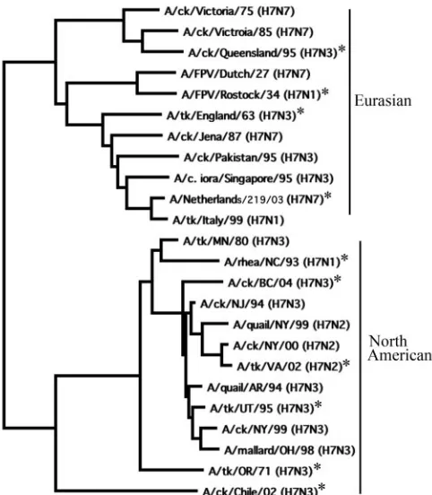
Selection of viruses. On phylogenetic analysis of the HA1 domain, the HA genes of 24 H7 subtype viruses clustered into Eurasian and North American lineages (Fig. 1). From these 24 H7 subtype viruses (Fig. 1 and Table 1), 10 viruses were se-lected on the basis of their phylogenetic relationships and geographic locations for evaluation in a mouse model and to identify viruses for vaccine development. Four viruses
be-longed to the Eurasian lineage, and six viruses bebe-longed to the North American lineage (Fig. 1). Amino acid sequence identity of all pairwise comparisons of the predicted HA protein se-quences ranged from 88.6 to 98%. The selected viruses in-cluded different subtypes of NA proteins, as shown in Table 2. The group of 10 viruses included six HP and four LP viruses on the basis of their previously established pathogenicity in chick-ens (Table 2) (1, 3, 8–10, 13, 18, 22, 24, 26, 28).
Lethality of H7 viruses for mice. Differences in lethality among the 10 viruses were noted for BALB/c mice (Table 2). The mice inoculated with 105TCID
50of the BC/04, NL/03, and
EG/63 viruses succumbed by 5 to 6 dpi. However, the HP viruses CH/02 and QL/95 were not lethal for mice, even at the maximum dose of virus tested (105TCID
50). In contrast, the
LP virus NC/93 was highly lethal for mice when it was used at 105 TCID
50 per mouse. On the basis of their phylogenetic
relationships and virulence for mice, BC/04 (an HP virus from the North American lineage), NL/03 and EG/63 (HP viruses from the Eurasian lineage), UT/95 (an LP virus from the North American H7N3 group), and VA/02 (an LP virus from the North American H7N2 group) viruses were selected for fur-ther study (Fig. 1 and Table 2). The three selected HP viruses, BC/04, NL/03, and EG/63, were lethal to mice at very low doses. Most of the mice that received 102TCID
50of the virus were dead
within 8 to 10 days (Fig. 2). On the basis of the lethality data for mice that received 102 TCID
50 and 103 TCID50, the LD50 of
BC/04 virus was determined to be 102.4TCID
[image:3.585.300.541.67.343.2]50(Fig. 2).Sixty FIG. 1. Phylogenetic relationships among selected avian influenza A H7 subtype viruses. The phylogram was generated by the neighbor-joining method, with the best tree option and midpoint rooting, using the MacVector 9.0 software package. The horizontal branch lengths are proportional to the nucleotide differences between the sequences. An asterisk indicates the viruses that were selected for further study.
on November 8, 2019 by guest
http://jvi.asm.org/
percent of the mice that received 101TCID
50of NL/03 died
(Fig. 2), and all of the mice that received 100 TCID
50of the
virus survived (not shown in Fig. 2), resulting in an LD50of
100.8for this virus. For the EG/63 virus, 60% of the mice that
received 101TCID
50died (Fig. 2), and only 50% of the mice
that received 100TCID
50of the virus survived (not shown in
Fig. 2), resulting in an LD50 of 100.5. The LP virus UT/95
caused significant weight loss by 3 dpi, but the mice recovered; the VA/02 virus did not cause weight loss during the study period (Fig. 3).
Kinetics of replication of H7 viruses in mouse tissues.The kinetics of replication of the five selected H7 viruses are
sum-marized in Fig. 4. With the exception of UT/95 virus, all of the viruses replicated to high titers in the upper respiratory tract (nasal turbinates) of mice. UT/95 showed only a moderate level of replication in the upper respiratory tract, but it repli-cated to a higher titer in the lungs (Fig. 4A and B). All of the H7 viruses replicated to high titers in the lungs. The LP viruses showed peak replication in the lungs by 4 dpi and declined by 6 dpi, whereas the HP viruses maintained high titers in the lungs until the mice succumbed (Fig. 4B).
HP H7 viruses also disseminated to the brain and spleen (Fig. 4C and D). In the brain, the titer of BC/04 peaked by 4 dpi. In the cases of NL/03 and EG/63, similar titers of virus were observed in the brain throughout the study period. In general, all of the HP viruses showed peak replication by 2 and 3 dpi in the spleen. The titers in the spleen were similar for NL/03 and EG/63 on days 2 and 3 postinfection. The BC/04 virus showed a moderate level of replication by 2 dpi in the spleen and showed limited replication by 4 and 6 dpi. The LP viruses UT/95 and VA/02 showed only limited and inconsistent replication in the brain, and neither was detected in the spleen (Fig. 4D).
Generation of postinfection sera in mice and ferrets. Postin-fection sera were generated in mice and ferrets for the 10
FIG. 2. Lethality of HP viruses BC/04 (A), NL/03 (B), and EG/63 (C) for mice following i.n. inoculation with 1,000 (䡺), 100 (f), and 10 (Œ) TCID50of the indicated virus per mouse.
[image:4.585.91.236.429.691.2]FIG. 3. Percent change in weight following i.n. inoculation with LP viruses UT/95 (F) and VA/02 (Œ). Mice that were mock infected (䡺) received L-15 medium.
TABLE 2. Pathogenicity of selected H7 viruses for different speciesa
Virus (subtype)
Pathogenicity forc:
HA cleavage site sequenceb
Chicken Mouse Human
A/ck/BC/CN-7/04 (H7N3) ⫹ ⫹ ⫹ ENPKQAV...RKRMTR/GLF
A/Netherlands/219/03 (H7N7) ⫹ ⫹ ⫹ EIPKRR...RR/GLF
A/tk/England/63 (H7N3) ⫹ ⫹ ⫺ ETPKRR...RR/GLF
A/Rostock/34 (H7N1) ⫹ ⫹ ⫺ EPSKKRE...KR/GLF
A/ck/Queensland/95 (H7N3) ⫹ ⫺ ⫺ EIPRKR...KR/GLF
A/ck/Chile/02 (H7N3) ⫹ ⫺ ⫺ EKPKTCSPLSRCRETR/GLF
A/rhea/NC/93 (H7N1) ⫺ ⫹ ⫺ ENPK...TR/GLF
A/tk/OR/71 (H7N3) ⫺ ⫺ ⫺ ENPK...TR/GLF
A/tk/UT/95 (H7N3) ⫺ ⫺ ⫺ ENPK...TR/GLF
A/tK/VA/02 (H7N2) ⫺ ⫺ ⫺ EKPK...PR/GLF
a
The pathogenicity for chickens and humans are historical data, and those for mice are part of the present study. b
The slash indicates the site of cleavage of HA into HA1 and HA2. c
A plus sign for chickens means that the virus is HP. A plus sign for mice indicates the virus is pathogenic as defined by mortality. A plus sign for humans indicates the ability of the virus to cause disease (illness and/or death). A minus sign indicates the absence of the ability to cause lethal infection in mice and chickens and the lack of a known ability to infect humans.
on November 8, 2019 by guest
http://jvi.asm.org/
selected H7 viruses. In an HI assay using postinfection mouse sera raised against NL/03, UT/95, NC/93, OR/71, EG/63, and RK/34, a two- to fourfold higher titer was observed when horse erythrocytes were used instead of turkey erythrocytes (Table 3). Therefore, horse erythrocytes were used in subsequent HI assays. Most of the H7 viruses induced serum HI antibody responses in mice that peaked by 42 or 56 dpi. The HI antibody
titers observed in ferret sera by 21 dpi were similar to or higher than the HI antibody titers observed in mouse sera by 42 or 56 dpi for all viruses except the NC/93 virus (Fig. 5).
Homologous neutralization titers also were determined by an MN assay. Most of the viruses elicited homologous neutral-ization titers that were within a twofold difference from the level found for mice and ferrets. However, EG/63 and NC/93 showed a 4- and 10-fold difference, respectively, in homolo-gous neutralization titers between mice and ferrets (Tables 4 and 5).
Cross-neutralization assay. (i) Mouse antisera. Antigenic relationships among selected viruses were assessed using an MN assay, and the levels of cross-reactivity of antibodies elic-ited by different viruses in mice are summarized in Table 4. A neutralization titer that is fourfold or lower than the homolo-gous neutralization titer was considered significantly different and indicated a lack of cross-reactivity. The Eurasian viruses elicited broadly cross-reactive antibodies that neutralized vi-ruses from both Eurasian and North American lineages, but the converse was not true. NL/03 and EG/63 from the Eurasian lineage and BC/04, UT/95, and NC/93 from the North Amer-ican lineage elicited postinfection mouse sera with a broader range of cross-reactivity than the other viruses from each group. Antibodies elicited by VA/02, CH/02, and OR/71 were poorly cross-reactive with other viruses, despite showing high homologous neutralization titers.
(ii) Ferret antisera. All of the selected viruses, with the exception of NC/93, induced a more rapid neutralizing anti-body response in ferrets than in mice. The levels of cross-reactivity of antibodies elicited by the different viruses in
fer-FIG. 4. Replication kinetics of H7 viruses in mice following i.n. inoculation with 105TCID
50/mouse. Shown are the upper respiratory tract (A),
lower respiratory tract (B), brain (C), and spleen (D) of mice at 1 (cross-hatched bars), 2 (open bars), 3 (diagonally hatched bars), 4 (black bars), 6 (heavily shaded bars), and 7 (lightly shaded bars) dpi. Virus titers are expressed as means⫾standard errors in log10TCID50/gram of tissue. The
[image:5.585.66.522.66.323.2]dashed horizontal line indicates the lower limit of detection. Organs were collected up to 4 dpi for NL/03 and EG/63 viruses and 6 dpi for BC/04 virus, because the mice did not survive beyond these time points.
TABLE 3. HI antibody titers induced by H7 subtype viruses in postinfection mouse sera using horse and turkey erythrocytesa
Virus (subtype)
HI titers in homologous postinfection mouse
sera using erythrocytes from:
Fold difference
Horse Turkey
A/Netherlands/219/03(H7N7) 320 80 4
A/tk/England/63 (H7N3) 320 80 4
A/rhea/NC/93 (H7N1) 640 160 4
A/tk/UT/95 (H7N3) 160 40 4
A/FPV/Rostock/34 (H7N1) 80 40 2
A/tk/OR/71 (H7N3) 320 160 2
A/ck/BC/CN-7/04 (H7N3) 160 160 0
A/tk/VA/02 (H7N2) 160 160 0
A/ck/Chile/02 (H7N3) 80 80 0
A/ck/Queensland/95 (H7N3) 80 80 0
aHI titers are expressed as the reciprocal of the highest dilution of serum that completely inhibited the agglutination of the indicated erythrocyte suspension by four HA units of the virus titrated. The HI titers in all of the prebleed sera were
⬍10 with both horse and turkey erythrocytes. Antisera were collected at 28 dpi for VA/02 and RK/34 viruses and at 56 dpi for the remaining eight viruses. Postinfection sera from three to four mice were pooled and used in the HI assay.
on November 8, 2019 by guest
http://jvi.asm.org/
[image:5.585.43.283.518.675.2]rets are summarized in Table 5. Seventy-four percent of the cross-MN titers were concordant between postinfection mouse and ferret sera. EG/63 and NC/93 viruses did not elicit the broad cross-reactivity in ferret sera that they did in the postin-fection mouse sera. In contrast to postinpostin-fection mouse serum collected at 56 dpi, CH/02 virus elicited a broadly cross-reac-tive postinfection ferret serum collected at 21 dpi. A compar-ison of the cross-reactivity of postinfection mouse and ferret sera is shown in Table 6.
DISCUSSION
Avian influenza H7 subtype viruses have caused large outbreaks of disease in domestic poultry and several human infections in recent years (8, 13, 28). There is a need for the development of vaccines against H7 subtype viruses, be-cause the ability of these viruses to transmit to humans may indicate pandemic potential. Animal models are needed for the study of pathogenesis to characterize the immune
re-sponse and for the evaluation of vaccines against H7 subtype viruses.
In this study, we used 10 H7 subtype viruses that were selected on the basis of phylogenetic relationships and loca-tions from which the viruses were isolated. On phylogenetic analysis of the HA genes, the viruses clustered into Eurasian and North American lineages, as has been reported previously (3). The percentage of amino acid sequence identity among the viruses ranged from 88.6 to 98%, and the VA/02 (H7N2) virus showed the lowest percentage of identity due to a deletion of eight amino acid residues in the HA1 domain of the HA protein (22).
[image:6.585.134.448.65.257.2]In order to develop a mouse model of infection for H7 subtype viruses, NL/03 and EG/63 viruses from the Eurasian lineage and BC/04, VA/02, and UT/95 viruses from the North American lineage were evaluated in greater detail. LP and HP viruses showed a marked difference in their patterns of repli-cation in the respiratory tract of mice. While the levels of replication of LP viruses increased and then declined in the
FIG. 5. HI antibody titers induced by H7 subtype viruses at 28 (open bars), 42 (black bars), and 56 (diagonally hatched bars) dpi in mice and at 21 (shaded bars) dpi in ferrets. HI titers were expressed as the reciprocal of the highest dilution of the serum that completely inhibited agglutination of horse erythrocytes by four HA units of the indicated virus. HI titers were converted to log2and were expressed as the arithmetic
means⫾standard errors from three to five mice. Sera for CH/02, QL/95, UT/95, and OR/71 viruses at 28 dpi and sera for VA/02 and RK/34 viruses at 42 and 56 dpi were not obtained from mice. HI titers for the ferret sera are from a single ferret.
TABLE 4. Titers of cross-neutralizing antibodies induced by H7 subtype viruses in postinfection mouse sera
Virus (subtype) Titers of mouse antisera raised against indicated virus a
:
NL/03 EG/63 RK/34 QL/95 OR/71 UT/95 NC/93 BC/04 CH/02 VA/02
A/Netherlands/219/03 (H7N7) 453 10 50 40b 16b 20b 254b 20b 63b 7b
A/tk/England/63 (H7N3) 254 202 16b 63 40b 28b 127b 25b 101b 10b
A/Rostock/34 (H7N1) 254 127 160 57 25b 28b 453 16b 99b 7b
A/ck/Queensland/95 (H7N3) 127 202 50 226 127b 226 202b 254 113b 8b
A/tk/OR/71 (H7N3) 640 905 160 403 1280 1016 2032 1016 453 160
A/tk/UT/95 (H7N3) 254 320 40b 202 403 320 640 320 226b 50b
A/rhea/NC/93 (H7N1) 1016 320 202 160 254b 254 1613 320 50b 50b
A/ck/BC/CN-7/04 (H7N3) 640 226 57 113 254b 320 640 254 160b 28b
A/ck/Chile/02 (H7N3) 806 320 101 226 202b 202 806 113 1016 25b
A/tk/VA/02 (H7N2) 160 127 50 101 254b 160 806 226 226b 320
aMouse antisera were collected at 28 dpi for VA/02 and RK/34 viruses and at 56 dpi for the remaining eight viruses. Postinfection sera from three or four mice were pooled and used in the MN assay. Homologous neutralization titers are indicated in boldface.
bNeutralization titers that are fourfold or lower than the homologous titers.
on November 8, 2019 by guest
http://jvi.asm.org/
[image:6.585.43.541.572.699.2]respiratory tract of mice, the HP viruses that were lethal to mice maintained a high titer until the mice succumbed. The HP H7 viruses also were characterized by their spread to ex-trapulmonary tissues, as evidenced by their presence in the brain and the spleen, which is seen with some H5N1 avian influenza viruses (16). However, the H7 viruses were detected in the brain as early as 1 dpi, while H5N1 viruses were detected at 4 to 6 dpi. The lethality and kinetics of replication of NL/03 in mice were consistent with those of previously published studies (6). The differences in the levels and patterns of repli-cation correlated with the differences in lethality of the viruses in mice. Very low doses of the HP viruses were lethal to mice, whereas LP viruses did not kill mice even at high doses. Inter-estingly, in contrast to the UT/95 virus, the VA/02 virus that replicated to a high titer in the upper and lower respiratory tracts of mice did not cause significant weight loss during the course of infection.
Most of the H7 subtype viruses were able to infect mice without adaptation and manifested different levels of lethality. The molecular basis of the difference between the pathogenic-ity of HP and LP avian influenza viruses for chickens has been attributed mainly to the cleavability of precursor HA0 into HA1 and HA2 subunits. The cleavability of the HA0 of LP viruses is restricted to tissues in which trypsin and trypsin-like proteases are present (respiratory and intestinal tracts), and this limits the tissue range of the virus. The HA0 of HP viruses that possess multiple basic amino acids at the cleavage site can be cleaved by ubiquitous intracellular proteases, like furins and
non-trypsin-like proteases. This enables the virus to dissemi-nate systemically and replicate in extrapulmonary organs, in-cluding the brain, causing fatal disease and death of the birds (12, 19). Our results indicate that the virulence motif of H7 viruses that makes viruses highly pathogenic to chickens may not be significant in mammals. Among the HP viruses, CH/02 and QL/95 were not lethal for mice, despite the presence of a multibasic amino acid cleavage site in the HA protein. In contrast, the LP virus NC/93, which does not have a multibasic cleavage site in the HA protein, was lethal for mice, and an-other LP virus, UT/95, caused significant weight loss in mice. A recent study also showed that an H7N2 avian influenza LP virus isolated from chickens in China was able to infect mice without adaptation, causing significant weight loss following i.n. inoculation (15). These observations suggest that some H7 viruses are capable of infecting mammals without adaptation, and a determinant(s) other than the presence of a multibasic cleavage site in the HA protein is involved in their pathoge-nicity for mammalian species. Further studies are required to understand the molecular basis for the lethality of H7 viruses in mice.
[image:7.585.45.542.80.207.2]In order to understand the extent to which cross-protective antibody responses were induced by viruses from the two line-ages, we generated postinfection mouse and ferret sera against 10 selected H7 viruses. Stephenson et al. (23) and Meijer et al. (17) have reported that the use of horse erythrocytes in the HI assay improved the sensitivity of detection of antibodies against H5 and H7 avian influenza subtype viruses, presumably
TABLE 5. Titers of cross-neutralizing antibodies induced by H7 subtype viruses in postinfection ferret sera
Virus (subtype)
Titers of ferret antisera raised against indicated virusa:
NL/03 EG/63 RK/34 QL/95 OR/71 UT/95 NC/93 BC/04 CH/02 VA/02
A/Netherlands/03/219 (H7N7) 254 57b 101b 63b 28b 28b 63b 101b 160b 8b
A/tk/England/63 (H7N3) 226 806 101b 127b 101b 63b 63b 50b 226b 8b
A/Rostock/34 (H7N1) 453 127b 453 226 57b 63b 113 80b 508 20b
A/ck/Queensland/95 (H7N3) 202 127b 80b
522 160b 160 13b 57b 202b 20b
A/tk/OR/71 (H7N3) 508 403 127 403 806 806 160 320 640 254
A/tk/UT/95 (H7N3) 226 202 40b 202 403
403 50b 202 453 113b
A/rhea/NC/93 (H7N1) 254 80b 127 80b 57b 80b 160 127 320 50b
A/ck/BC/CN-7/04 (H7N3) 403 254 101b 254 254 453 63b
453 1016 226
A/ck/Chile/02 (H7N3) 113 50b 101b 403 160b 226 28b 113b 905 32b
A/tk/VA/02 (H7N2) 320 160b 127 160 127b 202 113 127 508
508
a
Antisera were collected at 21 dpi from a single ferret. Homologous neutralization titers are indicated in boldface. b
Neutralization titers that are fourfold or lower than the homologous titers.
TABLE 6. Concordance of cross-neutralization assays between postinfection mouse and ferret sera
Virus (subtype) Antisera raised against indicated virus a
:
NL/03 EG/63 RK/34 QL/95 OR/71 UT/95 NC/93 BC/04 CH/02 VA/02
A/Netherlands/219/03 (H7N7) ⫹ ⫺ ⫺ ⫹ ⫹ ⫹ ⫹ ⫹ ⫹ ⫹
A/tk/England/63 (H7N3) ⫹ ⫹ ⫹ ⫺ ⫹ ⫹ ⫹ ⫹ ⫹ ⫹
A/Rostock/34 (H7N1) ⫹ ⫺ ⫹ ⫺ ⫹ ⫹ ⫹ ⫹ ⫺ ⫹
A/ck/Queensland/95 (H7N3) ⫹ ⫺ ⫺ ⫹ ⫹ ⫹ ⫹ ⫺ ⫹ ⫹
A/tk/OR/71 (H7N3) ⫹ ⫹ ⫹ ⫹ ⫹ ⫹ ⫹ ⫹ ⫹ ⫹
A/tk/UT/95 (H7N3) ⫹ ⫹ ⫹ ⫹ ⫹ ⫹ ⫺ ⫹ ⫺ ⫹
A/rhea/NC/93 (H7N1) ⫹ ⫺ ⫹ ⫺ ⫹ ⫺ ⫹ ⫹ ⫺ ⫹
A/ck/BC/CN-7/04 (H7N3) ⫹ ⫹ ⫺ ⫹ ⫺ ⫹ ⫺ ⫹ ⫺ ⫺
A/ck/Chile/02 (H7N3) ⫹ ⫺ ⫺ ⫹ ⫹ ⫹ ⫺ ⫺ ⫹ ⫹
A/tk/VA/02 (H7N2) ⫹ ⫺ ⫹ ⫹ ⫹ ⫹ ⫹ ⫹ ⫺ ⫹
aA plus sign indicates concordant results; a minus sign indicates discordant results.
on November 8, 2019 by guest
http://jvi.asm.org/
[image:7.585.45.542.588.716.2]due to the preference of avian HAs for␣(2,3)-galactase-linked sialic acid receptors present on horse erythrocytes. Consistent with these studies, 6 of the 10 H7 viruses in this study showed two- to fourfold higher HI titers when horse erythrocytes were used in HI assays instead of turkey erythrocytes. However, the source of erythrocytes did not influence the HI titers of the remaining four viruses.
This study also provided an opportunity to compare the im-mune responses of mice to avian influenza H7 subtype viruses to those of ferrets. The levels of cross-reactivity of antibodies elicited by the 10 selected viruses in mice and ferrets in virus neutraliza-tion generally were consistent. Although ferret antiserum is the WHO standard for antigenic characterization of influenza viruses, our study suggests that postinfection mouse antiserum provides comparable data and could be used for analysis of H7 subtype viruses.
A delayed maturation of the functional antibodies in mice following infection with avian influenza A H5N1 viruses has been demonstrated previously (25). As was seen with H5N1 viruses, HI assays using horse erythrocytes showed that most of the H7 viruses in this study also induced serum HI antibody responses in mice that gradually increased and peaked at 42 or 56 dpi. This observation has implications for the preclinical evaluation of H7 virus vaccines given to mice. Ferrets devel-oped a more rapid antibody response than mice, with similar or higher titers of HI antibodies by 21 dpi. Postinfection ferret and mouse sera were used in cross-HI and cross-MN assays to establish the extent of antigenic relatedness of the selected H7 viruses. The results from the two assays did not correlate well (data not shown). Since the HI assay using horse erythrocytes is not standardized and the MN assay may be a more relevant functional assay than the HI assay, the level of cross-reactivity of antibodies was assessed using MN titers.
Cross-neutralization studies showed that phylogenetic rela-tionships or the percentage of amino acid identity may not accurately reflect antigenic relationships among H7 subtype viruses. In this study, viruses from the Eurasian lineage elicited broadly cross-reactive antibodies that neutralized viruses from both Eurasian and North American lineages, but the converse was not true. These data suggest that vaccines generated using viruses from the Eurasian lineage may provide a broader range of protection than vaccines generated from North American strains. The cross-neutralization titers suggest that NL/03 is a suitable candidate virus for vaccine development, because it elicited broadly cross-reactive antibodies that neutralized vi-ruses from both lineages. Additionally, NL/03 was isolated from a fatal human infection during the H7N7 avian influenza outbreak in The Netherlands in 2003 (8, 13). However, it may be appropriate to select viruses from both lineages for the development of vaccines. Although the UT/95 and BC/04 vi-ruses elicited a broader range of cross-reactive antibodies among the viruses from the North American lineage, the BC/04 virus was selected for vaccine development, because it is a recently identified isolate that has caused human infections (9, 28). Taking these results together, we selected NL/03 (H7N7) and BC/04 (H7N3) viruses for vaccine development.
In summary, we have developed a BALB/c mouse model for the evaluation of pathogenicity and antibody responses to avian influenza A H7 subtype viruses that can be used for the preclinical evaluation of vaccines. We have shown that several
H7 viruses induced antibodies in postinfection mouse and fer-ret sera with comparable cross-reactivity. On the basis of phy-logenetic data and the induction of broadly cross-neutralizing antibodies in postinfection mouse and ferret sera, we have selected NL/03 (H7N7) and BC/04 (H7N3) viruses for the development of vaccines against H7 pandemic influenza.
ACKNOWLEDGMENTS
This research was supported by the Intramural Research Program of the NIAID, NIH. This research was performed as part of a Coopera-tive Research and Development Agreement (CRADA no. AI-0155) between the Laboratory of Infectious Diseases, NIAID, and Med-Immune, Inc.
We thank Jadon Jackson and the staff of SoBran, Inc., and the Comparative Medicine Branch, NIAID, for excellent technical support for animal studies. We thank Nick Nguyen of MedImmune, Inc., for assistance with ferret studies. We are grateful to Robert G. Webster, David Swayne, Nancy Cox, and John Pasick for providing the viruses used in this study.
REFERENCES
1.Akey, B. L.2003. Low-pathogenicity H7N2 avian influenza outbreak in Virgnia during 2002. Avian Dis.47:1099–1103.
2.Banks, J., E. Speidel, and D. J. Alexander.1998. Characterisation of an avian influenza A virus isolated from a human—is an intermediate host necessary for the emergence of pandemic influenza viruses? Arch. Virol.143:781–787. 3.Banks, J., E. C. Speidel, J. W. McCauley, and D. J. Alexander.2000. Phy-logenetic analysis of H7 haemagglutinin subtype influenza A viruses. Arch. Virol.145:1047–1058.
4.Campbell, C. H., R. G. Webster, and S. S. Breese, Jr.1970. Fowl plague virus from man. J. Infect. Dis.122:513–516.
5.Capua, I., and D. J. Alexander.2004. Avian influenza: recent developments. Avian Pathol.33:393–404.
6.de Wit, E., V. Munster, M. I. Spronken, T. M. Bestebroer, C. Baas, W. E. Beyer, G. F. Rimmelzwaan, A. D. Osterhaus, and R. A. Fouchier.2005. Protection of mice against lethal infection with highly pathogenic H7N7 influenza A virus by using a recombinant low-pathogenicity vaccine strain. J. Virol.79:12401–12407.
7.Fouchier, R. A., V. Munster, A. Wallensten, T. M. Bestebroer, S. Herfst, D. Smith, G. F. Rimmelzwaan, B. Olsen, and A. D. Osterhaus.2005. Charac-terization of a novel influenza A virus hemagglutinin subtype (H16) obtained from black-headed gulls. J. Virol.79:2814–2822.
8.Fouchier, R. A., P. M. Schneeberger, F. W. Rozendaal, J. M. Broekman, S. A. Kemink, V. Munster, T. Kuiken, G. F. Rimmelzwaan, M. Schutten, G. J. Van Doornum, G. Koch, A. Bosman, M. Koopmans, and A. D. Osterhaus.2004. Avian influenza A virus (H7N7) associated with human conjunctivitis and a fatal case of acute respiratory distress syndrome. Proc. Natl. Acad. Sci. USA
101:1356–1361.
9.Hirst, M., C. R. Astell, M. Griffith, S. M. Coughlin, M. Moksa, T. Zeng, D. E. Smailus, R. A. Holt, S. Jones, M. A. Marra, M. Petric, M. Krajden, D. Lawrence, A. Mak, R. Chow, D. M. Skowronski, S. A. Tweed, S. Goh, R. C. Brunham, J. Robinson, V. Bowes, K. Sojonky, S. K. Byrne, Y. Li, D. Kobasa, T. Booth, and M. Paetzel.2004. Novel avian influenza H7N3 strain outbreak, British Columbia. Emerg. Infect. Dis.10:2192–2195.
10.Jones, Y. L., and D. E. Swayne.2004. Comparative pathobiology of low and high pathogenicity H7N3 Chilean avian influenza viruses in chickens. Avian Dis.48:119–128.
11.Kendal, A. P., M. S. Pereira, and J. J. Skehel.1982. Concepts and procedures for laboratory-based influenza surveillance. Centers for Disease Control, U.S. Department of Health and Human Services, Atlanta, GA.
12.Klenk, H. D., and W. Garten.1994. Host cell proteases controlling virus pathogenicity. Trends Microbiol.2:39–43.
13.Koopmans, M., B. Wilbrink, M. Conyn, G. Natrop, H. van der Nat, H. Vennema, A. Meijer, J. van Steenbergen, R. Fouchier, A. Osterhaus, and A. Bosman.2004. Transmission of H7N7 avian influenza A virus to human beings during a large outbreak in commercial poultry farms in The Nether-lands. Lancet363:587–593.
14.Kurtz, J., R. J. Manvell, and J. Banks.1996. Avian influenza virus isolated from a woman with conjunctivitis. Lancet348:901–902.
15.Li, Y., C. Li, L. Liu, H. Wang, C. Wang, G. Tian, R. G. Webster, K. Yu, and H. Chen.2006. Characterization of an avian influenza virus of subtype H7N2 isolated from chickens in northern China. Virus Genes33:117–122. 16.Lu, X., T. M. Tumpey, T. Morken, S. R. Zaki, N. J. Cox, and J. M. Katz.1999.
A mouse model for the evaluation of pathogenesis and immunity to influ-enza A (H5N1) viruses isolated from humans. J. Virol.73:5903–5911. 17.Meijer, A., A. Bosman, E. E. van de Kamp, B. Wilbrink, R. van Beest Holle
Mdu, and M. Koopmans.2006. Measurement of antibodies to avian
on November 8, 2019 by guest
http://jvi.asm.org/
enza virus A (H7N7) in humans by hemagglutination inhibition test. J. Virol. Methods132:113–120.
18.Pasick, J., K. Handel, J. Robinson, J. Copps, D. Ridd, K. Hills, H. Kehler, C. Cottam-Birt, J. Neufeld, Y. Berhane, and S. Czub.2005. Intersegmental recombination between the haemagglutinin and matrix genes was responsi-ble for the emergence of a highly pathogenic H7N3 avian influenza virus in British Columbia. J. Gen. Virol.86:727–731.
19.Perdue, M. L., M. Garcia, D. Senne, and M. Fraire.1997. Virulence-asso-ciated sequence duplication at the hemagglutinin cleavage site of avian influenza viruses. Virus Res.49:173–186.
20.Puzelli, S., L. Di Trani, C. Fabiani, L. Campitelli, M. A. De Marco, I. Capua, J. F. Aguilera, M. Zambon, and I. Donatelli.2005. Serological analysis of serum samples from humans exposed to avian H7 influenza viruses in Italy between 1999 and 2003. J. Infect. Dis.192:1318–1322.
21.Reed, L. J., and H. Muench.1938. A simple method of estimation fifty per cent endpoints. Am. J. Hyg.27:493–497.
22.Spackman, E., D. A. Senne, S. Davison, and D. L. Suarez.2003. Sequence analysis of recent H7 avian influenza viruses associated with three different outbreaks in commercial poultry in the United States. J. Virol.77:13399– 13402.
23.Stephenson, I., J. M. Wood, K. G. Nicholson, and M. C. Zambon.2003. Sialic acid receptor specificity on erythrocytes affects detection of antibody to avian influenza haemagglutinin. J. Med. Virol.70:391–398.
24.Suarez, D. L., D. A. Senne, J. Banks, I. H. Brown, S. C. Essen, C. W. Lee, R. J. Manvell, C. Mathieu-Benson, V. Moreno, J. C. Pedersen, B. Panigrahy,
H. Rojas, E. Spackman, and D. J. Alexander.2004. Recombination resulting in virulence shift in avian influenza outbreak, Chile. Emerg. Infect. Dis.
10:693–699.
25.Suguitan, A. L., J. McAuliffe, K. L. Mills, H. Jin, G. Duke, B. Lu, C. J. Luke, B. Murphy, D. E. Swayne, G. Kemble, and K. Subbarao.2006. Live, atten-uated influenza A H5N1 candidate vaccines provide broad cross-protection in mice and ferrets. PLoS Med.3:e360.
26.Swayne, D. E., J. R. Beck, M. L. Perdue, M. Brugh, and R. D. Slemons.1996. Assessment of the ability of ratite-origin influenza viruses to infect and produce disease in rheas and chickens. Avian Dis.40:438–447.
27.Taylor, H. R., and A. J. Turner.1977. A case report of fowl plague kerato-conjunctivitis. Br. J. Ophthalmol.61:86–88.
28.Tweed, S. A., D. M. Skowronski, S. T. David, A. Larder, M. Petric, W. Lees, Y. Li, J. Katz, M. Krajden, R. Tellier, C. Halpert, M. Hirst, C. Astell, D. Lawrence, and A. Mak.2004. Human illness from avian influenza H7N3, British Columbia. Emerg. Infect. Dis.10:2196–2199.
29.Webster, R. G., W. J. Bean, O. T. Gorman, T. M. Chambers, and Y. Kawaoka.1992. Evolution and ecology of influenza A viruses. Microbiol. Rev.56:152–179.
30.Webster, R. G., J. Geraci, G. Petursson, and K. Skirnisson.1981. Conjunc-tivitis in human beings caused by influenza A virus of seals. N. Engl. J. Med.
304:911.
31.Wright, P. F., and R. G. Webster.2001. Orthomyxoviruses, p. 1533–1579.In D. M. Knipe and P. M. Howley (ed.), Fields virology, 4th ed. Lippincott Williams & Wilkins, Philadelphia, PA.