A STUDY ON NEW ONSET ARRHYTHMIAS IN
ACUTE MYOCARDIAL INFARCTION
Dissertation submitted to
The Tamilnadu Dr.M.G.R. Medical University
In partial fulfilment of the regulations
for the award of the degree of
M.D. General Medicine – [Branch - 1]
DEPARTMENT OF GENERAL MEDICINE
K.A.P.VISWANATHAM GOVERNMENT MEDICAL COLLEGE
&
M.G.M. GOVERNMENT HOSPITAL,
TIRUCHIRAPALLI.
THE TAMILNADU DR.M.G.R. MEDICAL UNIVERSITY
CHENNAI
ACKNOWLEDGEMENT
I express my sincere gratitude to the Prof. Dr. G. ANITHA M.D.,
DEAN, K.A.P.V. Government Medical College, Tiruchirappalli for allowing
me to undertake this study in this prestigious institution.
I am extremely grateful to Prof. Dr. N. K. SENTHILNATHAN, M.D.,
Professor and Head of the Department of General medicine, K.A.P.V. Govt
Medical College and Hospital for permitting me to carry out this study and for
his constant encouragement and guidance.
I take immense pleasure in expressing my sincere thanks to
Prof. Dr. T. BALASUBRAMANIAN DM., (CARDIOLOGY) Department of
Cardiology, K.A.P.V. Govt. Medical College, Trichy for permitting me to do
the required analysis in the patients admitted in ICCU and for his guidance.
I thank my Chief PROF.DR.V.USHA PADMINI MD., for her guidance
throughout the study.
I extend my thanks to my unit Assistant Professors Dr.Ranjith MD,
Dr.Rajamahendran MD for their motivation and guidance.
I sincerely thank all the Assistant Professors of the Department of
General Medicine for their co-operation and guidance.
I whole heartedly thank my parents, colleagues, friends and staff of our hospital for their support for this work.
CONTENT
S. NO TITLE PAGE NO
1 INTRODUCTION 1
2 AIMS AND OBJECTIVES 2
3 MATERIALS AND METHODS 3
4 REVIEW OF LITERATURE 6
5 STATISTICS 35
6 DISCUSSION 74
7 CONCLUSION 79
8 LIMITATIONS 81
9 BIBLIOGRAPHY 82
10 ANNEXURES 93
a. Data collection Proforma
b. Consent and Patient Information letter c. Ethical committee clearance certificate d. Plagiarism Report
1
INTRODUCTION
Acute myocardial infarction is one of the commonest emergencies in the
developed and developing countries. Death from arrhythmias especially ventricular
tachycardia has been one of the commonest causes of sudden cardiac death following
acute myocardial infarction. In the prefibrinolytic era, deaths reported after acute MI
were as high as 60 percent, which is usually seen within first twenty four hours,
especially in the first hour. This high death rate was attributable to usually ventricular
fibrillation1.
However in recent years, improvements in the diagnosis and treatment
modalities has improved the outcomes associated with acute MI including the
outcome of ventricular arrhythmias that occur following acute MI. These has led to
significant fall in mortality associated with the complications of acute MI.
Many studies have been done to evaluate the incidence of arrhythmias
complicating the peri-infarct period, to study their prognostic significance and
outcomes with various treatment modalities2. But comparison of these studies is not
easy because of the different study populations, different types of infarct, and also the
variations in the types of arrhythmias reported. Also there is not much documented
evidence regarding these periventricular arrhythmias in the population of Trichy,
Tamilnadu. Thus the purpose of this study is to study the incidence, the profile of
arrhythmias and the outcome associated with them in the population of Trichy.
2
AIMS AND OBJECTIVES
1. To study incidence and profile of various types of new onset arrhythmias in
acute MI- both STEMI and NSTEMI
2. To study various types of arrhythmias in relation to the wall involved
3. To study various types of arrhythmias in relation to time between the
admission and the onset.
3
MATERIALS AND METHODS
SOURCE OF DATA
The present study was done at Mahatma Gandhi Memorial Govt. Hospital
attached to K.A.P.V.Govt.Medical College, Tiruchirapalli.
STUDY DESIGN
Cross-sectional prospective study.
PERIOD OF STUDY
The study was conducted from June 2016 to June 2017.
INCLUSION CRITERIA
1
) Patients with ECG changes suggestive of acute MI
2) Patients presenting with in 24 hours of onset of symptoms suggestive
of acute MI
3) Acute MI patients lysed or not presenting within 24 hours of onset of
illness
4) Willingness of the patient
EXCLUSION CRITERIA
1) Patients with past history of MI
2) Patients with coronary artery disease on drugs
3) Patients who are known cases of arrhythmias on treatment.
4
5) Patients without willingness to participate
ETHICS COMMITTEE APPROVALApproval was obtained from Institutional Ethics Committee.
CONSENT
Informed consent was obtained from all the participants and their relatives
wherever necessary.
PROCEDURE OF STUDY
In this study,100 cases of acute myocardial infarction presenting to the
emergency room of Mahatma Gandhi Memorial Government Hospital, Trichy
attached to KAPV Government Medical College were studied for occurrence of
various new onset arrhythmias in relation to typ e of MI,wall involved and time
duration of illness.
Patients were included according to the criterias mentioned above, after
getting informed consent. These patients were closely monitored with serial ECG
done at 1,3,6,12,24,48,72 hours. Various investigations like urea, sugar,creatinine,
serum cholesterol, serum electrolytes, cardiac enzymes were done and assessed.
ECG parameters like heart rate, rhythm, p wave morphology, qrs morphology,
t wave morphology, PR interval, ST segment, QT interval, p wave axis, qrs axis,
chamber hypertrophy / dilatation, were assessed periodically and noted.
STATISTICAL ANALYSIS
All the parameters were tabulated. Mean, Standard deviation were analysed
using SPSS 20 software. All the biochemical parameters were correlated with serum
5
for qualitative variables to find the association between them. T test was the test of
significance used for comparing quantitative variables with qualitative variable.
One-way Anova is used as test of significance to assess various parameters with the
6
REVIEW OF LITERATURE
Complications of MI may be classified as mechanical, arrhythmic,
inflammatory (early pericarditis and post-MI syndrome) sequelae, and left ventricular
mural thrombus (LVMT). Other fatal complications are right ventricular (RV)
infarction and cardiogenic shock3.
ARRHYTHMIAS COMPLICATING MI
Arrhythmias following MI is more common immediately after or during the event.
Serious arrhythmias like ventricular fibrillation occurs more in the first hour following
the event, later the incidence decreases.ST elevation MI has more risk of arrhythmias
than Non ST Elevation myocardial infarction.
Usually peri- infarct arrhythmias are benign and self limiting. At times it can cause
hypotension,increase myocardial oxygen requirements and can lead to fatal ventricular
arrhythmias. These should be monitored and treated aggressively4.
PATHOPHYSIOLOGY OF ARRHYTHMIAS OCCURRING IN ACUTE MYOCARDIAL INFARCTION
Generalized autonomic dysfunction the myocardium in acute myocardial infarction
leads to enhanced automaticity of the myocardium and conduction system which leads
to arrhythmias. Electrolyte imbalances and hypoxia also contribute to arrhythmias.
The damaged myocardium is more prone for re-entrant circuit, making it more
7
Enhanced efferent sympathetic activity, increased catecholamines, play a role in the
pathogenesis of peri-infarction arrhythmias.Transmural infarction interrupt
sympathetic flow to the myocardium distal to the area of infarction5.
CLASSIFICATION OF PERI-INFARCTION ARRHYTHMIAS
Peri-infarction arrhythmias can be broadly classified into the following categories:
Bradyarrhythmias, including sinus bradycardia and junctional bradycardia
Atrioventricular (AV) blocks, including first-degree AV block, second-degree
AV block, and third-degree AV block
Intraventricular blocks, including left anterior fascicular block, right bundle
branch block (RBBB), and left bundle branch block (LBBB)
Supraventricular tachyarrhythmias, including sinus tachycardia, premature
atrial contractions, paroxysmal supraventricular tachycardia, atrial flutter, and
atrial fibrillation
Accelerated junctional rhythms
Ventricular arrhythmias, including premature ventricular contractions (PVCs),
accelerated idioventricular rhythm, ventricular tachycardia, and ventricular
fibrillation
Reperfusion arrhythmias6.
SUPRAVENTRICULAR TACHYARRHYTHMIAS
Increased sympathetic activity plays an important role in development of sinus
8
myocardial oxygen demand and worsens myocardial ischaemia by decreasing the
diastole. Persistent tachycardia may be due to pain, anxiety, anemia, heart failure,
hypoxia, hypovolemia, pulmonary embolism, pericarditis.
Sinus tachycardia following acute MI should be identified earlier and treated
appropriately because it worsens the ischaemia. Treatment includes use of beta –
blockers or nitrates to relieve ischaemia, adequate pain management, oxygenation,
volume repletion for hypovolemia, administration of anti- inflammatory agents to
treat pericarditis, dieresis to manage heart failure7.
PREMATURE ATRIAL CONTRACTIONS
Premature atrial contractions often precede paroxysmal supraventricular tachycardia,
atrial flutter, or atrial fibrillation . These extra impulses are usually due to atrial
distention causing increased left ventricular (LV) diastolic pressure or inflammation
associated with pericarditis. No specific therapy is indicated8.
PAROXYSMAL SUPRAVENTRICULAR TACHYCARDIA
Incidence of PSVT in the setting of AMI is less than 10 %. In hemodynamically stable
patients , adenosine can be used. In patients without LV failure, i.v. diltiazem or a
beta – blocker can be used. In hemodynamically unstable patients , synchronized
9
ATRIAL FLUTTER
Incidence of atrial flutter is only less than 5 %. Treatment strategy is similar to atrial
fibrillation. Synchronised electrical cardioversion may be needed in case of decreased
coronary blood flow and or hemodynamic compromise. In case of refractory atrial
flutter, overdrive atrial pacing is considered10.
ATRIAL FIBRILLATION
Incidence of atrial fibrillation is 10 – 15 %. Causes include LV failure, RV
infarction, pericarditis ,ischaemic injury to the atria, conditions causing elevated left
atrial pressure. Risk of mortality and stroke increases in AF following acute MI. If the
patient is unstable immediate electrical cardioversion is indicated. Synchronized
electrical cardioversion with 200 J is used.Continuous sedation or general anesthesia
should be given before the procedure.
If the patient is not responding to cardioversion i.v. amiodarone or i.v. digoxin
can be used. In case of stable patients, rate control is achieved by using i.v.metoprolol
or i.v. diltiazem. Diltiazem should not be used in patients with moderate – severe
heart failure. Conversion to sinus rhythm is considered in patients with new onset
sustained tachycardia. Anticoagulation with unfractionated heparin or LMWH is
considered in patients with atrial flutter and fibrillation, to reduce the risk of
10
ARRHYTHMIC COMPLICATIONS: ACCELERATED JUNCTIONAL
RHYTHM
Accelerated junctional rhythm Uaually seen following inferior wall MI. It is a
result of increased automaticity of the junctional tissue, with a heart rate of 70- 130
bpm. Treatment is directed at correcting the underlying ischaemia.
ARRHYTHMIC COMPLICATIONS: BRADYARRHYTHMIAS
SINUS BRADYCARDIA
Inferior or posterior acute myocardial infarction is commonly associated with
sinus bradycardia. Highest incidence is seen in the first two hours following the event.
Stimulation of cardiac vagal afferent receptors results in efferent cholinergic
stimulation of the heart which leads to sinus bradycardia. If untreated may lead to
reduced cardiac output and hypotension , which may result in ventricular arrhythmias.
It is not associated with increase in risk of mortality. Therefore therapy is indicated
only when adverse signs or symptoms is present.
In case of emergency, (HR <40 bpm, hypotension, ) i.v. atropine sulphate is
given at a dose of 0.5 – 1mg every 3 – 5 minutes to a maximum of 0.03–0.04mg/kg .
If the hypotension does not reverse volume depletion or RV infarction is considered.
If atropine therapy is ineffective, transcutaneous or transvenous pacing is indicated.
Denervated , transplanted hearts are non responsive to atropine, therefore cardiac
pacing is needed. Dopamine 5-20 mcg/kg/min given intravenously, epinephrine 2-10
11
JUNCTIONAL BRADYCARDIA
Junctional bradycardia is a protective AV junctional escape rhythm at a rate of
35-60 bpm in patients who have an inferior MI. This arrhythmia is not usually
associated with hemodynamic compromise, and treatment is typically not required.
ARRHYTHMIC COMPLICATIONS: AV AND INTRAVENTRICULAR BLOCKS
FIRST-DEGREE AV BLOCK
First degree AV block is most common in acute inferior wall myocardial
infarction. Incidence of first degree AV block in AMI is 15 %. First degree AV block
is defined as prolongation of PR interval more than 0.20 seconds. Usually patients
with first degree AV block have conduction disturbances above the bundle of His.
Treatment is indicated only when associated with hemodynamic compromise.
Progression of first degree AV block to ventricular asystole or complete heart block
is rare.
In case of Calcium channel blockers and beta-blocker usage, stopping the drug
is considered if hemodynamic impairment or progression of the block occurs.
Atropine is administered and continuous cardiac monitoring is done13.
SECOND-DEGREE AV BLOCK
Incidence of second degree AV block ( Mobitz type I or II ) following Acute
MI is 10% . Second degree AV block is usually associated with inferior wall MI .
12
MI. Treatment is not always needed for Mobitz type I AV block. If the heart rate is
very low, 0.5-1 mg atropine is administered intra venously . Rarely transcutaneous
or transvenous pacing is required.
Mobitz type II AV block accounts for 10% of all second degree AV block . Mobitz
type II block is usually associated with anterior wall MI. Second degree AV block
often progresses to complete heart block. Mor tality is high in Mobitz II AV block ,
as it can progress to Complete heart block. Therefore immediate treatment is needed
with transcutaneous pacing or atropine. A temporary transvenous pacemaker or a
permanent pacemaker will be the final solution14.
THIRD-DEGREE AV BLOCK
Incidence of third degree AV block following acute MI is 5 – 15%. Complete
heart block develop gradually from first or second degree heart blocks in patients with
anterior or inferior infarctions.If the block is supranodal or intranodal , the escape
rhythm usually has narrow QRS,with rates exceeding 40 bpm. If the block is below
the His bundle,the escape rhythm has wide QRS and the rate is slower than 40 bpm.
Complete heart block occurring in inferior wall MI is responsive to atropine.
These patients may recover without need for a pacemaker. Complete heart block
following isolated inferior wall MI has a mortality of 15%. In case of coexisting RV
MI mortality is higher. Immediate treatment with atropine is indicated. If the patient is
unresponsive, temporary transcutaneous or transvenous pacing is indicated. Permanent
13
Complete heart block occurring in acute anterior wall MI, is usually preceeded
by an intraventricular block or a Mobitz type II block. Immediate treatment with
atropine or transcutaneous pacing may be needed. Transvenous pacing may be
required in some patients. Permanenent pacemaker is the ultimate treatment in patients
with complete heart block following anterior wall MI15.
INTRAVENTRICULAR BLOCKS
His bundle divides into 3 fascicles and supply the ventricles, the anterior
division of the left bundle, the posterior division of the left bundle, and the right
bundle. Intraventricular blocks occurring in one of these fascicles constitute for 15%
of the conduction disturbances occurring in acute MI. Incidence of LAFB is 3-5 %,
LAFB is only 1-2 %. Posterior fascicle block usually occurs in large MI and has high
mortality rate.
Incidence of RBBB following acute MI is 2%. Since the right bundle receives
blood supply from the Left anterior descending artery, a new RBBB suggests a large
infarct territory. It is a major risk factor for cardiogenic shock, due to large size of the
infarct. Proximal LAD occlusion is charecterised by bifascicular block, combination
of RBBB and LAFB. Bifascicular block does not usually progress to complete heart
block. When it occurs along with first degree AV block, it is called a trifascicular
block. Progression of trifascicular block to complete heart block occurs in 40% of the
14
ARRHYTHMIC COMPLICATIONS: VENTRICULAR ARRHYTHMIAS
PREMATURE VENTRICULAR CONTRACTIONS
Premature ventricular contractions is commonly observed in post MI patients.
But these arrhythmias usually do not develop into ventricular fibrillation.primary
ventricular fibrillation arises denovo. Prophylactic suppression of PVCs is not
recommended. On the contrary prophylactic suppression has increased risk of asystole
or bradycardia. Therefore treatment is not aimed at giving antiarrhythmics, but on
correcting any associated electrolyte or metabolic abnormalities17.
ACCELERATED IDIOVENTRICULAR RHYTHM
Incidence of accelerated idioventricular rhythm is 20 % following acute MI .
Accelerated idio ventricular rhythm is characterized by wide QRS ventricular rhythm
with the regular escape rate faster than atrial rate but less than 100 beats per minute.
Slow non conducted P waves unrelated to the QRS complexes may be seen indicating
AV dissociation .
Usually the episodes are short and spontaneously terminating . Incidence is equal
in anterior and inferior infarctions . The pathogenesis may be a structural damage to
the SA node or the AV node or a ventricular ectopic focus that takes over as a
dominant pace maker .
AIVR does not have a prognostic value . Untreated cases do not show increase
in the incidence of VF or mortality. This rhythm is more frequently seen in patients
who develop early reperfusion. But this rhythm cannot be used as a marker of
reperfusion. In symptomatic patients who develop hypotension or ischaemia
15
significant bradycardia or asystole. Therefore an accelerated idioventricular rhythm
should be left untreated18.
NONSUSTAINED VENTRICULAR TACHYCARDIA
It is defined as three or more ventricular ectopic beats occurring at a rate greater
than 100 bpm and lasting for less than 30 seconds. In case of repeated episodes,
sudden hemodynamic collapse can occur. Nonsustained VT occurring within 48 hours
of acute MI does not increase the mortality.Antiarrhythmic treatment does not offer a
benefit. However nonsustained VT occurring after 48 hours of acute MI has increased
of sudden cardiac death. Treatment is mandatory in these patients. Electrolyte
disturbances should be treated promptly19.
SUSTAINED VENTRICULAR TACHYCARDIA
Sustained VT is characterized by 3 or more consecutive ventricular ectopics
occurring at a rate greater than 100 bpm and lasting longer than 30 seconds.
Myocardial scar gives rise to monomorphic VT. Polymorphic VT responds to
treatment of cardiac ischemia. Mortality rate is as high as 20 % in sustained
polymorphic VT. Sustained VT is an emergency, since it can easily deteriorate into
ventricular fibrillation.
In patients with hemodynamically unstable polymorphic VT, immediate
unsynchronized DC cardioversion of 200 J should be given. However monomorphic
VT should be treated with synchronized DC cardioversion of 100 J. If the patient is
hemodynamically stable, antiarrhythmic therapy is considered before cardioversion.
16
corrected. In patients with persistent or recurrent VT overdrive pacing can restore
sinus rhythm20.
VENTRICULAR FIBRILLATION
Incidence of primary VF is 4.5% in the first hour, 60% within 4 hours, 80 %
within 12 hours following MI. Secondary VF associated with cardiogenic shock has
very high mortality rate of 40 – 60 %. Usually secondary VF occurs more than 48
hours following MI. Complications like pump failure and cardiogenic shock sre
common.
VF needs emergency treatment with unsynchonized DC shock of 200 – 300 J as
early as possible. Extensive myocardial ischemia, necrosis or cardiac rupture should
be considered if there is no effective contraction evenafter restoration of synchronous
cardiac electrical activity. Antiarrhythmics like amiodarone or lidocaine helps to
prevent recurrent or refractory episodes. Continuous intravenous infusion of
antiarrhythmics for 12 – 24 hrs after electrical cardioversion improves the success rate
of treatment.
There is no role of prophylactic lidocaine infusion, because of associated
complications like bradycardia and asystole. Early use of beta – blockers is
recommended in acute MI to reduce the risk of VF 21.
ARRHYTHMIC COMPLICATIONS: REPERFUSION ARRHYTHMIAS
It was believed that new onset arrhythmias following acute MI are a marker of
17
whom reperfusion is unsuccessful. Therefore these reperfusion arrhythmias cannot be
used as a marker of coronary reperfusion and it should be treated like AIVR.
Arrhythmias are well recognized complication of acute myocardial infarction.
Acute MI is characterized by enhanced automaticity in ischemic zone, increased
tissue excitability, regional dispersion of repolarization. These changes play an
important role in the pathogenesis of ventricular arrhythmias. Incidence of ventricular
arrhythmias following acute MI is 2 – 20 %.
Initially it was believed that ventricular arrhythmias following acute MI does
not affect the outcome. Subsequent RCTs showed that sustained ventricular
arrhythmias may be associated with unresolved occlusion of the artery and may be
associated with early mortality22.
Sustained ventricular arrhythmias are associated with increased mortality
inspite of thrombolytic therapy. It remains a controversy whether patients undergoing
mechanical revascularization ,who develop arrhythmias are at increased risk of
adverse outcome.
In a study by Jasim et al in 2003 at Ibna – Sina Teaching Hospital in mosul,Iraq,
618 patients admitted to CCU were studied. MI with heart block was seen in 61
(9.9%) patients, of them 14 died (22.9%). In the control group was 60 patients with
acute MI without heart block, 5 of them died (8.3%). These evidence show that
prognosis is affected by the presence of heart block. And the mortality increases with
progressing degree of heart block, with maximum risk for complete heart block23.
In a study by Verma Y et al in 2001, 310 patients admitted to ICCU in Gandhi
18
myocardial infarction, conduction disturbances associated and the effect of
thrombolysis on prognosis. As per accepted ECG criterias,Territory-wise incidence of
acute MI was per anteroseptal MI 37%, anterior wall MI 20%, extensive anterior wall
MI 15%, inferior wall MI 28%. Incidence of conduction disturbance was 20% with a
distribution of: right bundle branch block (RBBB) - 58%, complete heart block
(CHB)-21%, left bundle branch block (LBBB)-12%, left anterior fascicular block
(LAFB)- 5.5% and Wenkebach’s - 3.5%. 25 patients of AMI with fresh bifascicular
block or RBBB were studied .The overall mortality in AMI with RBBB was 40%.
23 % mortality was seen in thrombolyzed patients with RBBB and 60 % mortality
was seen in non thrombolyzed patients with RBBB. It was therefore concluded that
significantly high mortality was seen among patients of AMI presenting with RBBB
and mortality is even higher in the sub group of nonthrombolyzed patients24.
In a study by Archbold RA et al in 1998, which was conducted in CCU of new
Ham General Hospital UK, 1225 patients were studied. Incidence of conduction
defects was 16%. 65 cases of AV nodal block (5.3%) , 29 cases of LBBB (2.4%)
and 44 cases of RBBB (3.6%) and 36 cases of bifascicular block(2.9%) and 20 cases
of complete heart block (CHB) (1.6%) was recorded. Percentage of mortality in
patients with conduction defects was as follows : 19% in patients with normal
conduction, 38% in patients with RBBB, 57% in patients with LBBB, 58% in patients
with bifascicular block and 60% in patients with complete heart block25.
In a study by David T G et al in 1988, 164 patients of acute MI admitted to CCU
of medical college Ludhiana were studied. Territorywise incidence was 57.2% -
19
cardiac failure in the presence of LBBB was improved by temporary pacing.
Complete heart block was more common in inferior wall MI (18.05%) than in anterior
wall MI (8.2%). Mortality was 15.8% and 50% respectively26.
In a study by Jones M E et al5 in 1976,556 patients with acute MI admitted to
CCU at Aberden Royal Infermary were studied. Incidence of conduction disturbances
was 34.9% (194 patients). Complete LBBB was observed in 23 patients and carried
61% mortality. Complete RBBB was seen in 8 patients with a mortality of 38% .
LAHB was observed in 72 patients with 13% mortality. Left posterior hemiblock was
observed in 32 patients with 19% mortality. Bifascicular block was observed in 59
patients with a mortality of 52%. Complete atrioventricular block was seen in 51
patient with a mortality of 87%27.
In a study by Patricia Jabre et al, 3220 patients hospitalized with incident MI
from 1983 to 2007 in Olmsted country were studied. Atrial fibrillation was identified
in these patients by diagnostic codes and ECG. 304 patients had AF before MI. 729
patients developed AF after MI (218 within 2 days, 119 between 3 and 30 days, and
392 more than 30 days post MI). Mortality risk was increased in patients with AF
irrespective of clinical characteristics and heart failure. Mortality risk varied according
to the timing of AF after MI, with maximum risk for MI occurring 30 days post MI28.
Martin St.John Sutton et al studied 263 subjects in whom Transthoracic 2D
Echocardiogram and arrhythmia monitoring were performed at baseline , 1 year and 2
years after MI. ECG was assessed for the prevalence of VT and VPC. The study
showed the prevalence of VT and PVC’s in 20% and 29% at baseline, 22% and 35%
20
LV architecture and function by post MI remodelling makes it a substrate for
ventricular arrhythmias29.
Jane S.Saczynski et al studied 7513 residents of the Worcester,
Massachusetts,metropolitan area, and found that the overall incidence of AF
complicating AMI was 13.3%. Mortality was high in patients who developed AF.
They concluded that AF remains a frequent complication of AMI and it has poor
prognosis30.
Keith.H.Newby et all did a huge study, which was conducted on 40,895 patients
with Ventricular Arrhythmia. 4,188(10.2%) had sustained Ventricular Tachycardia ,
Ventricular fibrillation or both. Occurrence of sustained Ventricular Tachycardia or
Ventricular Fibrillation, whether late or early, has a negative impact on patient
outcome. Mortality is high in Patients with both Ventricular Tachycardia and
Ventricular fibrillation31.
In a study conducted by Tom P Aufderheide, 90 % of acute MI patients have
some cardiac rhythm abnormality &cardiac conduction disturbance was seen in 25%
of patients. These abnormalities were recorded within 24 hrs of infarct onset.
Incidence of VF was high in the first hour following MI (4.5%). All MI patients have
increased ANS activity, which results in sinus bradycardia, atrioventricular block32.
In a study by Jonathan P.Piccini et all they concluded that of the 9015 patients
who underwent percutaneous coronary intervention for Acute MI, sustained
ventricular tachycardia or fibrillation before revascularization developed in 472
(5.2%) patients. Cardiogenic shock was an independent predictor of sustained VT/VF.
21
In a study conducted by Mohit J Shah, Nikita R. Bhatt, Ajay Dabhi, P.B. Thorat,
KetanChudasama, JigarPatel - 100 cases of acute MI were studied. Males (70%) had
higher incidence of MI than females (30%). Incidence of Anterior wall infarcts (69%)
was higher than inferior wall (26%). Incidence of VPC (36.23%) was the highest in
anterior wall MI. Incidence of complete heart block (26.9%) was the highest in
inferior wall MI. A large number of arrhythmias were terminated pharmacologically
(39%) whereas 13 % of the arrhythmias persisted in spite of treatment34.
Haitham Hreybe MD, Samir Saba MD did a huge study on patients with a
primary diagnosis of AMI from 1996 – 2003, in which they included 21,807 patients,
representing 2,632,217 hospital discharges in the United States. Complete heart block
was common in inferior wall MI than anterior wall MI (3.7% vs 1.0%, hazard ratio
[HR]=3.9, p<0.001), but these patients are less likely to die prior to hospital discharge (7.7% vs 11.3%, HR=0.65, p<0.001). Conduction disturbances are more likely to develop in patients with an inferior or posterior AMI when compared to
patients with an anterior or lateral AMI. But , anterior or lateral MI is a significant
predictor of in-hospital death35.
In a study by Joern Schmitt, Gabor Duray, Bernard J. Gersh, and Stefan H.
Hohnloser,they concluded that atrial fibrillation (AF), is the most common arrhythmia
complicating AMI (6 – 21 %). Advancing age, heart failure, depressed LV function
are the predictors of AMI. Evidence demonstrates that AF in hospitalized acute MI
patients affect the prognosis adversely. This apply for all patient populations studied
excluding the differences related to the treatment of AMI (i.e. no reperfusion therapy
22
congestive heart failure and/or a reduced left ventricular ejection fraction have high
mortality. AF complicating AMI not only increases the risk for ischaemic stroke
during hospitalization but also during follow-up. Transient AF which has reversed
back to sinus rhythm at the time of discharge also carries same risk . Prospective
studies are needed to evaluate optimal therapeutic approaches for AMI patients
complicated by AF36.
G. E. Honey and S. C. Truelove showed in their study that death occurs
commonly during the first 24 hours after infarction, and that the risk of death falls
after 48 hours, and that the chances of survival are high if the patient gets through the
first week safely. The percentage of mortality for patients admitted to hospital after
acute myocardial infarction lies between 30 and 40%. But many reach a hospital bed
after the most critical 24 hours have already passed. If the patients are admitted earlier
and continuous electrocardiographic monitoring is done, the incidence of recorded
arrhythmias will be higher.
Dr. E. Stock and his colleagues did a study in the coronary care unit at the Royal
Melbourne Hospital, and tried to elucidate the influence of particular arrhythmias on
mortality. The development of an ectopic rhythm or disturbance in conduction
occurring after AMI leads to grave deterioration in cardiac function. Supraventricular
tachycardia, flutter, or fibrillation shortens the diastole , which leads to reduction of
ventricular filling in diastole and further impairs blood flow in the coronary arteries.
In 20 % of patients sinus or nodal bradycardia occurs at some time, which leads to
23
which results in an even more profound fall in output . The loss of atrial function in
nodal rhythms or heart block causes further fall in stroke volume, and hence output.
Also slow heart is an unstable heart which can harbinge lot of arrhythmias,
especially when the rate is in 50s. Ectopic beats frequently presage major arrhythmias,
especially when they fall on the T wave of the preceding beat. This phenomenon
which is called “R on T” is the major predisposing factor for ventricular arrhythmias.
When ventricular fibrillation occurs, effective cardiac contraction ceases. The
application of external cardiac massage and electrical countershock has saved the lives
of many patients in hospital.
The incidence of major arrhythmias and conduction defects is high even in
cases of clinically mild infarction. This finding emphasizes the need to admit all
patients with a recent infarct to CCU, however well they may appear initially. Among
the patients at Melbourne with severe infarction 27 out of 47 patients with major or
multiple arrhythmias died, however in patients who had minor or no arrhythmias only
14 out of 46 patients died. Also the incidence of major and of multiple arrhythmias
rose in proportion to the severity of infarction. When the infarct is complicated by left
ventricular failure or a low output and hypotesion, major and recurrent arrhythmias
are common. This is because of the resulting derangement in the milieu interior of the
heart. When the cardiac output falls ,it is associated with an increased release of
adrenaline and noradrenaline, which tend to maintain the blood pressure by causing
vasoconstriction but which also increase cardiac irritability. As a result there is
24
Hypoxemia and acidosis are not related to the arrhythmias occurring in patients
with minor infarcts. Lignocaine has a role in preventing ventricular ectopic beats and
the recurrence of ventricular tachycardia or fibrillation after successful
electricalreversion. Digitalis is indicated in tachyarrhythmias occurring in the setting
of left ventricular failure and is the drug of choice for the treatment of recurrent
supraventricular arrhythmias. Atropine is the drug of choice for sinus or nodal
bradycardia, however immediate insertion of endocardial pacing catheter is needed in
case of atrioventricular block. Fall in cardiac output and the emergence of serious
ectopic escape rhythms is prevented by early pacing.
Ronald W F Campbell, Alan Murray, Desmond G Julian studied the prevalence
of ventricular arrhythmias in the first 12 hours following acute myocardial infarction.
They compared 17 patients who developed primary ventricular fibrillation and 21
apparently similar patients without primary ventricular fibrillation. This study showed
that different ventricular arrhythmias occur at different rate in acute myocardial
infarction. Primary ventricular fibrillation was closely associated with R-on-T
ventricular ectopic complexes37.
Nagabhushana S, Ranjith kumar GK, Ranganatha M, Virupakshappa –
studied 100 patients of Acute Myocardial Infarction (AMI) admitted to ICCU of Mc
Gann Hospital from April 2015 to June 2015. They recorded ECGs immediately after
admission, then four hourly till the hospital stay and whenever required. Cardiac
enzymes and 2D echocardiography were done to confirm MI. The mean age as per
their observation was 52.91 years, with male to female ratio being 2.9:1. Incidence of
25
in the first 24 hours. Sinus tachycardia (48%), ventricular premature beats (VPCs)
(24%), Sinus bradycardia (22%) and atrial premature complexes (15%) were the
commoner arrhythmias. Atrioventricular blocks (93.4%) were the commonest
arrhythmia occurring in inferior wall infarctions. Incidence of arrhythmias was
90.91% in anterior wall MIs, incidence of arrhythmias was 83.33% in inferior wall
MIs38.
Usefulness of the accerelated idioventricular rhythm as a marker for myocardial
necrosis and reperfusion during thrombolytic therapy in acute myocardial infarction
was studied by GorgelsAP, Vos MA ,LetschIS, Verschuuren EA, Bar FW, Janssen
JH, Wellens HJ. They concluded that reperfusion of left anterior descending fascicle
shows most configurations of accelerated idioventricular rhythm with QRS width
whereas reperfusion of circumflex branch never had a left bundle branch like
configuration. AIVR occurring during persistent ischemic pain can be used as a
marker of myocardial necrosis as well as reperfusion of infarcted vessel39.
Van der Weg K , Majidi M ,HaeckJD,TijssenJG,Green CL, Koch KT , Krucoff
MW, GorgelsAP , de Winter RJ did a study to determine whether Ventricular
arrhythmia can be used as an independent indicator of larger infarct size even in
optimal reperfusion in STEMI. They concluded that in those with optimal epicardial
and microvascular reperfusion ,Ventricular arrhythmia bursts were associated with
larger infarct size40.
BassanR , Mala IG , Bozza A, Amino JG , Santos M did a study to determine
whether Atrioventricular block in acute inferior wall myocardial infarction is
26
that patients presenting with inferior myocardial infarction and left anterior
descending artery obstruction have increased risk of developing heart block in the
acute phase of infarction which also explains the observations that the proximal AV
conduction system has a dual arterial blood supply from right &left anterior
descending coronary arteries41.
Six AJ ,Louwerenburg JH , Kingma JH , Robies de Medina EO , Van Hemel
NM did a study to assess the predictive value of ventricular arrhythmia for patency of
the infarct related coronary artery after thrombolytic therapy. Their sudy revealed that
81% of the patients with accelerated idioventricular rhythm or non sustained
ventricular tachycardia or both after thrombolysis had a patent infarct related vessel42.
Majidi M , Kosinski AS , Al-Khatib SM , Lemmert ME , Smolders L,
vanWeertA, Reiber JH, Tzivoni D, Bar FW, Wellens HJ, Gorgels AP, Krucoff M did
a study to assess whether Reperfusion ventricular arrhythmia bursts predict larger
infarct size despite TIMI 3 flow restoration with primary angioplasty for anterior ST
Elevation myocardial infarction. They have concluded that successful epicardial
reperfusion with primary percutaneous coronary intervention for ST elevation
myocardial infarction can paradoxically evoke myocardial reperfusion injury signalled
by ventricular arrhythmias43.
Gorenek B, Dogan V, Yasar B, Birdane A, Cavusoglu Y, UnalirA, Ata N,
Timuralp B have studied the Initiation patterns of monomorphic ventricular
tachycardia in acute myocardial infarction by analysing the rhythm strips. From the
27
most often preceded by ventricular ectopic beats in the acute phase of Myocardial
Infarction44
Smith PJ, Blumenthal JA, Babyak MA, Georgiades A, Sherwood A, Sketch MH Jr,
Watkins LL studied the impact of self reported stress after myocardial infarction.
Temporal analyses of the relationship between stress &ectopy showed that
psychological stress predicts increased arrhythmic activity during routine daily
activities in post MI patients45.
Talbot S studied the prognostic importance of ventricular extrasystoles in acute
myocardial infarction. He observed that three quarters of the severe arrhythmias
occurred in the first 24 hours following acute myocardial infarction and 60% of these
were preceded by either multiform ventricular extrasystoles or extrasystoles with
variable coupling46.
Khan A, Nadeem S, Kokane H, Thummar A, Lokhandwala Y, Mahajan AU,
Nathani PJ did a study to assess whether accelerated idioventricular rhythm is a good
marker for reperfusion after streptokinase. They concluded that Accelerated
Idioventricular Rhythm is a common arrhythmia in patients with ST segment
elevation myocardial infarction. Early AIVR can be used as an additive criterion to ST
segment resolution as a non invasive marker of successful thrombolysis with
streptokinase47.
Iakovlev VA, Tarasov VA studied the chronostructure of ectopic heart activity
in the acute phase of myocardial infarction and found that the probability of
ventricular arrhythmia is maximal in transmural MI and the most arrhythmogenic
28
the morning , afternoon and on the first 5 days after midnight. Atrial ectopic activity
in transmural MI significantly enhance in the morning ,afternoon and at night48.
Chiladakis JA, PashalisA,Patsouras N, Manolis AS studied the autonomic
patterns preceding and following accelerated idioventricular rhythm in acute
myocardial infarction. They concluded that reperfusion induced accelerated
idioventricular arrhythmias are modulated by sympathetic stimulatory effects and the
conterregulatory vagal response exert a profound effect upon its suppression49.
Suguira T, Iwasaka T, Koito H, Kimura Y, Inada M, Spodick DH studied the
supraventricular arrhythmias in the late hospital phase of acute Q wave myocardial
infarction . According to the study conducted, predictors of supraventricular
tachycardias were moist rales, digitalis, age,and cardiothoracic ratio. Aging ,
hemodynamic change imposed on the left ventricle and arrhythmic effects of digitalis
are the major factors associated with supraventricular arrhythmias in the late hospital
phase of acute MI50.
Sasikumar N, Kuladhipati I studied the possibility of spontaneous recovery of
complete atrioventricular block complicating acute anterior wall ST elevation
myocardial infarction. Complete AV block is one of the worst prognostic indicators
following acute myocardial infarction. However, Complete AV block produced by
reocclusion of an infarct related artery can be reversed by percutaneous coronary
angioplasty of the infarct related artery51.
Ricci JM, Dukkipati SR, Pica MC, Haines DE, Goldstein JA studied malignant
ventricular arrhythmias in patients with acute right ventricular infarction undergoing
29
Arrhythmias are common in right ventricular infarctions which commonly occur
before perfusion and are associated with larger infarcts52.
Fiol Sala M, Marrugat J, Bergada Garcia J, GuindoSoldevila J, Bayes de Luna
A studied the differential characteristics of early ventricular arrhyhmias following a
myocardial infarct in patients with and without ventricular fibrillation. This study
showed that R on T phenomenon or short prematurity index and fast runs of
ventricular tachycardia along with other parameters such as inferior site of infarct,
sum of ST -3 leads > 10 mm and basic heart rate > 100 beats per minute are the
characteristic ventricular arrhythmias preceding ventricular fibrillation episodes53.
Araszkiewicz A, Grygier M, Pyda M, Rajewska J, Lesiak M, Grajek S did a
study to assess whether postconditioning attenuates early ventricular arrhythmias in
patients with high risk ST segment elevation myocardial infarction. The study
demonstrated that postconditioning may reduce the occurrence of malignant
ventricular arrhythmias in patients with STEMI treated with primary PCI54.
Cricri P, Trachsel LD, Muller P, Wackerlin A, Reinhart WH, Bonetti PO
studied the Incidence and time frame of life threatening arrhythmias in patients
undergoing primary percutaneous coronary intervention. They found that most of the
life threatening arrhythmias occur during the primary percutaneous coronary
intervention procedure. Post procedural life threatening arrhythmias are limited to the
first 24 hours after PPCI55.
JurkovicovaO ,Cagan S studied the supraventricular arrhythmias and disorders
of atrioventricular and intraventricular conduction defects in patients with acute
30
arrhythmias are Atrial dilatation ,increase in intra atrial pressure ,autonomic
disturbances, acidosis , global hypoxia. SVA complicate the course of inferior ,
posterior and lateral myocardial infarction. But also occurs in right ventricular
myocardial infarction and pericarditis. AV block occurring in inferior myocardial
infarction is frequently reversible whereas that occurring in anterior myocardial
infarction is persistent and irreversible. Bundle branch block can sometimes be used
as a marker of multivessel disease56.
Gore JM , Ball SP, Corrao JM, Goldberg RJ did a study to assess arrhythmias as
a marker of coronary artery reperfusion following thrombolytic therapy. Conclusion
of the study showed that arrhythmias in general should not be used as markers for
coronary reperfusion. Only bradyarrhythmias can be used as a marker of reperfusion
of right coronary artery57.
BaroidSS ,Herweg B did a study in the name of second degree atrioventricular
block revisited. Type II AV block appears to be all or none conduction without visible
changes in the AV time before and after the blocked impulse. Also absence of sinus
slowing is an important criterion of type II AV block due to vagal surge causing
simultaneous sinus slowing and AV nodal block. Diagnosis of type II AV block can
only be made if the first postblock P wave is followed by shortened PR interval or the
P wave is not discernible58.
DurakI ,Kudaiberdieva G, Gorenek B studied the prognostic implications of
arrhythmias during primary percutaneous coronary interventions for ST elevation
myocardial infarction. They concluded that sustained ventricular arrhythmias
31
affect the prognosis.Ventricular arrhythmias which are associated with incomplete
revascularistion and ongoing ischemia, new onset atrial fibrillation ,high degree
atrioventricular block are associated with poor prognosis59.
Brenhardt G, SeipelL ,Loogen Fdid a study to assess prognostic significance of
arrhythmias in acute myocardial infarction. The study concluded that frequent ectopic
beats, multifocal ectopic beats, ventricular bigeminy, ventricular salvoes, ventricular
tachycardia and the R on T phenomenon are considered warning arrhythmias before
ventricular fibrillation develops following myocardial infarction. Recent studies show
that lidocaine can be used to prevent ventriculat fibrillation following MI60.
Wildi K, Cucult F, Twerenbold R, Marker T, RubiniGimenez M, Reichlin T,
Haaf P, Monsch R, Mersch S, Hunziker P, Bingisser R, Osswald S, Erne P, Mueller C
studied the incidence and timing of serious arrhythmias after early revascularisation in
non ST – elevation myocardial infarction. The study concluded that unlike STEMI ,in
NSTEMI the incidence of serious arrhythmias after successful early revascularisation
seems to be very low61.
Comerford TJ, Propert DB studied accelerated idioventricular rhythm in patients
without acute myocardial infarction. They concluded that Accelerated Idioventricular
Rhythm can be seen in patients with acute myocardial infarction, subarachnoid
haemorrhage, digitalis excess, and in those with rheumatic , primary myocardial ,and
hypertensive heart disease. Study shows that AIVR occurs infrequently in patients
without demonstrable heart disease62.
Jurkovicova O, Cagan s did a study in the name of Reperfusion arrhythmias.
32
frequent (> 30 episodes/hour) ,and repetitive (occurring during more than 3
consecutive hours) accelerated idioventricular arrhythmias. This study found that
Reperfusion arrhythmias is an important non invasive marker of coronary artery
recanalization. Arrhythmias such as frequent premature ventricular complexes,
increase in episodes of nonsustained ventricular tachycardia, sinus bradycardia, high
degree atrioventricular blocks are also considered as markers of reperfusion63.
Tatli E, Alicik G, Buturak A, Yilmaztepe M, Aktoz M studied arrhythmias
following revascularization procedures in the course of acute myocardial infarction to
assess whether they are indicators of reperfusion or ongoing ischemia. Study suggests
that ongoing vascular occlusion and ischemia may lead to arrhythmias which can not
be distinguished from reperfusion arrhythmias64.
Ilia R, Amit G, Cafri C, Gilutz H, Abu-Ful A, Weinstein JM, Yaroslavtsev S,
Gueron M, Zahger D did a study to assess whether reperfusion arrhythmias occured
during coronary angioplasty for acute myocardial infarction predict ST – segment
resolution. They concluded that Reperfusion arrhythmias following coronary
angioplasty for acute myocardial infarction are highly specific marker for ST
resolution and also indicates successful microvascular reperfusion65.
Gao R studied Reperfusion arrhythmias in acute myocardial infarction. This
study concluded that Reperfusion arrhythmias following acute myocardial infarction
remains an indicator of recanalization of infarcted coronary artery.Also these
arrhythmias are treated by anti arrhythmic agents, electric defibrillation, but could not
33
Maclellan- Tobert SG, Porter CJ studied whether Accelerated idioventricular
rhythm is a benign arrhythmia in childhood. This study showed that AIVR seems to
be a benign arrhythmia in childhood. But complete resolution of the arrhythmia
doesn’t occur. Also treatment was not effective in controlling the arrhythmia and is
considered unnecessary67.
Hoffman I, Zolnick MR, Bunn C studied Transient post reperfusion left bundle
branch block and accelerated idioventricular rhythm with paradoxical QRS narrowing.
They concluded that AIVR commonly follows coronary reperfusion whereas transient
left bundle branch block is only rarely seen after interventional reperfusion. Study
reports post perfusion AIVR and a simultaneous transient LBBB with fusion
complexes cause paradoxical QRS narrowing68.
Henriques JP, Gheeraert PJ, OttervangerJP, de Boer MJ, Dambrink JH,
Gosselink AT, van’t Hof AW, Hoorntje JC, Suryapranata H, Zijlstra F studied the
characteristics of Ventricular fibrillation in acute myocardial infarction before and
during primary PCI. Study shows that patients with early ventricular fibrillation before
reperfusion have characteristics which is different from the ventricular fibrillation
occurring after reperfusion. Also the Timing of ventricular fibrillation is determined
by infarct location69.
Osmancik PP, Stros P, Herman D studied the In – hospital arrhythmias in
patients with acute myocardial infarction and their relation to the reperfusion strategy
and their prognostic impact. The presence of AIVR along with non invasive markers
34
reperfusion. Early and successful reperfusion therapy is the best anti- arrhythmia
therapeutic measure in patients with myocardial infarction70.
Terkelssen CJ, Sorensen JT, Kaltoft AK, Nielsen SS, Thuesen L, Betker HE,
Lassen JF analysed the prevalence and significance of accelerated idioventricular
rhythm in patients with ST-elevation myocardial infarction treated with primary
percutaneous coronary intervention. They concluded that AIVR is the most frequent
arrhythmia occurring during primary percutaneous coronary intervention in patients
with ST- elevation myocardial infarction. But it cannot be taken as a marker of
successful reperfusion , also AIVR is associated with extensive myocardial damage
and delayed microvascular reperfusion71.
Piccini JP, Berger JS, Brown DL studied early sustained ventricular arrhythmias
complicating acute myocardial infarction. The study concluded that sustained
ventricular tachycardia /ventricular fibrillation remains a significant complication in
patients undergoing percutaneous coronary intervention for acute MI and is associated
35
STATISTICS
TABLE -1 AGE DISTRIBUTION
Particulars Frequency Percentage
Below 30yrs 4 4.0
31 to 39yrs 15 15.0
40 to 49yrs 33 33.0
50 to 59yrs 31 31.0
Above 60yrs 17 17.0
Total 100 100.0
In this study the total number of cases studied were 100. These cases were
divided into five age groups. 4 % of cases were in below 30 yrs age group. 15 % were
in 31 – 39 yrs age group. A majority of 33 % belong to 40 – 49 yrs age group. 31 %
were in 50 – 59 yrs age group.17 % were in above 60 yrs age group.
4%
15%
33% 31%
17%
Age
Below 30yrs
31 to 39yrs
40 to 49yrs
50 to 59yrs
36
TABLE - 2 SEX DISTRIBUTION
Particulars Frequency Percentage
Male 67 67.0
Female 33 33.0
Total 100 100.0
In this study, 67 % of cases were male. 33 % cases were female. Male sex
predominance was present in the study group.
0 10 20 30 40 50 60 70
Male Female
Frequency 67 33
A
xi
s Ti
tle
37
TABLE – 3 ALCOHOL INTAKE DISTRIBUTION
Particulars Frequency Percentage
No 32 32.0
Yes 68 68.0
Total 100 100.0
Incidence of alcohol intake in this study population was 68 %. 32 % were
non alcoholic. Predominance of alcoholism was present in the study group
0 10 20 30 40 50 60 70
No Yes
Alcohol Frequency 32 68
A
xi
s Ti
tle
38
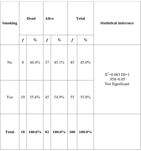
TABLE – 4 SMOKING DISTRIBUTION
Particulars Frequency Percentage
No 45 45.0
Yes 55 55.0
Total 100 100.0
Smoking frequency among the study population was 55 %. 45% of cases were
non smokers. Hence this study included predominantly smokers.
0 10 20 30 40 50 60
No Yes
Smoking Frequency 45 55
A
xi
s Ti
tle
39
TABLE – 5 HYPERTENSION FREQUENCY
Particulars Frequency Percentage
No 41 41.0
Yes 59 59.0
Total 100 100.0
In this study group, 59 % were hypertensives, 41 % were nonhypertensives.
This shows predominance of hypertension among the affected people.
41%
59%
HT Frequency
No
40
TABLE – 6 DIABETES MELLITUS FREQUENCY
Particulars Frequency Percentage
No 33 33.0
Yes 67 67.0
Total 100 100.0
In this study 67 % of cases included were diabetic. 33 % were non diabetic.
Predominantly diabetics were the affected population
33%
67%
DM Frequency
No
41
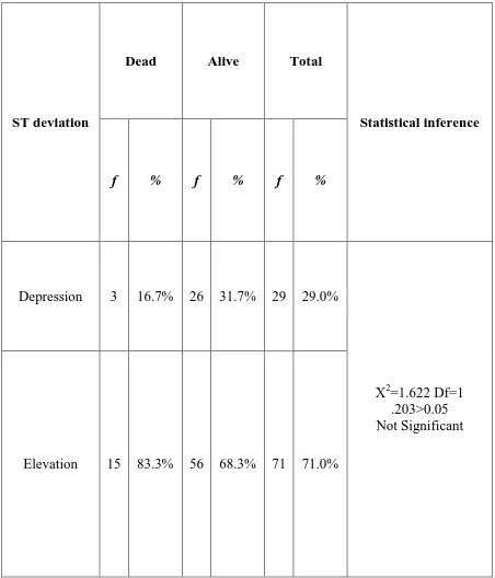
TABLE – 7 ST DEVIATION DISTRIBUTION
Particulars Frequency Percentage
Depression 29 29.0
Elevation 71 71.0
Total 100 100.0
Out of the 100 caes of acute MI studied, 71 % presented with ST elevation. 29 %
presented with ST depression. ST elevation MI was the most common presentation
Depression Elevation
ST deviation Frequency 29 71
0 10 20 30 40 50 60 70 80
A
xi
s Ti
tle
42
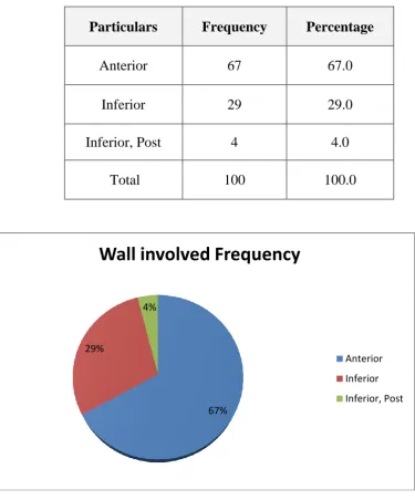
TABLE – 8 DISTRIBUTION OF WALL OF THE HEART INVOLVED IN ACUTE MI
Particulars Frequency Percentage
Anterior 67 67.0
Inferior 29 29.0
Inferior, Post 4 4.0
Total 100 100.0
Out of the 100 cases of acute MI studied, 67% had Anterior wall MI, 29 % had
inferior wall MI. 4 % had combined involvement of both inferior and posterior wall.
Hence anterior wall MI was the commonest, followed by inferior wall MI, and the
least was combination of inferior and posterior wall MI.
67% 29%
4%
Wall involved Frequency
Anterior
Inferior
43
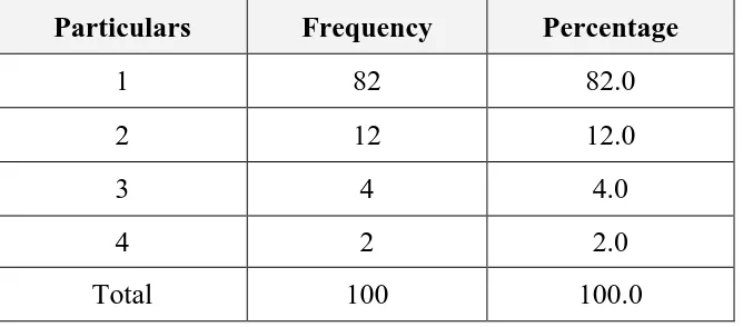
TABLE 9 TIME DURATION BETWEEN PRESENTATION AND ONSET OF ARRHYTHMIAS IN DAYS
Particulars Frequency Percentage
1 82 82.0
2 12 12.0
3 4 4.0
4 2 2.0
Total 100 100.0
Time interval between presentation and onset arrhythmias was studied in the
study population. It was found that majority of arrhythmias occurred in the first day
post MI which was 82%. 12 % cases developed arrhythmia in the second day. 4 %
developed in the third day. Only 2% of cases developed arrhythmia on day 4.
Timing frequency
1
2
3
4
82 % 12%
44
TABLE – 10 DISTRIBUTION OF THE TYPE OF ARRHYTHMIAS
Particulars Frequency Percentage
I DEGREE AVB 12 12.0
II DEGREE AVB 9 9.0
AF 4 4.0
AIVR 13 13.0
APC 5 5.0
CHB 6 6.0
LBBB 7 7.0
RBBB 5 5.0
SVT 1 1.0
VF 3 3.0
VPC 27 27.0
VT 8 8.0
45
12 different types of arrhythmias were observed in the study population. Each
occurred at a different frequency. VPC (27%)was clearly the most common
arrhythmia observed. Followed by AIVR-13%, I degree 12%, II degree
AVB-9%,VT – 8%, LBBB- 7%, CHB – 6%, RBBB, APC each 5%,AF – 4%,VF – 3%,
SVT- 1% respectively.
0 5 10 15 20 25 30
Arrhythmia Frequency
46
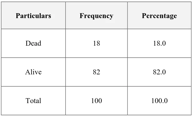
TABLE – 11 DISTRIBUTION OF THE OUTCOME
Particulars Frequency Percentage
Dead 18 18.0
Alive 82 82.0
Total 100 100.0
Out of the 100 cases of acute MI studied , 82 % of cases were alive, 18 % of
cases died.
0 10 20 30 40 50 60 70 80 90
Dead Alive
Outcome Frequency
47
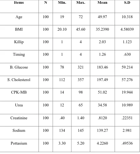
TABLE – 12 DESCRIPTIVE STATISTICS OF AGE, BMI, KILLIP CLASS, TIME INTERVAL , BLOOD GLUCOSE, SERUM CHOLESTEROL, CPK – MB, UREA, CREATININE, SODIUM, POTASSIUM.
Items N Min. Max. Mean S.D
Age 100 19 72 49.97 10.318
BMI 100 20.10 45.60 35.2390 4.58039
Killip 100 1 4 2.03 1.123
Timing 100 1 4 1.26 .630
B. Glucose 100 78 321 183.46 59.214
S. Cholesterol 100 112 357 197.49 57.276
CPK-MB 100 14 98 51.02 19.944
Urea 100 12 65 34.58 10.989
Creatinine 100 .40 1.40 .8120 .22351
Sodium 100 134 145 139.27 2.981
48
0 50 100 150 200 250 300 350 400
N
Min.
Max.
Mean
49
Various parameters like age, BMI, Killip class, time interval behetween the
presentation and the onset of arrhythmia, blood glucose, serum cholesterol, CPK –
MB, urea, creatinine, sodium, potassium were analysed.
Mean age was 49.97, standard deviation was 10.318. Mean BMI was 35.2390,
SD was 4.58039. Mean Killip class was II, SD was 10123. Mean time interval
between presentation and onset of arrhythmia was 1.26 days, with a SD of 0.630.
Mean B.glucose was 183.46 with a SD of 59.214.
Mean S.cholesterol was 197.49 with a SD of 57.276. Mean CPK-MB was
51.02 with a SD of 19.944. Mean urea was 34.58 with a SD of 10.989. Mean
creatinine was 0.8120 with a SD of 0.22351. Mean sodium was 139.27 with a SD of
50
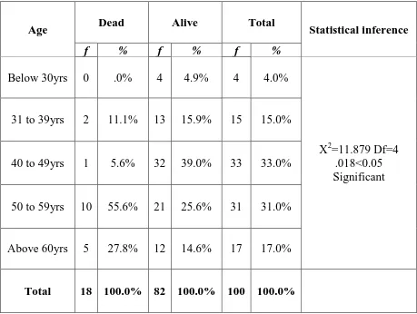
TABLE – 13 CHI-SQUARE TEST TO COMPARE THE OUTCOME IN DIFFERENT AGE GROUPS
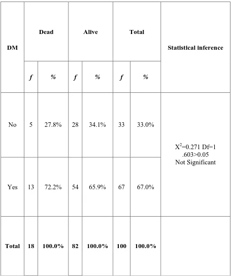
Age Dead Alive Total Statistical inference
f % f % f %
Below 30yrs 0 .0% 4 4.9% 4 4.0%
X2=11.879 Df=4 .018<0.05 Significant 31 to 39yrs 2 11.1% 13 15.9% 15 15.0%
40 to 49yrs 1 5.6% 32 39.0% 33 33.0%
50 to 59yrs 10 55.6% 21 25.6% 31 31.0%
Above 60yrs 5 27.8% 12 14.6% 17 17.0%
51
Using chi-square test overall outcome is compared with the different age groups
of the cases. It showed significant relationship between the age group and the
outcome. Death was highest in 50- 59 years age group, and least in below 30 years age
group.
0 5 10 15 20 25 30 35
1 2 3 4 5
Age f
Age %
Alive f
52
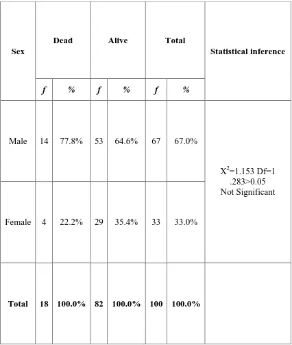
TABLE – 14 CHI-SQUARE TEST TO COMPARE OUTCOME IN DIFFERENT SEX GROUPS
Sex
Dead Alive Total
Statistical inference
f % f % f %
Male 14 77.8% 53 64.6% 67 67.0%
X2=1.153 Df=1 .283>0.05 Not Significant
Female 4 22.2% 29 35.4% 33 33.0%
53
Chi – square test was used to compare overall outcome in different sex groups.
It was found that sex had no significant effect on mortality in this study.
0 10 20 30 40 50 60
Male Female
Dead f
Dead %
Alive f
54
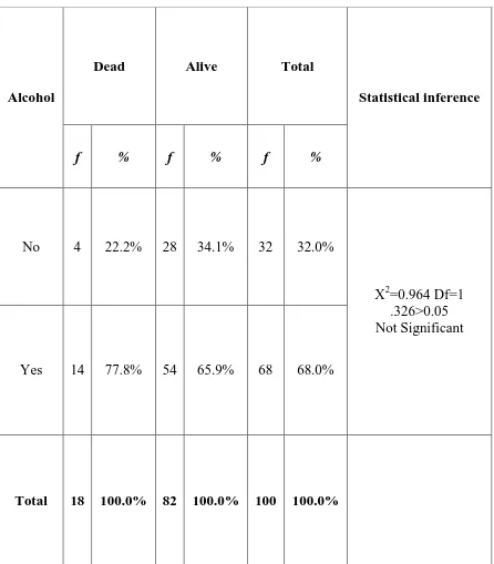
TABLE – 15 CHI-SQUARE TEST TO COMPARE OUTCOME WITH ALCOHOL INTAKE
Alcohol
Dead Alive Total
Statistical inference
f % f % f %
No 4 22.2% 28 34.1% 32 32.0%
X2=0.964 Df=1 .326>0.05 Not Significant
Yes 14 77.8% 54 65.9% 68 68.0%
55
By using chi-square test overall outcome was compared with alcohol intake. In
this study alcohol intake did not had significant effect on mortality.
0 10 20 30 40 50 60
No Yes
Dead f
Dead %
Alive f