DIGITALLY PLANNED 3D PRINTED
SURGICAL STENT- A CASE SERIES
Dissertation submitted to
THE TAMILNADU Dr. MGR MEDICAL UNIVERSITY
In partial fulfillment for the Degree of MASTER OF DENTAL SURGERY
BRANCH III
DECLARATION BY THE CANDIDATE
I hereb y decl are that t his di ssert ation titl ed
“EVALUATION OF ACCURACY OF DENTAL IMPLANT POSITIO NI NG PL ACE D USI NG DI GI TALLY PL ANNE D 3D PRINTED SURGI CAL STE NT - A CASE SE RIES ” i s a bonafi de record and genuine research work done b y me under the
guidance of Dr.M.VEERABAHU, M.D.S. ,FIBO MS P rofessor and Head, Departm ent of Oral & M axillofacial Surger y, R agas
Dental Col le ge and Hospital, C hennai.
Date:
Place: Chennai
Dr. KISHOK.R,
Post Graduate Student,
Department of Oral & Maxillofacial Surgery,
Ragas Dental College and Hospital,
“EVALUATION OF ACCURACY OF DENTAL IMPLANT POSITIO NI NG PL ACE D USI NG DIGI TALLY PL ANNE D 3D PRINTED SURGI CAL STE NT - A CASE SERI ES ” is a bonafi de record of work done b y Dr.KISHO K.R, under our guidance and to our sati sfaction duri ng his post graduat e st ud y
period 2016 -2019.
This Diss ertation i s submitt ed t o THE T AMILNADU Dr. MGR MEDICAL UNIVE RSIT Y, in partial fulfillm ent for the award of the Degree of MAST ER O F DENT AL SURGERY – ORAL AND MAXI LLO FACI AL SURGE RY, BRANCH I II. It has not been submi tted (part ial or full ) for the a ward of an y
other degree or diploma .
Gui de: H ead of the Departm ent:
Dr. M. Veerabahu, M.D.S.,FIBOMS., Dr. M. Veerabahu, M.D.S.,FIBOMS.,
Professor and Head Professor and Head
Department of Oral & Maxillofacial Surgery Department of Oral &maxillofacial Surgery Ragas Dental College & Hospital Ragas Dental College & Hospital
Chennai. Chennai.
Dr. N.S.Azhagarasan, M.D.S.,
Principal,
PLAGIARISM CERTIFICATE
This is t o certi f y t hat t he dissert ation titled “” done b y the candi dat e Dr.KISHO K.R , for the award of MASTER OF DENT AL S URGE RY in B RANCH I II – Oral and Maxill ofacial Surgery.
On veri fi cat ion wi t h the websi te unkund.com for t he
purpos e of pl agiar i sm check, the upl oaded thes is fil e from
introducti on to concl usion contains 8% of plagiaris m, as per the report generated and the same is encl osed in Annexure – II.
Date:
Place: Chennai Guide sign with seal:
Dr. KISHOK.R, Post Graduate Student,
Department of Oral & Maxillofacial Surgery Ragas Dental College and Hospital,
Chennai.
Dr.M.VEERABAHU, M.D.S., FIBOMS.,
Professor and head
wise and confident acknowledge this help with gratitude”
Keeping the above lines in mind the very least i could do is pen my gratitude to those who have made my post-graduation period and dissertation a success.
First and foremost, i thank God Almighty for giving me strength, skill, knowledge and a comfortable environment to complete this project satisfactorily, without whose blessings this achievement would not have been possible.
With immense satisfaction and lovable pleasure, i am presenting this work undertaken as a Post Graduate student specializing in Oral and Maxillofacial Surgery at Ragas Dental college and Hospital. I am acknowledging my work on this dissertation which is making me wonderful and enriching learning experiences.
A good teacher is everything a parent can ever be. I would take this as an opportunity to thank my mentor, my guide, my role model and Head of the department Prof. Dr. M. Veerabahu for enlightening me with
skills in execution and prioritizing perfection both in planning and action. These three years of my post graduation period under his mentorship and guidance, will be the most enchanting, enriching and enlightening period of my life. This made me to develop an attitude of constantly learning each day in this field of developing maxillofacial surgery. I thank you sir from the bottom of my heart for introducing me in to the world of virtually planned 3D surgery and assure you that i will do my best to meet your expectations. I thank him for guiding me in my thesis work and i am indebting him always forever for all considerations to me. These memories definitely cherish me throughout my life .
I sincerely thank my and Prof. Dr. Nathan for his discipline and sincerity in the field of maxillofacial surgery. His extensive knowledge, enthusiasm and numerous stories from experiences in the field has kindled my passion towards surgery even more and has always been a source of encouragement. I am extremely lucky to have studied under you sir and thank you for the fulfilling experience you have given me in this journey. I also take this chance to thank my professor Dr. Malini Jayaraman for providing support and encouraging thoughts during my period of post-graduation. Her care and compassion towards every student will always be remembered.
I am extremely grateful to our beloved principal
Prof. Dr. Azhagarasan for his support and for allowing me to access the research facilities of the college.
me a more disciplined and responsible student over the last 3 years.
I sincerely thank Prof. Dr. Radhika Krishnan, her support and work as the Anesthetist is the main reason we post graduates were lucky to observe, assist and perform numerous surgical procedures without fear. I am grateful to her for sharing her knowledge in medicine with us.
I honestly thank Dr. Satheesh who has been extremely generous and supportive throughout my studies. I am extremely indebted to him for always being there for me and for being extremely encouraging. His down to earth nature and surgical skill has been a source of inspiration. I thank you very much sir for the last 3 years.
am so grateful to Dr. Harish, for taking me under his wing when I first started my thesis. You have been an exemplary and visionary mentor. You have been such an integral part of my career. Thank you sir for being there for me and teaching me so much.
I am extremely fortunate to have Dr. Stephen, Dr. Manoj, Dr. Arun Vignesh, Dr. Ajit and Dr. Deepan as my batch mates. Thanks to their constant support, help and reciprocity this journey has been a breeze. I wish them a bright future ahead.
I thank my juniors Dr. Veera, Dr. Alka, Dr. Diana, for all their constant support. I thank my sub-juniors, Dr. Badrudeen, Dr. Abinaya, Dr. Priyanka, Dr. Hema, Dr. Priyadardhini, Dr. Moni Vikasini for their help.
I thank Dr. Arjun, department of prosthodontics for helping me in prosthetic rehabilitation of my thesis patient.
I take this opportunity to thank Dr. Deiva Nayagi from the department of oral medicine and radiology who guided me to analyze cone beam computed tomography and 3D implant planning.
I sincerely thank Proto 3D solutions and Graft 3D for providing me accurate digitally planned surgical guides.
I am grateful to the sisters of the department Mrs. Deepa, Mrs. Leema, Mrs. Laila and Mrs. Mala and our OT assistants Mr. Venugopal
and Mrs. Malathi for their invaluable help during my post-graduation period.
I thank Mr. S. Kothandapani for giving me his precious time for the documentation of my thesis and Mr. Thavamani for editing and printing my thesis.
mother Mrs.Suganya Rajkumar for the sacrifices which made my foundation of my life and for being the most wonderful parents to me. I thank my wife Tmt. Ponmani kishok for being the pillars of my life and showering me her love, encouragement and for making my ordinary moments extroordinary. I thank my sister Dr. R. Deepika Rajkumar who is studying MS,(OG) in Chidambaram always giving me the extra push I needed and for sharing my crazy thoughts. I sincerely thank my grandmother Mrs.Arpudhavalli and My grant father Mr. M..Pichappa
for believing me & supporting me unconditionally through their blessings . Also special welcome to my lovable Son K. Deivik Meerath and mySweet S. Amirtha Shanaya Sai..
I would like to dedicate this dissertation to my family who always wish for me to reach great heights and achieve greater goals in my life. I am always surrendering and submitting my life to God.
positioning placed using digitally planned 3D printed surgical guide.
MATERIALS AND METHOD: The study was conducted in the department of Oral and Maxillofacial Surgery, Ragas Dental college, Chennai, Tamilnadu.
15 implant sites in 4 patients irrespective of gender (two males and two
females) were selected based on the inclusion criteria. These patients, who
presented to the department of Oral and maxillofacial surgery Ragas Dental
College and hospital with a complaint of partial or complete edentulism with a
desire of fixed prosthesis, were included in the study. Pre-operative CBCTs
were taken. Virtual planning was performed through blue sky bio software and
surgical guide was prepared. Guides were fabricated through Stratasys Eden
260vs computer 3D printer by polyjet printing technology. SS316 cylindrical
drill sleeves were prepared. Drilling was done by placing the surgical guides
through the information gained from virtual planning. Depth control system
was used. A drill was inserted in the drill sleeve of master sleeve upto the
physical stop of drill shaft and the next size sleeve was placed where the drill
was completely inserted in to the master sleeve for achieving the implant
depth. This was done in a sequential order and the final drill was placed
directly in to the master sleeve. After six months, postoperative CBCT were
taken. A Virtual planning was performed in preoperative CBCT and post
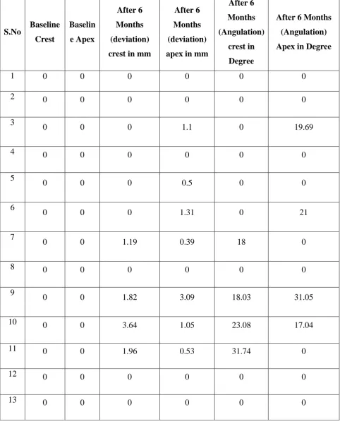
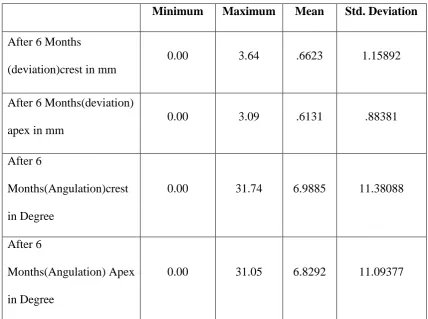
RESULTS: The results from the study revealed that the mean value for deviation of the angulation at crest was 6.9885 degrees and the standard
deviation was 11.38088 degrees. The mean value for deviation of the
angulation at apex was 6.8292 and the standard deviation was 11.09377. The
mean value for the deviation at crest was 0.6623mm and the standard
deviation was 1.15892mm. The mean value for the deviation at apex was
0.6131mm and the standard deviation was 0.8831mm.
CONCLUSION: From the results of the study we would like to conclude that the placement of dental implants using digitally planned surgical guides
is within clinically acceptable limits. It further ensures minimal or virtually
nil damage to the relevant anatomical structures and makes implant
placement less difficult for beginners.
1. INTRODUCTION 1
2. AIMS AND OBJECTIVES 10
3. REVIEW OF LITERATURE 11
4. MATERIALS AND METHODS 33
5. RESULTS 42
6. DISCUSSION 45
7. SUMMARY AND CONCLUSION 57
8. BIBILOGRAPHY 58
TABLE. No.
TITLE
1.
DEVIATION OF IMPLANTS AT CREST AND APEX IN
ANGULATION (°) AND DISTANCE (MM)
2.
CUMULATIVE ASSESSMENT OF DEVIATION OF IMPLANT IN
GRAPH No.
TITLE
1
DEVIATION (IN MM) OF IMPLANTS AT VARIOUS
SITES
2
ANGUALTION (IN DEGREE) OF IMPLANTS AT
1
INTRODUCTION
The term Edentulism as per glossary of prosthodontics terms (GPT) is
defined as the state of being edentulous without natural tooth.
Tooth loss can lead to the following consequences, which may include
the following:
Dissatisfaction with appearance and loss of confidence.
Mental anxiety.
Changes in facial features, loss of bone and gingival tissue, difficulty
eating and speaking, poor nutrition.
Pain, distress and relaxation problems.
Embarrassed and/or ashamed to seek dental treatment.
Financial burden for costs of possible treatment options.
There are various methods to replace the tooth loss or to treat edentulism
namely
COMPLETE DENTURE: It is a removable dental prosthesis that replaces the single or multiple missing teeth; the removable denture can be readily
2
REMOVABLE PARTIAL DENTURE: It is a removable dental prosthesis that replaces the single or multiple missing teeth; the removable partial
denture can be readily inserted and removed from the mouth by the patient.
FIXED PARTIAL DENTURE is referred to any dental prosthesis that is luted, screwed, or mechanically attached or otherwise securely retained to
natural teeth, tooth roots, and/or dental implants/abutments that furnish the
primary support for the dental prosthesis and restoring teeth in a partially
edentulous arch; it cannot be removed by the patient.
IMPLANT is referred to any object or material, such as an alloplastic substance or other tissue, which is partially or completely inserted or grafted
into the body for therapeutic, diagnostic, prosthetic, or experimental purposes.
DEFINITION
Dental Implant is defined as a prosthetic device made of alloplastic
material implanted into the oral tissues beneath the mucosal or/ & periosteal
layer &/or within the bone to provide retention & support for a fixed or
removable dental prosthesis; a substance that is placed into or / & upon the
jaw bone to support a fixed or removable dental prosthesis.
Advantages of dental implants
Lack of involving adjacent teeth in prosthetic management.
Preventing root canal treatment and tooth preparation in adjacent
3
It has better longevity when compared with other restorative
procedures if properly placed and maintained.
It restores bite force.
It is biocompatible.
It prevents bone loss.
It enables natural speech.
It provides better esthetics.
Disadvantages of dental implants
Require a higher up-front cost than a bridge or dentures. However, the
longevity of results makes this approach more cost-effective.
In order to replace missing teeth with implants and provide implant
supported restorations such as or dentures, it is necessary to undergo a
minor oral surgery.
Time period for delivering the final prosthesis is conventionally longer
than FPD/RPD; although immediate loading is possible with dental
implants, the placed implants fulfills certain stringent clinical
parameters.
Lack of bone at implant site will require additional bone augmentation
procedure.
4
1. I.V bisphosphonate,
2. Uncontrolled diabetes,
3. Smoking,
4. Children whose growth is yet to increase.
IMPLANT PLANNING CONSIDERATIONS
Bone quality and quantity plays the major role in implant planning and
placement. Implants would fail if a patient has;
Inadequate labiolingual width.
Inadequate vertical maxillary alveolar bone.
Inadequate vertical mandibular alveolar bone.
Angulation of the implant is another crucial factor that has to be
considered while placing an implant. If the angulation is inappropriate the
implant may damage the neighbouring vital anatomic structures and the root
of the adjacent teeth. Improper angulation leads to lack of esthetic replacement
of prosthetic crown or bridge in addition it may contribute to gingival
recession as well.
The minimum required distance between the implant and the buccal
plate is 1mm, lingual plate is 1 mm, maxillary sinus is 1 mm, nasal cavity is
1mm and in incisive canal midline of the maxilla is to be avoided.
5
In relation to the inferior alveolar canal, the implant has to be placed 2mm
from superior aspect of bony canal.
In relation to mental nerve, the implant has to be placed 5mm anteriorly to
mental foramen.
Orofacial dimension:
Anterior region: The implant shoulder is positioned 1 mm palatal to the point of emergence of adjacent teeth, aiming for at least 1.5 mm bone thickness at
the labial side.
Posterior region: The implant is positioned in the centre of the future restoration, aiming for at least 1.5 mm of bone thickness at the buccal and
palatal/lingual side.
Corono-apical dimension: The implant shoulder is positioned 3 mm apical to the emergence of the future restoration with an expected biologic width of also
3 mm, being the equivalent of 1 mm apical of the cementoenamel junction of
the contralateral tooth in patients without gingival recession.
The minimum integration time in the region of anterior mandible is 3 months,
posterior mandible is 4 months, anterior and posterior maxilla is 6 months &
6
CHALLENGES IN IMPLANT PLANNING
Imaging of the implant site will have to be done to determine bone
density and bone quantity, to evaluate the approximation of vital anatomical
structures and to estimate the dimensions, number, location, orientation,
prognosis of the implant to be inserted, identify underlying bony pathologies,
undercuts and concavities. The requirement for additional bone augmentation
should be evaluated.
In 2D imaging, certain parameters cannot be assessed such as:
a) Labiolingual width,
b) Exact position of vital anatomical structures,
c) Quality and quantity of available bone.
The objectives of diagnostic imaging depend on the amount and type
of information required and the treatment period rendered. The timing and
type of imaging modality to be used depends on the integration of the phases
mentioned below:
PHASE I (Pre-surgical implant imaging)
To determine the bone quantity and quality and an approximation of
the implant site with the critical structures, as well as to plan implant
7
PHASE II (Surgical and intraoperative implant imaging)
Along with analysis the optimal position and orientation of the
implant, intraoperative implant imaging helps evaluate the healing and
integration of surgery sites. The correct position of the abutment and
prosthesis fabrication will have to be ensured in this phase.
PHASE III (Post-prosthetic implant imaging)
This phase starts from the placement of the implant and lasts as long as
the implant remains in the jaw. The radiographic sequence for phase III
imaging is post-prosthetic imaging, followed by recall and maintenance
imaging and an evaluation of alveolar bone change.
SURGICAL GUIDES:
Cast-based Guided Surgical Guide
The surgical guide is a combination of an analog technique done along
with bone sounding and the use of periapical radiographs in a conventional
flapless guided implant surgery. The periapical radiograph is modified using
digital software to help in transposition of root structure onto the cast. The cast
is then sectioned at the proposed implant site, and bone-sounding
measurements are transferred to help in orientation of the drill bit to perform a
cast osteotomy. A laboratory analogue is placed in the site, and a guide sleeve
8
create a framework around the teeth. Vinyl polysiloxane occlusal registration
material is used to form the super structure.
CAD/CAM-based surgical guide
They are appliances that are computer designed and are fabricated
from acrylic resin by a process called Stereolithography. The surgical guides
contain steel sleeves with a predefined diameter to guide the drills during the
osteotomy process. There are different types of surgical guides such as a pilot
guide, which allows the clinician to create the initial osteotomy. After the pilot
osteotomy is created, the guide is removed and the rest of the process is done
free handed. Other guides allow completing all the osteotomy processes by
using the entire drill sequence; however, the implant is placed free handed,
whereas there are others that require a guide that allows going through all the
drilling sequence and placement of the implant. Some protocols include depth
control systems that allow the surgeon to control vertical preparation by using
9
be categorized according to the type of stabilization they have such as teeth,
bone, or soft tissue.
The advantages of CAD-CAM based surgical guide are as follows,
With good technique, guided surgery using a tooth, mucosal or bone
borne guide or a combination of these approaches can be a predictable
method for dental implant placement.
Although it increases the number of steps compared to freehand
placement, guided implant surgery allows clinicians to develop a
restoratively driven surgical plan, with the ultimate goal of
patient-centered, positive outcomes.
Surgical guides can assist the practitioner in avoiding damage to
anatomic structures, as well as limiting fenestration and dehiscence of
the alveolar ridge at potential implant sites.
The purpose of this study is to evaluate the accuracy of angulation of
the implants placed using surgical guides by superimposing the cone beam
computed tomography scan of actual implant that was placed on the patient
10
AIMS AND OBJECTIVES
The aim of study is to evaluate the accurac y of dent al im pl ant
positi oning placed using di gi tall y pl anned 3D pri nted surgical
11
REVIEW OF LITERATURE
David R. Burns, D.M.D., Donald G. Crabtree, D.D.S., and Dewey H. Bell, D.D.S. (1988) described a technique for fabrication of a surgical guide that gives the surgeon the appropriate location and angulation of the
submerged implant relative to the opposing dentition. Presurgical planning for
submerged implant location and angulation within bone relative tothe
opposing occlusion was important for the restorative dentist. This information
would be accurately communicated to the surgeon by using a surgical
template.[1]
Igor J. Pesun, DMD and F. Michael Gardner, DDS, MA (1995)
described a technique in which a guide was fabricated for radiographic
evaluation of implant placement and also serves as a surgical guide for
placement of the implants for a patient with severely worn dentition. They also
added that it provides for visualization of the final restoration and can be
modified to be used for implant placement. Chair and laboratory time were
reduced because one guide could be used for both radiographic evaluation and
surgery. The guide was easily fitted to the existing dentition and allows
evaluation of the contours of the final restoration in the patient's mouth by the
patient, the restoring dentist, and the surgeon. The use of a known diameter
gutta-percha plug could determine the distortion factor of the radiographic
12
Naitoh M, Ariji E, Okumura S, Ohsaki C, Kurita K, Ishigami T (1999) conducted a study to propose a method for evaluating the placement accuracy using a specific surgical template. Their results showed that the
template used in our procedure had a sufficient quality in identifying the
proposed direction, because the difference could be recovered by the greater
access hole of the set-screw (Parel & Sullivan 1989) and the use of the
angulated abutment. Although this method was difficult to apply to individual
cases, it may be useful to evaluate a procedure for implantation.[3]
Roger A. Sollow(2001) stated that, Radiographic surgical template related the position of the planned implant site to the remaining bone.. A
surveyor - mounted handpiece was not required. Clean enlargement of the
access opening was possible without shredding plastic or metal tubing. The
pilot drill aligned was controlled. A surveyor was used to orient the composite
channel to the ideal position or to match the emergence profile of adjacent
teeth. Correction of pilot drill channel was possible after radiographic
evaluation. The desired correction in angulation was measured with a
protractor on the tomogram. The composite channel was enlarged, and the bur
shank was rebonded with composite to this new angle by placing the
protractor on the surveyor rod. Any desired horizontal change was measured
with Calipers from the tomogram. This dimension was transferred to the new
bur shank position in the enlarged channel, and the composite is rebonded.
13
represents the central axis of the prosthetic tooth and is a better indicator of the
tooth to-bone relationship than a radiopaque outline of the tooth or denture
base. The access opening in the radiographic surgical template that confines
the composite should be perpendicular to the occlusal plane or parallel to the
adjacent teeth. The marker angulation was apparent in the vertical slices of the
tomogram, could be used in fully edentulous patients by extending the acrylic
resin to cover the palate, tuberosity, or retromolar pads on the duplicate
diagnostic cast and retained as a guide to locate healing screws at the second
stage surgery. The author summarized that versatile radiographic-surgical
template for multiple parallel implant placement has been described. Simple
materials and methods allow the restorative dentist to evaluate the final
prosthetic contours, implant locations, and treatment plan before surgical or
patient consult. The precise guide for implant pilot drills could be modified
easily after radiographic evaluation or during surgery.[4]
Kıvan Aka, DDS, PhD, Haldun I plikioglu, DDS, PhD, and Murat C. ehreli, DDS, PhDc (2002) conducted a study which describes a modified surgical stent that serves as a guide to proper mesiodistal paralleling of dental
implants. They concluded that when computerized tomography was not
required to evaluate the buccolingual angulation of available bone, the simple
design and function of the modified stent make it advantageous for use in the
14
Kevin C. Kopp, BS, DDS, Alyson H. Koslow, BS, DDS, and Omar S. Abdo, BDS, MSc (2003) presented an article about precise dental implant placement. A barium-coated template with external guide wires used in
conjunction with a computed tomography scan and interactive software may
provide superior presurgical diagnostics, treatment planning, and
prosthetically directed implant placement. Measurements predetermined on
the computed tomography scan could be transferred accurately to the
diagnostic/surgical template by use of a precision milled cylinder placed into
the template at the proper angulation and linear dimensions. The
diagnostic/surgical template demonstrates the surgeon the optimal position for
implant placement, thus establishing greater clinical confidence when placing
implants. They concluded that the location of implant placement was often
critical to the success or failure of a particular restoration. .Placing a wire
buccally allows for the marker to remain intact and not be obliterated when
preparing the template for the surgical phase. With this method, the
buccolingual and mesiodistal positions could be maintained throughout the
surgery. This was accomplished by affixing an external guide wire that
remains with the template from diagnostic CT imaging to surgical placement
of the implant.
The use of a diagnostic/surgical template may allow predictable
15
Antonio Rocci, DDS, Massimiliano Martignoni, DDS, Jan Gottlow, DDS, PhD (2003), conducted a study to evaluate an immediate-loading treatment protocol, which included flapless surgery, implants placed in
predetermined positions and connected to prefabricated provisional
restorations, and the 3 year clinical results. They concluded that the feasibility
of an immediate-loading treatment protocol in the maxilla, which included
flapless surgery, implants and abutments placed in predetermined positions,
and prefabricated provisional restorations. All failures occurred within the first
2 months of loading. The unchanged survival rate and the low average bone
loss found during the following 3-4month study period indicate a good
long-term prognosis for the performed immediate-loading treatment.[7]
Mohammed Zaheer Kola, Altaf H Shah, Hesham S Khalil, Ahmed Mahmoud Rabah, Nehad Mohammed H Harby, Seham Ali Sabra, Deepti Raghav (2004) mentioned that Improvements in surgical reconstructive methods, as well as increased prosthetic demands, require a highly accurate
diagnosis, planning, and placement. Identification of the bony anatomy with
respect to the teeth, prior to surgery, allows the clinician to place implants in
areas where the implant bone interface could be maximized, and the
prosthetic result was optimized. As discussed earlier, the completely limiting
design was considered far superior design concept, most clinicians still adopt
the partially limiting design due to its cost effectiveness and credibility in the
16
templates that are based on cross sectional imaging to facilitate accurate
planning and guidance during the surgical phase. Computer aided planning
and image guided surgery can be carried out, when implant positioning is to
be precisely executed, and when safe positioning of implants with optimal use
of available bone, and whenever a CT scan was recommended as a diagnostic
means evidence based research still needs to be conducted to evaluate the
applications of the completely limiting design and its effect on the treatment
outcome in oral implantology.[8]
Jakob Brief, Dieter Edinger ,Stefan Hassfeld Georg Eggers (2005)
conducted a study to assess accuracy of two commercially available systems
for image-guided dental implant insertion based on infrared tracking cameras
was compared with manual implantation. Phantoms of partially edentulous
mandibles were used. In a master phantom, pilot boreholes for dental implants
were placed. These boreholes were reproduced in slave phantoms using either
of the two image-guided systems and manual implantation. The resulting
positions were determined using a coordinate measurement machine and
compared with the master model. In comparison with manual implantation, the
difference of borehole positions to the master phantom was significantly lower
using either of the systems for image-guided implant insertion. They
concluded that the Image-guided insertion of dental implants is significantly
more accurate than manual insertion. However, the accuracy that can be
17
Giovanni A.P. Di Giacomo,Patricia R. Cury, Ney Soares de Araujo, Wilson R. Sendyk,and Claudio L. Sendyk (2005) concluded that computer-aided rapid prototyping of surgical guides may be useful in implant
placement. However, the technique requires improvement to provide better
stability of the guide during the surgery, in cases of unilateral bone-supported
and non-tooth-supported guides. Further clinical studies, using greater number
of patients, were necessary to evaluate the real impact of the stereolithographic
surgical guide on implant therapy.[10]
Alan L rosenfeld,George A.mandelaris,philippe B.Tardieu (2006)
conducted a study to discuss the use of scanning appliances to transfer
clinically relevant prosthetic outcome information to a ct data set. This
information could be used to provide a pretreatment outcome analysis using
implant software which could be used for fabrication of models and surgical
drilling guides used during osteotomy preparation. They also stated that
interactive CT technology allows the incorporation of prosthetic information
into a CT study which could be analysed against the patients regional anatomy
before surgical intervention. Surgical requirements could be established prior
to treatment. This technology allows for the process of informed consent to be
18
Gerlig Widmann, MD/Reto Josef Bale, MD (2006) presented an article to review the different factors and limitations influencing the accuracy
of computer-aided implant surgery. They mentioned that the accuracy of
image-guided systems for oral implant surgery depends on all cumulative and
interactive errors involved, from the data-set acquisition to the surgical
procedure. A safety distance at least equivalent to the maximum deviation of
the individual system was necessary. Similar accuracy data had been reported
for bur tracking and image-guided template production, and both methods
allow precise positioning of oral implants. Compared to the conventional
technique, computer-aided implant surgery was superior on account of its
potential to eliminate error and systematize reproducible treatment success. It
also enables the protection of critical anatomic structures and the esthetic and
functional advantages of prosthodontics - driven implant positioning.
Based on clinical data, image guidance was not required for easy cases
of sufficient anatomic orientation and bone height, but whenever a CT scan is
recommended as a diagnostic means, when prosthodontic-driven implant
positioning was to be precisely executed, and when safe positioning of
implants with maximum length was desired for optimal use of the available
bone, the patient could fully benefit from the advantages of complete
3-dimensional imaging, computer aided planning, and image-guided surgery.
19
success, to confirm the value of this strategy, and to justify the additional
radiation dose, effort and costs.[12]
Dov M. Almog, DMD B. W. Benson, DDS, MS L. Wolfgang, DDS N. L. Frederiksen, DDS, PhD S. L. Brooks, DDS, MS (2006) stated that report reviews recent developments in CT-image–based information and
surgical guidance systems and attempts to provide an argument for the
development to evidence-based research on the utility of such systems and
their effect on the outcome in oral implantology. Outcomes assessment
research, including cost-to-benefit analysis, difficult but was critical in
answering these and other questions about implant site assessment and
surgical guidance. Furthermore, the recent introduction of numerous
associated commercial CT-based imaging and surgical guidance platforms and
its expected effects on the way the profession views and practices oral
implantology provides sufficient argument for the necessity of large
prospective clinical trials on the quantitative relationship between successful
dental implant treatment outcomes and CT-based dental imaging coupled with
surgical guidance systems.[13]
Kunal Lal, DDS, MS, George S. White, DDS Dennis N. Morea, DDS and Robert F.Wright, DDS (2006) presented an article about the use of Stereolithographic templates for Surgical and Prosthodontic Implant Planning
and Placement. They concluded that the stereolithographic templates could be
20
could be entirely supported either by soft tissue,bone,or remaining teeth. The
cost associated was higher than with conventional templates, but in more
complexes, fully edentulous cases, the benefit could justify the additional
expense. The use of this approach could make the goal of ideal surgical and
prosthodontic implant placement a distinct possibility. Considering the
possible benefits and implications of achieving this goal, it would be prudent
to direct further clinical research endeavors toward validating the accuracy and
effectiveness of the system.[14]
Van Assche N, van Steenberghe D, Guerrero ME, Hirsch E, Schutyser F, Quirynen M, Jacobs R (2007) conducted a study to evaluate the precision of transfer of a Computer - based three-dimensional (3D)
planning, using re-formatted cone-beam images, for oralimplant placement in
partially edentulous jaw. In his study four formalin-fixed cadaver jaws were
imaged in a 3D Accuitomo FPDs cone-beam computed tomography (CT).
Data were used to produce an accurate implant planning with a transfer to
surgery by means of stereolithographic drill guides. Pre-operative cone-beam
CT images were subsequently matched with post-operative ones to calculate
the deviation between planned and installed implants. The surgical procedure
was performed uneventfully. Finally they stated that the ability of cone-beam
CT images to offer a reliability data set for planning and further transfer of
oral implant placement in partial edentulism. The study also indicates that the
21
template fit and template stability and concluded that cone-beam images could
be used for implant planning, taking into account a maximal 4 degree angular
and 2.4mm linear deviation at the apical tip.[15]
Loong Tee Yong, BDS, MD, FRACDS;Peter K. Moy, DMD (2007)
conducted a study to evaluate early clinical results of computer-aided design
(CAD)/computer-aided machining (CAM)-guided surgical implant placement
(NobelGuide™, Nobel Biocare,Yorba Linda, CA, USA) with focus on
surgical and/or prosthetic complications, management, and prevention.
Thirteen patients rehabilitated between March 2003 and October 2006 with
CAD/CAM-guided dental implants and immediate loading were evaluated.
The treatment planning and procedures were carried out in accordance to the
system protocol. The complications encountered in this case series were
classified and assessed according to early (planning and procedural – surgical;
prosthetic) and late complications (surgical; prosthetic). They finally stated
that the prosthetic complications Out numbered surgical complications both in
the early and late treatment phases and the Nobel Guide system is a reliable
treatment modality but not without its complications.[16]
Natalie Y. Wong, DDS, Heather Huffer-Charchut, DMD, and David P. Sarment, DDS, MS (2007) stated that new technological advances in CAD/CAM implant treatment planning and guidance may offer significant
clinical advantages over traditional implant rehabilitation. In this report, the
22
transfer the CT-based implant plan to actual surgery.A detailed,
comprehensive approach to complex implant treatment planning and surgical
implant placement offers advantages to both clinician and patient.[17]
Philippe B. Tardieu, DDS/Luc Vrielinck, DMD/Eric Escolano, DMD(2007) presented a case of immediate loading of mandibular implants using a 5-step procedure. The first step progress in medical imaging and data
processing was creating profound changes in the way that professional
practice was perceived. Treatments are becoming more precise, faster, and
safer for patients. Surgeons were now well equipped in controlling and carry
them out with precision of implant positioning also helps in reducing the cost
of prosthetic restorations, avoiding the use of additional abutments to realign
implants. Implant treatment with immediate loading using a bone-supported
stereolithographic drill guide has been demonstrated in this treatment
situation. Implant treatment with immediate loading using a bone-supported
stereolithographic drill guide has been demonstrated in this treatment
situation. This drill guide may be called the “missing link” in implant
treatment.[18]
O. Ozan, I. Turkyilmaz & B. Yilmaz (2007) presented a clinical study to compare the survival rates of early loaded implants placed using
flapless and flapped surgical techniques and to determine the bone density in
the implant recipient sites using computerized tomography (CT). The study
23
group consisted of five patients referred for the placement of 14 implants and
treated with a flapless procedure. The other group consisted of seven patients
referred for the placement of 45 implants with a conventional flapped
procedure. Patients were selected randomly. CT machine was used for
pre-operative evaluation of the jaw bone and the mean bone density value of each
implant recipient site was recorded in Hounsfield units (HU). All implants
were placed using CT guided surgical stents. The early loading protocols
included 2 months of healing in the mandible and 3 months of healing in the
maxilla. Single-implant crowns, implant-supported fixed partial dentures, and
implant-retained over dentures were delivered to the patients. The results of
this study show that the early loading of implants placed utilizing flapless
surgical technique with CT-guided surgical stents may be possible.[19]
Hans Joachim NICKENIG, Stephen EITNER (2007), presented a study to assess the reliability of planning software system that allows transfer
of virtual planning data to a surgical template that is then used as a drill guide
during surgery. They finally mentioned that implant placement after virtual
planning of implant positions using CBCT data and surgical templates could
be reliable for pre-operative assessment of implant size, position and
anatomical complications. It is also indicative of cases amenable to flapless
surgery.[20]
24
edentulous jaws following computer-assisted virtual treatment planning
combined with flapless surgery.They concluded that the patient’s
post-operative discomfort, such as pain and swelling was almost negligible in
successfully treated cases. However, compared with conventional treatment
protocols and the Branemark Novums, the occurrence of surgical and
technical complications was higher in edentulous mandibles. Thus, the method
of computer-assisted treatment planning and immediate loading must still be
regarded to be in an exploratory phase. Further investigations were needed to
validate the accuracy in the method taking into account each step in the
clinical procedure. The positioning of implants after guided surgery and 3D
virtual planning still had to be verified in both maxilla and mandible.
Continued investigations along these lines were in progress in both our clinics
and laboratories.[21]
Leonard Spector (2008) mentioned that technologic advances in computer based planning of implants as allowed the clinician to plan more
accurately and place dental implants precisely. With the planning software, the
virtual treatment plan was used to create a surgical template that guides the
placement of implants during surgery. The minimally invasive surgery was
performed without raising the flap, thereby minimizing surgery time,
post-operative pain and swelling and recovery time. The guided implant surgery
was very helpful to plan, place and restore implants accurately with the level
25
David Schneider, Ronald E. Jung (2009), conducted a study to analyse the dental literature regarding accuracy and clinical application in
computer-guided template-based implant dentistry. For this, an electronic
literature search complemented by manual searching was performed to gather
data on accuracy and surgical, biological and prosthetic complications in
connection with computer-guided implant treatment. For the assessment of
accuracy meta-regression analysis was performed. Complication rates are
descriptively summarized. They concluded that, Computer-guided
template-based implant placement showed high implant survival rates ranging from
91% to 100%. However, a considerable number of technique related
perioperative complications were observed. Preclinical and clinical studies
indicated a reasonable mean accuracy with relatively high maximum
deviations. Future research should be directed to increase the number of
clinical studies with longer observation periods and to improve the systems in
terms of perioperative handling, accuracy and prosthetic complications.[23]
U. S. Pal, Pooran Chand, Neeraj Kumar Dhiman, R. K. Singh, Vimlesh Kumar (2010) demonstrated the extreme accuracy of the conventional surgical stent. If each step of this protocol was followed
precisely, it was possible to deliver an optimum implant installation in terms
of position and diameter and later on function at a very much reduced rate and
time, as taken in CT and CAD/CAM technology which are the most accepted
26
Christoph Vasak, Georg Watzak, Andre ´ Gahleitner, Georg Strbac, Werner Zechner (2010) conducted a retrospective study to evaluate the overall deviation in a clinical treatment setting to provide for quantification
of the potential impairment of treatment safety and reliability with
computer-assisted, template-guided transgingival implantation. The patient population
enrolled (male/female¼10/8) presented with partially dentate and edentulous
maxillae and mandibles. Overall, 86 implants were placed by two experienced
dental surgeons strictly following the NobelGuide protocol for
template-guided implantation. All patients had a postoperative computed tomography
(CT) with identical settings to the preoperative examination. Using the triple
scan technique, pre- and postoperative CT data were merged in the Procera
planning software, a newly developed procedure – initially presented in 2007
allowing measurement of the deviations at implant shoulder and apex. They
concluded that template-guided implantation will ensure reliable transfer of
preoperative computer assisted planning into surgical practice. With regard to
the required verification of treatment reliability of an implantation system with
flapless access, all maximum deviations measured in this clinical study were
within the safety margins recommended by the planning software.[25]
Curtis M. Becker, DDS, MSD, and David A. Kaiser, DDS, MSD (2010) conducted a study for describing a procedure for constructing a precise surgical guide that ensures appropriate placement of dental implants. If
27
This would result in a functional and esthetically pleasing restoration.
Although the procedure is time-consuming during the diagnostic phase of
treatment, it is worth the effort to ensure a long-term esthetic and functional
result.[26]
Volkan Arısan, Z. Cuneyt Karabuda, and Tayfun O zdemir (2010)
conducted a study in which accuracy of two stereolithographic surgical guide
systems by support type (i.e., bone, tooth, and mucosa) was analyzed and
compared. After enrolling 54 eligible patients, 294 implants were planned on
cone-beam computerized tomography CBCT-derived images.
Sixty guides, both single- and multiple-type, were produced using two
commercial systems.
Mucosa-supported guides were fixed with osteosynthesis screws.
Implants were inserted, and at the end of osseointegration period, a new CBCT
scan was performed. Preoperative planning was merged with the new CBCT
data to identify the deviations between the planned and placed implants for
each support type and manufacturer. The Kruskal-Wallis and
Mann-Whitney U tests were used for comparison (P <0.05). There were no
damage-related complications in any critical anatomy. Implants that were
placed by bone supported guides had the highest mean deviations (5.0–
1.66angular, and 1.70 – 0.52 mm and 1.99 – 0.64 mm for implant shoulder and
28
that were placed by mucosa-supported guides (2.9–0.39 angular, and 0.7 –
0.13 mm and 0.76–0.15 mm for implant shoulder and tip, respectively). They
concluded that Computer-aided planning and manufacturing surgical guides in
accordance with CBCT images may help clinicians place implants. Rigid
screw fixation of a single guide incorporating metal sleeves and a special drill
kit further minimizes deviations.[27]
Manikandan Ramasamy, Giri, Ramesh Raja, Subramonian, Karthik, Rachuri Narendrakumar (2013) stated that to achieve a successful final treatment outcome, a position at least equivalent to the
maximum deviation of the implant placement was necessary. This had been
best achieved clinically with the help of a computer aided surgical guide. But
compared to the conventional technique, limitation with computer aided
implant surgery requires substantially greater investment and effort. Based on
clinical data, image guidance is not required for cases with sufficient anatomic
orientation and bone height. Computer aided planning and image guided
surgery could be carried out, when implant positioning was to be precisely
executed, and when safe positioning of implants with optimal use of available
bone, and whenever a CT scan is recommended as a diagnostic means.[28]
Bruno Ramos Chrcanovic, Tomas Albrektsson,, Ann Wennerberg (2014) conducted a study to test the null hypothesis of no difference in the implant failure rates, postoperative infection, and marginal bone loss for
29
surgical procedure versus the open flap technique, against the alternative
hypothesis of a difference. They concluded that the difference between the
procedures (flapless vs. flapped) statistically affected the implant failure rates.
However, the results must be interpreted carefully, as a sensitivity analysis
revealed differences when the groups of studies of high and low risk of bias
were pooled separately. No statistically significant effects of open flap surgery
or flapless surgery on the occurrence of postoperative infection and on the
marginal bone loss were observed.[29]
Maria Romero-Ruiz, Regina Mosquera-Perez, Jose-Luis Gutierrez-Perez, Daniel Torres-Lagares (2015) mentioned that flapless technology in implantology allows to make intervention with a minimum
aggression to both the bone and soft tissues, shortening the surgery time and
achieving high levels of satisfaction by the patient. Patients treated with
anticoagulant drugs or medically compromised equally can get benefitted by
this minimal invasion technique. They also stated that the main obstacle of
flapless surgery was the fact of limited visibility of the drilling and during
implant placement, so the risk of causing wrong bone directions or damaging
neighbour structures is higher than with the conventional technique.[30]
Scott ganz (2015) summarizes that technology continues to improve, and imaging modalities become widely available, clinicians worldwide were
increasingly adopting guided surgical applications for dental implants.
30
universally taught in dental schools at either the undergraduate or postgraduate
levels, clinicians were becoming more aware of the benefits of proper
planning through advanced imaging modalities and interactive treatment
planning applications. It should be emphasized that all aspects of the planning
phase were based on sound surgical and restorative fundamentals. However, it
must also be stated that “It’s not the Scan, it’s the PLAN,” meaning that
guided surgery applications are dependent on careful diagnosis using the
advanced tools that 3-dimensional imaging offers in combination with
advanced interactive treatment planning software. Yet this alone is not
sufficient. Clinicians who wish to achieve true restoratively driven implant
dentistry must be aware that the diagnostic phase often begins before the scan
is taken. The use of diagnostic wax-ups, radiopaque scanning appliances, and
the incorporation of intraoral optical scanners could significantly enhance the
process and improve accuracy. Perhaps the most exciting aspect about the new
digital workflow was the ability to take the information from the diagnostic
wax-up as confirmed by the 3-dimensional planning and combine this with
CAD CAM software to design first a transitional restoration and then the final
restoration.[31]
Marjolein Vercruyssen, Isabelle Laleman Reinhilde, Jacobs Marc Quirynen (2015) conducted a study to give an overview of the workflow
from examination to planning and execution, including possible errors and
31
data base. The transfer of the implant planning (in a software program) to the
operative field remains however the most difficult part. Guided implant
surgery clearly reduces the inaccuracy, defined as the deviation between the
planned and the final position of implant in the mouth. It might be
recommended for the following clinical indications: need for minimal invasive
surgery, optimization of implant planning and positioning (i.e. aesthetic
cases), and immediate restoration was his result and concluded that the digital
technology rapidly evolves and new developments have resulted in further
improvement of the accuracy. Future developments include the reduction of
the number of steps needed from the preoperative examination of the patient to
the actual execution of the guided surgery. The latter will become easier with
the implementation of optical scans and 3D printing.[32]
Rosen borisov (2016) mentioned about literary sources concerning the use of radiological templates and surgical guides were reviewed in the dental
implant treatment. On comparable bases, modern digital concepts have been
explored in their preparation. The advantages and problems associated with
their use have been analysed in his study.[33]
Boyoung Ma, Taeseok Park, Inkon Chun, Kwidug Yun (2018)
conducted a study to assess the accuracy of the implants placed using a
universal digital surgical guide. Among 17 patients, 28 posterior implants
were included in this study. The digital image of the soft tissue acquired from
32
location, length, diameter of the implant fixture. Then digital surgical guides
were created using 3D printer. Each of angle deviations, coronal, apical, depth
deviations of planned and actually placed implants were calculated using
CBCT scans and casts. They finally stated that the deviation obtained by the
plaster cast is significantly smaller, which could be useful in evaluation of
implant placement accuracy and concluded that this study showed
significantly smaller deviation values using cast model analysis than those
measured using the CBCT superimposition method. The angle and length
deviation value of the universal digital guide stent were clinically
33
MATERIALS AND METHODS
Source of Data:
The study was conducted in the department of Oral and Maxillofacial
Surgery, Ragas Dental college, Chennai, Tamilnadu. 15 implant sites in 4
patients irrespective of gender (two males and two females) were selected
based on the inclusion criteria. These patients, who presented to the
department of Oral and maxillofacial surgery Ragas Dental College and
hospital with a complaint of partial or complete edentulism with a need of
fixed prosthesis, were included in the study.
INCLUSION CRITERIA:
Partially edentulous alveolar ridge.
Completely edentulous alveolar ridge.
Maxilla and mandible with D1, D2, D3 or D4 bone.
Age group- 18 to 70 years.
Both sexes.
Patient with good oral hygiene.
34
EXCLUSION CRITERIA: Bruxism.
Smoking.
Patients on I.V, Bisphosphonates.
Patients taking Corticosteroids.
Uncontrolled diabetes mellitus.
Patients with psychiatric illness.
DESIGN OF STUDY:
PROSPECTIVE Study.
Study of 15 implants in clinical series of 4 patients.
METHODS OF COLLECTION OF DATA:
Ethical approval was obtained from the Institutional Review Board of
Ragas dental college, Chennai, Tamilnadu.
Prospective study was done in 4 patients who required implant supported
prosthesis for single tooth/ partial/ complete edentulism.
Patients were systemically evaluated and clinically examined.
The planned procedure was explained to patients in detail. Patients were
35
CASE PROFORMA AND INFORMED CONSENT FORM: A standard case sheet was used to record data on each subject during the course of the
study. Information about the study was provided to all patients in a language
understandable to them both orally and in writing.Informed consent was given
by the subject in writing before the start of the study.
ARMAMENTARIUM:
2% lignocaine with 1:80,000 Epinephrine. (Fig 1)
27 gauge, 40 x 35 mm disposable needle. (Fig 2)
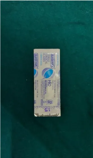
No.15 Bard Parker Blade. (Fig 3)
Bard Parker Handle No.3. (Fig 4)
Molt no 9 periosteal elevator. (Fig 5)
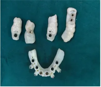
Surgical guiding stent. (Fig 6)
Tissue biopsy punch. (Fig 7,8)
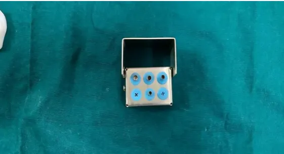
Cylindrical drill sleeves. (Fig 9)
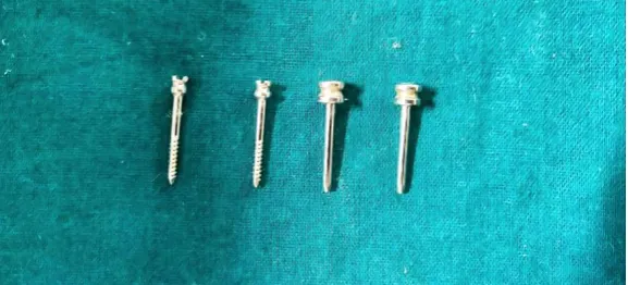
Anchoring pins. ( Fig 10)
Physiodispenser motor and contra-angled hand piece. (Fig 11)
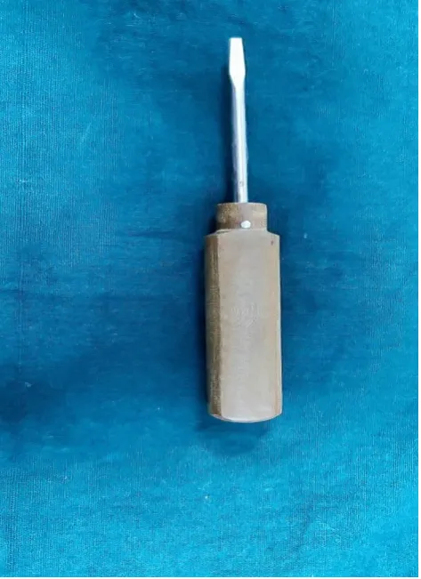
Surgical screw drivers. (Fig 12)
Surgical Implant kit which belongs to ADIN DENTAL IMPLANT
SYSTEM. (Fig 13)
3.0 silk suture.( Fig 14)
36
All these patients underwent routine blood investigations followed by
complete medical evaluation.
PREOPERATIVE IMPLANT PLANNING: For partially edentulous patient
Diagnostic impressions were made with alginate and diagnostic models
were poured by orthokal. Wax up of teeth was done in the region of missing
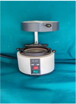
teeth of diagnostic models. Duplicate models prepared. Thermoplast sheet of
2mm thickness placed in the model through pressure moulding machine.
Radiographic markers were placed on the upper part of the splint where wax
up done (Fig 16). Then splint was placed in patient’s mouth and CBCT was
taken by carestream scanner(resolution upto 90 µm and volume sizes from
17cm*13.5cm to 5cm*5cm,Atlanta,GA). A virtual planning was obtained
through blue sky bio software(version 4.3,USA) and surgical guide was
prepared. Guides were fabricated through stratasys eden 260vs computer 3D
printer by polyjet printing technology.SS316 cylindrical drill sleeves were
fabricated in different diameters.
For completely dentulous patient
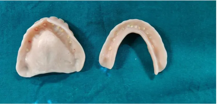
Complete dentures were fabricated. Teeth were drilled and gutta percha
sticks were inserted into the teeth. CBCT scan of the patient was taken with
complete denture in patients mouth (Fig 17) .Virtual planning was performed
37
fabricated through Stratasys Eden 260vs computer 3D printer by polyjet
printing technology. SS316 cylindrical drill sleeves were prepared.
INTRA OPERATIVE SURGICAL IMPLANT PLACEMENT:
Under local anaesthesia (2% lignocaine with adrenaline) implant sites
were marked with a probe by placing surgical guides. Mucosal tissues were
removed by tissue biopsy punch. Surgical guides were placed on the teeth.
Drilling was done by placing the surgical guides through the information
gained from virtual planning. Depth control system was used. A drill was
inserted in the drill sleeve of master sleeve upto the physical stop of drill shaft
and the next size sleeve was placed where the drill was completely inserted in
to the master sleeve for achieving the implant depth. This was done in a
sequential order and the final drill was placed directly in to the master sleeve.
The same procedure was followed for completely edentulous patients whereas
additionally anchoring pins were used to stabilise the surgical guides .The
implants were placed according to the above mentioned protocol and cover
screws were placed.
POST-OPERATIVE EVALUATION OF IMPLANT PLACEMENT:
After six months, postoperative CBCT were taken. A Virtual planning
performed in a preoperative CBCT and post op CBCT were converted into
virtual 3D models using DICOM to Print (D2P) software. These 3D models
were superimposed using Geomagic Freedom software and accuracy of the
38
POST-SURGICAL PROTOCOL:
All patients were administered with a single dose of analgesic (Inj.
diclofenac 75mg) intramuscularly immediately after the procedure. They were
prescribed with a following regimen of oral antibiotics Cap.Amoxicillin
500mg TDS, Tab.Metronidazole 400mg TDS, analgesics Tab.paracetomol
650mg TDS and antacid Tab. Ranitidine hydrochloride 150mg BD before
food for a period of 5 days.
Regular oral hygiene maintenance was advised.
SECOND STAGE SURGERY:
Implants were uncovered (3months in case of mandible and 6 months
in case of maxilla).
Cover screws were removed.
Healing abutments were placed for 1 week.
Further prosthetic procedures were undertaken.
IMPLANT SYSTEM:
The implant system used in the study was Adin dental
implant system(Israel). The sizes of the implants were selected according to
39
PROFORMA
NAME : AGE:
SEX :
MOB NUMBER : OP.NO:
FATHER/GUARDIAN NAME :
POSTAL ADDRESS :
OCCUPATION :
CHIEF COMPLAINT :
HISTORY OF PRESENTING ILLNESS :
PAST MEDICAL HISTORY :
PAST SURGICAL HISTORY :
40 PERSONAL HISTROY
HABITS :
DRUGS :
BRUSHING :
GENERAL EXAMINATION
HEIGHT :
WEIGHT :
BLOOD PRESSURE :
ANAEMIA :
PULE RATE :
CYANOSIS :
CLUBBING :
LOCAL EXAMINATION
EXTRAORAL EXAMINATION
FACIAL ASYMMETRY:
TMJ MOVEMENTS :
MOUTH OPENING :
41 INTRA ORAL EXAMINATION
SOFT TISSUE :
ORAL HYGIENE STATUS:
GINGIVA:
FRENAL ATTACHMENT
UPPER :
LOWER:
TONGUE SIZE :
HARD TISSUE TEETH PRESENT D- M- F- INVESTIGATION STUDY CASTS:
CBCT :
CLINICAL PHOTOGRAPHS :
DIAGNOSIS:
ARMAMENTARIUM
[image:63.595.216.380.422.661.2]Fig 1 2%LIGNOCAINE WITH 1:80000 EPINEPHRINE.
Fig 3 NO.15 BARD PARKER BLADE.
[image:64.595.210.388.442.673.2]Fig 5MOLT NO9 PERIOSTEAL ELEVATOR.
[image:65.595.136.461.405.684.2]Fig 7TISSUE BIOPSY PUNCH (MOTOR DRIVEN).
[image:66.595.149.447.292.443.2]Fig 8 TISSUE BIOPSY PUNCH( MANUAL).
[image:66.595.152.447.472.655.2]
Fig 10 ANCHORING PINS.
[image:67.595.115.485.298.535.2]