A thesis presented in panial fulfilment of the requirements for the degree of Masters of Science in Molecular Genetics
at
Massey University, Palmerston North New Zealand
Anita Therese Bidlake
Abstract
Acknowledgements
Firstly my sincerest appreciation must go to my supervisor Dr. Rosie Bradshaw. Rosies optimism, encouragement, patience and organisation have played a huge part in guiding me to the completion of this momentous task.
Special thanks to all the people in my lab who helped and supported me. For Carmel who got me started, Dianne who keeped me going on my transformations, and Linda and Paul who were always around for advice and a chat. Thanks also to Branwyn and Brendon who brought some new life into our lab. Brendon must also be thanked for his technical assistance along with Shalome Campbell and Tash Forester. A big thank you must also be extended to the "MGUs personal" technichan Carolyn Young. To everyone else in the MGU and the department thankyou all very much for advise and friendship, I really appreciated it.
Gratitude must then be expressed to my friends and family outside the department. Thanks for friends and flatmates who have provided an ear for listening and sources of entertainment. Thanks to my family especially Dad and Judy for giving me lots of love. And special thanks to Granny and Grandpop and their garden for keeping me healthy. A special acknowledgment must be given to someone who can not read, Squeak whose antics always keep me laughing and cuddles keep me smiling.
I would especially like to thank Paul whose love and support have been really special. Without his help this thesis would have been an even longer time coming.
ABSTRACT ... ii
ACKNOWLEDGEMENTS... iii
TABLE OF CONTENTS ... iv
LIST OF TABLES ... ix
LIST OF FIGURES ... x
Chapter 1. INTRODUCTION ... 1
1.1 General Characteristics of Dothistroma pini.................... 1
1.2 Dothistroma Infection... 2
1.3 Chemical Control. ... 4
1.4 Resistant Strains ... 5
1.5 Dothistromin Toxin ... 7
1.6 Inactivation of the Dothistromin Toxin ...... 8
1.7 Transformation of Fungal Plant Pathogens ... 9
1.8 Isolation of a Dothistromin pini P-tubulin Gene for use as an Endogenous promoter ... 10
1.9 Aims and Objectives ... 13
Chapter 2. METHODS AND MATERIALS... 15
2.1 Fungal, Bacterial Strains and Plasmids... 15
2.2 Media... 15
2.2.1 Bacterial Media ... 15
2.2.1.1 Liquid Media ... 15
2.2.1.2 Solid Media ... 15
2.2.1.3 Media Supplements... 17
2.2.2 Fungal Media... 17
2.2.2. l Liquid Media... 17
2.3.l Bacterial Cultures ... 18
2.3.2 Fungal Cultures ... 18
2.4 Common Solutions ... 18
2.4.1 2.4.2 2.4.3 2.4.4 2.4.5 2.4.6 2.4.7 2.4.8 2.4.9 2.5 2.5.1 2.5.2 2.5.3 2.5.4 2.6 2.6.1 2.6.2 2.7 2.8 2.8.1 2.8.2 2.9 lOx TAE Buffer ... 18
TE Buffer ... 18
20x SSC ... 18
1 Ox Gel Loading Dye... 19
Phenol. ... 19
OM Buffer ... 19
ST Buffer ... 19
STC Buffer ... 19
SM Buffer ... 19
DNA Preparations ... 19
Small Scale Alkaline Lysis Plasmid DNA Preparation ... 19
Cesium Chloride-Ethidium Bromid Density Gradient Plasmid Preparation 20 Fungal DNA Extraction... 20
Lambda Phage DNA Preparation ... 21
Purification of DNA ... 22
PhenoVChloroform Extraction... 22
Commercial Kits... 22
Ethanol or Isopropanol Precipitation of DNA. ... 22
Determination of DNA Concentration and Purity ... 22
Spectrophotometric Determination of DNA Concentration ... 22
Determination of DNA Concentration using Concentration Standards ... 23
DNA Manipulations ... 23
2.9.1 Restriction Enzyme Digests of DNA ... 23
2.9.1.1 Digests of Lambda and Plasmid DNA. ... 23
2.9.1.2 Digests of Genomic DNA ... 23
2.9.2 Agarose-gel Electrophoresis ... 24
2.9.3 DNA Extraction from SeaPlaque Agarose ... 24
2.10 Cloning Procedures ... 24
2.10.1 Preparation of Insert DNA ... 24
2.11 2.11.1
Preparation of Dothistroma pini Protoplasts.... 26
Protoplast Protocol 1... 26
2.11.2 Protoplast Protocol 2... 26
2.11.3 Protoplast Protocol 3 ... 26
2.12 Transformation of Dothistroma pini ... 27
2.12.1 Transformation Protocol A... 27
2.12.2 Transforamtion Protocol B ... 27
2.12.3 Transformation Protocol C ... 27
2.13 Subculturing of Transformants... .. ... 28
2.14 Southern Blotting and Hybridisation ... 28
2.14.1 Southern Blotting ... 28
2.14.2 Radioactive [a-32p]dCTP labelling of the DNA Probe ... 29
2.14.3 Hybridisation of Probe DNA to Southen Blots ... 30
2.14.4 Autoradiography of Southern Blots ... 30
2.14.5 Stripping the Hybridised probe from Southern Blots ... 30
2.15 Library Screening ... 30
2.15.1 Determination of library Titre and Primary Round of Library Screening .... 30
2.15.2 Plaque Lifts ... 31
2.15.3 Hybridisation of a [a-32P]dCTP Labelled Probe to Phage A. DNA ... 31
2.15.4 Second and Third Round of Library Screening ... 32
2.16 DNA Sequencing ... 32
2.16.1 Sequencing Reactions... 32
2.16.2 Polyacrylamide Electrophoresis ... 32
Chapter 3 OPTIMISATION OF PROTOPLAST ISOLATION FROM 3.1 3.1.l 3.1.2 3.1.3 3.1.4 3.1.5 DOTHISTROMA PINI ............... 35
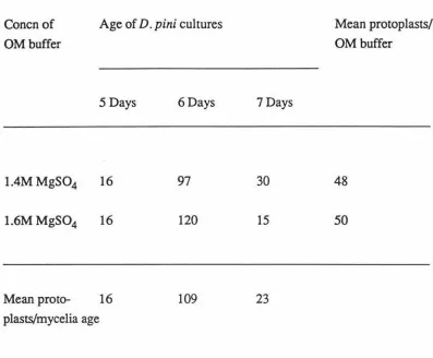
Investigation into the Parameters which Affect Protoplast Isolation... 35
Analysis of Various Osmotic Stabilisers ... 35
Investigation into Varying Conentrations of MgS04, NaCl and Novozyme per ml of Osmotic Stabiliser ... 36
Assesing Different pHs of OM Buffer ... 38
Influence of Mycelium Age on Protoplast Numbers ... 38
3.2 Regeneration of Protoplasts ... 43
3.3 Examination of Different Colony Morphologies of Regenerated Protoplasts ... 44
3.4 Further Attempts to Improve Protoplast Isolation and Harvesting/Purity ... 46
3.4.1 Filtration of the Hyphal Debris Through a Sintered Glass Filter... 46
3.4.2 Increasing Mycelia Concentration in the Preparations ... 46
3.4.3 Trying another Centrifuge for Harvesting ... 48
Chapter 4. DEVELOPING A TRANSFORMATION SYSTEM... 49
4.1 4.1.1 4.1.2 4.2 4.2.1 4.2.2 4.2.3 4.3 4.4 4.5 4.6 Preparatory Work ... 49
Investigating the Antibiotic Concentrations which Inhibit D. pini.. ... 49
Preparation and Molecular Analysis of pAN7-1 and pANS-1 ... 49
Analysis of Different Transformation Methods... 50
Attempts at Transforming D. pini with Transformation Protocol A... 50
Attempts at Transforming D. pini with Transformation Protocol B ... 52
Attempts at Transforming D. pini with Transformation Protocol C ... 56
Abortive Transformants... 62
Suggestions to Further Improve Transformation Frequencies... 62
Molecular Analysis of Transformants... 67
Further Transformant Analysis ... 72
Chapter 5. DEVELOPING A HOMOLOGOUS TRANSFORMATION SYSTEM FOR 5.1 5.1.1 5.1.2 5.2 5.2.1 5.2.2 5.2.3 5.2.4 DOTHISIROMIN PINI ... 79
Analysis of Unsuccessful Clones A. BTl, BT2 and BT3 ... 79
Restriction Digestion of A. Clones and Southern Hybridisation ... 79
Computer Analysis of the A. Clones... 87
Analysis of Positive A. Clones AB 1 to AB5 ... 88
Library Screening ... 88
Restriction Enzyme Analysis of A. Clones and Southern Hybridisation... 89
Mapping of A. Clone AB!... 95
APPENDICES ... 102
Appendix 1: Vector Maps ... 102
Appendix II: Sequence Data ... 104
PrettyOut of 1.3 XhoI D. pini P-tubulin Gene... 104
BESTFIT of 1.3 XhoI D.pini P-tubulin Gene with N. crassa P-tubulin Gene ... 107
Table 1. Table2. Table 3. Table4.
Table 5. Table 6. Table 7. Table 8. Table 9. Table 10. Table 11.
Selectable Genes for Transforming Plant Pathogenic Fungi... 11
Fungal and Bacterial Strains, A. Clones and Plamids ... 16
Primers Used in Sequencing Reactions ... 34
Protoplast Numbers with Differing Concentrations of MgS04, NaCl and Novozyme ... 37
Protoplast Numbers from 1.4 versus 1.6M MgS04 ... 39
Protoplast Numbers Using Differing pHs of OM Buffer... 39
Protoplast Numbers Obtained from Varying Aged Mycelia ... 40
Protoplast Numbers Formed at 30 min Intervals... 42
Regeneration (%) of D. pini Protoplasts ... 45
Protoplast Numbers Obtained using Different Amounts of Mycelium ... 47
Transformation of Fungal Protoplasts with pAN7-1 or pAN8-1 using Transformation Protocol
A...
51Table 12. Transformation of Fungal Protoplasts with pAN7-1 orpAN8-1 using Transformation Protocol B... 54
Table 13. Transformation of Fungal Protoplasts with pAN8-1 and Various forms of pAN7-1 using Transformation Protocol C ... 58
Table 14. Transformation of Protoplasts with Linear and CircularpAN7-1 from Two Separate CsCl-density Preparations and with Two Concentrations of pAN7-1 from CsCl Preparation 2... ... ... .. .. ... .... .... ... 60
Table 15. Resistance levels shown by transformants AB 1 to 8 when plated on increasing concentrations of hygromycin B ... 76
Figure 1. Transformants ABl and AB2 plated on selective and non-selective DM ... 53
Figure 2. Stable and abortive transformants ... 63
Figure 3. The number of transformants/µg of DNA generated during various
transformations ... 64 Figure 4. Hybridisation of pAN7-1 to transformants AB 1 to ABS... 68
Figure 5. Diagram of integration events as described by Hinnen et al. ( 1978).... 73
Figure 6. Sensitivity of wild-type D. pini and four independent transformants to various
concentrations of hygromycin B ... 75 Figure 7. Restriction enzyme analysis of A.BTl, A.BT2 and A.BT3 ... 80
Figure 8. Autoradiograph of hybridisation results washed at two different
stringencies... 82
Figure 9. Restriction enzyme analysis of A.BT3 ... 84
Figure 10. Autoradiograph of hybridisation results from blot of A. clone BT3
washed at two different stringencies... 85
Figure 11. Restriction enzyme analysis of A. clones AB 1, AB2, AB3, and AB4 ... 90
Figure 12. Autoradiograph of hybridisation results from blot of A. clones ABl,
AB2, AB3 and AB4 ... 92 Figure 13. Restriction map of the A. clone AB 1 from a Dp-1 genomic library that
hybridised to Neurospora crassa ~-tubulin gene ... 97
Chapter
1.
Introduction
1.1
General Characteristics of
Dothistroma pini
The fungus D. pini is a needle pathogen found mainly on many pine species including Pinus
radiata. Infection begins with chlorosis and necrosis at the base of the crown (Philips and Burdekin, 1982) and is seen as red tinged lesions or bands. Necrosis often extends
throughout the needle leading to premature defoliation, followed by a reduction in photosynthesis and eventually wood yield (Franich et al., 1982). In extreme cases this can be followed by death of the tree (Gallagher, 1971 ).
D. pini has been recorded in Europe, South and East Africa, North and South America and Australasia. It was first identified in the central North Island of New Zealand in 1962 and is
now found in all of the North Island except the Northern tip and Great Barrier Island. In the South Island Nelson, Malborough, North of the Wairou river, Westland. Southland and Otago are all sites of infection (Gadgil, 1984). Most species of genus Pinus are susceptible
to D. pini attack including Pinus radiata, P. ponderosa, P. nigra (Gallagher. 1971) although other species such as Pseudotuga menziesii and Larix decidua are also slightly susceptible (Philips and Burdekin, 1982).
Indigenous New Zealand trees are slow growing and would take rotations of hundreds of
years to achieve the desired crop. Consequently introduced softwoods, which are fast growing are used in our commercial plantations, of which radiata pine makes up 93%. Accordingly because of the important role the forestry industry has in New Zealand and the role P. radiata plays in it. D. pini infection is of major economic significance to New Zealand. In fact plantation losses in New Zealand by disease and insects is only exceeded by
losses caused by wind. Direct costs in controlling D. pini which can cause 10-25% periodic advancement of growth loss. since 1967 in New Zealand have totalled $35.3 million (1988)
(New, 1989).
D. pini caused lesions are band-like and contain 1-12 stromata which vary in size from 300-750 x 150-400µm. Asexual sporation inside the stromata is good. The black conidia, which
are produced as sticky masses vary in size and are septate when mature. Below the stromata
Fortunately for New Zealand the perfect stage is only found in North America (Vancouver) and Europe for its readily produced airborne spores would have dire consequences for our industry (Gadgil, 1984).
Water is required for rain-splash dispersal, as asexual spores are liberated from fruiting bodies in a film of water on the needle surf ace. As the water droplets hit the ground the spores are released to the air where they normally infect only the neighbouring trees. Germination of the spores is nearly complete within 48 hours. Mycelium then grows on the surface of the needle for 7-10 days, producing secondary conidia, before penetrating the stomata between the guard cells where its lateral spread is limited to a few millimeters from the point of infection (Gadgil, 1967, 1984). The first macrosopic sign of needle infection is yellow flecks which extend to become "red bands". Three to four months after the initial inoculation stroma primordia develop and push there way to the needle surface within these bands. Necrosis develops eventually leading to premature sheding of infected needles (so the crowns may appear thin with tufts of needles at the tips of the branches) and to a litter layer bearing a large number of D. pini stromata (Gadgil, 1970).
1.2
Dothistroma pini Infection
The sources of inoculum and the factors which influence infection by Dothistroma have
undergone extensive investigation. In one study Gadgil (1970) looked at the survival of D. pini on fallen needles of P. radiata. The litter layers around infected trees are comprised of
needles bearing large numbers of D. pini stromata. If D. pini could survive for long periods
in competition with other micro-organisms, then the litter layer could be a major source of inoculum of fresh foliage. However it was found that D. pini was a poor saprophyte
because the proportion of D. pini conidia compared to other fungi decreased rapidly over
time. The viability period of the stromata was shown to differ throughout the year. The infective period was shown to depend on the position of the infected needles and whether the stand was thinned or left untreated, not on actual seasons (Gadgil, 1970).
It has been observed that along with the actual number of the infective spores landing on the leaf surface (usually several thousand) rainfall and other environmental parameters influence the rate and intensity of Dothistroma infection. In unpublished work Gadgil used a Hirst spore trap (which efficiently collects dry spores), operating continuously for six months (1974-1975), in a heavily infected P. radiata stand but caught very few conidia. This
therefore hydrated when they fall on to a susceptible host It has therefore being shown that
germination and penetration of the host by the fungi will occur with relatively short wetness periods, as the conidia which are deposited on the surface of the host are always hydrated
(Sheridan et al., 1970; Gadgil, 1977). However stromatal development was shown to be reduced until the foliage was moistened and the longer the dry period following deposition
of conidia the lower the severity of the infection.
If the inoculum is sufficient infection will occur at a wide range of temperatures, between 5 and 30oC, with an optimum at 17°C. A study of meterological data shows that the
temperatures suitable for infection occur from November to March in all radiata pine
growing areas in New Zealand (Gadgil, 1974; Sheridan et al., 1970). Field observations
indicated that shaded foliage of radiata pine is markedly less infected than foliage exposed to the full light Though neither germination or growth of the fungus on needle surfaces was
affected by light intensities, this was believed to be a response of the host to low light
intensities that reduced the infection (Gadgil and Holden, 1976).
The genus Pinus shows varying resistance to the pathogen. P. radiata is susceptible when
young, but mature trees older than 15 to 20 years show little infection, suggesting increased resistance (Franich et al., 1982). Other species for example P. ponderosa remain equally
susceptible at all age (Philips and Burdekin, 1982). Entry of the pathogen was examined and
direct entry of the epidermis was only seen when macerated mycelium was sprayed as the
inoculum in the absence of spores. So it seems penetration of the stomata by hyphae between the guard cells, although not common (seen 5 times in about 20000 sections of
inoculated needles) is the only way for infection to develop inside the needles naturally
(Gadgil, 1967).
Scanning electron microscopy testifies that stomata of young, vulnerable trees to be open
-,.
pores, 15-20µm long of which the guard and subsidiary cells have an epidermis covered with
fine microtubular wax. In contrast the mature trees have stomata! of 10-15µm which are
often occluded by an amorphous wax. It has been suggested that this wax could present a
mechanical barrier to ingress of hyphae, or act by masking possible chemotactic or chemotrophic stimuli experienced by the hyphae during stomata! penetration (Franich et al.,
1977; 1983).
The actual chemical nature of this epitcuticular wax may be of importance. In young trees it
resin acid derivatives. An in vitro test on D. pini showed these oxygenated resin acids inhibited both pore gennination and mycelium growth. An in vivo test of artificial inoculation showed plants treated with acetone (depletes epicuticular and stomata! pore fatty
and resin acids) had a mean infection level about two times that of the control. These
experiments suggest that the resin acid derivatives could be pre-infection factors
contributing to resistance in mature trees and once stomata! penetration and hyphae growth
in the mesophyll has occurred the extent of the tissue damage and the rate of fruiting body
fonnation is dependent on other factors such as sensitivity of the needle tissue to the toxin
dothistromin (Franich et al., 1983).
The pH buffering capacity and monoterpene (volatile compounds consisting mainly of 13
monoterpene hydrogen compounds) levels in young and mature trees have been looked at in
attempts to associate them to differences in the level of resistance to D. pini. Measuring the pH buffering capacity, (at pH 6.2) of one-year-old needles of trees increasing in age, showed
the level of buffering capacity increased with maturity. But because Dothistroma in culture tolerates a wide pH range, the pH buffering capacity of the P. radiata needles was thought unlikely to be directly linked to increased mature tree resistance (Franich and Wells, 1977).
As with monoterpenes, although the yields of volatile compounds from young trees were twice that from 20 to 40 year-old trees, and the volatile mixes stimulated the gennination of
spores and growth of the mycelium in liquid culture; monoterpene concentration does not
bear any simple relationship to mature tree resistance (Franich et al., 1982).
1.3
Chemical Control
East African work earlier showed that the needle blight by D. pini could be controlled by copper-based fungicides and subsequent New Zealand work showed that the disease could
be controlled by aerial applications of copper oxychloride (Dick, 1989). Consequently aerial
application of copper fungicide has been the main method for control of D. pini in New Zealand over the last three decades. Pruning of the branches of the infected foliage also
helps and can postpone the need for a fungal application for several years (Gadgil, 1984).
Stands in the age class susceptible to the blight (less than 16 years) are surveyed from the
air, every 2 to 3 years in mid-winter and the average percent of crown infection in each stand
is observed. This is important as 23% of the areas of susceptible age classes of P. radiata are infected. The major consequence of this is that it leads to a loss in wood volume. On
example 10% infection is equal to 10% loss). The overall effect of infection is apparent after
about 2 years (Gadgil, 1984).
Copper fungicide as 50% cuprous oxide (Cu20) reacts with aqueous substances on the needles and with D. pini metabolites to give in aqueous solution cupric ion (Cu2+) at concentrations sufficient to inhibit germination of Dothistroma conidia. The cupric ions also
stimulate dothistromin toxin biosynthesis, which forms a brown water-soluable complex with
cu2+ probably making the toxin unavailable to needle tissue and also removing some of the
Cu2+ on the needle surface by chelation. Interaction of Cu20 fungicide with geothermal sulphur (H2S) gases in the air gives CuS and CuS04 which also contribute to solubilising the fungicide. It is this combination of solubilisation and redistribution of Cu2+ and its
complexes, along with their ready uptake of D. pini conidia which explains why copper
fungicides give good control of this needle blight (Franich, 1988).
Spraying is usually undertaken in November which kills most of the inoculum at the time it is multiplying (Gadgil, 1984). After a couple of months of rain in the North Island the copper
is washed off but reinfection is quite slow due to the rapid reduction with time in the amount
of inoculum, produced by the stromata, on fallen needles. This short viable conidia time (
4-6 months) has other benefits because as trees over 20 years are seldom infected, and the
shortest possible rotation for P. radiata in New Zealand is 22 years, there should be no danger that regenerating seedlings will become infected as a direct result of growing on an
area where a previous crop was infected (Gadgil, 1970).
Even though the average spray frequency per stand is higher than initial research indicated because of the wet summers, deep gullies and extensive mists in infected areas (Dick, 1989)
the cost per hectare for New Zealand spraying of copper fungicide has reduced from >$60
(1960s) to $15 (1988). 11ris reduction is due to the careful assessment of the optimum time
to spray, improvements in spraying techniques (therefore less spray and reduction in flying
time) and reduction in fungicide doses (less chemical costs) (Dick, 1989; Franich, 1988).
Further savings now have to be found by reducing disease levels using Dothistroma resistant
breeds of trees.
1.4
Resistant Strains
Thirty years ago all seed for the industry in New Zealand was unimproved bulk seed.
Research Institute (FRI) in Rotorua, by selection for growth and form. The FRI maintain an
extensive breeding population from which parent trees are selected and crossed and their
progeny tested in trials. This has lead to three main seed and plant classifications: GF
(Growth and Form - improved growth and more merchantable volume), LI (Long Intemode - more clearwood and minimal pruning) and DR (Dothistroma Resistant - better growth rate
on high D. pini risk sites) (FRI, 1987).
The best Dothistroma resistant seedlots are expected to reduce stand mean infection by 15%
and the effects of spraying and resistance are expected to be additive (Carson and Carson,
1991). As an example of estimated saving in terms of spraying costs, if the resistant breed
of radiata pine is established in Kinleith forest, in place of existing breeds, savings of 56%
are expected. But a prerequisite for improvement in disease resistance in a particular area, is
the need to determine the economic significance of the disease relative to other selection traits. A 6% difference in growth-rate was suggested by Carson (1989) between the best general breed and her proposed resistant breed. Therefore where past history indicates
frequent spraying, then the resistant breed should be established but on healthier sites, the
standard breeds should be used (Dick, 1989).
D. pini resistance behaves as a classical additively inherited trait with little dominance
variance. Estimates of genetic correlation of D. pini disease symptoms across sites have
generally been high. Subsequently there appears no reason to regionalise a selection
program for Dothistroma resistance as resistant seedlots will probably be resistant on all
sites where D. pini infection is present (Carson and Carson, 1991).
The nature of resistance seems to be the property of the whole tree and variability in
selecting reliable resistant selections could be due a number of reasons including disease
escape, variable symptom expression and non-optimal time of original assessment for
resistance. Due to different weather patterns field assessment needs to be carried out for 3
to 5 consecutive years (Frainch et al., 1986). Earlier research showed cuttings overall
seemed to have more resistance than seedlings hence chemical control could be reduced if
new plantations were established with cuttings rather than seedlings in areas where needle
blight appear prevalent (Gadgil and Holden, 1976).
P. radiata has a broad genetic variability base as the original trees are from 2 to 3
independent American populations. Monoclonal propagation of P. radiata could potentially
decrease and more virulent strains of Dothistroma could arise and overcome current
resistant mechanisms. Because of this possibility and since spraying with copper will only
control the disease and not eliminate it, it seems necessary to know more about the pathogen - host relationship. In particular the mechanism resulting in needle death which involves the D. pini toxin, dothistromin.
1.5
Dothistomin Toxin
D. pini produces the mycotoxin dothistromin as a major metabolic byproduct (Gallagher and
Hodges, 1972). This red pigmented metabolite was first isolated from D. pini and subsequently from several Cercospora spp. and Mycosphaerella larcina. Early evidence of
phytotoxicity of dothistromin was demonstrated by the injection of acetone and dothistromin
into pine needles: within 5 days dothistromi~ 'red band' symptoms typical of those produced by D. pini appeared (Franich et al., 1986; Stoessl et al., 1990).
Up to 90% of the injected dothistromin is metabolised by needle cells or phytolytically
degraded to C02 and oxalic acid within · the first 24 hours, while simultaneously the formation of a small necrotic lesion appears. The mechanism, based on the formation of
metabolites, is thought to involve peroxidase catalysed oxidation of dothistromin by
hydrogen peroixde. Cells adjacent to those killed by dothistromin die and the lesions expand
over 2-3 days. Since most of the dothistromin is degraded or metabolised, the cause of the
expanding lesion is thought to be due to other toxic substances rather than dothistromin. It is found that the cells adjacent to those initially killed, synthesise and accumulated benzoic
acid, which is toxic to P. radiata needle mesophyll cells and is highly fungistatic. The length
of D. pini induced lesions were found to be proportional to the amount of benzoic acid
injected into the needles and the necrotic tissue adjacent to the bands also accumulated
benzoic acid which prevents colonisation of that tissue by the fungus. Benzoic acid is also
bound tightly to lignin polymers which are found in disproportionably high amounts in dark
green tissue adjacent to the dothistromin induced lesions. Benzoic acid therefore has been
put forward as the phytoalexin of P. radiata needles (Franich et al., 1986).
D. pini produces dothistromin as a major metabolic byproduct (Gallagher and Hodges,
1972). [13C]-labelling pattern shows that dothistromin is a difuroanthraquine fused to a
substituted tetrahydro-2-hydroxy-bisfuran ring system (Shaw, 1978). It is structurally
related to the mycotoxins, sterigatocystin and aflatoxin B 1 from Aspergillus flavus. It is
and potential human carcinogenicity associated with this toxin (Elliot et al., 1989; Harvey et
al., 1976).
On the strength of the remarkable similarity between dothistromin and aflatoxin B 1,
mutagenicity studies were initiated with dothistromin. Harvey et al., (1976) demonstrated that dothistromin inhibited RNA synthesis, as measured by [3H]-uridine incorporation in
Chiarella pyrenoidosa and Bacillus megaterium. It was observed that dothistromin was 10 times more active in inhibiting the growth of B. megaterium. Various field samples, extensive environmental monitoring and human exposure studies were carried out by both
the Department of Health and the FRI. Dothistromin was tested for mutagenicity in a wide
variety of in vitro bioassays, most of which were positive. For example, chromosome damage in human peripheral blood lynphocyte cultures, accompanied by lysis of red blood
corpuscles and for complete risk assessment a mouse in vivo mutagenicity assay. This work suggests that although it acts in a slightly different way, dothistromin may be just as hazardous as aflatoxin B 1. However neither are much of health risk unless there is some
underlying pathology present, for example smoking or the Hepatitus B Virus (Elliot et al., 1989; Stoessl et al., 1990).
1.6
Inactivation of the Dothistroma Toxin
Copper fungicide is only a control mechanism and resistant varieties are only successful as
long as the pathogen retains its present virulence levels (Gallagher, 1971). So when the
much shorter life-cycle of the plant pathogen is compared with its hosts, it is plausible that
more virulent strains of D. pini could arise which would overcome the current resistant mechanisms. For this reason different possibilities of Dothistroma control such as
monoclonal antibodies and development of a transformation system to produce D. pini isolates which are specifically blocked in dothistromin biosynthesis, must be investigated.
A team of research scientists at the Hort Research Immunology Group (led by Dr Paul
Reynolds), situated in Palmerston North, have prepared monoclonal antibodies to
dothistromin. They have cloned the gene encoding this dothistromin-specific antibody, and
prepared a single-chain antibody epitope. They aim to produce transgenic plants which
express this antibody, thus inactivating the dothistromin toxin if D. pini infects them and hence eliminating the need for spraying by copper fungicides. In in vitro experiments so far the antibody competes quite well for dothistromin binding, but preincubation of the antibody
assumption underlying this approach is that the dothistromin toxin is the primary cause of disease symptoms. This is suggested by experimental work in which purified dothistromin
toxin was injected into pine needles (Franich et al., 1986).
However, genes required for toxin-biosynthesis have recently been cloned from several plant
pathogenic fungi which, when disrupted (leading to loss of toxin production), had no effect
on fungal pathogenicity in two out of five cases (VanEtten et al., 1994). These results lead
us to question whether D. pini would also still be pathogenic to pine trees in the absence of
dothistromin toxin: if so, the antibody approach to "immunising" pine trees may be ineffectual. We aim to answer this question by generating non-dothistromin producing
isolates of D. pini so that their pathogenicity can be assessed. This will be achieved by disruption of dothistromin pathway genes.
The biosynthetic pathway of dothistromin has been investigated and intermediates have been
identified which also occur in the biosynthesis of aflatoxin. Clones of some of the aflatoxin
biosynthetic genes from Aspergillus are being used as hybridisation probes in the hope that
they will be sufficiently homologous to D. pini biosynthetic genes to enable their detection. Once the dothistromin biosynthetic genes are cloned, a transformation system is required so
the genomic copies can be disrupted or replaced to produce isolates of D. pini which are specifically blocked in dothistromin biosynthesis. The ploidy of D. pini is unknown. Consequently experimental work to detennine the ploidy will have to be completed prior to gene disruption. Once disrupted the isolates will be checked for the production of toxic pathway intermediates, or other toxic products formed by branching pathways. These isolates can then be tested in the field to determine whether they have lost their
pathogenicity.
1. 7
Transformation of Fungal Plan_t Pathogens
The accessibility of a DNA-mediated transfonnation system is one of the first requirements
to carrying out modem molecular biological research. Gene transformation of filamentous
fungi is a relatively new area. The first description of such a system was in Neurospora
crassa (Case et al., 1979), followed soon after with gene transfer in another ascomycete, Aspergillus nidulans (Ballance et al., 1983). Many other transformation systems have been developed for a range of commercially and agriculturally important fungal species
To permit selection of transformed cells markers are used that are capable of complementing
a mutation such as auxotrophic markers (negative selection) or that provide a new property
to the host cell, for example antibiotic resistance (dominant or positive selection). An advantage of using a 'negative' selectable marker is that transformants for potential field
release would be wild-type for the selectable marker rather than acquiring an additional
characteristic. Dominant selection however has the advantage that mutant strains are not
required so a genetically uncharacterised species can also be transformed (Punt and van den Hondel, 1992). Examples of genes used as markers in positive (dominant) or negative
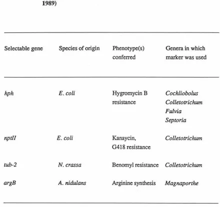
selection systems for transformation of plant pathogenic fungi are given in Table 1 (Fincham,
1989; Hargreaves and Turner, 1992).
In developing a transformation system positive selectable markers which confer resistance to hygromcin and phleomycin will be used. Hygromycin B (HmB) is an aminoglycosidic
antibiotic which disturbs protein synthesis by interfering with peptidyl-tRNA translocation,
causing misreading. Hygromycin B resistance genes encode the enzyme hygromycin B
phosphotransferase which phosphorylates the antibiotic HmB. HmB resistant genes are
isolated from Streptomyces hygroscopicus and from Escherichia coli and these genes have
been used with many pathogenic fungi (see Table 1).
A less toxic and cheaper alternative to hygromycin is phleomycin. Phleomycin is a
metalloglycopeptide antibiotic causing DNA strand scission. Phleomycin resistance genes
isolated from Streptoalloteichus hindustanus and E. coli encode proteins which bind to and
inactivate the antibiotic
1.8
Isolation of a
Dothistroma pini
P-tubulin Gene for use as an
Endogenous Promoter
Due to a low success rate when transforming D. pini using promoters from Aspergillus
nidulans to drive the expression of selectable markers, an alternative approach was investigated. One reason for this low transformation efficiency may be poor expression of
the selectable markers which are under the control of the Aspergillus nidulans gpd.A
promoter, when placed in the D. pini genome. In the literature the use of endogenous promoters has been reported to both increase (Skatrud et al., 1986) and decrease (Smith et
Table 1. Selectable Genes for Transforming Plant Pathogenic Fungi (Fincham, 1989)
Selectable gene Species of origin Phenotype(s) conferred
Genera in which
marker was used
hph E.coli Hygromycin B Cochliobolus
resistance Colletotrichum
Fulvia
Septoria
nptll E.coli Kanaycin, Colletotrichum
G4 l 8 resistance
tub-2 N. crassa Benomyl resistance Col/etotrichum
promoter greatly increased the transformaiton efficiency (Gruber et al. 1990).
Due to the very low transformation efficiency in D. pini we decided to isolate a gene from
D. pini with a view to using an endogenous promoter in the hope of increasing expression
and transformation efficiency. The most widely used homologous promoter which has been
used to increase transformation rates of dominant selection systems, is the
constitutively-expressed glyceraldehyde-3-phosphate dehydrogenase (gpd) gene. This gene is highly
conserved throughout the kingdoms of organisms (Jungehulsing et al., 1994). Another gene
which is also highly conserved (so the gene can easily be isolated using heterologous probes)
and usually constitutively expressed is 13-tubulin. The 13-tubulin gene was chosen for use in
our system, over the gpd gene, as it is slightly more variable and consequently could have
other uses, for example, as a control probe for Northern blots and for phylogenetic studies.
Microtubules are distinct fibrous structures found in all eukaryotic cells. They play a role in
a variety of intracellular processes including intracellular transport, mitosis and meiosis, and
maintenance of cell shape. The constituent protein of microtubules is tubulin and it is composed of two similar sub-units a- and 13-tubulin. A central question concerning the regulation of microtubule function is how microtubules can play a role in such a variety of
cell processes. Two differing hypothesis have been suggested to explain this. The
multi-tubulin hypothesis (Fulton and Simpson, 1976) states that microtubules with different
functions are composed of different types of tubulins. The other hypothesis proposes that tubulin genes are functionally equivalent but different sets of microtubule-associated proteins
are associated with microtubules having different functions (Weatherbee and Morris, 1984).
Multiple J3-tubulin genes are seen in many organisms, for example A. nidulans contains three
(Weatherbee and Morris, 1984) whereas two have now been identified in Colletotrichwn gloeosporioides (Buhr and Dickman, 1994).
Most mutations conferring resistance to the fungicide benomyl in fungi (such as A. nidulans
and Saccharomyces cerevisiae, Orbach et al., 1986) have been mapped in the f3-tubulin
structural genes. Consequently the mutational change in the 13-tubulin genes responsible for
benomyl resistance has been determined, and is then able to be used as a dominant selectable
marker in transformation experiments. An example of cloning and characterising a gene for
13-tubulin from a benomyl-resistant mutant of Neurospora crassa and its subsequent use as a
Using a plasmid (pBT6) which contains the J3-tubulin gene from N. crassa we aim to isolate
the J3-tubulin promoter from D. pini. We are hoping that the isolation of an endogenous
promoter may facilitate the development of a more efficient transformation system.
1.9
Aims and Objectives
Transformation protocols vary between different fungi and different laboratories. Therefore,
when developing a transformation system for D. pini it is necessary to try different protocols
under many conditions to develop a method which is efficient, reliable and hopefully yields
high numbers of transformants.
The simplest place to start developing a transformation system for D. pini is to try positive selection of transformants for phleomycin resistance. The first step requires the growing of
Dothistroma in liquid culture or on cellophane discs without contamination and developing
methods for optimising the isolation of protoplasts. There is a choice of enzymes for
digesting the cell wall, including helicase and chitinase, but Novozyme234 prepared from the
fungus Trichoderma viride and used at concentrations of 5-25mg/ml seems to be the one
most commonly used. Young cultures are used because young hyphae are more susceptible
to the cell wall digesting enzyme and the protoplasts are more easily separated. Protoplasts
must be prepared and maintained in the presence of an osmotic stabiliser to prevent bursting.
For this purpose a variety of osmotic stabilisers can be used, including sorbitol, mannitol,
potassium chloride and magnesiun sulphate, which are used at concentrations of 0.6-1.2M
and pH 5-6 for an incubation time from 5 minutes to 3 hours. Again all these conditions need to be optimised (Fincham, 1989; Hargreaves and Turner, 1992).
After the protoplasts have been purified and numbers of > 10 7 protoplasts/ml have been
obtained, the protoplasts are induced to take up DNA molecules by treatment with calcium
ions and poly ethylene glycol (PEG: causes the treated cells to clump which may facilitate in
trapping of the DNA). For the regeneration of the protoplasts the osmotic stabiliser has to
present in the growth media until the cell wall has been regenerated: this can be the same, or
a different, osmotic stabiliser to that used for the protoplast isolation (Fincham, 1989).
For positive selection of transformed cells a concentration of the inhibitory substance (in this
case phleomycin or hygromycin) that completely blocks growth of sensitive,
non-transformed cells must be used. To test the resistant levels of D. pini to the antibiotic,
0-lOOOµg/ml hygromycin or 1-lOOµg/ml of phleomycin will be used. The composition of the growth media may influence resistant levels, for example, phleomycin has reduced antibiotic activity in acidic media (pH<.5) and both phleomycin and hygromycin antibiotic activity is
decreased in a hypertonic media (such as the one used for protoplast regeneration) (Punt and van den Hondel, 1992). The ploidy of D. pini is unknown but this will not interfere when selecting transformants as we will be using dominant selectable markers. Once the transformants are selected they will be analysed for stability by being taken through several rounds of subculturing. Analysis of location of the plasmid DNA, copy number and site or sites of integration of the plasmid in individual transformants will be performed by Southern blotting.
This project will establish the basic procedures of transformation for the first time in D. pini. The work is complementary to that of Ms C. Gilman and Ms B. Morgan who are currently isolating genes involved in the biosynthesis of dothistromin. The ultimate aim is to use the transformation system methodologies developed in this project to disrupt the wild-type copies of dothistromin biosynthetic genes to create a toxin-minus mutant. This will be useful for two reasons. The first reason being that although dothistromin is implicated as the primary cause of disease symptoms, the extent of the damage to the tree caused by the invasion of the fungus itself is unknown. The benzoic acid which is produced by the plant when it is challenged with the toxin, is a major source of tissue and needle damage: benzoic acid may still be produced by invasion of the fungal mycelium alone. Maybe the dothistromin is the elicitor which initiates the hypersensitive response of the plant and without it D. pini could become fully pathogenic. This information is very important to the Hort Research group who are developing monoclonal antibodies against dothistromin.
Chapter
2.
Materials and Methods
2.1
Fungi, Bacterial Strains and Plasmids
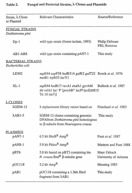
Fungal and bacterial strains,
A
clones, and plasmids used in this study are listed in Table 2.2.2
Media
2.2.1 Bacterial Media
2.2.1.1 I .iqnid Media
Luria Broth (LB)
(g/1): Tryptone, 10.0; NaCl, 5.0; Yeast Extract, 5.0. pH 7.5 (Miller, 1972). SOC Medium
(gll): Tryptone, 20.0; Yeast Extract, 5.0; NaCl, 0.6; KCL, 0.2; MgC12 , 0.95; MgS04, 2.5; Glucose, 3.6 (Dower et al., 1988).
NZCYM
(gll): NZ Amine, 10.0; NaCl, 5.0; Casamino Acids, 1.0; Bacto Yeast Extract, 5.0; MgS04.7H20, 2.0. NaOH to pH 7.5.
2.2.1.2 Solid Media
Luria Agar
LB containing 15 g/1 agar.
Top Agarose
Table 2. Fungal and Bacterial Strains, A. Clones and Plasmids
Strain, A. Clone or Plasmid FUNGAL STRAINS Dothistroma pini Dp-1 AB1-AB8 Relevant Characteristics
wild type strain (forest isolate, 1993)
wild type strain containing pAN7-1
Source/Ref ere nee
Philip Debnam FRI, Rotorua This study BACTERIA!. STRAINS Escherichia coli LE392 XL-1 A.CI.ONES A.GEM-12 A.ABl-5 ELASMIDS pAN7-1 pAN8-1 pBT6 pUC118 pABl
supE44 supF58 hsdR514 galK2 gaIT22 Borek et al. 1976 metBl trpR55 lacYl
supE44 hsdR.17 recAl endAl gyrA46 Bullock et al. 1987 thi relAl lac- F' [proAB+ laclq lacUM15
Tn 10 (tetr)]
A replacement library vector based on Frischauf et al. 1983
A.GEM-12 clones containing genomic This study
DNAfrom Dothistroma pini homologous to f3-tubulin from Neurospora crassa
6.5 kb HmBR AmpR Punt et al. 1987
5.9 kb PhleoR AmpR Mattern and Punt 1988
5.8 kb based on pBT3 containing the Marc Orbach
N. crassa BmlR f3-tubulin gene University of Arizona
3.2kb AmpR Messing 1983
pUCl 18 containing a l.3kb XhoI This study
2.2. 1.3 Media Supplements
When required the antibiotic concentration used for selection was: lOOµg/ml Ampicillin from a stock solution of lOOmg/ml.
When required isopropylthio-J3-galactoside (IPTG) and
5-bromo-4-chloro-3-indolyl-J3-D-galactoside in dimethylformamide (X-gal) were also supplemented: 30 µg/ml of IPTG
60µg/ml X-gal.
lOmM MgS04 and 0.2% (w/v) maltose were supplemented to LB in which an overnight culture of LE392 (plating cells) for library screening were grown.
2.2.2 Fungal Media
2.2.2.1 Liquid Media
D. pini Liquid Media (DM Broth)
(g/l): Malt Extract, 50.0; Nutrient Broth, 20.0.
2.2.2.2 Solid Media
D. pini Media (DM)
(gll): Malt Extract, 50.0; Nutrient Agar, 28.0.
D. pini Top Media (DM Top)
(gll): Malt Extract, 50.0; Nutrient Agar, 11.2; Sucrose, 273.9 (0.8M).
Osmotically Stabilised DM (DM Sue)
(g/l): Malt Extract, 50.0; Nutrient Agar, 28.0; Sucrose, 273.9 (0.8M).
D. pini Sporulation Media (DSM)
(g/l): Malt Extract, 50.0; Yeast Extract, 20.0; Agar, 15.0.
2.2.2.3 Antibiotic Concentration
When required the antibiotic concentration used for selection was:
60-70 µg/ml Hygromycin B from a 50mg/ml H20 stock
2.3
Growth of Cultures
2.3.1 Bacterial Cultures
E. coli strains were maintained on LB plates supplemented as required. The cultures were
grown at 37°C, then stored at 4°C with regular subculturing.
Alternatively for plasmid DNA preparations, 2-5mls of LB supplemented appropriately, was
inoculated with a single bacterial colony and incubated with shaking (300rpm) overnight at
37°C.
2.3.2 Fungal Cultures
A (8.0mm x 8.0mm) chunk (cut with scapple) of D. pini mycelia was ground in lml of
sterile milliQ (MQ) water using a plastic grinder in an eppendorf tube. From this 200µ1 was
spread onto DM plates (with or without cellophane discs) and incubated at 20°C for 6-14
days. The cultures were then stored at 4°C for up to 6 months before subculturing.
DM broth cultures were inoculated in the same way with lml of inoculum/lOOml of DM
broth in a 1 litre siliconised flask. These were grown at 20oC with gentle shaking (1 OOrpm).
Note: the inoculum siz.e is not strictly quantitative due to varying siz.es (10.0mm x
5.0-10.0mm) of mycelia chunks being unavoidably cut depending on the morphology and
thickness of the mycelial material.
2.4
Common Solutions
2.4.1 lOx TAE Buffer (Tris Acetate EDTA buffer)
400mM Tris, 11.4ml glacial acetic acid, 20mM EDTA (pH 8.5).
2.4.2 TE Buffer (Tris EDTA buffer)
lOmM Tris-HCUlmM EDTA (10:1.0 TE) or lOmM Tris-HCUO.lmM EDTA (10:0.1 TE).
2.4.3 20x SSC (Standard Saline Citrate)
2.4.4 lOx Gel Loading Dye
50.0% (w/v) glycerol, Ix TAE, I2.0% (w/v) urea, 0.4% (w/v) bromophenol blue.
2.4.5 Phenol (Tris-equilibrated)
Phenol was liquified by heating at 50oC prior to hydroxyquinoline being added to a final
concentration of O. I% ( w/v ). The phenol was washed three times with an equal volume of O.lM Tris-HCl (pH 8.0). The equilibrated phenol was stored under O.IM Tris-HCl, at 4°C,
in a dark bottle.
2.4.6 OM Buffer
l.6M MgS04.7H20, with IOmM Na2HP04'IOOmM NaH2P04 buffer (pH 5.8).
2.4. 7 ST Buff er
I.OM sorbitol, IOOmM Tris-HCl (pH 8.0).
2.4.8 STC Buff er
l.2M sorbitol, 50mM Tris-HCl (pH 8.0), 50mM CaC12.
2.4.9 SM Buffer
IOOmM NaCl, 8mM MgS04, IM Tris.
2.5
DNA Preparations
2.5.1 Small Scale Alkaline Lysis Plasmid DNA Preparation
This method is based on that of Sambrook et al., (1989). 2ml of LB + ampicillin was
inoculated with a single bacterial colony and shaken overnight at 37°C. 1.5ml of the culture
was pelleted by centrifugation for I minute (min) in a 1.5ml eppendorf tube. The
supernatant was removed and the pellet resuspended in IOOµl of TEG containing 50mM
glucose, 25mM Tris (pH 8.0) and lOmM EDTA for 5 min at room temperature. 200µ1 of a
solution containing 0.2M NaOH and 1.0% (w/v) SOS was added, mixed rapidly by inversion several times and stored for 5 min on ice. A 150µ1 ice cold solution of potassium acetate
(60ml of 5M potassium acetate, I 1.5ml of glacial acetic acid and 28.5ml of MQ water) was
2.6.1) followed by an ethanol precipitation (Section 2.7) were performed before resuspension of the pellet in TE. RNase was added to restriction digests at 0.5µg/µ1.
2.5.2 Cesium Chloride-Ethidium Bromide Density Gradient Plasmid Preparation
E.coli cells were grown overnight with shaking at 37°C, in 250ml of LB+ ampicillin (1/100
inoculum), and harvested at 10400g (8000rpm, GSA) for 10 min. The cells were washed by resuspending in lOOml of TE and then pelleted as above. Resuspension was achieved in 30ml of a solution containing 50mM glucose, 25mM Tris-HCI (pH 8.0), lOmM EDTA and 150mg lysozyme. After 10 min incubation at room temperature 60ml of a solution containing 0.2M NaOH and 1.0% (w/v) SDS was mixed by inversion with the suspension, incubated on ice for 10 min, then 45ml of a solution containing 3M potassium acetate and l l.5ml glacial acetic acid per lOOml was added, mixed by inversion then incubated for a further 10 min on ice. After a further 10 min centrifugation the supernatant was transfered to a fresh tube and the DNA was precipitated with isopropanol (Section 2.7). 1his method was based on that of lsh-Horowicz and Burke, (1981). The DNA was resuspended in 3.5ml TE and Cesium Chloride (CsCl) was added in a ratio of 1.05g/ml of DNA solution. Ethidium bromide was added in a ratio of 75µ1/ml of DNNCsCl solution (from lOmg/ml stock), mixed well and left at 4°C overnight The solution was then spun at 17300g (12000rpm, SS34) for 10 minutes and the refractive index of the supernatant was checked to be between n
=
1.3860- 1.3920, and adjusted if necessary. The solution was then ultracentrifuged for 5 hours (hr) at 223000g (55000rpm, S01-vall combi TV865). The plasmid band was removed with a 18 gauge hypodermic needle and syringe and the ethidium bromide was subsequently removed by extraction with equal volumes of SSC saturated isopropanol (prepared by stirring equal volumes of 20x SSC and isopropanol for several hours). CsCl was then removed by dialysis against TES (10/1/100, lOmM Tris-HCl (pH 8.0), 1 mM EDTA (pH 8.0) and lOOmM NaCl) with 4 changes. Dialysis was performed with stirring at 4°C. After dialysis the DNA was quantitated as in Section 2.8.2.5.3 Fungal DNA Extraction
(w/v) SOS) was added (lmVO.lg dry weight of mycelia) and vortexed thoroughly. Then 0.7 volumes of phenol (Section 2.4.5) and 0.3 volumes of chloroform were added, with thorough vortexing between each addition. Following centrifugation at 17300g (12000rpm, SS34) for 1 hr, the supernatant was again extracted with phenol/chloroform (Section 2.7.1) and centrifuged for 15 min. 250µg/ml of RNase was added to the supernatant and incubated for 30 min at 37oC, before a further phenoVchloroform extraction (with 15 min centrifugation) and a 1 volume chloroform extraction (20 min centrifugation). Following precipitation with isopropanol (Section 2.7) the pellet was resuspended fully in lml lM NaCl and transferred to an eppendorf in which polysaccharides were precipitated by centrifugation for 5 min. The supernatant was transferred to a fresh tube and an isopropanol precipitation was performed (Section 2.7) before resuspending the DNA in TE (20-200µ1) overnight
2.5.4 Lambda Phage DNA Preparation
The method used was a modification of the Liquid Lysate method of phage preparation based on Current Protocols in Molecular Biology, (1994). 100µ1 of an overnight culture (Section 2.3.1) of LE392 in LB supplemented with maltose and MgS04 (Section 2.2.1.3) was combined with 100µ1 of titred eluted phage (106-107) and incubated at 37oC for 30 min. The phage mixture was added to 50ml of NZCYM, shaken vigorously at 37oC until lysis occurred (6-8 hr) and then harvested immediately. The solution was centrifuged for 10 min at 16300g (10000rpm, GSA) and the lysate removed and stored overnight at 4oC. RNase and DNase were added to lOµg/ml and the solution was incubated for 1 hr at 37oC, then 0.5M NaCl and 10% (w/v) PEG 6000 were added and dissolved using a magnetic stirrer prior to precipitating on ice for 2 hr. The phage were pelleted by centrifugation at 4920g (5500rpm, GSA) for 10 min and the supernatant drained completely. Resuspension of the pellet was in lrnl SM buffer and the solution was then transferred to an eppendorf before centrifugation for 10 min at 12000rpm to pellet remaining bacterial debris. O.lmg/ml of Proteinase K was added to the phage suspension followed by an incubation at 37oC for 30 mm. The phage suspension was then extracted twice with an equal volume of phenoVchloroform (each with 20 min of vortexing), followed by a chloroform extraction (Section 2.6.1) with a 5 min vortex. An ethanol precipitation was then undertaken (Section 7) followed by a resuspension in TE 10/0.1 for 20 min at 65oC. After a final 10 min
2.6
Purification of DNA
2.6.1 Phenol/Chloroform Extraction
DNA samples were extracted when an equal volume of Tris equilibrated phenol (Section
2.4.5) and chloroform were added, the sample was vortexed and then centrifuged for 5 min at 12000rpm in an eppendorf centrifuge, or for larger samples, 15 min at 17300g
(12000rpm, SS34). The aqueous phase was removed and re-extracted. Any residual traces
of phenol were then removed with a final extraction of 1 volume of chloroform. The DNA
was then ethanol precipitated as described in Section 2.7.
2.6.2 Commercial Kits
The usage of commercial kits was confined to extraction of DNA from Seaplaque agarose
(Section 2.9.3). The kits used were:
The GENECLEAN Kit; manufactured by BIO 101 Inc.
The GLASSMAX DNA Isolation Spin Cartridge System; manufactured by
GibcoBRL Life Technologies, Inc.
The kits were used as per the kit manufacturers instructions.
2. 7
Ethanol or Isopropanol Precipitation of DNA
To precipitate DNA 0.1 volume of 3M Na acetate and either 2.5 volumes of 95% ethanol or 0.6 volumes of isopropanol were added to the DNA, mixed by inversion and incubated at room temperature for 5 min. The DNA was then pelleted via centrifugation for 15-30 mins
at 12000rpm in an eppendorf centrifuge, or SS34 or GSA rotors. The pelleted DNA was washed once with cold (-20°C) 70% ethanol to remove excess salt and the pellet was then dried in the vacuum desiccator, before resuspension in water or TE.
2.8
Determination of DNA Concentration and Purity
2.8.1 Spectrophotometric Determination of DNA Concentration
DNA concentration was estimated by measuring the light absorption of diluted DNA at
absorbs maximally at 280nm, so the purity of the DNA is estimated by the ratio of
OD26dOD280. Pure DNA has the ratio of approximately 1.8.
2.8.2 Determination of DNA Concentration using Concentration Standards
For plasmids, linerarised pBR322 concentration standards of 2.5ng, 5ng, IOng and 20ng/5µ1
were run on 1 % TAE agarose minigels beside an aliquot of the DNA (diluted if necessary) to be quantitated. The concentration of the DNA of interest was then estimated by visual
comparison with the standards.
Genomic DNA concentration was estimated visually in comparison with uncut lamda
standards of !Ong, 20ng, 50ng, lOOng and 200ng/5µ1 which were used instead of the
pBR322 standards.
2.9
DNA Manipulations
2.9.1 Restriction Enzyme Digests of DNA
2.9.1.1 Digests of Lambda and Plasmid DNA
0.5-5.0µg of DNA was digested in a commercially prepared buffer specifically matching the
appropriate enzyme, which was added in excess (eg. 3-4 fold), to a 10-50µ1 reaction mix.
The digestion was carried out at 37oC for 1-5 hr. Where necessary RNase was added at a
concentration of 0.5µg/µl at the completion of the digestion, and the reaction was continued
for another 15 min. The digests were then checked for completion on an agarose gel
(Section 2.9.2).
2.9.1.2 Digests of Genomic DNA
5.0µg of D. pini genomic DNA was digested in a 30µ1 total reaction mix with the appropriate restriction enzyme added at 3U/µg of DNA. Bovine serum albumin was added
to a final concentration of lmg/ml and the digestion was performed overnight at 37°C.
2.9.2 Agarose-gel Electrophoresis
DNA samples were size fractionated through a 1.0-1.2% (w/v) agarose gel in lx TAE buffer, immersed in lx TAE buffer. 1/10 volume of lOx Gel Loading Dye (Section 2.4.4)
was added to each sample of DNA before loading in the wells. Life Technologies Horizon
minigel boxes were run at 80-95V at room temperature for 30-60 min whereas medium
horizon gel boxes (110 x 140mm) or Biorad DNA Sub-Cell boxes (150 x 200mm) were run
at 34V at 4°C overnight. The gels were stained in 5µg/ml ethidium bromide solution for
10-20 min, before briefly destaining in water. Bands were visualised under short wave UV light
and photographed on Polaroid type 667 film.
DNA fragment sizes were determined by running a Lambda EcoRl!HindIII ladder alongside
the DNA sample. Large gels intended for hybridisation experiments were photographed
beside a ruler so the mobility of the DNA could be measured accurately. The molecular
weights of the DNA samples were then determined using a graph plotted on semi-log paper
of relative mobility versus molecular weight markers or by a computer program such as Cricket graph or Gel Frag Sizer (Gilbert, 1989 - Version 1.4 on HyperCard).
2.9.3 DNA Extraction From SeaPlaque Agarose
A 1 % SeaPlaque agarose gel, in lx TAE, was poured in the medium horizon gel box
(Section 2.9.2) was run at 4°C for 2-4 hr at 60-90V. After staining the DNA, the
fragment(s) of interest were viewed under a long wave UV light, excised with a scapel and
placed into pre-weighed l.5ml eppendorf tubes. The DNA was then extracted from the
melted agarose using a commercial kit (Section 2.6.2). The resulting DNA concentration
was determined by checking on a minigel (Section 2.8.2).
2.10 Cloning Procedures
2.10.1 Preparation of Insert DNA
The DNA was digested with the appropriate restriction enzyme(s) (Section 2.9.1.1) to
release the fragment of interest. The DNA was then electrophoresed through a SeaPlaque
agarose gel (Sections 2.9.2 and 2.9.3), the fragment of interest excised and then purified
2.10.2 Linearisation and CAP-Treatment of Vector DNA
Approximately 5.0µg of vector DNA was linearised with the appropriate enzyme (Section 2.9.1.1), then dephosphorylated by the addition of 0.5 units of calf intestinal alkaline phosphatase (CAP, Boehringer lU/µl) in a 30 min incubation at 37oC. 5mM EDTA, 0.5% (w/v) of SDS and 50µg/ml Proteinase K were added to the reaction mix, mixed by inversion and incubated for 30 min at 56oC. A phenol/chloroform extraction and ethanol precipitation (Sections 2.6.1 and 2.7) were then performed and the resulting DNA was resuspended in TE (10/1) at a concentration of 20ng/µl. This method was based on Sambrook et al. (1989).
2.10.3 Ligation
Ligations were carried out using 2µ1 of (New England Biolabs) 5x ligation buffer, 20ng of DNA insert, 20ng of vector, 1µ1of1/10 (40 units) T4 DNA ligase (NEB) and MQ water to 10µ1, overnight at 4°C. This was based on a modification of the method proposed by Dugaiczyk et al (1975).
To check whether ligation had occurred a 1µ1 aliquot of the ligation mix was removed before and after adding the T4 DNA ligase and checked on an agarose gel (Section 2.9.2) alongside a vector only, before and after T4 DNA ligase, control
2.10.4 Transformation of E.coli by Electroporation
Plasmids were transferred into E. coli XL-1 cells by the method of Dower et al., (1988)
using a Biorad Gene Pulser Transfection Apparatus set to 25µF and 2.5kV and Pulse Controller set to 2oon.
Transformants were screened via informative restriction digests (Section 2.9.1.1) of plasmid DNA isolated by the alkaline lysis method (Section 2.5.1) followed by gel electrophoresis
(Section 2.9.2).
2.11 Preparation of
Dothistoma pini
Protoplasts
2.11.1 Protoplast Protocol 1 (most successful)
Protoplasts of D. pini were prepared using a modification of the methods described by Punt
and van den Hondel, (1992) and Yelton et al., (1984). Young fungal mycelia (grown as in
Section 2.3.2) were collected after 6 days growth by filtration through a sterile nappy liner,
(standard brand) rinsed and suspended in osmotic medium (OM buffer: 1.6M MgS04, pH 5.8) at lg wet weight mycelium/20ml. Filter-sterilised Novozyme 234 (Interspex, 5mg/ml
OM buffer) were added, and protoplasts were prepared by incubation at 37°C with gentle
shaking (50-lOOrpm). Protoplast formation was checked under a microscope, and harvested
after 2.5-3.0 hrs when many free protoplasts were observed but the mycelia had not
completely broken down. Protoplasts were separated from most of the mycelium by
filtration through a sterile nappy liner and harvested in a 15ml corex tube by overlaying 5ml of the protoplast solution with lml of ST buffer (Section 2.4.7) and then centrifuging for 5
min at 1085g (3000rpm, SS34). The protoplasts formed a white band at the interface of the
two solutions and were removed and washed twice in 5ml of STC buffer (Section 2.4.8),
being pelleted in between washes by centrifuging as above. Fmally, the pellet was
resuspended in STC buffer and the concentration of the protoplasts (typically 107-108/ml
STC buffer) estimated using a haemocytometer.
2.11.2 Protoplast Protocol 2
This method is based on protocol 1 (Section 2.11.1) with the differences being: 4 mycelial
covered cellophane discs were placed, face down in the Novozyme/OM buffer solution and
were harvested directly via the flotation method with no prior separation of mycelial
fragments by filtering through gauze.
2.11.3 Protoplast Protocol 3
The main difference between protoplast protocol 3 and protocol 1 is that harvesting of
filters. 240ml of DM broth (Section 2.2.2.2) inoculated with D. pini and grown as in section 2.3.2, was harvested by centrifugation for 10 min in 8x 30ml sterile Naglcne tubes at 27000g (15000rpm, SS34). Mycelia were resuspended in approximately 15ml sterile MQ water and combined into 4 tubes before futher centrifugation as above. 5ml of Novozyme solution (5mg/ml in OM buffer) was added to the mycelial pellet in each tube, vortexed gently to resuspend the pellet and gently shaken (80-lOOrpm) for 2.5 hr (or until protoplasts were formed) at 30°C. The solution was distributed into 15ml falcon tubes (5ml in each) and overlaid with I .OM ST buffer. A protoplast band at the buffer interface was formed by centrifugation in the Heraeus centrifuge 2500rpm (1085g) for 5 min. The protoplasts were pooled after removal from the interface, washed in STC buffer and resuspended and counted as in Section 2.11.1.
2.12 Transformation of Dothistroma pini
2.12.1 Transformation Protocol A
Transformations were performed based on the method described by Murray et al., (1992). To 80µ1 of protoplasts in STC buffer (1.25x10⁸ protoplasts/ml) 20µ1 of a 40% poly-ethylene glycol (PEG) 4000 solution (in 50mM CaC12, IM sorbitol, 50mM Tris-HCI pH 8.0), 2µ1 of sperrnidine (50mM) and 5µg of DNA were added. The solution was mixed and incubated on ice for 30 min. Then, 900µ1 of 40% PEG solution was added to the protoplast solution, mixed and incubated at room temperature for 15-20 min. Aliquots (100µ1 of a 1/10 dilution
in STC buffer) of this mixture were spread onto DM plates before overlaying with D. pini
top media (containing the appropriate antibiotic) immediately or 24 hr later.
2.12.2 Transformation Protocol B
This transformation method was almost identical to protocol A (Section 2.12.1). The changes were concerned with the method of mycelia growth (on DM plates covered with cellophane discs) and harvesting (Section 2.11.3). Other factors were also investigated such as the protoplast regeneration media and subculturing techniques.
2.12.3 Transformation Protocol C (most successful)