Transient Receptor Potential Vanilloid 1 (TRPV1)
in Haematological Malignancies
Sofia Atif (Moh’dAli) Omari
B.Sc., M.Sc. (Medical Laboratory Sciences) Jordan University of Science and Technology
A thesis submitted in fulfilment of the requirements for the
Degree of Doctor of Philosophy
School of Human Life Sciences
University of Tasmania
i
Dedication
To my parents, husband Asal, son Awsam and my newborn Karam.
ii
Declaration of Originality
This thesis contains no material which has been accepted for a degree or diploma by the University or any other institution, except by way of background information and duly acknowledged in the thesis, and to the best of my knowledge and belief no material previously published or written by another person except where due acknowledgement is made in the text of the thesis, nor does the thesis contain any material that infringes copyright.
Full Name
Sofia Atif (Moh’dAli) Omari
Signed ………. Date ……….
Authority of Access
This thesis is not to be made available for loan or copying for two years following the date this statement was signed. Following that time the thesis may be made available for loan and limited copying and communication in accordance with the Copyright Act 1968.
Full Name
Sofia Atif (Moh’dAli) Omari
Signed ………. Date ……….
Statement of Ethical Conduct
The research associated with this thesis abides by the international and Australian codes on human and animal experimentation, the guidelines by the Australian Government's Office of the Gene Technology Regulator and the rulings of the Safety, Ethics and Institutional Biosafety Committees of the University. This study was approved by the Human Research Ethics Committee Network, Tasmania (Approval No. H0011050).
Full Name
Sofia Atif (Moh’dAli) Omari
iii
Acknowledgments
I would like to express my sincere gratitude and appreciation to everyone who has been involved in the work of this thesis, with special thanks to the following
Dr. Murray Adams and Prof. Dominic Geraghty, my supervisors, not only for being the initiators and facilitators of this research, but also for their invaluable guidance, flexibility, patience, support and ability to help me through the tough times. They always kept focussing on the optimistic side and encouraged me to get through any frustration. I can’t say enough to thank you both! You maintained the smile on my face even when I wasn’t in the mood.
Assoc. Prof. Alhossain Khalafallah, my research advisor, for the support and help he provided in recruiting patients, and for his enthusiasm and dedication to research, and to the in-need. I would also like to thank the participants involved in this study without whom, this research would not have been possible.
Dr. Dale Kunde for his technical support and troubleshooting, Dr. Anthony Cook for his invaluable advice, Prof. Peter McIntyre for his gift of TRPV1-transfected HEK293 cells and Dr. Rajaraman Erifor his support and RAW264.7 cells.
The Clifford Craig Medical Research Trust and University of Tasmania for generous financial support, and Australian government for the Australian Postgraduate Award.
iv
Many thanks to my postgraduate research colleagues, Dr. Safa Al-Maghrabi, Katharine Herbert, Waheeda Basheer and all in the postgraduate room, for the nice times we spent together and for your support.
Special thanks are given to Dr. Scott Ragg and his team at the Royal Hobart Hospital for sharing their knowledge and advice with regard to flow cytometry, Launceston General Hospital Pathology, and Holman Clinic for their helping hands and support.
My parents had to grudgingly learn to accept my separation from them and still gave me nothing but support, day after day, my love and gratitude for them can hardly be expressed in words. I would also like to thank my loved ones, my two sisters Dareen and Noor, my three brothers Mohammad, Yahya and Islam, who have supported me throughout the entire process, by keeping me harmonious and believing in me.
Next in the list is my beloved, wonderful 3 year old son Awsam, who I gave birth to during my PhD candidature. Thank you sweetheart for your unconditional love and support, and for dealing with my frustrations during this journey. And to my newborn Karam, the most wonderful surprise in my life, who decided to come to life early and before I submit this thesis. I love you baby, you are my little angel!
v
Table of
Contents
Dedication ... i
Declaration of Originality... ii
Authority of Access ... ii
Statement of Ethical Conduct ... ii
Acknowledgments ... iii
Table of Contents ... v
List of Figures ... viii
List of Tables ... x
List of Abbreviations ... xi
Presentations at Conferences during PhD Candidature ... xv
Abstract ... 1
1. Chapter 1: Literature Review ... 5
1.1 Introduction ... 6
1.2 Overview of TRP Channels ... 6
1.3 Transient Receptor Potential Vanilloid 1 (TRPV1) ... 11
1.3.1 Structure ... 11
1.3.2 Activation of TRPV1... 12
1.3.3 Expression of TRPV1 ... 14
1.4 TRPV1 and Pain ... 15
1.4.1 TRPV1 and nociceptive pain ... 16
1.4.2 Sensitisation and desensitisation of TRPV1 receptors ... 16
1.5 TRPV1 and Disease ... 20
1.5.1 Neuropathic Pain Syndromes ... 20
1.5.2 Neurogenic Inflammation ... 21
1.5.3 Systemic Diseases ... 21
1.5.4 Vanilloid-induced Apoptosis and Cancer ... 23
1.6 TRPV1 Expression and Function in Immune System Cells... 25
1.6.1 Lymphocytes ... 25
1.6.2 Macrophages ... 27
1.6.3 Neutrophils ... 29
1.7 The TRPV1: Role in Haematological Malignancies ... 29
1.7.1 Leukaemic cell lines ... 31
1.7.2 Adult T-cell leukaemia ... 32
1.7.3 Multiple Myeloma ... 33
1.8 Project Aims ... 34
1.9 Hypotheses ... 34
2. Chapter 2: Capsaicin-Induced Death of Human Haematological Malignant Cell Lines is Independent of TRPV1 Activation ... 36
2.1 Abstract ... 37
vi
2.3 Materials and Methods ... 40
2.3.1 Materials ... 40
2.3.2 Methods ... 40
2.3.3 Cryopreservation ... 43
2.3.4 Cell metabolic activity assays ... 43
2.4 Results ... 45
2.5 Discussion ... 53
3. Chapter 3: Validation and Optimisation of a Western Blotting Method to Detect TRPV1 Protein in Human Peripheral Blood Mononuclear Cells and Malignant Haematological Cell Lines ... 59
3.1 Abstract ... 60
3.2 Introduction ... 61
3.3 Materials and Methods ... 61
3.3.1 Materials ... 61
3.3.2 Cells ... 62
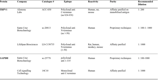
3.3.3 Antibodies ... 62
3.3.4 Ethical Approval ... 62
3.3.5 Cell Processing ... 64
3.3.6 Protein Assay ... 65
3.3.7 Blocking Solutions Optimisation ... 65
3.3.8 Protein Quantity Optimisation ... 66
3.3.9 Western Blotting (Optimised Protocol) ... 66
3.4 Results ... 69
3.4.1 Optimisation of the Western Blotting Protocol ... 69
3.4.2 Detection of TRPV1 in Human PBMCs using the Optimised Method ... 77
3.5 Discussion ... 78
4. Chapter 4: Development and Optimisation of a Flow Cytometric Method for the Detection of TRPV1 Expression in Human Leukocytes ... 85
4.1 Abstract ... 86
4.2 Introduction ... 87
4.3 Materials and Methods ... 88
4.3.1 Materials ... 88
4.3.2 Methods ... 89
4.4 Results ... 93
4.4.1 Flow Cytometry Optimisation setup ... 93
4.4.2 Fixation and Permeabilisation Optimisation ... 94
4.4.3 Assessment of the Primary Antibodies ... 95
4.4.4 Secondary Antibody Assessment ... 98
4.4.5 Detection of TRPV1 in Human Normal Leukocytes using the Optimised Method ... 101
5.4 Discussion ... 102
5. Chapter 5: TRPV1 Expression in Human Haematological Malignancy Cell Lines ... 109
vii
5.2 Introduction ... 111
5.3 Materials and Methods ... 112
5.3.1 Cells and Cell Culture ... 112
5.3.2 Western Blotting and Flow Cytometry Experiments ... 113
5.3.3 Data Collection and Analysis ... 114
5.4 Results ... 114
5.4.1 TRPV1 Expression in Malignant Haematological Cell lines ... 114
5.4.2 TRPV1 Expression in Other Cell lines: A Control Study ... 117
5.5 Discussion ... 120
6. Chapter 6: TRPV1 Expression in Patients with Haematological Malignancies ... 127
6.1 Abstract ... 128
6.2 Introduction ... 129
6.3 Materials and Methods ... 129
6.4 Results ... 130
6.4.1 General Characteristics of Patients and Controls ... 130
6.4.2 Detection of TRPV1 using Flow cytometry ... 131
6.4.3 TRPV1 Detection Using Western blotting ... 139
6.5 Discussion ... 141
7. Chapter 7: Conclusions and Future Studies ... 148
Appendix I: Preliminary Flow Cytometry Optimisation Using BD FACScalibur™ ... 157
Appendix II: Patients and Control Subjects Consent Forms ... 166
Appendix III: Experiments Sheets and Protocols ... 175
viii
List of Figures
Figure 1-1: Topological model of TRPV1 ... 12
Figure 1-2: TRPV1 signal transduction ... 18
Figure 1-3: TRPV1 status. ... 20
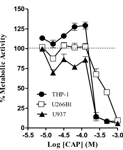
Figure 2-1: Differential response of THP-1, U266B1 and U937 cells to CAP... 45
Figure 2-2: Effect of CAP and the TRPV1 antagonist, SB452533, on the metabolic activity (resazurin reduction) of THP-1, U266B1 and U937 cells ... 47
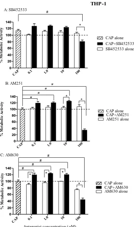
Figure 2-3: Effect of SB452533, AM251 and AM630 on CAP-induced change in metabolic activity (resazurin reduction) in THP-1 cells ... 50
Figure 2-4: Effect of SB452533, AM251 and AM630 on CAP-induced metabolic activity (resazurin reduction) of U266B1 cells ... 51
Figure 2-5: Effect of SB452533, AM251 and AM630 on CAP-induced metabolic activity (resazurin reduction) of U937 cells ... 52
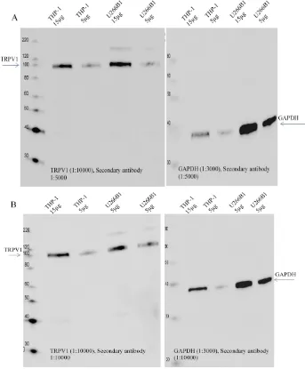
Figure 3-1: TRPV1 detection attempt using the Biotin-Streptavidin detection system in THP-1 cells ... 70
Figure 3-2: ECL detection method with secondary antibody dilution study, 1:5000 vs. 1:10000 of Santa Cruz Biotechnology in THP-1 cells ... 70
Figure 3-3: Protein quantity study (10, 20 and 30 µg) of THP-1 cell lysate to detect TRPV1 with Santa Cruz anti-TRPV1 using the ECLmethod ... 71
Figure 3-4: TRPV1 detection using Alomone Labs anti-TRPV1 antibody ... 72
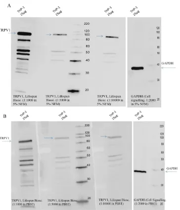
Figure 3-5: Detecting TRPV1 using LifeSpan Biosciences antibody ... 73
Figure 3-6: Titration & blocking studies for LifeSpan Biosciences Anti-TRPV1.. .. 74
Figure 3-7: Secondary antibody (Santa Cruz Biotechnology) titration study. ... 75
Figure 3-8: Secondary antibody (Cell Signalling Technology) dilution Study. ... 76
Figure 3-9: TRPV1 detected in normal human PBMCs protein. ... 77
Figure 4-1: Example of optimised Attune® Cytometer settings using AbC™ beads. ... 94
Figure 4-2: FSC and SSC electronic optimisation for fixed/ permeabilised cells using the Attune® Flow Cytometer ... 94
Figure 4-3: Isotype control overlapping with TRPV1 signal in human leukocyte. ... 95
Figure 4-4: Santa Cruz Biotechnology anti-TRPV1 blocking optimisation using flow cytometry... 96
Figure 4-5: Comparison between two isotype controls vs. Santa Cruz Biotechnology anti-TRPV1 ... 97
Figure 4-6: Alomone Labs anti-TRPV1 signal ... 97
Figure 4-7: Blocking step optimisation for anti-TRPV1 (LifeSpan Biosciences, USA) signal ... 99
Figure 4-8: Dilution study of LifeSpan Biosciences anti-TRPV1 ... 99
Figure 4-9: Secondary antibody titration for FITC-goat anti rabbit (Santa Cruz Biotechnology) using the Attune® Cytometer ... 100
ix
Figure 5-1: TRPV1 expression was detected in THP-1 cells using Western blotting
and flow cytometry ... 115
Figure 5-2: TRPV1 expression was detected in U266B1 cells ... 115
Figure 5-3: TRPV1 expression was detected in U937 lymphoma cells ... 116
Figure 5-4: Relative expression of TRPV1 in three haematological malignant cell lines ... 116
Figure 5-5: TRPV1 expression in TRPV1-transfected HEK293 cells (tetracycline (tet) on/off) by flow cytometry ... 117
Figure 5-6: TRPV1 expression in TRPV1-transfected HEK293 cells (tetracycline off) by Western blotting. ... 118
Figure 5-7: TRPV1 expression in untransfected HEK293 cells by Western blotting. ... 118
Figure 5-8: TRPV1 expression in HEK293 cells was confirmed by flow cytometry. ... 118
Figure 5-9: TRPV1 was detected in RAW264.7 cells. ... 119
Figure 6-1: TRPV1 expression in patients with haematological malignancies. ... 132
Figure 6-2: TRPV1 expression for all patients with haematological malignancies vs. controls ... 133
Figure 6-3: TRPV1 expression in patients with B-NHL. ... 133
Figure 6-4: TRPV1 expression in patients with MM ... 137
Figure 6-5: TRPV1 expression in patients with other blood cancers... 138
Figure 6-6: TRPV1 MFI ratio between males and females ... 138
x
List of Tables
Table 1-1: Overview of TRP family subgroups ... 8
Table 1-2: Summary of studies investigating the role of TRPV1 in systemic diseases and conditions ... 22
Table 1-3: Some non-haematological cell lines that undergo vanilloid-induced cell death ... 24
Table 1-4: Summary of TRPV1-expression and function in malignant haematological cell lines ... 30
Table 2-1: Characteristics of the studied haematological malignant cell lines ... 41
Table 2-2: EC50/IC50 for CAP-induced metabolic activity in THP-1, U266B1 and U937 cells ... 46
Table 3-1: Characteristics of the primary rabbit anti-TRPV1 and anti-GAPDH antibodies ... 63
Table 3-2: Characteristics of secondary antibodies used in Western blot ... 64
Table 3-3: Western blotting protocols tested to detect TRPV1 in human malignant cell lines and PBMCs ... 67
Table 3-4: Summary of some studies detecting TRPV1 by Western blot ... 80
Table 4-1: Characteristics of the isotype controls used in the study ... 89
Table 4-2: Mean-MFI values for different normal leukocytes subpopulations ... 102
Table 4-3: Some published studies on TRPV1 expression detected by flow cytometry ... 103
Table 5-1: Comparison of TRPV1 Mean-MFI in THP-1, U266B1 and U937 cell lines and normal leukocytes... 116
Table 5-2: TRPV1 MFI in some cell lines compared to normal leukocytes from healthy control ... 119
Table 6-1: General characteristics of patients with haematological malignancies .. 131
Table 6-2: Characteristics of B-NHL patients compared to control subjects used for analysis of TRPV1 expression by flow cytometry ... 134
Table 6-3: Characteristics of MM patients compared to control group subjects used for analysis of TRPV1 expression by flow cytometry ... 135
Table 6-4: Other haematological malignant cancers patients compared to control subjects used for analysis of TRPV1 expression by flow cytometry ... 136
Table 6-5: Patients with detected TRPV1 bands on Western Blotting ... 140
xi
List of Abbreviations
A-425619 1-isoquinolin-5-yl-3-(4-trifluoromethyl-benzyl)urea A-778317 1-((R)-5-tert-butyl-indan-1-yl)-3-isoquinolin-5-yl-urea
AA Arachidonic acid
ACA N-(p-amylcinnamoyl)anthranilic acid
ADP Adenosine diphosphate
AEA N-arachidonoylethanolamine (anandamide)
ALL Acute lymphocytic leukaemia
AMG628 (R)-N-(4-(6-(4-(1-(4-fluorophenyl)ethyl)piperazin-1-yl)pyrimidin-4-yloxy)benzo[d]thiazol-2-yl)acetamide
AML Acute Monocytic Leukaemia
AMP Adenosine monophosphate
AMPK AMP-activated protein kinase
AP-1 Activator protein-1
ATL Adult T-cell leukaemia
ATP Adenosine triphosphate
Bax Bcl-2–associated X protein
Bcl-2 B-cell lymphoma 2
Bcl-xL B-cell lymphoma-extra large
BCTC N-(4-tertiarybutylphenyl)-4-(3-chloropyridin-2-yl) tetrahydropyrazine-1(2H)-carbox-amide.
BDNF Brain-derived neurotrophic factor bFGF Basic fibroblast growth factor
BTP2 4-methy-4′-[3,5-bis(trifluoromethyl)-1H-pyrazol-1-yl]-1,2,3-thiadiazole-5-carboxanilide
C- terminus Carboxy terminus
CaM Calmodulin
CaMKII Ca2+-calmodulin-dependent kinase II
cAMP Cyclic adenosine monophosphate
CAP Capsaicin
CAZ Capsazepine
CD Cluster of Differentiation
CDK Cyclin-dependent kinase
CGRP Calcitonin gene-related peptide
CLL/SLL Chronic lymphocytic leukaemia/ small lymphocytic lymphoma
CML Chronic myelogenous leukaemia
CMML Chronic Myelomonocytic Leukaemia
CNS Central Nervous System
COPD Chronic Obstructive pulmonary disease
CRP C- reactive protein
DAG Diacylglycerol
DC Dendritic cell
DMEM Dulbecco's Modified Eagle Medium
DMSO Dimethyl sulfoxide
DPTHF Diphenyltetrahydrofuran
DPBA Diphenylboronic anhydride
xii
ECL Enhanced Chemiluminescence
eIF2α Eukaryotic translation initiation factor 2, subunit 1 (α, 35kDa)
eIF2αK3 Eukaryotic translation initiation factor-2α kinase-3
EIPA Ethylisopropyl amiloride
ER Endoplasmic reticulum
ET Essential thrombocythaemia
ETC Electron transport chain
FBS Foetal Bovine Serum
FITC Fluorescein isothiocyanate
FSC Forward Scatter
GADD153 Growth arrest- and DNA damage-inducible transcript 3 GAPDH Glyceraldehyde 3-phosphate dehydrogenase
GARD Gastroesophegal reflux disease
GM-CSF Granulocyte-macrophage colony stimulating factor GTPγS Guanosine gamma thiophosphate
HCL Hairy-Cell Leukaemia
HEK293 Human Embryonic Kidney cells
HEPES 4-(2-hydroxyethyl)-1-piperazineethanesulfonic acid HL-60 Human myelocytic leukaemia
HRP Horseradish Peroxidase
ICDA Inhibitor of caspase activated DNase
IDN Identification number
IFN-γ Interferon-gamma
IL-1/ 2/ 6 Interleukin-1/ 2/ 6 ILD Interstitial lung disease Ins(1,4,5)P3 Inositol 1,4,5-trisphosphate
IP3 Inositol triphosphate
JNJ17203212
4-(3-trifluoromethyl-pyridin-2-yl)-piperazine-1-carboxylic acid (5-trifluoromethyl-pyridin-2-yl)-amide JYL1421
N-(4-tert-butylbenzyl)-N′-[3-fluoro-4-(methylsulfonylamino)benzyl]thiourea
KB-R7943 2-[2-[4-(4-nitrobenzyloxy)phenyl]ethyl]isothiourea methanesulfonate
LGH Launceston General Hospital
LNCaP Androgen-dependent prostate cancer cells MAPK Mitogen-activated protein (MAP) kinases MFI Median Fluorescence intensity
ML-9 1-(5-chloronaphtalene-1-sulphonyl) homopiperazine
MM Multiple myeloma
MPD Myeloproliferative Disorder
MTT 3-(4,5-dimethylthia-zol-2-yl)-2,5-diphenyltetrazolium bromide
M.W Molecular weight
N- terminus Amino- terminus
NADA N-arachidonoyldopamine
xiii
NF-κB Nuclear factor kappa-light-chain-enhancer of activated B cells
NFM Non-Fat Milk
NGF Nerve growth factor
NHBE Normal human bronchial epithelial
NHL Non-Hodgkin’s Lymphoma
OAG 1-oleoyl-2-acetyl-sn-glycerol
p21 WAF1/CIP1 Cyclin-dependent kinase inhibitor PBMC Peripheral blood mononuclear cell
PBS Phosphate buffer saline
PBSA Phosphate buffer saline-sodium azide PBST Phosphate buffer saline tween- 20
PC3 Androgen-independent prostate cancer cells
PHB2 Prohibitin
PI3K Phosphatidylinositol 3-kinase
PIP2 Phosphatydyl-inositol-4,5-bisphosphate
PKA Protein kinases A
PKC Protein kinases C
PLC Phospholipase C
PMA Phorbol 12-myristate 13-acetate
PMT Photo multiplier tube
PgE2 Prostaglandin E2
PP Peyer’s patch
PTCL-NOS Peripheral T-cell lymphoma/ not otherwise specified PVDF Polyvinylidene difluoride
Q-PCR Quantitative real-time PCR RCF Relative Centrifugal Force
RHC80267 1,6-di[O-(carbamoyl)cyclohexanone oxime]hexane
ROS Reactive oxygen species
RT-4 Human well-differentiated low-grade papillary RT-PCR Reverse transcription polymerase chain reaction
RTX Resiniferatoxin
SB366791 N-(3-methoxyphenyl)-4-chlorocinnamide
SDS Sodium Dodecyl Sulphate
SSC Side Scatter
STAT Signal transducer and activator of transcription
TBMC 6-tert-butyl-m-cresol
THC Δ9-tetrahydrocannabinol TNF-α tumour necrosis factor-alpha
TRAIL Tumour necrosis factor-related apoptosis-inducing ligand
TRIM 1-(2-(trifluoromethyl)phenyl) imidazole
trkA Tyrosine kinase A
TRP Transient receptor potential
TRPV1 Transient receptor potential vanilloid type 1 URB597 3′-carbamoylbiphenyl-3-yl cyclohexylcarbamate.
UTAS University of Tasmania
Va
xiv
WS-12 2-isopropyl-5-methyl-cyclohexanecarboxylic acid (4-methoxy-phenyl)-amide
2-APB 2-aminoethoxydiphenyl borate 4α-PDD 4α-phorbol 12,13-didecanoate
5-HT Serotonin
5(6)-EET 5’,6’-epoxyeicosatrienoic acid
5-(S)-HETE 5-(S)-hydroxyeicosatetraenoic acid 12-(S)-HPETE and
15-(S)-HPETE
12- and 15-(S)-hydroperoxyeicosatetraenoic acids
xv
Presentations at Conferences during PhD Candidature
Conference Presentations
Omari, S and Adams, MJ and Khalafallah AA and Mohamed, M and Geraghty, DP, TRPV1 expression in haematological malignancies, Annual Combined ASM of APSA and ASCEPT, 1 - 4 December, Melbourne, Australia (2013) [Conference Extract].
Omari, S and Geraghty, DP and Kunde, DA and Adams, MJ, Inhibition of Human Haematological Malignant Cell Lines by Capsaicin is not TRPV1-Mediated, Annual Combined ASM of the HSANZ/ANZBT/ASTH and the APSTH, 28 – 31 October, Melbourne, Australia (2012) [Conference Extract].
Omari, S and Kunde, DA and Adams MJ and Geraghty DP, Inhibition of human haematological malignant cell line growth by capsaicin is not TRPV1-mediated, Annual Combined ASM of APSA and ASCEPT, 2-5 December, Sydney, Australia (2012) [Conference Extract].
Presentations related to but not directly arising from this thesis Conference Presentation
1
Abstract
Transient receptor potential vanilloid-1 (TRPV1) is a member of the TRP family of channels that are responsible for nociceptive, thermal and mechanical sensations. It is primarily associated with neuronal cells, but has been detected in different non-neuronal cells, including leukocytes. Capsaicin (CAP), the active ingredient of hot chilli peppers, is one of a number of related endogenous and plant-derived compounds (broadly termed ‘vanilloid-like agents’) that have been shown to induce apoptosis and inhibit cell proliferation in some cancer cells, through both TRPV1-dependent and -inTRPV1-dependent mechanisms. The expression and function of TRPV1 in haematological malignancies however, has not been extensively investigated. Specific targeting by vanilloid-like agents toward TRPV1 on cancerous cells in patients with haematological malignancies may represent a novel therapeutic approach to treating these diseases.
2
The thesis begins with a comprehensive review and discussion on TRPV1 structure and function, as well as its expression and role in health and disease. In particular, there is a focus on the role of TRPV1 in cancer, including haematological malignancies (Chapter 1).
In Chapter 2, the effect of CAP on the metabolic activity of three malignant haematological cell lines, THP-1, U266B1 and U937, was investigated. Metabolic activity assays were performed using the alamarBlue® method. CAP induced cytotoxicity in all three cell lines in a concentration-dependent manner. A biphasic effect on metabolic activity was observed on THP-1 cells [EC50, IC50 (95% CI) = 32.9 (19.9-54.3), 219 (144-246) µM]. U266B1 cells were more resistant to CAP-induced death than THP-1 and U937 cells. TRPV1 and CB1 antagonists (SB452533 and AM251, respectively) suppressed the CAP-induced increase in THP-1 cell metabolic activity (P<0.001). These experiments suggest that CAP inhibits the metabolic activity of malignant haematological cells through a non-TRPV1-dependent mechanism.
3
protocols were then used to investigate the expression of TRPV1 in human malignant haematological cell lines (Chapter 5) and leukocytes obtained from patients with blood cancers (Chapter 6).
Increased expression of TRPV1 protein was observed in THP-1, U266B1 and U937 cells compared to normal leukocytes. Furthermore, a TRPV1 dimer was detected in U266B1 cells. Interestingly, TRPV1 was detected in non-haematological cell lines that have previously been used as TRPV1-negative cells for Western blotting, including untransfected- and TRPV1-transfected (without tetracycline to switch TRPV1 transcription off) HEK293 and RAW264.7 cells. This latter finding highlights the need for appropriate negative (and positive) controls in both flow cytometric and Western blotting studies of TRPV1.
Expression of TRPV1 in leukocytes obtained from patients with a range of haematological malignancies, including multiple myeloma (MM) and B-cell non-Hodgkin’s Lymphoma (B-NHL), was then investigated (Chapter 6). TRPV1 expression was detected in all patients and controls using flow cytometry, but not Western blotting. Using flow cytometry, a sub-group of patients (4/49=8.2%, MM=2, B-NHL=2) demonstrated increased expression of TRPV1 relative to the remainder of the cohort. TRPV1 was found to be similar to the control group for 91.8% of all patients. There were no significant differences in TRPV1 expression (assessed using flow cytometry) between patients with MM and B-NHL, or between
4
although C-reactive protein was elevated (≥5 mg/L) in 25% of all patients, it was not associated with higher TRPV1 expression. These results indicate that TRPV1 expression in leukocytes is relatively increased in a small subset of patients with blood cancers, and is not associated with inflammation. Furthermore, some patients may have a unique isoform of TRPV1 that warrants further investigation.
5
6
1.1
Introduction
Natural compounds have been used over decades for treating various diseases and for relieving symptoms, although the mechanisms explaining their effects have largely not been described. Capsaicin (CAP), the active ingredient of ‘hot chilli peppers’, has been proposed as an anticancer agent. It has been shown to induce apoptosis and inhibit cell proliferation in some cancer cells, including haematological malignancies (Ito et al. 2004; Zhang et al. 2003a).
The CAP receptor, transient receptor potential vanilloid-1 (TRPV1), originally described as an afferent neuron nociceptive receptor, is expressed in neuronal and non-neuronal cells (Cortright and Szallasi 2004; Nagy et al. 2004). However, there are few published studies of TRPV1 expression in haematological malignancies (Bhutani et al. 2007; Gertsch et al. 2002). The focus of this thesis was to therefore investigate the expression and role of TRPV1 in malignant haematological cell lines, as well as in patients with blood cancers and compared to normal subjects.
1.2
Overview of TRP Channels
TRP channels and more specifically, TRPV1, are not only important in many sensory systems, they are crucial components for the function of non-sensory neurons, such as in epithelial, blood and smooth muscle cells (Minke 2006).
7
intracellular and extracellular messengers, exogenous chemicals, temperature, and mechanical stress (Table 1-1).
TRP channels are involved in multiple functions, including nociception, temperature, mechanical sensations, renal Ca2+/Mg2+ handling, lysosomal function, cardiovascular regulation, control of cell growth and proliferation, perception of pungent compounds (e.g., chilli, mustard, garlic), taste perception, smooth muscle tone and blood pressure regulation (Christensen and Corey 2007; Clapham et al. 2003; Ramsey et al. 2006). TRP channels consist of six transmembrane domains segments (S1–S6) and a pore region between S5 and S6, with both the carboxy (C-) and amino (N-) termini located intracellularly (Clapham et al. 2005; Minke 2006). Despite the structural similarities between the TRPs and the voltage-gated K+ channels, these group of channels are quite different (Clapham et al. 2005). At least 28 different TRP subunit genes have been identified in mammals, comprising six subfamilies, namely the classical or canonical TRPs, TRPV (vanilloid), TRPM (melastatin), TRPA (ankyrin), TRPP (polycystin), and TRPML (mucolipin). Each subfamily comprises several channel subtypes, which differ in their selectivity for Ca2+, activation mechanisms, and interacting proteins (see Table 1-1) (Clapham 2007; Holzer 2008; Nilius 2007).
8
TRP PCa/PNa Agonists/ Activators Role Antagonists References
TRPC1 - Depletion of int. Ca2+ stores, PLC, DAG, OAG, orexin-A, TRK-PLCγBDNF, bFGF, thapsigargin,carbachol, Gq/11-coupled
receptors, membrane stretch, int. NO-mediated cysteine S-nitrosylation
Vertebrate mechanosensitive Ca2+ permeable channel that is gated by tension developed in the lipid bilayer
2APB, Gd3+, La3+, SKF96365, Ca2+ -CaM, GsMTx-4
(Abramowitz and Birnbaumer 2009; Alexander et al. 2007; Bollimuntha et al. 2006; Maroto et al. 2005; Venkatachalam et al. 2003)
TRPC2 2.7 DAG, Ca2+ stores depletion, AA Pheromone and odours sensing in mice
Unknown (Abramowitz and Birnbaumer
2009; Gailly 2012; Zufall 2005) TRPC3 1.6 DAG, OAG, orexin-A, IP3, Ca2+ store
depletion,
TRK-PLCγ, BDNF, PLC, Gq/11-coupled
receptors, Ins(1,4,5)P3, Ca 2+
, PIP2
BDNF mediated neuronal
differentiation, vasomotor function, resistance vessel, airway regulator, antigen(Ag) stimulation lymphocytes
Gd3+, La3+, Ni2+, 2-APB, SKF96365, KB-R7943, BTP2
(Abramowitz and Birnbaumer 2009; Alexander et al. 2007; He et al. 2005; Hofmann et al. 1999; Kiyonaka et al. 2009; Numaga et al. 2010)
TRPC4 1.1 GTPγS, La3+ (at µM range), Gq/11-coupled
receptors, ext.H+, thapsigargin, F2v peptide and calmidazolium by antagonism of Ca2+ -CaM, NO-mediated cysteine S-nitrosylation,
Vasoregulation, lung microvascular permeability, GABAergic input lateral geniculate nucleus
Pyr3, BTP2, La3+(high
concentrations), 2-APB, SKF96365
(Abramowitz and Birnbaumer 2009; Alexander et al. 2007; Jung et al. 2011; Nilius et al. 2005) TRPC5 9 GTPγS, Gq/11-coupled receptors, Ins(1,4,5)P3,
adenophostin A, thapsigargin, La3+, Gd3+, high int.Ca2+, lysophosphatidyl choline, ext.H+, Riluzole, lead, genistein, rosiglitazone
Growth cone morphology, brain development, innate fear
La3+ (high concentrations, enhanced at low), 2-APB, SKF96365, KB-R7943, BTP2, flufenamic acid, chlorpromazine
(Abramowitz and Birnbaumer 2009; Alexander et al. 2007; Gross et al. 2009; He et al. 2005; Majeed et al. 2011; Riccio et al. 2009; Richter et al. 2014; Semtner et al. 2007; Sukumar and Beech 2010; Wong et al. 2010; Xu et al. 2005b) TRPC6 5 DAG, AlF4-, Gq/11-coupled receptors,
membrane stretch, GTPγS, 20-HETE, OAG (independent of PKC) and inhibition of DAG lipase with RHC80267, flufenamate,
hyperforin
Vasomotor and cardiac function, airway resistance, platelet aggregation.
2-APB, BTP2, La3+ (IC50 = 6µM),
Gd3+, amiloride, SKF96365, ACA, KB-R7943, ML-9 (independent of MLCK), ext.H+,GsMTx-4
(Abramowitz and Birnbaumer 2009; Alexander et al. 2007; Clapham 2003; Nilius et al. 2005; Xu et al. 2005b)
TRPC7 2 DAG, exocytosis, Ca2+ store depletion, Gq/11
-coupled receptors, OAG
Respiratory rhythm activity, conducts mono- and divalent- cations with a preference for divalents
La3+, SKF96365, amiloride, 2-APB (Abramowitz and Birnbaumer 2009; Alexander et al. 2007; Ben-Mabrouk and Tryba)
TRPV1 10 CAP, resiniferatoxin, olvanil, PKC,
depolarisation, heat (≥ 43C), low pH (≤ 5.9), AEA, eicosanoids, 12, 15-(S)-HPETE, 5-(S)-HETE, leukotriene B4, NADA, adenosine and 2-APB
Selective for Ca2+ and Mg2+, inflammation, pancreatitis, sensing hot chilli, pain, noxious temperature, bladder distension and more.
Ruthenium red, 5′-IRT, 6-iodo-nordihydroCAP, BCTC, CAZ, A-425619, A-778317, AMG517, AMG628, SB-705498, JNJ17203212, JYL1421, SB366791, SB452533
[image:24.842.32.823.72.522.2]9 TRPV2 1-3 Heat (>53°C, rodent), 2-APB (rodent), DPBA,
THC, probenecid, cannabidiol, growth factors i.e. IGF-1
Sensing thermal pain, phagocytosis, axonoutgrowth regulation
Ruthenium, SKF96365, amiloride, TRIM, La3+
(Caterina et al. 1999; Hu et al. 2004; Link et al. ; Shibasaki et al.) TRPV3 2.6 Heat (> 23– 39°C), 2-APB, camphor, TBMC,
carvacrol, eugenol, thymol, menthol, incensole acetate, DPBA
mechanosensor in vascular smooth muscle cells
Ruthenium red (<1 mM), DPTHF (Beech et al. 2004; Hu et al. 2004; Moqrich et al. 2005; Smith et al. 2002)
TRPV4 6
Heat (> 24°- 32°C), 4α-PDD,
bisandrographolide A, anandamide, AA, 5(6)-EET, cell swelling,
Osmosensing, warm sensing,
nociception, pressure sensing in DRG
Ruthenium red (voltage-dependent), La3+,Gd3+
(Clapham 2003; Harteneck and Schultz 2007; Kanzaki et al. 1999; Nilius et al. 2004; Vriens et al. 2004; Watanabe et al. 2002) TRPV5 >100 Low [Ca2+]i, hyperpolarization Ca2+ reabsorption in the kidney Ruthenium red, econazole,
miconazole, Pb2+ = Cu2+= Gd3+> Cd2+> Zn2+> La3+> Co2+> Fe2+, Mg2+
(Alexander et al. 2007; den Dekker et al. ; Nilius and Voets 2005) TRPV6 >100 Low [Ca2+]i, hyperpolarization, 2-APB Ca
2+
reabsorption in the intestine Ruthenium, Cd2+, Mg2+, La3+ (Alexander et al. 2007; den Dekker et al. ; Nilius and Voets 2005) TRPA1 0.8–1.4 Cooling (< 17°C), isothiocyanates, THC,
allicin, menthol, thymol, cinnamaldehyde (100µM), carvacrol, formalin, 4-HNE, methylparaben, URB597, cyclopentone, prostaglandins, 1,4-dihydropyridines, isoflurane, desflurane, propofol, etomidate, Camphor (100µM)
Mechanically gated transduction channel required for the auditory response in mammals. Reception of pungent painful stimuli (mustard oil, wasabi, horse radish, garlic and onions)
Camphor (1mM), cinnamaldehyde (3mM), ruthenium red, gentamicin, Gd3+ , amiloride, menthol (mouse), HC-030031, blockers of the mechanosensory channels
(Alexander et al. 2007; Alpizar et al. 2013; Corey et al. 2004; Macpherson et al. 2005; Sukharev and Corey 2004)
TRPP2 1-5 Mechanical stress, int.Ca2+, constitutive activity suppressed by co-expression of TRPP1
Signalling complex with TRPP1, cilia movement, development of heart, skeletal muscle and kidney, fertility.
La3+ , Gd3+, amiloride (Delmas 2004; Delmas et al. 2004; Giamarchi et al. 2006; Volk et al. 2003)
TRPP3 4 Int.Ca2+, low constitutive activity, depolarisation, cell swelling
Hair cell, Kidney, retinal developments
Phenamil, benzamil, EIPA, amiloride, La3+, Gd3+, flufenamate
(Delmas 2005; Shimizu et al. 2009) TRPP5 1-5 Int.Ca2+ Cell proliferation and apoptosis,
Ca2+homeostasis, spermatogenesis
Unknown (Chen et al. 2008; Guo et al. 2000; Xiao et al. 2010)
TRPML1 ~ 1 TRPML1Va: constitutively active, ext. high H+ (equivalent to Intralysosomal high H+), Ca2+
Role in late endosomes pathway that is necessary for lysosome formation and recycling
Amiloride, low pH (LaPlante et al. 2002; Piper and Luzio 2004; Raychowdhury et al. 2004; Xu et al. 2007a)
TRPML2 -
TRPML2Va: constitutively active, ext. high H+ (equivalent to intralysosomal high H+), ADPR, oxidative stress
Mediating cation (Ca2+/Fe2+) efflux from endosomes and lysosomes
Cu2+, clotrimazole, flufenamic acid, 2APB, ACA
(Jia et al. 2011; Montell 2005; Moreau et al. 2013; Xu et al. 2007a; Zeng et al. 2012)
TRPML3 - TRPML3va: constitutively active. TRPML3WT: activated by Na+-free ext. solution, depolarization
Hair cell, stereocilia maturation, and int. vesicle transport
TRPML3va & TRPML3WT: Gd3+, ext.acidification (intralysosomal acidification),TRPML3
10 TRPM1
non-selective cation
Translocation, constitutively active Tumour suppressor, sensor of cellular redox status, oxidant stress sensor in immune and glia cells, respiratory bursts in neutrophils
La3+ ,Gd3+ (Alexander et al. 2007; Duncan et al. 2001; Hara et al. 2002)
TRPM2 0.5–1.6 Oxidative and nitrosative stress, NAD, radicals, oxidative, int. ADPR, cADPR, int. Ca2+, AA, heat ~ 35°C
Regulates endothelial barrier function, cell proliferation
Clotrimazole, miconazole, econazole, flufenamic acid, ACA, 2-APB, ADPR and cADPR blocked by AMP and 8-bromo-cADPR
(Alexander et al. 2007; Clapham 2003; Fonfria et al. 2004; Hecquet et al. ; Perraud et al. 2005) TRPM3 1–2 Cell swelling, Ca2+store depletion,
pregnenolone sulphate, nifedipine, depolarisation, D- erythrosphingosine, dihydrosphingosine, eicosanoids
Sphingolipid signalling, renal volume regulation (osmosensor)
La3+,Gd3+, 2-APB, int.Mg2+, ext.Na+ (TRPM3α2 only)
(Alexander et al. 2007; Grimm et al. 2003; Grimm et al. 2005; Harteneck and Schultz 2007) TRPM4 - Int.Ca2+, ATP, decavanadate, depolarization,
heat, PIP2, BTP2
Negative-feedback regulation of Ca2+ fluctuation, release of IL-2 from T-cell. Impermeable to Ca2+
ATP4, ADP, AMP, AMP-PNP, adenosine, int.spermine, flufenamic acid, ext. clotrimazole, 9-phenanthrol
(Alexander et al. 2007; Launay et al. 2004; Ullrich et al. 2005) TRPM5 - Gq/11-coupled receptors, Ins(1,4,5)P3, int.Ca2+,
membrane depolarization, heat, PIP2
Taste receptor of the tongue;
transduction of sweet, amino acid and bitter stimuli, impermeable to Ca2+
Int. spermine, flufenamic acid, ext. Protons
(Alexander et al. 2007; Ullrich et al. 2005; Zhang et al. 2003b)
TRPM6 - Reduction of Mg2+, ext. H+, 2-APB High permeability for Mg2+, influx pathway for divalent cations
Ruthenium red, inward current by monovalent cations blocked by Ca2+ and Mg2+
(Alexander et al. 2007; Voets et al. 2004b)
TRPM7 3 Reduction of Mg2+, G-proteins, PKA, int.ATP, PIP2, ext. H+
Mg2+ and Ca2+ entry & trace metal ion uptake. Role in neuronal cell death
Spermine (permeant blocker), carvacrol, La3+, Mg2+, 2-APB
(Aarts et al. 2003; Alexander et al. 2007; Monteilh-Zoller et al. 2003; Nadler et al. 2001)
TRPM8 1-3 menthol, depolarisation, Ca2+, cooling (< 22– 26°C), PIP2; WS-12, menthol, icilin
Voltage-dependent channel, pain and cold sensor
CAZ, BCTC, and thio-BCTC, low pH, La3+, clotrimazole, 2-APB, ACA, NADA, anandamide, linoleic acid, cannabinoids
(Alexander et al. 2007; Behrendt et al. 2004; McKemy et al. 2002; Peier et al. 2002; Reid 2005; Voets et al. 2004a)
11
which is distinct from taste and smell (Bandell et al. 2007). TRP channels are opened or closed by conformational changes in the channel protein (Bandell et al. 2007; Dhaka et al. 2006). The ion selectivity differs markedly among the same family of TRP channels, as most of the channels are non-selective cation channels, which is particularly true for TRPV1 although with a preference for Ca2+ (Julius and Basbaum 2001).
1.3
Transient Receptor Potential Vanilloid 1 (TRPV1)
Interest in TRPV1 began when pharmacological aspects of CAP were first recognised. CAP, which is derived from Capsicum spp., is responsible for the sensation of ‘hot’ and ‘burning’ when exposed to chilli peppers. It acts specifically on nociceptive afferent neurons (Jancso´ 1960). Researchers found that the selectivity of CAP’s action on afferent neurons could only be explained by an action on specific CAP receptors which led to the revelation of TRPV1 as a CAP-specific receptor (Caterina et al. 1997; Nagy et al. 2004; Szolcsanyi et al. 1975).
1.3.1 Structure
12
generate the different sensitivity and regulate the function of TRPV1 (Phelps et al. 2010). The intracellular domain of TRPV1 contains multiple binding sites for phosphatydyl-inositol-4,5-bisphosphate (PIP2) on the N- and C-termini (Grycova et al. 2012), and two other sites for a protein-kinase C and Ca2+/CaM-dependent protein kinase II (CAM-kinase II) which are important proteins for channel regulation ( see
Figure 1-1) (Ferrer-Montiel et al. 2004; Jeske et al. 2009).
1.3.2 Activation of TRPV1
The initial detection of noxious signals occurs predominantly at the peripheral terminals of primary afferent neurons, called polymodal nociceptive neurons (Fields, 1987). TRPV1 has been shown to be the polymodal nociceptive receptor, that is also allosterically modulated by many proalgesic agents (Holzer 2008).
The TRPV1 channel is activated by vanilloids, which are present in a variety of plants that are normally ingested through the diet. As previously mentioned, they are
C-terminus N-terminus
Figure 1-1: Topological model of TRPV1. TRPV1 subunit exhibiting six putative transmembrane domains, an intracellular N-and C-terminal domains, amino acid residues specific for CAP and proton activation, as well as residues important for phosphorylation by PKA, PKC and CaMKII.Kinases are highlighted at both intra-cytoplasmic regions. The TRP-like, CAM- and PIP2- binding domains are also depicted. Reproduced from Planells-Cases et
13
found in spicy foods, such as hot chilli pepper, and belong to the family of
Solanaceae, genus Capsicum, in which the molecules responsible for the ‘burning’ taste are CAP and other capsaicinoids (Monsereenusorn et al. 1982). In addition to capsaicinoids, TRPV1 channels are activated by a variety of other plant-derived vanilloids including, camphor and resiniferatoxin (RTX), and putative endogenous vanilloids such as the endocannabinoid, anandamide, some lipoxygenase products of arachidonic acid (AA) such as 12-(S)- and 15-(S)-hydroperoxyeicosatetraenoic acid (12S- and 15S-HPETE), and arachidonoyldopamine (NADA) and its congener, N-oleoyldopamine (Chu et al. 2003; Van Der Stelt and Di Marzo 2004; Xu et al. 2005a). Vanilloids bind to intracellular sites on the TRPV1 channel, and the amino acids critical for CAP/RTX binding include Arg-91, Tyr-511, Ser-512, Ile 514, Val-518 and residue 547 (Met in rat, Leu in human), which are part of transmembrane segments 3 and 4 (Jordt and Julius 2002). CAP is a key activator of TRPV1 and acts by lowering the heat threshold required to open the TRPV1 channel (Szallasi and Blumberg 1999). Vanilloid-like agents may affect cellular function through two mechanisms; 1) the interaction with the TRPV1 receptor, or 2) direct interaction with the cell due to their ability to cross the plasma membrane bilayer because of their lipophilicity (Ziglioli et al. 2009).
14
2001) and PAR-2 agonists (Amadesi et al. 2004). NGF appears to act via phospholipase C (PLC) to hydrolyse PIP2, leading to the inhibition of the channel (Chuang et al. 2001). Sustained exposure to agonists increases the Ca2+ permeability of TRPV1 and causes pore-dilation (10.1 A˚ initial to 12.3 A˚ after 3 min of exposure) (Chung et al. 2008). Neurons that express TRPV1 are eventually overloaded by Ca2+, which in conjunction with other factors, can result in mitochondrial swelling, long-lasting defunctionalisation or even degeneration of the neurons (Szoke et al. 2002; Szolcsanyi et al. 1975). In addition, TRPV1 allows protons to enter the cell in an acidic environment, which results in intracellular acidification (Hellwig et al. 2004). Researchers have reported that TRPV1(-/-) null mice were defective in nociceptive, inflammatory and hypothermic responses to vanilloid compounds, supporting the notion that TRPV1 contributes to acute thermal nociception and hyperalgesia after tissue injury (Caterina et al. 2000).
1.3.3 Expression of TRPV1
15
Once believed to be exclusively neuronal, many studies subsequently reported TRPV1 expression in non-neuronal cells in all organs (Cortright and Szallasi 2004; Fernandes et al. 2012), such as urothelium and urinary tract (Avelino and Cruz 2006; Birder et al. 2001); human smooth muscle, keratinocytes (Jaggar et al. 2001); myenteric ganglia, muscle layer, mucosa and epithelial cells of the gastrointestinal tract (Geppetti and Trevisani 2004); mouse epidermis and keratinocytes (Bode et al. 2009); airway epithelial cells (Reilly et al. 2003), human umbilical vein endothelial cells (Himi et al. 2012), leukocytes (Saunders et al. 2009; Saunders et al. 2007) and microglia (Kim et al. 2006b). In addition, elevated TRPV1 expression was identified in some malignancies including, prostatic cancer cell lines, e.g., PC3 and LNCaP (Sánchez et al. 2006; Ziglioli et al. 2009), pancreas (Hartel et al. 2006; Mergler et al. 2012), colon (Domotor et al. 2005) and urinary bladder (Lazzeri et al. 2005). The wide distribution of TRPV1 emphasises its role as a universal sensor of many chemical stimuli.
1.4
TRPV1 and Pain
16 1.4.1 TRPV1 and nociceptive pain
The body’s reaction to nociceptive stimuli, including thermal, mechanical and chemical, is a natural process that minimises the physical harm to the body, and is not considered as a diseased state (Nilius et al. 2007).
1.4.2 Sensitisation and desensitisation of TRPV1 receptors
17
TRPV1 potentiation by proalgesic substances also involves the activation of intracellular signalling pathways (Figure 1-2) (Planells-Cases et al. 2005). Two major mechanisms have been found to be responsible for TRPV1 potentiation that leads to nociceptor sensitisation, (1) chemical modification of the intracellular domains of the channel, which is induced by protein kinases or phosphatases, and/or by hydrolysis of phosphoinositides produces a higher TRPV1 activity, and (2) an increase of TRPV1 receptor expression in peripheral terminals which enhances nociceptor responses. Both mechanisms are believed to occur in parallel (Figure 1-2) (Planells-Cases et al. 2005).
18
phosphatidylinositol 3-kinase (PI3K) dependent pathway (Nilius 2007; Stein et al. 2006; Zhang et al. 2005). Furthermore, the release of neuropeptides such as substance P and CGRP from central and peripheral neurons also have a role in TRPV1 activation (Planells-Cases et al. 2005). Another sensitisation mechanism is TRPV1 pore dilation following prolonged exposure to agonists (Chung et al. 2008). However, repeated or prolonged application of CAP evokes a transient current leading to insensitivity of TRPV1 to subsequent noxious stimuli, the phenomenon of desensitisation (Rohacs et al. 2008). TRPV1 desensitisation protects cells from potential excitotoxicity that arises from excessive activation of the receptor that may lead to Ca2+ overload (Bhave et al. 2002). Although phosphorylation causes sensitisation, dephosphorylation promotes desensitisation of TRPV1, which is a major mechanism of inhibitory regulation by protein phosphatases (Mohapatra and Nau 2005).
Figure 1-2: TRPV1 signal transduction. A) TRPV1 is activated by noxious heat (≥ 43 °C), low pH (≤ 5.9), eicosanoids, anandamide, and CAP. Ligands of guanine nucleotide (G) binding GPCR that activate PLC leading to hydrolysis of PIP2 disinhibit the TRPV1 channels. (B) TRPV1 sensitisation resulting in
neuropathic pain depends on channel phosphorylation by PKC or PKA and is triggered by products of inflammation, including prostaglandin E2 (PgE2),
19
Desensitisation of TRPV1 to CAP is a Ca2+-dependent process (Koplas et al. 1997; Mohapatra and Nau 2003). The Ca2+ sensor CaM activates the protein phosphatase calcineurin leading to TRPV1 desensitisation (Docherty et al. 1996). Calcineurin greatly reduces CAP-induced desensitisation of sensory neurons (Jung et al. 2004). It has been suggested that a rise in cytosolic Ca2+ levels caused by TRPV1 activation results in the activation of Ca2+/CaM-dependent protein phosphatases that mediate channel desensitisation (Docherty et al. 1996). Furthermore, desensitisation of TRPV1 to CAP involves a number of intracellular components including PKA, ATP and CaM (Bhave et al. 2002; Lishko et al. 2007; Mohapatra and Nau 2003). It appears that there is a dynamic balance between phosphorylation/dephosphorylation of TRPV1 by CaMKII and calcineurin, respectively. This balance controls the activation/desensitisation state of the channel by regulating ligand binding (Figure 1-3, A), as CaMKII recovers TRPV1 from the desensitised state (Docherty et al. 1996; Jung et al. 2004; Mohapatra and Nau 2005; Suh and Oh 2005). TRPV1 has five phosphorylation sites for CaMKII, in which Ser 502 and Thr 704 are targeted for phosphorylation (Jung et al. 2004).
20
phosphorylated in the resting state, PKA can phosphorylate TRPV1 more obviously in the desensitised state at Ser116 amino acid (Bhave et al. 2002).
1.5
TRPV1 and Disease
TRPV1 appears to be involved in either the pathogenesis or progress of many diseases and conditions. Here, the role of TRPV1 in different diseases is reviewed.
1.5.1 Neuropathic Pain Syndromes
Neuropathic pain syndromes are a group of heterogeneous conditions caused by a lesion or disease that affects the peripheral or CNS and are often clinically characterised by spontaneous burning pain. In some patients, the nerve lesion enhances molecular dysfunction that evokes pathological activity of nociceptive neurons and thus, hypersensitivity of TRPV1 that causes pain (Baron 2006). The hyperactivity in nociceptors induces secondary changes in processing neurons in the
CAP
Phosphate molecule A
B
Desensitised Open
Closed
Desensitised PLC Activation
TRPV1
21
spinal cord and brain, whereby input from normal mechanical stimuli is sensed as pain.
1.5.2 Neurogenic Inflammation
Neurogenic inflammation is a process where inflammation is provoked by the nervous system, leading to the release of inflammatory mediators (Meggs 1993). The most common features of neurogenic inflammation are redness and warmth (secondary to vasodilation), swelling (secondary to plasma extravasation), and hypersensitivity (Richardson and Vasko 2002). It is triggered by overstimulation of peripheral nociceptor terminals as a consequence of injury, or by non-neurogenic inflammation (Proudfoot et al. 2006). Many studies have reported the role of TRPV1 in the neurogenic inflammatory process. Tissue damage induces inflammation, which is characterised by hypersensitivity to noxious stimuli or even allodynia (pain due to a stimulus which does not normally provoke pain) (Julius and Basbaum 2001). This provokes the release of neurotransmitters such as substance P and CGRP, and in the case of tissue injury, ATP and H+ from damaged cells. This in turn leads to vasodilation, increased capillary permeability at the site of injury and ultimately TRPV1 sensitisation (Nilius 2007).
1.5.3 Systemic Diseases
22
TRPV1 in Systemic Diseases TRPV1 role/effect References
Urinary tract:
Bladder cystitis Hyperalgesia and hyperreflexia, increase anandamides concentration.
(Avelino and Cruz 2006; Dinis et al. 2004; Nilius et al. 2007)
Painful bladder syndromes. Inactivation of bladder nociceptors i.e. treatment approach (Habler et al. 1990; Sengupta and Gebhart 1994)
Urinary incontinence due to bladder overactivity caused by spinal cord lesions
Vanilloids suppresses involuntary bladder contractions leading i.e. treatment approach
(de Groat 1997)
End-stage kidney disease Activation of the channel leads to PBMC death (Saunders et al. 2009)
Gastrointestinal tract:
Gastrointestinal inflammation Aids in the mechanism of intestinal inflammation (Massa et al. 2005; Sanger 2007)
Erosive esophagitis Increase TRPV1 expression (Matthews et al. 2004)
Functional dyspepsia CAP therapy (Allescher 2006; Rosch et al. 2006)
Inflammatory bowel disease Regulates neurogenic inflammation and protect from colon cancer.
(Vinuesa et al. 2012)
Cardiovascular system:
Salt-sensitive hypertension Prevention action through vasodilatory and natriuretic–diuretic actions
(Inoue et al. 2006; Watanabe et al. 2009)
Depressor effect on cardiac tissue
Myocardial ischemia
Changes in heart rate and contractility
Protect the heart through the release of substance P, CGRP and other neurokines
Respiratory tract:
Cough and airway obstruction.
Acid- induced cough in guinea pigs.
Causes cough through endogenous inflammatory mediators such as PgE2, bradykinin, and histamine
(Fox et al. 1996; Jia et al. 2002)
Chronic obstructive pulmonary disease, interstitial lung disease, gastroesophegal reflux disease, upper airway cough syndrome, bronchiectasis and airway infections.
CAP cough hypersensitivity (Higenbottam 2002; Lalloo et al. 1998; Niimi et al. 2004; O'Connell et al. 1996; Plevkova et al. 2006)
Guinea pig and rat ozone-induced airway inflammation and hyperreactivity
Reduction by CAP (Kaneko et al. 1994; Koto et al. 1995)
Airway response to formaldehyde, cigarette smoke in rats CAP decreased airway response to such substances. (Lundberg and Saria 1983; Lundblad and Lundberg 1984; Morris et al. 1999)
Guinea pigs sulphur dioxide-provoked bronchoconstriction Reduction by CAP (Bannenberg et al. 1994; Lalloo et al. 1995) Table 1-2: Summary of studies investigating the role of TRPV1 in systemic diseases and conditions
23
protecting the heart in myocardial ischemia (Inoue et al. 2006), or is directly involved in the inflammatory process (Table 1-2).
1.5.4 Vanilloid-induced Apoptosis and Cancer
Vanilloids, including CAP and their role in tumoral growth and cell transformation have been of great interest over the past few years. It has been shown that vanilloids play a role in cell death (apoptosis) as well as the inhibition of tumour proliferation through TRPV1-dependent and -independent mechanisms (Ziglioli et al. 2009). Several vanilloids have been reported to induce cell death in a variety of cell types (Table 1-3). Apoptosis, defined as a programmed cell death, is an important function in the cell as it controls cell proliferation (Khan et al. 2007). However, the apoptosis pathway is often dysfunctional in tumour cells (Thompson 1995). The mechanisms of apoptosis are potentially active in every cell, either tumoral or normal, though some studies have demonstrated the ability of vanilloids to target apoptosis in tumour cells rather than normal cells (Ito et al. 2004; Macho et al. 1999).
24
Cell type Vanilloid Mechanism of apoptosis and/or associated observation
TRPV1-mediated
References
Glioma cell lines: Ge227, U87,G2258, U251
AEA Unknown Yes (Contassot et al. 2004b)
A172 CAP Down-regulation of Bcl-2 and activation of caspase-3, reduce ROS level, upregulation of DR5
No (Gil and Kang 2008; Kim et al. 2010; Lee et al. 2000)
Rat C6 glioma cells AEA Influx of Ca2+, activation of the ceramide pathway, TRPV1 involve with CB1&2 Partially (Jacobsson et al. 2001)
CAP Peroxynitrite – mediated apoptosis No (Qiao et al. 2005)
U373 (III) CAP Ca2+ influx and p38 MAPK activation Yes (Amantini et al. 2007)
Hepatic cells: Chang liver cell (non-tumour)
AEA Lipid rafts interaction: p38/JNK , ceramide pathways, AP-1, caspase3 activity No (Giuliano et al. 2006) HepG2 ( hepatoma) CAP Mediated by NADPH oxidase-mediated increase in ROS or by activation of the
PLC-dependent [Ca2+ ]i release pathway
No (Huang et al. 2009; Kim et al. 2005; Lee et al. 2004; Reilly et al. 2003)
Bronchiolar cells: BEAS-2B, BEAS-2B (TRPV1+/+), NHBE, A549
CAP Unknown Yes (Reilly et al. 2005; Reilly et al.
2003; Thomas et al. 2007) Nonivamide
RTX, AEA
Activation of ER-bound TRPV1 stimulates GADD153 expression via EIF2αK3/ EIF2α pathway
Neuroblastoma: CHP100 SHSY-5Y
AEA CAP
Increase [Ca2+ ]i , mitochondrial uncoupling, and cytochrome c release
Inhibited protein synthesis IC50=60mM, induced DNA strand breaks
Yes Unknown
(Maccarrone et al. 2000) (Richeux et al. 1999)
Squamous cell carcinoma:
COLO16, SRB-12
CAP, RTX Rapid induction of hydroperoxide generation, inhibition of mitochondrial respiration
Unknown (Hail Jr and Lotan 2002)
Embryonic kidney cells:
TRPV1-HEK293 CAP Increase in [Ca2+ ]i , apoptosis mechanism unknown Unknown (Grant et al. 2002) Cervical cancer: CC299,
caski, HeLa cells KB cells
AEA CAP
Cleavage of caspace-7
Disruption of mitochondrial potential and activation of caspase 9, 3 and poly-(ADP-ribose) polymerase
Yes Unknown
(Contassot et al. 2004a) (Lin et al. 2013)
Spermatogenic stem: Gc-5spg, Rat Gc-6spg
CAP Caspace-3 activation, DNA fragmentation Unknown (Mizrak et al. 2008)
Urothelial cell cancer: RT4 CAP Fas/CD95 clustering, caspase-3, -8 activation, BID cleavage Yes (Amantini et al. 2009)
Pancreatic cancer: BxPC-3, AsPC-1
CAP Mediates ROS production which leads to the inhibition of mitochondrial complex-I and III activity
Unknown (Pramanik KC 2011; Zhang et al. 2008)
Melanoma: A375 ( mouse), A375-S2
B16-F10 (murine)
CAP Inhibition of NADH oxidase, high nitric oxide, down- regulate the ICDA Down-regulation the Bcl-2 and caspase-3 activation
Unknown (Gong et al. 2005; Jun et al. 2007; Kim 2012; Morre et al. 1996)
Table 1-3: Some non-haematological cell lines that undergo vanilloid-induced cell death
25
tumoral cells with CAP, which antagonises coenzyme Q, inhibits the run of the ETC from complex I to complex II and produces more reactive oxygen species (ROS) in tumour cells than in normal cells (Ziglioli et al. 2009).
1.6
TRPV1 Expression and Function in Immune System Cells
Few studies have investigated TRPV1 expression in immune cells. TRPV1 mRNA has been detected in the peripheral blood mononuclear cells (PBMCs) of rats (Schumacher et al. 2000), and humans using RT-PCR (Saunders et al. 2007). In the next few pages, TRPV1 expression and role in the leukocytes have been extensively reviewed.
1.6.1 Lymphocytes
26
production of various cytokines (Inada et al. 2006). In the transient [Ca2+]i increase phase, the IP3-receptor is activated by IP3 cleaved from PIP2, causing the release of Ca2+ from the ER into the cytoplasm. In the subsequent phase, different ion channels induce Ca2+ influx across the plasma membrane and maintain high [Ca2+]i levels (Inada et al. 2006).
NF-κB is a protein complex that controls the transcription of DNA. It involves cellular response to stimuli such as stress, cytokines, free radicals, bacterial or viral antigens, and inflammation (Gilmore 2006). In inactive lymphocytes, NF-κB dimers are isolated in the cytoplasm in an inactive form by association with a suppressor 1κB subunit, mainly 1κ-Bα. Upon activation, multiple kinases lead to phosphorylation of 1κ-Bα and proteosome-mediated degradation. Thus, active NF-κB complex is released and translocated to the nucleus. In the nucleus, NF-NF-κB activates many genes that participate in inflammation, immune responses and cellular proliferation regulation (Baldwin 1996). It also causes blockade of the nuclear translocation of p65, leading to the inhibition of apoptosis (Han et al. 2002; Singh et al. 1996). CAP has been reported to inhibit the release of the inflammatory cytokines (Han et al. 2002; Sancho et al. 2002), and this effect is TRPV1-mediated (Chen et al. 2003). Two human transcription factors, NF-κB and activator protein 1 (AP-1), which have been implied in many inflammatory diseases and apoptosis were inhibited by CAP and RTX (Han et al. 2002; Portis et al. 2001; Sancho et al. 2002; Singh et al. 1996).
27
lymphocytes. Its gene expression and its receptor have been detected on human lymphocytes (Lai et al. 1998). CAP has been shown to have immunomodulatory effects through its ability to modulate lymphocyte proliferation and immunoglobulin A, E and G production (Nilsson et al. 1991; Takano et al. 2007). In addition, CAP inhibits T-helper cell cytokine production in cultured murine Peyer’s patch (PP) cells, in vitro, whereas oral injection of capsicum extract and CAP enhances the production of T-helper 1 cytokines such as IL-2 and interferon-gamma in response to the mitogen, concanavalin A (Takano et al. 2007). Capsicum extracts and CAP had an opposite effect on immune responses in vitro and ex vivo, indicating the presence of an indirect effect in vivo. Furthermore, direct treatment of PP cells with 3 and 30 μM of CAPreduced cell viability (Takano et al. 2007). TRPV1 has been detected on T-lymphocytes in PP and it mediates CAP-induced T-cell reduction which was partly inhibited with capsazepine. A study found that CAP, if administered subcutaneously to neonatal rats, it would cause a marked reduction in thymus weight and a lack of thymocyte differentiation by triggering apoptosis (Santoni et al. 2000). This indicates a negative effect of CAP on the thymus and lymphocytes which contributes to apoptosis in these cells.
1.6.2 Macrophages
28
suppression in microglia, the tissue macrophages function mainly in the CNS. In addition, membrane depolarisation, the main characteristic of NADPH oxidase activity in phagocytes, was reported to promote TRPV1 activity in microglia (Schilling and Eder 2009a).
1.6.2.1 Dendritic cells
Dendritic cells (DCs) are a set of leukocytes widely distributed in all tissues, especially in those that interface with the external environment. They are potent antigen presenting cells, that have the ability to stimulate T-cells and differentiate B-cells (Satthaporn and Eremin 2001).
29
have shown that human peripheral blood monocytes, immature and mature DCs express functional TRPV1 using flow cytometry, Ca2+-imaging and quantitative real-time PCR (Q-PCR). In addition, TRPV1 expression dramatically increased during cytokine-induced in vitro differentiation of monocytes to immature DCs. CAP has been reported to suppress phagocytosis by a TRPV1-mediated pathway, and did not induce apoptosis in immature DCs. In contrast to mouse DCs, CAP did not also promote DC maturation in the absence of cytokines, in humans (Toth et al. 2009).
1.6.3 Neutrophils
Neutrophils play a central role in the inflammatory response and comprise an essential part of the innate immune system (Wang et al. 2005). An increase in cytosolic [Ca2+]i is attributed to the neutrophils in response to chemoattractants (Krause et al. 1990), and induces synthesis and release of prostaglandins (Krump et al. 1995). CAP affects neutrophil function and increases [Ca2+]i in rat neutrophils by inducing extracellular Ca2+ influx and Ca2+ release from the internal Ca2+ pool (Wang et al. 2005). TRPV1 mRNA has been detected in human and rat neutrophils and in HL-60 (neutrophilic promyelocytic leukaemia) cell line using RT-PCR, however the role of TRPV1 in these cells remains poorly understood (Heiner et al. 2003; Wang et al. 2005).
1.7
The TRPV1: Role in Haematological Malignancies
30
Haematological cancer cell line TRPV1 expression
Vanilloid tested
Vanilloid effect
TRPV1-mediated? References Acute myeloid leukaemia:
Kasumi-1 Unknown CAP Apoptosis Unknown (Ito et al. 2004)
THP-1 Expressed CAP Stimulation at low, lethal at high concentrations Yes (Kunde et al. 2010)
Acute lymphoid leukaemia:
CEM Unknown THC Apoptosis Unknown (Powles et al. 2005)
HPB-ALL Unknown CAP Apoptosis Unknown (Zhang et al. 2003a)
Jurkat T-cell line Expressed Arvanil, CAP
Apoptosis
Immunomodulatory and anti-inflammatory, apoptosis is preceded by a reduction of ΔΨm and increase in ROS
No Unknown
(Dou et al. 2011; Gertsch et al. 2002; Macho et al. 1999; Sancho et al. 2003; Zhang et al. 2003a)
Human promyelocytic leukaemia:
HL-60
Unknown
CAP >50mM induced Apoptosis, cytotoxicity
Unknown
(Tsou et al. 2006)
(Ito et al. 2004; Powles et al. 2005; Roy et al. 2002)
THC Apoptosis
UF-1, NB4 CAP Apoptosis
ATL cells:
HPB-ATL-T, HPB-CTL-I, HUT-102 Unknown CAP Apoptosis, down-regulates Tax and Bcl-2 expression, inhibits NF-κB
Unknown (Zhang et al. 2003a)
Erythroblastic leukaemia: HEL-92 Unknown THC Apoptosis Unknown (Powles et al. 2005)
CML: KU812, K562 Unknown CAP Apoptosis Unknown (Ito et al. 2004; Roy et al.
2002)
MM cells: U266, MM.1S Unknown CAP > 5mM inhibited cell proliferation, Apoptosis, inhibits STAT3, potentiates chemotherapy
Unknown (Bhutani et al. 2007)
Lymphoma: U937 Expressed AEA
CAP
Apoptosis Apoptosis
Yes Unknown
(Ito et al. 2004; Maccarrone et al. 2000)
Table 1-4: Summary of TRPV1-expression and function in malignant haematological cell lines
31 1.7.1 Leukaemic cell lines
Few studies have investigated the expression and function of TRPV1 and its agonists in malignant haematological cells. As TRPV1 has been reported to be expressed in monocytes (Schilling and Eder 2009b), Ca2+ influx via TRPV1 was important for strengthening the adhesion between THP-1 cells (acute monocytic leukaemia cell line), and human umbilical vein epithelial cells. Mechanical stress caused by monocyte adhesion might activate TRPV1 channels (Himi et al. 2012).
Some groups have studied the effect of CAP on leukaemic cell lines in vitro and in vivo. Ito et al (2004) have reported that CAP suppressed the growth of leukemic cell lines i.e. Kasumi-1, UF-1 and NB4, but not normal bone marrow mononuclear cells, via induction of G0-G1 phase cell cycle arrest and apoptosis. This effect was
associated with increased ROS production, therefore CAP-induced cell death signalling is thought to be mediated by a mitochondrial-dependent pathway (Table 1-4) (Ip et al. 2012; Ito et al. 2004).