cells is down regulated in patients with acne vulgaris. : IL-10 is down-regulated in acne
patients.
.
White Rose Research Online URL for this paper:
http://eprints.whiterose.ac.uk/46795/
Version: Published Version
Article:
Caillon, Fabienne, O'Connell, Michael, Eady, Anne et al. (3 more authors) (2010) IL-10
secretion from CD14+ peripheral blood mononuclear cells is down regulated in patients
with acne vulgaris. : IL-10 is down-regulated in acne patients. British Journal of
Dermatology. pp. 296-303. ISSN 0007-0963
DOI 10.1111/j.1365-2133.2009.09420.x
eprints@whiterose.ac.uk
https://eprints.whiterose.ac.uk/
Reuse
Items deposited in White Rose Research Online are protected by copyright, with all rights reserved unless
indicated otherwise. They may be downloaded and/or printed for private study, or other acts as permitted by
national copyright laws. The publisher or other rights holders may allow further reproduction and re-use of
the full text version. This is indicated by the licence information on the White Rose Research Online record
for the item.
Takedown
If you consider content in White Rose Research Online to be in breach of UK law, please notify us by
C L I N I C A L A N D L A B O R A T O R Y I N V E S T I G A T I O N S
BJD
British Journal of DermatologyInterleukin-
10
secretion from CD
14
+
peripheral blood
mononuclear cells is downregulated in patients with acne
vulgaris
F. Caillon,* M. O’Connell, E.A. Eady, G.R. Jenkins,àJ.H. Cove,* A.M. Layton and A.P. Mountfordà
*Institute of Molecular and Cellular Biology, Faculty of Biological Sciences, University of Leeds, Leeds LS2 9JT, U.K.
Department of Dermatology, Harrogate District Hospital, Harrogate HG2 7SX, U.K.
àCentre for Immunology and Infection, Department of Biology and Hull York Medical School, University of York, York YO10 5YW, U.K.
Correspondence
Adrian Mountford. E-mail: apm10@york.ac.uk
Accepted for publication
13 July 2009
Key words
acne, CD14, interleukin-10
Conflicts of interest
The authors state no conflict of interest.
DOI 10.1111/j.1365-2133.2009.09420.x
Summary
BackgroundAcne is a common chronic inflammatory dermatosis of the piloseba-ceous unit. It is characterized by seborrhoea, comedone formation and an inflam-matory response consistent with defective cellular immunity to Propionibacterium acnes.
ObjectivesThe objective of this study was to investigate the immune reactivity of patients with acne compared with healthy controls by examining the response of peripheral blood mononuclear cells (PBMCs) to stimulation withP. acnes. Particu-lar focus was placed upon measuring the production of interleukin (IL)-10, which has an established immunoregulatory role.
Patients and methodsVenous blood was collected from 47 patients with acne and 40 age- and sex-matched healthy controls with no prior history of acne. PBMCs were cultured and their cytokine response toP. acnesinvestigated.
ResultsPro-inflammatory IL-8 and tumour necrosis factor (TNF)-a secretion
from PBMCs was higher in patients with acne when stimulated with P. acnes. In contrast, a statistically significant reduction in PBMC secretion of anti-inflammatory IL-10 in patients with acne was identified. The impaired production of IL-10 by PBMCs from patients with acne was confined to CD14+ cells presumed to be monocytes. The ability of CD14 cells from
patients with acne to phagocytose P. acnes bacteria was also observed to be defective but the addition of exogenous IL-10 to PBMC cultures restored phago-cytic activity.
ConclusionsThese data suggest that patients with acne have a pro-inflammatory cytokine milieu and crucially are unable to contain early inflammatory changes due to a specific defect in immunosurveillance, namely low monocyte IL-10 pro-duction. Our observations raise the possibility that acne therapeutics might profit-ably target IL-10 both as a regulator of pro-inflammatory cytokines and in augmenting the CD14+cell phagocytic response.
Acne vulgaris is a very common dermatosis1 characterized by an inflammatory reaction in the pilosebaceous follicles of the face and trunk. It is associated with androgen-mediated sebor-rhoea, ductal hypercornification and the production of inflam-matory mediators by Propionibacterium acnes and⁄or P. granulosum
trapped within the duct by a cornified plug.2 The cellular
infiltrate around early inflamed lesions and also healthy-look-ing follicles in acne-prone skin comprises predominantly CLA+T-helper (Th) 1 cells and CD68+macrophages.3
Kerati-nocyte-derived interleukin (IL)-1a is a key early mediator of
inflammation and comedogenesis.4 However, P. acnes appears capable of inducing the production of a variety of pro-inflammatory cytokines such as IL-8, tumour necrosis factor (TNF)-a, granulocyte–monocyte colony-stimulating factor and IL-12p40 by peripheral blood mononuclear cells (PBMCs)5,6
and⁄or keratinocytes.7
These findings have led to the perception that acne is asso-ciated with the dysregulation of the pro-inflammatory response to P. acnes. One potential counter-regulatory cytokine is IL-10, which profoundly inhibits many macrophage and
B J D
9
4
2
0
B
Dispatch: 14.9.09 Journal: BJD CE: MahindranJournal Name Manuscript No. Author Received: No. of pages: 8 PE: Revathi
dendritic cell functions by downregulating antigen presenta-tion as well as the producpresenta-tion of cytokines, chemokines, nitric oxide, reactive oxygen species and co-stimulatory mole-cules.8,9 Specifically, IL-10 is a strong inhibitor of IL-12 pro-duction in the skin and likely acts to contain excessive dermal inflammation.10Consequently, there is considerable interest in the role of IL-10 in a number of skin disorders both as a po-tential mediator of pathological change and as a therapeutic agent.11
The main cellular sources of IL-10 include CD14+
macro-phages, as well as Th2 and T-regulatory (Treg) lymphocytes,
which mediate peripheral tolerance.12,13 IL-10 levels increase
in acne comedones after ultraviolet irradiation and induce a functional deficiency of antigen-presenting cells including Lan-gerhans cells.14 This may explain why sunlight has beneficial
effects on acne in some patients. Although IL-10, as well as TNF-a and IL-8, is reported to be upregulated in established inflamed lesions,15 its role in the control of early inflamma-tory events by newly recruited cells from the blood is not known.
Thus, there is a need for a greater understanding of the in-flammatory response in acne and particularly the role of immunoregulatory IL-10. This study focused on the extent and cellular source of IL-10 secretion by PBMCs from patients with acne and healthy controls in response to P. acnes. The study demonstrates that IL-10 production by PBMCs, in partic-ular CD14+ cells, is significantly lower in patients with acne.
This highlights a potentially important role for this cytokine in regulating the inflammatory response to P. acnes within follicles.
Patients and methods
Patients and controls
Individuals participating in this study were 47 patients with acne (27 males and 20 females with a mean age of 19, range 16–26 years) and 40 controls (15 males and 25 females with a mean age of 20, range 16–26 years). Patients were graded for their acne according to the Leeds scale16 and were free of
topical or systemic therapy for at least 2 weeks prior to blood collection. Ethical approval for this project was given by the Research Ethics Committee of Harrogate District Hospital (REC number: 05⁄Q1107⁄78). The study was conducted according
to the principles of the Declaration of Helsinki and written informed consent was obtained from all participants.
Bacterial culture
A single colony population of P. acnes(NCTC 737) was incu-bated at 37C for 2–3 days in Wilkins Chalgren anaerobic
broth (Sigma-Aldrich Ltd, Poole, U.K.), washed three times, resuspended in NaCl (0Æ9%) and then frozen at)80C until
further use. The viability ofP. acnesafter freezing was verified and it was determined that 100lg bacteria was equivalent to 2·106CFU.
Isolation of peripheral blood mononuclear cells and
in vitroculture
Venous blood (20 mL) was layered onto an equal volume of Histopaque (Sigma-Aldrich Ltd) and centrifuged for 30 min at 400g. The PBMC interface layer was washed and then
cultured at 2·105cells per well in RPMI 1640 supplemented
with 10% low endotoxin fetal calf serum (Biosera, Ringmer, East Sussex, U.K.), 1% L-glutamine (200 nmol L)1) and 1% penicillin⁄streptomycin (10 000 U mL)1) (Gibco, Paisley, U.K.). Cells were stimulated withP. acnes (0Æ1–100lg mL)1), anti-CD3 monoclonal antibody (mAb); (0Æ5lg mL)1; R & D Systems, Abingdon, U.K.), or lipopolysaccharide (LPS) (from
Escherichia coli 0111 B4, 500 ng mL)1, Sigma-Aldrich Ltd) at 37C and 5% CO2. The lowest concentrations of P. acnes(0Æ1 and 1Æ0lg mL)1) were tested on only a limited number (~16–20) of subjects, hence the variable number of patients
given for different bacterial doses. PBMCs were cultured with liveP. acneswithout antibiotics.
Analysis of peripheral blood mononuclear cell proliferation and cytokine detection
Cell supernatants were collected at 24 and 72 h, and following storage at )20C, were analysed for the presence of IL-8,
IL-10, IL-12⁄23p40 and TNF-a using enzyme-linked
immu-nosorbent assay (ELISA) detection kits (IL-8 kit from Biosource Europe SA, Nivelles, Belgium; others from R & D Systems). After 72 h culture, cells were incubated for a further 18 h in the presence of 3[H]-thymidine at 0Æ2lCi per well
(Amer-sham plc, Amer(Amer-sham, Bucks, U.K.). Cell cultures were then harvested using a Filtermate Harvester (Packard Bioscience BV, Groningen, the Netherlands) and the incorporation of radio-isotope into cellular DNA determined using a TopCount NXTTM scintillation counter (Packard Bioscience BV) and expressed as mean stimulation index (± SEM) of c.p.m. for stimulated cells over unstimulated cells for each control and patient.
Intracellular interleukin-10staining and analysis by flow cytometry
PBMCs were cultured for 48 h, the last 5 h of which were in the presence of 0Æ5lL per well Golgi Stop (BD Pharmingen, Oxford, U.K.). Cells were then washed and stained with fluorochrome-conjugated antibodies [diluted in phosphate-buffered saline (PBS) containing 10% bovine serum albumin] for 30 min at 4C. Antibodies were as follows: Pacific
Blue-anti-human CD4 (clone OKT4), fluorescein–isothiocyanate-anti-CD19 (clone HIB19), biotin–anti-CD14 (clone 61D3; all BD Pharmingen) followed by PECy7-labelled streptavidin (eBioscience, Hatfield, U.K.), or the appropriate labelled iso-type control antibodies. Cells were then treated with Cytofix⁄
Cytoperm kit (BD Pharmingen) and stained with Alexa Fluor 647-anti-human IL-10 mAb (clone JES3-9D7) or the corresponding isotype control, for 30 min at 4C. Cells were
analysed using a Cyan II flow cytometer (DakoCytomation, Ely, U.K.) with SummitTMsoftware.
Phagocytosis ofPropionibacterium acnes in vitroby peripheral blood mononuclear cells
P. acnes cells were stained using Alexa Fluor 488 TFP (Alexa Fluor
488; Invitrogen, Paisley, U.K.) for 30 min in the dark at 37C. Following 1 h incubation in cold PBS in the dark to
remove unbound dye, labelled P. acnes were added to PBMCs for 45 min in the dark. Cells were then labelled with anti-CD14 antibody, and phagocytosis determined as the number of CD14+ cells also positive for Alexa Fluor 488. In some
experiments, PBMCs were pre-incubated with recombinant human IL-10 (10 ng mL)1; R & D Systems) for 2 h prior to the addition of P. acnes at 10 or 100lg mL)1. Cell culture supernatants were analysed for IL-8, IL-12p40 and TNF-a by ELISA as above after 24 h stimulation.
Statistical analysis
Statistical comparison between patient and control samples for each experiment was made using a Mann–WhitneyU-test. Values ofP< 0Æ05 were considered significant.
Results
Levels of interleukin-12, tumour necrosis factor-aand
interleukin-8are similar, or elevated in patients with acne
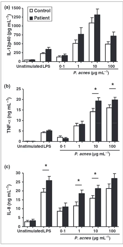
In healthy controls and patients with acne, the secretion of pro-inflammatory IL-12p40 by PBMCs was actively stimulated by increasing concentrations ofP. acneswith a peak response at 10lg mL)1 (492 ± 68 pg mL)1, Fig. 1a). However, there was no significant difference in the secretion profile between patients and controls. There was also little difference in the secretion of TNF-ain response to either LPS or low concentra-tions ofP. acnes between patients with acne and healthy con-trols, although when stimulated with 10 or 100lg mL)1
P. acnesthe level of TNF-adetected was significantly higher for PBMCs from patients with acne (both P< 0Æ05). In response to P. acnes, IL-8 secretion was produced in a dose-dependent manner (Fig. 1c). Moreover, PBMCs from patients with acne in the presence of LPS or P. acnes (at 1lg mL)1 and 10lg mL)1) secreted significantly more IL-8 than PBMCs from healthy controls (P< 0Æ05).
Secretion of the downregulatory cytokine interleukin-10 by peripheral blood mononuclear cells in response to
Propionibacterium acnesis significantly lower in patients
with acne
The peak of IL-10 production was detected at 72 h (data not shown) and secretion in response to P. acnes increased in a dose-dependent manner and reached a plateau at 10lg mL)1
in patients with acne and a maximum at 100lg mL)1in con-trols (Fig. 2a). However, IL-10 secretion was significantly lower in patients with acne compared with controls (e.g. at 10lg mL)1, 976 ± 102 cf. 766 ± 112 pg mL)1; and at 100lg mL)1, 1498 ± 210 cf. 807 ± 103 pg mL)1 for patients with acne and healthy controls, respectively; both
P< 0Æ05). Indeed, the level of the IL-10 response was on average 46% lower in patients with acne compared with con-trols for stimulation with P. acnes at 100lg mL)1. Similarly, significantly less IL-10 was also secreted in response to LPS in
(a)
(b)
(c) 1500
Control
0 250 500 750 1000
1250 Patient
IL-12p40 (pg mL
–1
)
P. acnes (µg mL–1)
Unstimulated LPS 0·1 1 10 100
TNF-α
(ng mL
–1
)
10 15 20
25
*
*
P. acnes (µg mL–1)
Unstimulated LPS 0·1 1 10 100
0 5
P. acnes (µg mL–1)
30
*
*
Unstimulated LPS 0·1 1 10 100
0 5 10 15 20
25
*
IL-8 (ng mL
–1
)
Fig 1. Peripheral blood mononuclear cells (PBMCs) from patients with acne secrete similar or greater amounts of pro-inflammatory cytokines than PBMCs from controls. (a) The secretion of interleukin (IL)-12p40 in patients with acne (n= 26–41) and controls
(n= 16–36); (b) tumour necrosis factor (TNF)-a(patientsn= 20–28; controls,n= 12–25); and (c) IL-8 (patients,n= 21–32; controls,
n= 11–28) was measured by enzyme-linked immunosorbent assay wherenvalues are greatest forP. acnesconcentration of 10 and 100lg mL)1and lowest for 0Æ1 and 1
lg mL)1. Results are expressed
as the mean ± SEM. *P£0Æ05.
IL-10is downregulated in patients with acne, F. Caillonet al. 3
[image:4.595.312.523.51.474.2]patients with acne when compared with the control PBMC response (541 ± 66 cf. 895 ± 122 pg mL)1; P< 0Æ05). No sex- or age-related difference in the production of IL-10 was demonstrated (data not shown).
In order to identify a possible correlation between acne dis-ease severity and IL-10 secretion in response toP. acnes, patient PBMCs were divided into two groups according to the severity of acne: mild acne with a grade £1, and moderate to severe
acne with a grade > 1 (scored according to the scale of Burke and Cunliffe16). No difference in the level of IL-10 secretion was observed between patients with mild acne (n= 19) and those with moderate to severe acne (n= 16) (data not shown).
Interleukin-10+peripheral blood mononuclear cells are less abundant in patients with acne
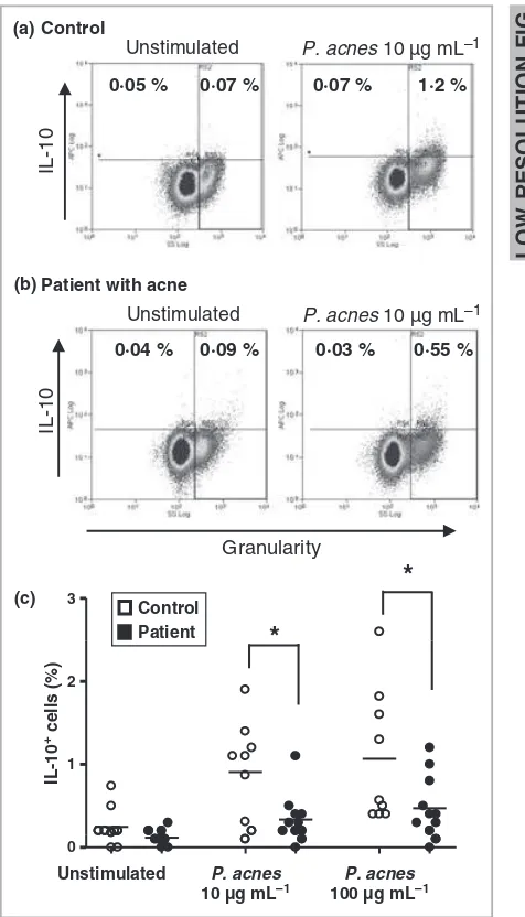
In resting healthy PBMCs, IL-10 was not detected, or at only a very low level (~0Æ2% Fig. 3a, c). However, when stimulated
with 10lg mL)1 of P. acnes, the detection of IL-10+ PBMCs was increased four-fold such that ~1Æ0% of the total PBMC population was positive for this cytokine. Although these values appear low, over 50 000 events were analysed by flow cyto-metry giving confidence that the observed changes are signifi-cant. The percentage of IL-10+ PBMCs in patients with acne was low (0Æ3%) after stimulation with 10lg mL)1P. acnes, and was significantly lower when compared with PBMCs from healthy controls (P< 0Æ05; Fig. 3b, c). Similar results were obtained using a higher stimulation dose of 100lg mL)1
P. acnes(P< 0Æ05). Overall, there were about 60% fewer IL-10+
PBMCs from patients with acne than healthy controls (Fig. 3c).
Proliferation of peripheral blood mononuclear cells from patients with acne and healthy controls is similar
The proliferative response of PBMCs toP. acneswas evaluated in order to determine whether the difference in IL-10 production
was a reflection of PBMC proliferation. P. acnes induced the proliferation of PBMCs from healthy controls and patients with acne to a similar extent, with mean stimulation indices, respectively, of 7Æ6 ± 1Æ1 and 9Æ2 ± 1Æ2 over unstimulated cells for a dose of 10lg mL)1 but they were not statistically different (allP> 0Æ05; Fig. 4). In comparison, anti-CD3 mAb induced far higher levels of cell proliferation leading to stimu-lation indices of 65Æ1 ± 8Æ7 and 77Æ7 ± 7Æ2 for control and patient PBMCs, respectively, but again they were not signifi-cantly different.
500 750 1000 1250 1500 1750
*
*
*
Control Patient
IL-10 (pg mL
–1
)
0 250
Unstimulated LPS 0·1 1 10 100
P. acnes (µg mL–1)
Fig 2. Interleukin (IL)-10 secretion by peripheral blood mononuclear cells (PBMCs) is significantly lower in patients with acne than in healthy controls. PBMCs incubated for 72 h in the absence or presence of lipopolysaccharides (LPS) orP. acnes(0Æ1, 1, 10 or 100lg mL)1).
IL-10 levels were detected by enzyme-linked immunosorbent assay. Results are expressed as the mean ± SEM (n= 20–40 for controls,
n= 25–39 for patients). *P£0Æ05.
(a)
(b)
(c) 3
*
*
Control Patient
1 2
IL-10
+ cells (%)
0
Unstimulated P. acnes
10 µg mL–1
P. acnes
100 µg mL–1
Control
Unstimulated
0·05 % 0·07 % 0·07 % 1·2 %
IL-10
P. acnes 10 µg mL–1
Patient with acne
IL-10
Unstimulated
Granularity
P. acnes 10 µg mL–1
0·04 % 0·09 % 0·03 % 0·55 %
Fig 3.
1 Interleukin (IL)-10+cells are less abundant in patients with acne than in healthy controls. Intracellular IL-10 in peripheral blood mononuclear cells (PBMCs) from (a) a healthy control, and (b) from a patient with acne, determined following 48 h incubation with or withoutP. acnes. The percentage of IL-10+cells (minus the isotype control) is indicated. (c) IL-10+cells were significantly less abundant in PBMCs from patients with acne (n= 11) than healthy controls (n= 9) stimulated with P. acnes. *P£0Æ05.
LOW
RESOLUTIO
N
FIG
[image:5.595.310.548.51.467.2] [image:5.595.58.295.52.198.2]Patients with acne have significantly fewer
interleukin-10+CD14+peripheral blood mononuclear cells
In healthy controls, the vast majority of IL-10+ PBMCs were CD14+ (51Æ6 ± 9%). The percentage of IL-10+ PBMCs that were CD4+or CD19+remained small, even after stimulation
with P. acnes (15Æ9 ± 4Æ8% and 7Æ8 ± 1Æ5%, respectively; Fig. 5). The remaining 25% were CD14) CD4) CD19), and possibly represent inactivated B cells, CD8+ cells, NK cells or
CD14)macrophages. Examination of the CD14+PBMC popu-lation from healthy individuals showed that when stimulated with 10lg mL)1 P. acnes, 5Æ05 ± 0Æ83% were IL-10+
(Table 1). This is significantly higher than in patients with acne where only 2Æ07 ± 0Æ77% were IL-10+(P< 0Æ05).
How-ever, no significant change between patients with acne and healthy controls in the proportion of IL-10+ PBMCs were
observed for CD19+ or CD4+ populations (both P> 0Æ05).
This suggests that the deficiency in IL-10 production in patients with acne is confined to the CD14+ cell population
and is not evident in CD4+T or CD19+B lymphocytes.
The phagocytic activity of CD14+peripheral blood mononuclear cells is significantly reduced in patients with acne, but is restored by the addition of exogenous interleukin-10
The phagocytic activity of CD14+ PBMCs was examined in light of their ability to take up Alexa Fluor 488 labelled
P. acnes. It was observed that CD14+ cells from patients with acne phagocytosed significantly fewer labelled P. acnes than cells from healthy controls (i.e. ~20% cf. 40%) (Fig. 6a–c,
P< 0Æ01). Furthermore, the addition of IL-10 to PBMC cultures increased the uptake of P. acnes by CD14+ cells from
patients with acne by approximately two-fold (P< 0Æ01), to a level similar to that seen in healthy controls (Fig. 6a–c). Sup-plementation of IL-10 to cultures of PBMCs from healthy con-trols had no significant impact on their ability to phagocytose
P. acnes(P> 0Æ05). The addition of IL-10 to PBMCs from acne and healthy controls stimulated with P. acnes (10lg mL)1) also led to a reduced secretion of IL-8 (Fig. 6d) while the effect of IL-10 supplementation on TNF-a secretion by PBMCs stimulated with P. acnes (10lg mL)1) was more pronounced with a reduction of 78% and 62% for PBMCs from patients with acne and healthy controls, respectively (Fig. 6e).
Discussion
The aim of this study was to investigate the inflammatory cytokine response of PBMCs from patients with acne vulgaris following stimulation withP. acnes. The production of the pro-inflammatory cytokines IL-8, TNF-aand IL-12p40 were either Control
5 10 15
Patient
35 45 55
Stimulation index
0
αCD3 LPS 0·1 1 10 100
P. acnes (µg mL–1)
Fig 4. Proliferation of peripheral blood mononuclear cells (PBMCs) from patients with acne and healthy controls in response to mitogen andP. acnesstimulation. PBMCs were cultured for 72 h in the presence or absence ofaCD3 monoclonal antibody, lipopolysaccharide (LPS) or
P. acnes(0Æ1, 1, 10 and 100lg mL)1). The incorporation of [3H]
thymidine was measured in c.p.m. Results are expressed as the mean ± SEM (n= 20–40 for controls,n= 19–33 for patients). *P£0Æ05.
Healthy control
CD4 15·9 4·8%
CD4 51·6 5·9% Unknown
4·6 1·3%
CD19 7·8 1·5%
Fig 5. The majority of interleukin (IL)-10+cells are CD14+. Relative contribution of CD4+, CD19+and CD14+cells in the detection of IL-10 in healthy controls peripheral blood mononuclear cells (PBMCs) treated withP. acnes(10lg mL)1). CD14+cells are the major source
[image:6.595.60.288.50.206.2]of IL-10. Similar proportions were observed for patients with acne PBMCs.
Table 1 The percentage of IL-10+CD14+cells cultured in the absence or presence ofP. acnesis lower in patients with acne
CD14+ CD19+ CD4+ Medium only
Controlsa 0Æ92 ± 0Æ25 0Æ54 ± 0Æ10 0Æ31 ± 0Æ08 Patientsa 1
Æ10 ± 0Æ17 0Æ32 ± 0Æ08 0Æ28 ± 0Æ08
P. acnes(10lg mL)1)
Controls 5Æ05 ± 0Æ83 0Æ61 ± 0Æ14 0Æ39 ± 013 Patients 2Æ07 ± 0Æ77* 0Æ65 ± 0Æ15 0Æ33 ± 0Æ09
P. acnes(100lg mL)1)
Controls 4Æ36 ± 0Æ72 0Æ68 ± 0Æ17 0Æ19 ± 0Æ04 Patients 3Æ09 ± 0Æ57 0Æ53 ± 0Æ19 0Æ36 ± 0Æ12
a
n= 8; *P< 0Æ05 for patients compared with controls; bold, statistically significant.
IL-10is downregulated in patients with acne, F. Caillonet al. 5
[image:6.595.88.261.482.647.2]slightly elevated or unchanged in patients with acne when compared with healthy controls. However, a statistically significant reduction in PBMC secretion of anti-inflammatory IL-10 in patients with acne was identified. The impaired production of IL-10 by PBMCs from patients with acne was confined to CD14+cells that were also defective in their ability
to phagocytose P. acnes bacteria. The addition of exogenous IL-10 to PBMC cultures from patients with acne restored phagocytic activity.
To our knowledge, this study is the first to report a significant imbalance in the production by PBMCs of pro- and
anti-inflammatory cytokines between patients with acne and healthy controls. The secretion of both IL-8 and TNF-a by PBMCs was significantly elevated in patients with acne in agreement with studies elsewhere.17 The production of these cytokines in response to P. acnes stimulation is linked to the expression of innate immune response pattern recognition receptor TLR2.5,18,19 However, no difference was detected in
the secretion of the p40 subunit of the pro-inflammatory cytokine complex IL-12⁄IL-23. This conflicts with findings
that IL-12 originating from monocytes was increased follow-ing stimulation withP. acnesvia TLR2,5,19but agrees with the
observation that IL-12p40 mRNA is not upregulated in acne lesions compared with uninvolved skin.15
In contrast to the upregulation of pro-inflammatory cyto-kines, the production of the anti-inflammatory cytokine IL-10 was markedly downregulated in patients with acne in this study. IL-10 is a well-described regulatory cytokine that acts to harness the release of several pro-inflammatory cytokines.20 In our study, it appears to act primarily on TNF-a but has ittle effect upon IL-12. Therefore, we speculate that in patients with acne, early activation of the immune response is not controlled, due to the relative paucity of IL-10. As a result, inflammation is pathologically sustained in patients with acne contributing to the events that lead to the initiation and⁄or
persistence of inflammatory lesions. On the other hand, there was no association between the severity of acne and the abso-lute quantity of secreted IL-10. This might suggest that a rela-tive deficiency of IL-10 predisposes a patient to acne, but cannot be used to predict the severity of the clinical lesions. It may also suggest that a lack of IL-10 cannot solely predict the severity of acne. Indeed, it is well known that acne is multi-factorial and it would be surprising if it could be completely explained through the actions of a solitary cytokine.21
IL-10 can derive from CD4+ T cells with regulatory func-tion (so-called Tregs).12,22 However, aCD3 mAb stimulation
did not affect the differential secretion of IL-10 between PBMCs derived from patients with acne and controls. We also show that CD4+ cells are only a minor (~16%) source of
IL-10 and the proportions of IL-10+CD4+cells do not differ
between control and patient PBMCs. Therefore, we conclude that IL-10-producing Tregs among the PBMC population are
unlikely to have a major role in determining the propensity to develop acne.
The most abundant IL-10+ cell population among PBMCs
were CD14+ (> 50%) and these are under-represented in
patients with acne. CD14+PBMCs are a well-described source
of IL-10.23In the skin, the main sources of IL-10 in the early phase of inflammation are CD14+cells,15 possibly originating from the peripheral blood. TLR2 expression on CD14+cells is also increased in cells localized within acne lesions.5,24The role of IL-10 is likely the downregulation of pro-inflammatory cytokines as demonstrated by the decreased production of IL-8 and TNF-aafter the addition of exogenous IL-10 to PBMC cul-tures. However, IL-10 is known to be markedly upregulated in established acne lesions.15This appears to contradict the
find-ings presented here. We speculate that the CD14+ PBMCs
Acne patient 16% Untreated CD14+ (c) (d) (e) Healthy control (a) (b) 36% Untreated Labelled P. acnes CD14+ 50 60 ** ** 0 10 20 30 40
P. acnes P. acnes + IL-10
P. acnes P. acnes + IL-10
CD14
+ Alexa Fluor 488 + cells (%)
Control Patient
10 000 20 000 30 000 Control
Control + IL-10 Patient Patient + IL-10
IL-8 (pg mL
–1
)
0
Unstimulated P. acnes
TNF (pg mL
–1 ) 10 000 15 000 20 000 25 000 30 000 35 000 40 000 Control Control + IL-10 Patient Patient + IL-10
** **
Unstimulated P. acnes
0 5000
Fig 6.
2 CD14+peripheral blood mononuclear cells (PBMCs) from patients with acne phagocytose lessP. acnesthan cells from healthy controls, and levels of phagocytosis are restored by the addition of exogenous interleukin (IL)-10. PBMCs from healthy controls and from patients with acne (n= 4) were stimulated with Alexa Fluor 488 labelledP. acnesin the presence or absence of recombinant IL-10. The level of phagocytosis was determined by the uptake of labelledP. acnes
by CD14+PBMCs from (a) a healthy control or (b) an acne patient as determined by flow cytometry. (c) The addition of IL-10 to PBMCs from patients with acne restored levels of phagocytosis ofP. acnesto levels seen in healthy controls and reduced the production of (d) IL-8 and (e) TNF-a. Data were normalized by a log10 for statistical analysis (P-values: **P£0Æ01).
[image:7.595.39.292.53.428.2]represent a set of circulating monocytes⁄immature macrophages
that will be the first to infiltrate the tissue around a develop-ing lesion. Dysregulation in IL-10 production by these circulat-ing CD14+ cells may facilitate the development of intense inflammatory lesions driven by an imbalance towards pro-inflammatory cytokines such as TNF-a and IL-8. Therefore, a deficit in the number of IL-10-producing CD14+monocytes, as
seen in our patients with acne, would favour the development of an inflammatory cascade characteristic of acne lesions. In established lesions, CD68+mature macrophages will
predomi-nate3and only then is IL-10 finally produced in an attempt to
regulate the overactive inflammatory response.
The cause of the differences in the production of IL-10 between healthy controls and patients with acne is uncertain. There is known to be considerable inter-individual variation in the capacity of human PBMCs to produce IL-10, with at least 50% likely due to genetic factors.25 Polymorphisms of
the 5¢-flanking region of the IL-10 gene26 and single
nucleo-tide polymorphisms within the promoter region have been linked with a number of chronic dermatoses including psoria-sis.27,28Unfortunately, a detailed genetic analysis was beyond the scope of the current study but the familial inheritance of acne may be partly explained by this mechanism. Our study also raises questions about the onset and resolution of acne. Onset commonly occurs during adrenarche when output of dihydroepiandrosterone (DHEA) starts to rise.29 The
relation-ship between DHEA, IL-10 and progression to acne has not been investigated, although data in humans indicate there may be a negative correlation between serum IL-10 levels and DHEA concentration.30,31The resolution of acne in individuals
may represent upregulation of IL-10 expression and it would be important to perform a longitudinal study to determine if the course of acne relates to changes of IL-10 with age. Alter-natively, other mechanisms of immune regulation may evolve with time and act independently of IL-10 released by CD14+ cells. For example, Tregcells may be involved in the resolution
of acne21 and have been implicated in other skin
condi-tions,32,33 while the delivery of T
reg cells can dampen skin
inflammation in a model of psoriasis.34 Therefore, it is
reasonable to assume that Treg cells may downregulate the
CD4+T-cell response to P. acnesand thus explain why
resolu-tion can occur even in the presence of reduced IL-10 produc-tion by PBMCs.
Administration of exogenous IL-10in vivo is reported to be an effective therapeutic strategy in controlling skin disorders such as psoriasis.35 This study raises the possibility that the
same treatment strategy may be a feasible approach in acne. Moreover, and perhaps counterintuitively in light of its down-regulatory functions, administration of IL-10 toin vitrocultures of PBMCs from patients with acne increased the phagocytic process allowing CD14+ cells to take up more P. acnes. Thus, in addition to limiting the levels of pro-inflammatory cyto-kines and so preventing a ‘hyper-immune response’, as seen in patients with acne, IL-10 also acts to facilitate phagocytosis by CD14+ cells36 and presumably results in more effective
clearance ofP. acnes.
In summary, these data suggest that acne lesions may develop because of a defect in immunosurveillance. We pro-pose that an immune response toP. acnesbacteria in the follicle is usually dampened by PBMC-derived IL-10 before visible in-flammation develops. Reduced production of IL-10 by CD14+ PBMCs, most likely monocytes, predisposes to unchecked pro-inflammatory changes. This interpretation raises the possibility that acne therapeutics might profitably target IL-10 both as a regulator of pro-inflammatory cytokines and in augmenting the CD14+ cell phagocytic response. Future studies should
seek to characterize the cellular sources of IL-10 in evolving and healing acne lesions and compare the skin distribution of IL-10 in subjects with acne and subjects whose acne has resolved spontaneously.
Acknowledgments
The authors would like to thank the following individuals: Joe D. Turner, Peter C. Cook, Ross A. Paveley, Sarah A. Aynsley and Ann Bamford for technical and logistical support, as well as all individuals who donated blood either as patients of the Department of Dermatology in Harrogate District Hospital, or as students of the University of York. The project was funded by Roche.
References
1 Stathakis V, Kilkenny M, Marks R. Descriptive epidemiology of acne vulgaris in the community.Australas J Dermatol1997;38:115– 23.
2 Eady EA, Cove JH. Is acne an infection of blocked pilosebaceous follicles? Implications for antimicrobial treatment.Am J Clin Dermatol
2000;1:201–9.
3 Jeremy AH, Holland DB, Roberts SGet al.Inflammatory events are involved in acne lesion initiation.J Invest Dermatol2003;121:20–7. 4 Ingham E, Eady EA, Goodwin CEet al.Pro-inflammatory levels of
interleukin-1 alpha-like bioactivity are present in the majority of open comedones in acne vulgaris.J Invest Dermatol1992;98:895–901. 5 Kim J, Ochoa MT, Krutzik SRet al.Activation of toll-like receptor 2 in acne triggers inflammatory cytokine responses.J Immunol 2002; 169:1535–41.
6 Sugisaki H, Yamanaka K, Kakeda M et al. Increased interferon-gamma, interleukin-12p40 and IL-8 production inPropionibacterium acnes-treated peripheral blood mononuclear cells from patient with acne vulgaris: host response but not bacterial species is the deter-minant factor of the disease.J Dermatol Sci2009;55:47–52. 7 Schaller M, Loewenstein M, Borelli Cet al.Induction of a
chemoat-tractive proinflammatory cytokine response after stimulation of keratinocytes withPropionibacterium acnesand coproporphyrin III.Br J Dermatol2005;153:66–71.
8 Ma J, Chen T, Mandelin J et al.Regulation of macrophage activa-tion.Cell Mol Life Sci2003;60:2334–46.
9 Moore KW, de Waal Malefyt R, Coffman RLet al. Interleukin-10 and the interleukin-10 receptor. Annu Rev Immunol2001; 19:683– 765.
10 Trinchieri G. Interleukin-12 and the regulation of innate resistance and adaptive immunity.Nat Rev Immunol2003;3:133–46.
11 Weiss E, Mamelak AJ, La Morgia Set al.The role of interleukin 10 in the pathogenesis and potential treatment of skin diseases.J Am Acad Dermatol2004;50:657–75.
IL-10is downregulated in patients with acne, F. Caillonet al. 7
12 Belkaid Y, Rouse BT. Natural regulatory T cells in infectious disease.Nat Immunol2005;6:353–60.
13 Birch KE, Vukmanovic-Stejic M, Reed JRet al.The immunomodula-tory effects of regulaimmunomodula-tory T cells: implications for immune regula-tion in the skin.Br J Dermatol2005;152:409–17.
14 Suh DH, Kwon TE, Youn JI. Changes of comedonal cytokines and sebum secretion after UV irradiation in acne patients.Eur J Dermatol
2002;12:139–44.
15 Kang S, Cho S, Chung JH et al. Inflammation and extracellular matrix degradation mediated by activated transcription factors nuclear factor-kappaB and activator protein-1 in inflammatory acne lesionsin vivo.Am J Pathol2005;166:1691–9.
16 Burke BM, Cunliffe WJ. The assessment of acne vulgaris – the Leeds technique.Br J Dermatol1984;111:83–92.
17 Basal E, Jain A, Kaushal GP. Antibody response to crude cell lysate ofPropionibacterium acnesand induction of pro-inflammatory cytokines in patients with acne and normal healthy subjects.J Microbiol2004; 42:117–25.
18 Vowels BR, Yang S, Leyden JJ. Induction of proinflammatory cyto-kines by a soluble factor of Propionibacterium acnes: implications for chronic inflammatory acne.Infect Immun1995;63:3158–65. 19 Liu PT, Phan J, Tang Det al.CD209(+) macrophages mediate host
defense againstPropionibacterium acnes.J Immunol2008;180:4919–23. 20 Couper KN, Blount DG, Riley EM. IL-10: the master regulator of
immunity to infection.J Immunol2008;180:5771–7.
21 Wilcox HE, Farrar MD, Cunliffe WJet al.Resolution of inflamma-tory acne vulgaris may involve regulation of CD4+T-cell responses toPropionibacterium acnes.Br J Dermatol2007;156:460–5.
22 Dudda JC, Perdue N, Bachtanian Eet al.Foxp3+ regulatory T cells maintain immune homeostasis in the skin. J Exp Med 2008; 205:1559–65.
23 Keller CC, Yamo O, Ouma C et al. Acquisition of hemozoin by monocytes down-regulates interleukin-12 p40 (IL-12p40) tran-scripts and circulating IL-12p70 through an IL-10-dependent mechanism: in vivo andin vitrofindings in severe malarial anemia.
Infect Immun2006;74:5249–60.
24 Jugeau S, Tenaud I, Knol ACet al.Induction of toll-like receptors byPropionibacterium acnes.Br J Dermatol2005;153:1105–13.
25 Reuss E, Fimmers R, Kruger Aet al.Differential regulation of inter-leukin-10 production by genetic and environmental factors – a twin study.Genes Immun2002;3:407–13.
26 Eskdale J, Kube D, Gallagher G. A second polymorphic dinucleo-tide repeat in the 5¢ flanking region of the human IL10 gene.
Immunogenetics1996;45:82–3.
27 Hensen P, Asadullah K, Windemuth C et al. Interleukin-10 pro-moter polymorphism IL10.G and familial early onset psoriasis.Br J Dermatol2003;149:381–5.
28 Kingo K, Koks S, Silm H et al. IL-10 promoter polymorphisms influence disease severity and course in psoriasis.Genes Immun2003; 4:455–7.
29 Stewart ME, Downing DT, Cook JS et al.Sebaceous gland activity and serum dehydroepiandrosterone sulfate levels in boys and girls.
Arch Dermatol1992;128:1345–8.
30 Tabata N, Tagami H, Terui T. Dehydroepiandrosterone may be one of the regulators of cytokine production in atopic dermatitis.Arch Dermatol Res1997;289:410–14.
31 Suzuki T, Suzuki N, Engleman EG et al. Low serum levels of de-hydroepiandrosterone may cause deficient IL-2 production by lymphocytes in patients with systemic lupus erythematosus (SLE).
Clin Exp Immunol1995;99:251–5.
32 Werfel T, Wittmann M. Regulatory role of T lymphocytes in atopic dermatitis.Chem Immunol Allergy2008;94:101–11.
33 Kuhn A, Beissert S, Krammer PH. CD4(+)CD25(+) regulatory T cells in human lupus erythematosus. Arch Dermatol Res 2009; 301:71–81.
34 Teige I, Hvid H, Svensson Let al.Regulatory T cells control VEGF-dependent skin inflammation.J Invest Dermatol2009;129:1437–45. 35 Friedrich M, Docke WD, Klein A et al. Immunomodulation by
interleukin-10 therapy decreases the incidence of relapse and pro-longs the relapse-free interval in psoriasis. J Invest Dermatol 2002; 118:672–7.
36 Lingnau M, Hoflich C, Volk HDet al.Interleukin-10 enhances the CD14-dependent phagocytosis of bacteria and apoptotic cells by human monocytes.Hum Immunol2007;68:730–8.
Author Query Form
Journal:
BJD
Article:
9420
Dear Author,
During the copy-editing of your paper, the following queries arose. Please respond to these by marking up your proofs with the necessary changes/additions. Please write your answers on the query sheet if there is insufficient space on the page proofs. Please write clearly and follow the conventions shown on the attached corrections sheet. If returning the proof by fax do not write too close to the paper’s edge. Please remember that illegible mark-ups may delay publication.
Many thanks for your assistance.
Query reference Query Remarks
1 AUTHOR: Figure 3 has been saved at a low resolution of 248 dpi. Please resupply at 600 dpi. Check required artwork specifications at http://www.blackwellpublishing.com/authors/digill.asp