Dissertation on
A CLINICOPATHOLOGICAL STUDY TO ANALYSE THE IMPORTANCE OF HISTOPATHOLOGICAL EXAMINATION IN DIAGNOSIS OF EXCISED
CONJUNCTIVAL LESIONS OF BULBAR CONJUNCTIVA
THE TAMILNADU
Dr. M.G.R. MEDICAL UNIVERSITY CHENNAI
CERTIFICATE
This is to certify that this dissertation entitled “A CLINICOPATHOLOGICAL STUDY TO
ANALYSE THE IMPORTANCE OF HISTOPATHOLOGICAL EXAMINATION IN DIAGNOSIS OF EXCISED CONJUNCTIVAL LESIONS OF BULBAR CONJUNCTIVA”
is a bonafide record of research work done by Dr.N.SUDHA PRIYADHARSINI, Post Graduate
Resident in Department of Ophthalmology, Madurai Medical College, Madurai.
She has submitted this in partial fulfillment of the regulations laid down by The Tamil Nadu Dr.
M.G.R. Medical University, for the award of Master of Surgery Degree Branch III
(Ophthalmology), under our guidance and supervision during the academic years 2015-2018.
DR.S.V.CHANDRAKUMAR MS.,D.O
HOD and Professor of Ophthalmology,
GRH, Madurai Medical College,
Madurai.
DR.MARUTHU PANDIAN M.S.,F.I.C.S.,
The Dean,
GRH, Madurai Medical College,
CERTIFICATE FROM GUIDE
This is to certify that this dissertation entitled “A CLINICOPATHOLOGICAL STUDY TO
ANALYSE THE IMPORTANCE OF HISTOPATHOLOGICAL EXAMINATION IN DIAGNOSIS OF EXCISED CONJUNCTIVAL LESIONS OF BULBAR CONJUNCTIVA”
is a bonafide record of research work done by Dr.N.SUDHA PRIYADHARSINI, Post Graduate
Resident in Department of Ophthalmology, Madurai Medical College, Madurai.
DR.S.V.CHANDRAKUMAR M.S., D.O,
HOD and professor of ophthalmology
GRH, Madurai medical college
DECLARATION
I, Dr.N.SUDHA PRIYADHARSINI hereby solemnly declare that, this dissertation titled “A
CLINICOPATHOLOGICAL STUDY TO ANALYSE THE IMPORTANCE OF HISTOPATHOLOGICAL EXAMINATION IN DIAGNOSIS OF EXCISED CONJUNCTIVAL LESIONS OF BULBAR CONJUNCTIVA” was done by me.
I also declare that this bonafide work / a part of this work was not submitted by me / anyone
else, for any award, for Degree / Diploma to any other University / Board either in India / abroad.
This is submitted to The Tamilnadu Dr. M. G. R. Medical University, Chennai in partial fulfilment
of the rules and regulations for the award of Master of Surgery degree Branch -III (Ophthalmology)
to be held in April 2018.
Place: Madurai (Dr.N.Sudha Priyadharsini)
ACKNOWLEDGEMENT
I express my sincere thanks and gratitude to Prof. Dr.MARUTHU PANDIAN M.S.,F.I.C.S., The
Dean, GRH and MMC Madurai for permitting me to conduct this study. I am extremely grateful
to Dr.S.V.CHANDRAKUMAR M.S, D.O, HOD, Professor of Ophthalmology and Dr. S. KAVITHA M.S, DNB Associate Professor of Ophthalmology, GRH, MMC, Madurai, for his valuable suggestions and guidance throughout the course of my study. I have great pleasure in
thanking my beloved co-guide DR.E.RAJESWARI, M.S, Assistant Professor and all my
Assistant Professors of Ophthalmology department at Madurai Medical College, Madurai, for their
constant source of cheer and encouragement throughout the study
I express my sincere thanks to Prof. Dr.T.GEETHA MD., Head of the department of Pathology
for their constant support, guidance, cooperation in this study.
I thank all my dear friendsfor their timely help and encouragement to do my study .I express my
heartfelt love to my parents and husband for endless affection and support.
I thank the patients of our hospital for their extreme patience and cooperation without whom the
project would have been a distant dream.
INDEX
S.NO CONTENTS PAGE NO.
PART I
1. INTRODUCTION 1
2. ANATOMY OF CONJUNCTIVA 1
3. SURGICAL TECHNIQUE OF EXCISION BIOPSY 4
4. CONGENITAL LESIONS 9
5. BENIGN TUMOURS OF SURFACE EPITHELIUM 13
6. MELANOCYTIC TUMOURS 15
7. MISCELLANEOUS TUMOURS 22
8. DEGENERATIONS 32
9. MALIGNANT TUMOURS 38
S.NO CONTENTS PAGE NO. PART II
1. AIMS AND OBJECTIVES 59
2. MATERIALS AND METHODS 59
3. RESULTS AND INTERPRETATION 62
4. SUMMARY 87
5. DISCUSSION 89
6. CONCLUSION 92
ANNEXURES
1. BIBLIOGRAPHY
2. PROFORMA
3. MASTER CHART
4. LIST OF ABBREVIATIONS,
5. ETHICAL COMMITTEE CLEARANCE
1
A CLINICOPATHOLOGICAL STUDY TO ANALYSE THE IMPORTANCE OF HISTOPATHOLOGICAL EXAMINATION IN DIAGNOSIS OF EXCISED CONJUNCTIVAL LESIONS OF BULBAR CONJUNCTIVA
INTRODUCTION
The conjunctiva is readily visible and so the tumors and other lesions in the conjunctiva are
generally recognized at an early stage. Clinical diagnosis can often be made by ocular
examination and slit- lamp bio microscopy, if features are characteristics. A biopsy is not
necessary in cases of smaller tumors that appear benign. Small tumours can be better
removed completely in one setting (excisional biopsy). Larger lesions, remove a portion of
the tumor (incisional biopsy) to get a histopathologic diagnosis prior to more extensive
therapy.It is rarely needed to do exfoliative cytology or fine- needle aspiration biopsy, as
incisional biopsy is readily available. Slit- lamp examination of the cornea is needed in
patients with suspected conjunctival tumors. Rule out any corneal involvement in squamous
cell carcinoma and melanoma of conjunctiva before planning for surgery.
ANATOMY OF CONJUNCTIVA:
The conjunctiva has 3 geographic zones: palpebral, forniceal, bulbar. The palpebral
conjunctiva starts from the mucocutaneous junction of lid and covers the inner surface of lid
and attached firmly to the tarsus. The tissue is freely movable in the fornices, in the upper lid
it is enmeshed with fibrous elements of the levator aponeurosis and Muller muscle. In the
lower lid, fibrous expansions from the inferior rectus muscle join with the inferior tarsal
muscle. The bulbar conjunctiva is freely mobile.It fuses with the Tenon capsule and gets
inserted into the limbus. Blood supply to the bulbar conjunctiva is by anterior ciliary arteries
2
arcade, along the upper border of the lid, gives branches proximally to the forniceal and the
bulbar conjunctiva via the posterior conjunctival arteries. The ciliary arteries via the anterior
conjunctival arteries gives the limbal blood supply. The innervation of the conjunctiva is
from the ophthalmic division of trigerminal nerve.
The conjunctiva is a mucous membrane made of non-keratinizing squamous epithelium with
lot of goblet cells and richly vascularized substantia propria having lymphatic vessels, plasma
cells, macrophages, mast cells. A lymphoid layer is presents from the bulbar conjunctiva to
sub tarsal folds.Specialized aggregations of conjunctiva-associated lymphoid tissue (CALT)
correspond to mucosa associated lymphoid tissue (MALT) and have collections of T and B
lymphocytes under the epithelium. They help in antigen processing. The conjunctival
epithelium is 2 to 5 cells thick. The basal cells are cuboidal in shape and become flattened
polyhedral cells at the surface. The goblet cells (mucous glands) are found in the inferior and
medial part of the conjunctiva more in the region of the caruncle and plica semilunaris. They
are sparsely found in the remainder of the conjunctiva and absent in the limba1 area.
3
MANAGEMENT
Management of a conjunctival tumor may be
• serial observation
• incisional biopsy
• excisional biopsy
• cryotherapy
• chemotherapy
• radiotherapy
• modified enucleation
• orbital exenteration or
• Various combinations of the above.
If large conjunctival defect present-mucous membrane grafts of the other eye
conjunctiva, buccal mucosa, amniotic membrane graft may be done.
Observation
Most benign lesions like pingueculum, dermolipoma, and nevus are just observed. Anterior
segment slit lamp photos are taken for photographic evidence for follow up every 1 year to
look for any growth, malignant transformation, or compression on normal surrounding
tissues.
Incisional Biopsy
For suspicious lesion like squamous cell carcinoma, PAM, melanoma, and conjunctival
spread of sebaceous gland carcinoma if these tumours occupy >4 clock hours in conjunctiva.
For larger lesions incisional wedge biopsy or punch biopsy done. Treatment planned based on
4
Excisional Biopsy
In Intermediate and small tumors that are symptomatic or suspected to be malignant,
excisional biopsy is preferred over incisional biopsy to avoid inadvertent tumor seeding.
Excision biopsy needed in limbal dermoid, epibulbar osseous choristoma, steroid- resistant
pyogenic granuloma, squamous cell carcinoma, and melanoma.in lesions of conjunctival
fornix it is totally excised and the conjunctiva reconstructed with absorbable sutures , fornix
deepening sutures, symblephron ring used. If defect is large mucous membrane graft used.
Most malignant tumours are limbal in location. Limbal neoplasms invades the corneal
epithelium, sclera, anterior chamber and the soft tissues of the orbit. If sclera is involved thin
sclera biopsy taken to achieve tumour clearance. Friable tumors may cause seeding of tumour
and so “no touch technique” followed.
SURGICAL TECHNIQUEOF EXCISION BIOPSY: Done under microscope
The operative field should be left dry
Avoid wetting the field with balanced salt solution to prevent seeding of cells. Retrobulbar
anesthesia used
The corneal epithelial component is approached first and the conjunctival component is
dissected next so that the whole specimen resected in one piece. Absolute alcohol in an
applicator is gently applied to the entire corneal component. Epithelial cellular devitalization
and easy release of the tumor cells from Bowman’s.
The malignancy outlined with beaver blade using a delicate epithelial incision or
5
With the beaver blade the corneal epithelium is sweeped from centre to limbus. A pentagonal
or circular conjunctival limbal based incision is made outside the tumor margin.
The incision is carried through the underlying Tenon’s fascia until the sclera is seen and full
thickness conjunctiva and Tenon’s fascia is included into the excisional biopsy. Cautery done
to control bleeding. A second incision outlined by a superficial scleral groove approximately
0.2 mm in depth and 2.0 mm outside the base of the overlying adherent conjunctival mass.
This groove is continued anteriorly up to the limbus. This area outlined by the scleral groove
is cut by flat dissection of about 0.2-mm thickness within the sclera in an attempt to remove a
superficial lamella of sclera, overlying Tenon’s fascia and conjunctiva with tumor- free
margins is removed in one piece without touching the tumor itself (no- touch technique). The
removed specimen is held flatly on a piece of cardboard from the surgical tray and kept in
fixative and given for histopathologic studies. This prevents the specimen from folding and
so helps in better assessment of the tumor margins .Now new set of instruments used, to
avoid contamination of healthy tissue with possible tumor cells. After excision of the
specimen, cryotherapy is applied to the margins of the remaining bulbar conjunctiva. This is
performed by freezing the surrounding bulbar conjunctiva by lifting it away from the sclera
using the cryoprobe. When the ice ball reaches a size of 4 to 5 mm, it is allowed to thaw and
the cycle repeated once. The cryoprobe is then moved to an adjacent area of the conjunctiva
and the cycle is repeated until all of the margins have been treated by this method. Corneal
margins need not be treated with cryoapplication. The tumor bed treated with absolute
alcohol wash on cotton- tip applicator and bipolar cautery, to avoid direct cryotherapy to the
sclera. Using clean instruments, the conjunctiva is then mobilized for closing the defect by
making the intermuscular septum loose with Steven’s scissors spreading and creating Trans
positional conjunctival flaps. Closure is done with interrupted absorbable 6–0 or 7–0 sutures.
6
promotes better healing and helps in further surgery in case the patient develop recurrence.
The patient is given topical antibiotics and corticosteroids for 2 weeks and followed up at 3-
to 6-month intervals.
Cryotherapy
In the management of conjunctival tumors, cryotherapy can be used as a supplemental
treatment. It can eliminate microscopic tumor cells and prevent recurrence of malignant
tumors. It can also be used as a mainstay of treatment for primary acquired melanosis and
pagetoid invasion of sebaceous gland carcinoma. If cryotherapy can devitalize the malignant
or potentially malignant cells, radical surgery like orbital exenteration can be avoided.
Chemotherapy
Topical eyedrops composed of mitomycin C, 5-fluorouracil, interferon, or cidofovir are
effective in treating epithelial malignancies such as squamous cell carcinoma, primary
acquired melanosis, and pagetoid invasion of sebaceous gland carcinoma.
Mitomycin C or 5-fluorouracil are employed most successfully for squamous cell carcinoma,
especially after tumor recurrence following previous surgery. This medication is prescribed
topically four times daily for a 1-week period followed by a 1-week hiatus to allow the ocular
surface to recover. This cycle is repeated once again so that most patients receive a total of 2
weeks of the chemotherapy topically. Both mitomycin C and 5-fluorouracil are most effective
for squamous cell carcinoma and less for primary acquired melanosis and pagetoid invasion
of sebaceous gland carcinoma. Toxicities include most commonly dry- eye findings,
superficial punctate epitheliopathy, and punctal stenosis. Corneal, scleral melt, and cataract
can develop if these agents are used with open conjunctival wounds or used more. Topical
interferon can be effective for squamous epithelial malignancies and is less toxic to the
7
Other topical antiviral medications including cidofovir can be employed with little toxicity
for squamous epithelial tumors.
Radiotherapy
Two forms of radiotherapy are employed for conjunctival tumors, namely external beam
radiotherapy and custom designed plaque radiotherapy. External beam radiotherapy to a total
dose of 3000 to 4000 cGy is used to treat conjunctival lymphoma and metastatic carcinoma if
lesions are too large or diffuse to excise locally. Side effects are dry eye, punctate epithelial
abnormalities, and cataract. Custom- designed plaque radiotherapy to a dose of 3,000 to
4,000 cGy can be used to treat conjunctival lymphoma or metastasis. A higher dose of 6,000
to 8,000 cGy can be employed to treat the more radiation- resistant melanoma and squamous
cell carcinoma. The two designs for conjunctival custom plaque radiotherapy include a
conformer plaque technique with six fractionated treatment sessions or a reverse plaque
technique with the device sutured onto the episclera. Plaque radiotherapy to a low dose of
2,000 cGy is employed for benign conditions such as steroid resistant pyogenic granuloma
that show recurrence after surgical resection.
Modified Enucleation
Modified enucleation is a treatment modality for primary malignant tumors of the conjunctiva
that have invaded through the limbal tissues into the globe, causing secondary glaucoma. The
mucoepidermoid variant of squamous cell carcinoma of the conjunctiva has a greater
tendency for such invasion. At the time of enucleation, it is necessary to remove the involved
conjunctiva intact to avoid spreading tumor cells. Thus, the initial peritomy should begin at
the limbus, but when the tumor is approached, the incision should proceed posteriorly from
the limbus to surround the tumor- affected tissue by at least 3 to 4 mm. The tumor will
8
surrounding conjunctiva into the episclera to secure the tumor to the globe. The remaining
steps of enucleation are gently performed .The globe is removed with tumor adherent after
cutting the optic nerve from the nasal side. The margins of the remaining, unaffected
conjunctiva are treated with double freeze thaw cryotherapy.Residual conjucnctiva present
for closure. A mucous membrane graft or amniotic membrane graft may be necessary for
adequate closure and to provide fornices for a prosthesis. A simple horizontal inferior
forniceal conjunctival incision from canthus to canthus may suffice, as long as the conformer
is constantly worn as a template so the new conjunctival fornix grows deep and around this
structure.
Orbital Exenteration
Orbital exenteration is the treatment of choice for primary malignant conjunctival tumors
with orbital involvement. Either an eyelid- removing or eyelid- sparing exenteration is done,
depending on the extent of eyelid involvement. The eyelid- sparing technique is done due to
better cosmetic appearance and they heal within 2 or 3 weeks.If the anterior lamella of the
eyelid is uninvolved with tumor, an eyelid- sparing (eyelid- splitting) exenteration may be
accomplished .
Mucous Membrane Graft
Mucous membrane grafts are occasionally necessary to replace vital conjunctival tissue after
removal of extensive conjunctival tumors. The best donor sites include the forniceal
conjunctiva (ipsilateral or contralateral eye) and buccal mucosa from the posterior aspect of
the lower lip or lateral aspect of the mouth. Such grafts are removed by a freehand technique,
fashioned to fit the defect, and fit into place with cardinal and running absorbable 6–0 or 7–0
sutures. The tissue is delivered frozen and must be defrosted for 20 minutes. The fine,
9
side up, and sutured into place with absorbable sutures. Topical antibiotic and steroid
ointments are applied following all conjunctival grafting procedures. For graft harvest and
placement, the surgeon should always use clean, sterile instruments at both the donor and the
recipient sites. Free tumor cells can rest on instrument tips and later implant and grow in
previously uninvolved areas if such precautions are not taken.
I.CONGENITAL LESIONS
A variety of tumors and related conditions may be present at birth and become clinically
apparent shortly after birth. Most of the lesions are choristomas, consisting of tissue elements
that are not normally present at the involved site.
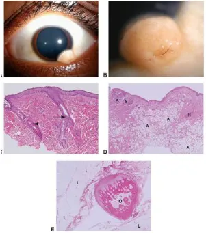
DERMOID
Conjunctival dermoid is a congenital well- circumscribed yellow- white solid mass that
involves the bulbar conjunctiva or at the corneoscleral limbus. It occurs near the limbus
inferotemporally, and often this tumor has fine hairs, best seen with slit- lamp biomicroscopy.
It may extend to the central cornea or be located in other quadrants on the bulbar surface. It
may be an isolated lesion or can be associated with Goldenhar’s syndrome. The patient
should be evaluated for ipsilateral or bilateral preauricular skin appendages, hearing loss,
eyelid coloboma, and orbitoconjunctival dermolipoma, and cervical vertebral anomalies that
comprise this uninheritable syndrome.
Histopathologically, the conjunctival dermoid is a choristomatous malformation that is made
up of dense fibrous tissue lined by conjunctival epithelium with dermal elements like hair
follicles and sebaceous glands. The management of dermoid includes observation if the
lesion is small and visually non symptomatic. We can excise the lesion for cosmetic reasons,
but the remaining corneal scar may be cosmetically unacceptable. Larger and symptomatic
10
keratosclerectomy and primary closure of overlying tissue if the defect is superficial or
closure with corneal graft if the defect is deep or full thickness. The cosmetic appearance may
improve, but the refractive and astigmatic error and visual acuity may not change .When the
lesion is in the central cornea, a lamellar or penetrating keratoplasty may be needed and long-
term amblyopia may be a problem. Rarely, extensive dermoids involve the lateral canthus,
[image:17.595.71.372.292.633.2]and planned excision with lateral canthal repair is necessary.
FIGURE 2: DERMOID – GROSS AND HISTOPATHOLOGICAL APPERANACE
DERMOLIPOMA
Dermolipoma is congenital and present at birth, but it remains asymptomatic and may not be
11
superotemporally. It appears as a pale yellow, soft, fluctuant, fusiform mass from the
palpebral lobe of the lacrimal gland, best visualized with the eye in inferonasal gaze.
It extends for a variable distance into the orbital fat and on the bulbar conjunctiva, and
occasionally it can extend anteriorly till the limbus. Unlike herniated orbital fat, dermolipoma
can contain fine white hairs on its surface and it could not be reduced with digital pressure
into the orbit. In computed tomography (CT) or magnetic resonance imaging (MRI),
dermolipoma is similar to orbital fat. Histopathologically, it is lined by conjunctival
epithelium on the surface and the subepithelial tissue has collagenous connective tissue. Most
of dermolipomas need no treatment, but larger symptomatic ones and cosmetically
unappealing can be managed by excision of the whole orbitoconjunctival lesion through a
conjunctival forniceal approach or simply removing the anterior part of lesion similar to
removal of prolapsed orbital fat.
12
EPIBULBAR OSSEOUS CHORISTOMA
Epibulbar osseous choristoma is a rigid deposit of bone generally seen in the bulbar
conjunctiva superotemporal quadrant. It is congenital and typically remains undetected till
personally palpated by the patient. It is clinically rock- hard consistency on palpation, though
fibrous tissue tumors can be similar. The diagnosis can be made with USG or CT to illustrate
the calcium component. This tumor is managed by periodic observation. Sometimes patients
have a foreign- body sensation, and such lesions can be excised with a circumtumoral
conjunctival incision followed by dissection to bare sclera and full- thickness conjunctival
resection done. For tumors that are adherent to the sclera, a superficial sclerectomy might be
needed.
LACRIMAL GLAND CHORISTOMA
Lacrimal gland choristoma is a congenital lesion in young children as an asymptomatic pink
stromal mass, mostly in the inferior bulbar or forniceal conjunctiva. It is found that this lesion
presents in this location due to the pathway the lacrimal gland takes during embryogenesis
from the inferior to superotemporal region. The lacrimal gland choristoma can mimic a focus
of inflammation because of its pink color.A cystic appearance comes from this secretory
mass if there is no attachment to the conjunctival surface. Excisional biopsy is usually done
to confirm the diagnosis.
RESPIRATORY CHORISTOMA
Rarely a cystic choristoma, appearing as congenital sclerocorneal ectasia, is noted
COMPLEX CHORISTOMA
The conjunctival dermoid and epibulbar osseous choristoma are simple choristomas as they
13
as dermal appendages, lacrimal gland tissue, cartilage and bone. It has variable in its clinical
appearance and may cover epibulbar surface or form a circumferential growth pattern around
the limbus. The complex choristoma has association with the linear nevus sebaceous of
Jadassohn which has the cutaneous features such as sebaceous nevus in the facial region and
neurologic features such as seizures, mental retardation, arachnoid cyst, and cerebral atrophy.
The ophthalmic features of this syndrome are epibulbar complex choristoma and posterior
scleral cartilage. The management of the complex choristoma is dependent on the extent of
lesion. Observation and wide local excision with mucous membrane graft reconstruction may
be done. In extensive lesion, where the lesion causes dense amblyopia and no hope for visual
acuity, modified enucleation and ocular surface reconstruction may be done.
II. BENIGN TUMORS OF SURFACE EPITHELIUM
PAPILLOMA
Squamous papilloma is a benign tumor that originates from human papillomavirus infection
of conjunctiva. This tumor can occur in children and adults, and has a pink fibrovascular
frond of tissue arranged in a sessile or pedunculated pattern. In children, the lesion is small,
multiple, and found in the inferior fornix. In adults, it is solitary, more extensive, and extend
to cover the full corneal surface resembling malignant squamous cell carcinoma.
14
KERATOACATHOMA
The conjunctiva can produce benign reactive inflammatory lesions that resemble carcinoma
such as pseudocarcinomatous hyperplasia and variant, keratoacanthoma. Sometimes a distinct
nodule may be found. This lesion appears gelatinous or leukoplakic, similar to squamous cell
carcinoma of conjunctiva, but its onset is more rapid. Acanthosis, hyperkeratosis, and
parakeratosis are seen histopathologically.
HEREDITARY BENIGN INTRAEPITHELIAL DYSKERATOSIS
Hereditary benign intraepithelial dyskeratosis (HBID) is a condition seen in isolate of
Caucasians, African Americans, and American Indians. It is an AD disorder and has bilateral
elevated fleshy plaques on nasal or temporal perilimbal conjunctiva. Similar plaques can be
seen on buccal mucosa. It can remain asymptomatic or can cause severe redness and foreign
body sensation. Sometimes it can extend onto the cornea.
DACRYOADENOMA
Dacryoadenoma is a rare tumor, seen in children or young adults as a pink mass in inferior
bulbar or palpebral region. It is uncertain if it is congenital or acquired. This benign tumour
originates from the surface epithelium and proliferate to the stroma, forming glandular
lobules.
KERATOTIC PLAQUE
Keratotic plaque is white limbal or bulbar conjunctival mass, seen in the interpalpebral region
made of acanthosis and parakeratosis and keratinization of the epithelium. It is similar to
15
ACTINIC KERATOSIS
Actinic keratosis is a frothy, white lesion seen over a chronically inflamed pingueculum or
pterygium. Histopathologically, it is made of a proliferation of surface epithelium with
keratosis. Clinically, it is similar squamous cell carcinoma of the conjunctiva.
III. MELANOCYTIC TUMORS
Tumors arise from the melanocytes of the conjunctiva and episclera .Benign pigmented
lesions include conjunctival nevus and racial melanosis. Ocular melanocytosis, a benign
pigmentation of the sclera, is misdiagnosed as a pigmented lesion of the conjunctiva.
Malignant or potentially malignant pigmented lesions include primary acquired melanosis
and malignant melanoma
MELANOSIS
Epithelial Melanosis
Epithelial/ racial melanosis of the conjunctiva is a primary melanotic condition affecting
blacks more than whites. Racial melanosis appears in early childhood and stabilizes in early
adulthood. Flat patches of pigment are scattered in the conjunctival epithelium, mostly in the
interpalpebral and perilimbal areas. Both eyes are affected, the amount of pigment may be
asymmetric. The lesions fade near the fornices.Due to their intraepithelial location, these
pigmented lesions are freely mobile over the globe. The pigmentation extend into the
peripheral cornea and may be pronounced around the perforating branches of the anterior
ciliary nerves. Histopathologic examination of epithelial melanosis shows an increased
deposition of melanin granules in basal layer of the conjunctival epithelium. The conjunctival
epithelium has normal morphology and maturation. The pigment does not extend than the
16
Epithelial melanosis is benign. Treatment is periodic observation. Care taken to distinguish
racial melanosis and primary acquired melanosis, especially in dark pigmented patients, in
whom distinction is difficult. Biopsies are useful to confirm the histopathologic diagnosis.
FIGURE 5: BENIGN MELANOSIS – HISTOPATHOLOGICAL APPEARANCE
SUBEPITHELIAL MELANOCYTOSIS
Clinical Presentation
Subepithelial (congenital) melanocytosis of the deep conjunctiva, episclera or superficial
sclera is congenital condition that is common in African Americans, Asians, and Hispanics.
The pigmented lesions appear bluish or slate-gray and usually unilateral. These lesions are
deep and immobile. The overlying conjunctiva is unpigmented. The melanocytosis may
affect the uvea, meninges, and soft tissues of orbit. Ipsilateral dermal melanocytosis in
distribution of the ophthalmic and maxillary branches of trigeminal nerve is found in
approximately 50% of patients with congenital melanocytosis. This is known to as
oculodermal melanocytosis or nevus of Ota .
Histopathology
The classic histopathologic finding is focal proliferation of subepithelial melanocytes. These
melanocytes are elongated and fusiform with prominent branching processes than the
17
FIGURE 6: SUBEPITHELIAL MELANOSIS – GROS AND HISTOPATHOLOGICAL APPEARANCE
Treatment and Prognosis
The prognosis is good.But white patients with lesion have more risk of developing uveal
melanoma. Glaucoma with hyperpigmentation of the trabecular meshwork develops in about
10% of patients. Yearly ophthalmic review is needed.
CONJUNCTIVAL NEVI
Classification and Clinical Presentation
Nevi are common lesions of conjunctiva. Their colour may be from tan to dark brown. 30%,
however, may be light pigmented or nonpigmented. Pigmentation increases during puberty,
and nonpigmented lesions can become pigmented, and seems as if the lesion has grown. Nevi
can be congenital, but commonly arise during childhood or adulthood. Like skin nevi,
conjunctival nevi are classified by layer in which they are seen into intraepithelial
(junctional), compound, or subepithelial type of nevi. It is tough to distinguish layer involved
on clinical examination alone. Nevi have little/ no malignant potential. Conjunctival nevi are
solitary, well-circumscribed, flat/ raised, brown, pigmented, free mobile lesions most
commonly found near the limbus.
Nevi can be focal or diffuse, but never multifocal. Many nevi have small cysts. Blue nevi are
18
and appear early in childhood. Blue nevi are benign, but have malignant potential if they are
hypercellular. Split nevus of the eyelid, may be found with malignant melanoma of the
conjunctiva.
FIGURE 7: CONJUNCTIVAL NEVI – GROSS AND HISTOPATHOLOGICAL APPEARANCE
Histopathologic examination of conjunctival nevi has spindle-shaped or multipolar dendritic
cells with fine melanin granules (nevus cells). The location of these cells detects if the nevus
is junctional, subepithelial, or compound. In junctional, nests of nevus cells are present at the
junction between the epithelial and subepithelial tissues. Junctional nevi may be tough to
differentiate from PAM with atypia or melanomas histologically. Junctional nevi usually
occur during childhood and pagetoid (intraepithelial) spread does not occur usually.
Compound nevi of the conjunctiva show nevus cells within and beneath the epithelium. In
subepithelial nevi, the cells are beneath the epithelium. Another variant, the combined nevus,
is contains blue nevus and a junctional, compound, or subepithelial nevus. The blue nevus
part of a combined nevi is smaller and deeper than other component.
Treatment and Prognosis
Nevi are benign and most do not need treatment or surgical excision. Sometimes nevi may be
19
rapid growth occurs around puberty and most commonly found in compound nevi near the
limbus. Patients with allergies can be prone. Histopathology shows the benign, inflammatory
nature of nevi. Nevi of the palpebral conjunctiva, fornix, caruncle, and cornea should be
suspicious of being malignant and an excisional biopsy needed.
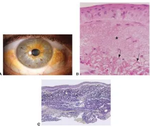
PRIMARY ACQUIRED MELANOSIS
Clinical Presentation
PAM of the conjunctiva made of unilateral, multiple, flat, indistinct areas of golden to dark
brown colour with irregular margins. The size and colour of PAM lesions changes over time.
The lesions are freely mobile and involve any part of the conjunctiva. Slit-lamp
biomicroscopy, includes lid eversion with careful inspection of the palpebral conjunctiva.
Double eversion of the upper lid is needed to see the entire upper fornix. The lacrimal gland
and lacrimal sac may be involved rarely by PAM. PAM is common in middle-aged /elderly
whites and is rare in blacks of all ages. The prevalence of PAM in the general population
ranges from 10% to 36%. The melanosis in PAM is due to an increase in melanin production
(with or without melanocytosis). Malignant transformation of PAM lesions occurs when
histologic atypia is present. 50% of patients with PAM with atypia may transform to
melanoma. This rate is 90% for lesions that have epithelioid cells or a pagetoid growth.
Malignant transformation is suspected in enlarging, highly vascularized lesions, and lesions
more than 7.5 to 10 mm, or in lesions having patchy pigmentation. Development of nodules
20
FIGURE 8: PAM – GROSS AND HISTOPATHOLOGICAL APPEARANCE
Histopathology
Clinical features cannot distinguish precancerous PAM (with histologic atypia) and benign
PAM without atypia. Suspicious lesions, should undergo an excisional biopsy, because this is
the only way to find the presence or absence of atypia. Lesions without histologic atypia
never become malignant. Histologically, PAM lesions without atypia may have increased
melanin production with or without melanocytosis. The melanocytosis is restricted to the
basilar regions of conjunctival epithelium. Nuclear hyperchromasia is absent and nucleoli are
not prominent. Patients with PAM without atypia tend are younger than patients with PAM
and atypia. PAM without atypia become PAM with atypia. PAM with atypia has an increased
chance of malignant transformation. Five patterns of atypical cells have been described with
21
large dendritiform melanocytes, epithelioid, or polymorphous (mixture). The atypia degree
increases with size of the nucleus and prominence of nucleoli. Lesions composed of
epithelioid cells or showing pagetoid spread have highest rate of malignant transformation.
Immunohistostaining with the monoclonal antibodies MIB-1 and PC-10 staining for the
proliferation markers Ki-67 and the proliferating cell nuclear antigen (PCNA), may help
differentiate between PAM with / without atypia.
Treatment and Prognosis
Complete excision of all lesions with atypia is goal of treatment, obtaining tumor-free
margins. In diffuse PAM, excision of any nodular areas is important. Multiple map biopsies
of the conjunctiva, and areas where there is no pigment, is needed in assessing the extent of
the disease. Cryotherapy, radiotherapy, or topical mitomycin C are useful adjunctives.
Topical mitomycin C is useful in patients with diffuse disease, with the entire ocular surface.
Extensive cryotherapy of the limbus can affect the stem cell population. Cryotherapy causes
necrosis of anterior segment. Six weekly cycles of topical mitomycin C 0.04% has good
response, ] but cytologic changes in the conjunctiva mimicking malignancy after giving
topical 0.02% to 0.04% mitomycin C drops is also noted. These changes localized to the
superficial layers of the conjunctival epithelium and include enlarged nucleus, chromatin
smudging-hyperchromasia, cytoplasmic eosinophilia, single cell necrosis, and subepithelial
inflammation. Primary acquired melanosis can recur after excision, and new lesions develop
elsewhere on the conjunctiva. Due to risk of malignant transformation and the possibility of
recurrences after excision, patients with PAM should have careful ocular examination and
22
IV. VASCULAR TUMORS
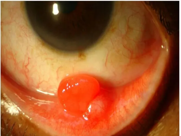
PYOGENIC GRANULOMA
Pyogenic granuloma is proliferative fibrovascular response to tissue insult by either
inflammation, surgery, or any nonsurgical trauma. It is classified as a polypoid form of
acquired capillary hemangioma. It appears as an elevated red mass, often with a good blood
supply. Microscopically, it is composed of “granulation tissue” with chronic inflammatory
cells and small calibre blood vessels. As the lesion is neither pyogenic nor granulomatous, the
name pyogenic granuloma is a misnomer. Pyogenic granuloma responds to topical
corticosteroids, but most cases ultimately require surgical excision. In recurrence, low- dose
plaque radiotherapy can be applied.
[image:29.595.78.384.434.665.2]23
FIGURE 9b: PYOGENIC GRANULOMA – GROSS AND HISTOPATHOLOGICAL APPEARANCE
CAPILLARY HEMANGIOMA
Capillary hemangioma of conjunctiva generally occurs in infancy, or several weeks following
birth, as red stromal mass, associated with cutaneous / orbital capillary hemangioma. The
conjunctival mass may enlarge over several months and spontaneously involute. Management
[image:30.595.79.504.138.419.2]–observation, surgical resection /local /systemic prednisone can be given.
24
CAVERNOUS HEMANGIOMA
Cavernous hemangioma is rare. This benign tumor is a red or blue lesion in the deep stroma
in young children. It is similar to the orbital cavernous hemangioma that is diagnosed in
young adults.Managed by local resection.
RACEMOSE HEMANGIOMA
Dilated arteriovenous communication without intervening capillary bed (racemose
hemangioma) is found in conjunctiva. It remains stable for years and is monitored
conservatively. Rule out Wyburn- Mason syndrome in these cases.
LYMPHANGIOMA
Conjunctival lymphangioma can occur as isolated conjunctival lesion or, is a superficial
component of deeper diffuse orbital lymphangioma .It becomes clinically apparent in first
decade and appears as multiloculated mass containing variable- sized clear dilated cystic
channels .Blood is seen in most of the cystic space called “chocolate cysts.” The treatment of
conjunctival lymphangioma is difficult because surgical resection or radiotherapy cannot
eradicate the mass.
25
VARIX
Varix is a venous malformation found in orbit and the conjunctiva. It is a mass of dilated
venous channels that enlarges with Valsalva manoeuvre. Treatment is cautious observation. If
painful, cold compresses and aspirin may be used. Surgical resection should be cautious due
to the risk for prolonged bleeding.
HEMANGIOPERICYTOMA
Hemangiopericytoma is made of pericytes that surround blood vessels. It shows both benign
and malignant cytologic features. Appears as a red conjunctival mass originating from the
stroma. Wide surgical resection with tumor- free margins is needed.
KAPOSI’S SARCOMA
Kaposi’s sarcoma is a cutaneous malignancy that occurs in elderly /immunosuppressed
patients.This tumor has become more common in AIDS and affects mucous membranes, such
as conjunctiva. Clinically it appears as reddish vascular masses that resembles a hemorrhagic
conjunctivitis. It is moderately responsive to chemotherapy and marked response to low- dose
radiotherapy.
26
V. FIBROUS TUMORS
FIBROMA
Fibroma is a rare conjunctival tumor which appears as a white stromal mass, unifocal or
multifocal. Surgical resection is needed.
Fibrous Histiocytoma
It is a rare mass of the conjunctiva and made of fibroblasts and histiocytes. Clinically and
histopathologically it is similar to many other amelanotic stromal tumors. In the conjunctiva
it may be benign, locally invasive, or malignant. Wide excision with tumor- free margins is
needed.
NODULAR FASCIITIS
It is a benign proliferation of connective tissue that commonly occurs in the skin and lesser in
the eyelid, orbit, and conjunctiva. Clinically and histopathologically it resembles fibro
sarcoma. The lesion is a solitary white mass in Tenon’s fascia. Complete excision is needed
as the lesion tend to recur.
VI. NEURAL TUMORS
Neural tumors of the conjunctiva are rare. They manifest a more yellow appearance than
fibrous tumors.
NEUROFIBROMA
It can occur in the conjunctiva as solitary /a diffuse / plexiform variety. The former is not
associated with systemic conditions and the latter is a part of von Recklinghausen’s
27
managed by complete surgical resection. The plexiform type is difficult to surgically excise,
and debulking procedures are necessary.
FIGURE 13: NEUROFIBROMA – GROSS APPEARANCE
NEURILEMOMA
Also known as schwannoma, is a benign proliferation of Schwann cells surrounding the
peripheral nerves. This tumor commonly from the orbit, rare in the conjunctiva. Clinically,
this lesion is yellowish- pink, nodular mass in conjunctival stroma. Complete excision is
needed.
GRANULAR CELL TUMOR
It is a rare tumor it is of Schwann cell origin. This benign tumor and clinically appears
smooth, vascular, and pink, and located in the stroma or within Tenon’s fascia.
Histopathologically, it is made of large round cells with granularity to the cytoplasm.
Complete excision is needed.
VII. HISTIOCYTIC TUMORS
XANTHOMA
Xanthoma most common in the cutaneous dermis, near extensor surfaces and on the
28
epibulbar surfaces. Bilateral conjunctival involvement has been noted and is termed
xanthoma disseminatum. Histopathologically, subepithelial infiltrate - lipidized histiocytes/,
eosinophils, / Touton giant cells are noted.
JUVENILE XANTHOGRANULOMA
It is a relatively common cutaneous condition that is painless, pink skin papules with
spontaneous resolution, in children under the age 2 years. Conjunctival, orbital, and
intraocular involvement is seen. The conjunctival mass appears as an orange- pink stromal
mass, typically in young adults. If the classic skin lesions are present, treatment with
observation or topical steroid ointment is provided. Else, biopsy is needed, and recognition of
the typical histopathologic features of histiocytes admixed with Touton’s giant cells helps to
confirm the diagnosis.
RETICULOHISTIOCYTOMA
It is a rare tumor, seen as part of a systemic multicentric reticulohistiocytosis. Clinically, the
tumor is seen as a pink, vascular limbal mass in adult. Histopathologically, it is made of large
histiocytes with granular cytoplasm.
VIII. MYXOID TUMORS
MYXOMA
Myxoma is a rare tumor that appears as orange- pink mass within stroma. This is associated
with Carney complex, a syndrome of cardiac / systemic myxomas, cutaneous lentigines, and
Sertoli cell tumor of testicles. Histopathology shows slender stellate and spindle cells
29
IX. LIPOMATOUS TUMORS
LIPOMA
It is rare and generally is found in adults as yellowish- pink stromal mass.Most are the
pleomorphic type having large lipid vacuoles surrounded by stellate cells.
FIGURE 14 – LIPOMA – GROSS APPEARANCE
HERNIATED ORBITAL FAT
Orbital fat presents in the conjunctiva as a herniation from superotemporal orbit. The
condition is bilateral and represents deficiency in orbital connective tissue to maintain the
proper location of orbital fat. Clinically, it is deep to Tenon’s fascia and is prominent on
inferonasal gaze. Digital reposition of the fat into the orbit is possible, but only temporary.
Management is observation.If dry eye occurs resection of the herniated fat and resuspension
of the orbit position of the fat is done. Histopathologically, the tissue is made of large lipid
cells.
LIPOSARCOMA
It has been rarely seen and shows features similar to lipoma. Histopathologically, neoplastic
30
X. LYMPHOID TUMORS
Lymphoid tumors can be isolated lesions or manifestation of systemic lymphoma.
Clinically, appears as a diffuse, slightly elevated pink mass in the stroma or deep to Tenon’s
fascia, most common in the forniceal region. This is similar to the smoked salmon; and so
called the “salmon patch”. It is tough to differentiate between a benign and malignant
lymphoid tumor.So, biopsy is needed to establish the diagnosis, and systemic evaluation
needed in all affected patients to exclude the possibility of systemic lymphoma.
Histopathologically, “sheets of lymphocytes” are found & classified as reactive lymphoid
hyperplasia or malignant lymphoma. Mostly they are B- cell lymphoma (non- Hodgkin’s
type). Rarely T- cell lymphoma is seen .Treatment of the conjunctival lesion includes
chemotherapy if systemic lymphoma is present or external beam irradiation (2,000 to 4,000
cGy) if it is localized to the conjunctiva. Other options are excisional biopsy & cryotherapy,
[image:37.595.73.487.494.732.2]local interferon injections/ observation.
31
XI. LEUKEMIA
Leukemia manifests in the ocular region as hemorrhages due to anemia and
thrombocytopenia rather than leukemic infiltration.Leukemic infiltration can be found with
CLL. The tumor appears as a pink smooth mass in the conjunctival stroma at the limbus/ the
fornix, similar to a lymphoid tumor. Biopsy shows sheets of large leukemic cells. Treatment
of the systemic condition is needed with secondary resolution of conjunctival infiltration.
XIII. METASTATIC TUMORS
These rarely occur in the conjunctiva, can be from breast carcinoma, cutaneous melanoma,
and other primary tumors. Metastatic carcinoma is one or more fleshy pink vascularized
conjunctival stromal masses. Metastatic melanoma to the conjunctiva is pigmented.
XIV. SECONDARY CONJUNCTIVAL INVOLVEMENT FROM ADJACENT TUMORS
The conjunctiva may be secondarily involved by adjacent structures tumours, by direct
extension from tumors of eyelids. The most important tumor causing is sebaceous gland
carcinoma of the eyelid. This shows pagetoid invasion & extends into the conjunctival
epithelium & causes clinical picture similar to chronic unilateral blepharoconjunctivitis.
Uveal melanoma in the ciliary body can go extrasclerally to the subconjunctival tissues,
producing a primary conjunctival tumor. Rhabdomyosarcoma of orbit, a tumor found in
children, presents first with conjunctival lesion before the orbital mass is discovered.
XV. CARUNCULAR TUMORS AND CYSTS
The caruncle is a unique that contains elements of conjunctiva and skin. The tumors in the
caruncle are similar to those in mucous membranes and skin. By histopathologic, 95%
32
and nevus. Other lesions are pyogenic granuloma, inclusion cyst, and sebaceous hyperplasia,
& sebaceous adenoma, oncocytoma. Malignant tumors like squamous cell carcinoma,
melanoma, lymphoma, & sebaceous carcinoma are rare in the caruncle. The oncocytoma is
benign tumor occurs more commonly in lacrimal / salivary glands. In caruncle it arises from
accessory lacrimal gland tissue &often has a blue cystic appearance. The treatment of
caruncular masses is observation / local resection
XVI. OTHER MISCELLANEOUS LESIONS THAT CAN SIMULATE
CONJUNCTIVAL NEOPLASMS
A few nonneoplastic conditions can be similar to neoplasms. These include pinguecula,
pterygium, foreign body, inflammatory granuloma, amyloidosis. In most cases, the history
and clinical finding, and in some excision of the mass may help to exclude a neoplasm.
XVII. DEGENERATIONS
PINGUECULA AND PTERYGIUM
A pinguecula is small, yellowish nodule, bilateral and located at the nasal / temporal limbus.
A manifestation of actinic damage “exposure to sunlight” or environmental factors, like dust
and wind, ageing- this growth is more common. On histology, the stromal collagen has
fragmentation and basophilic degeneration known as elastotic degeneration as the
degenerated collagen stains positively when using histochemical stains for elastic fibre like
the Verhoeff–van Gieson stain. A pterygium is like the pinguecula in etiology & location but
differs in its invasion of the ‘superficial cornea’ as a vascular, wing-shaped growth. Histology
shows elastotic degeneration; prominent blood vessels may be seen if vascularity seen
clinically and variable amount of chronic inflammation. Recurrent pterygia may not have
histologic feature of elastotic degeneration and are exuberant fibroconnective tissue response.
33
Thus, with actinic damage to the skin, there is the possibility for future malignant
transformation, though this occurs in rare cases with pingueculae and pterygia. If conjunctival
squamous neoplasia occurs, it occurs over an area of preexisting elastotic degeneration. If
epithelial hyperplasia, nuclear hyperchromasia and pleomorphism, & excess mitotic figures
[image:40.595.73.347.250.482.2]are seen in an excised pinguecula or pterygium, a diagnosis of OSSN should be assigned.
FIGURE 16: PINGECULA GROSS APPEARANCE
SENILE SCLERAL PLAQUES
These occur in the sclera rather than the cornea / conjunctiva. These lesions areYellow, gray,
/black vertical bands anterior to insertion of the medial & lateral rectus muscles in elderly.
They are more common after 60 years, like pinguecula and pterygium, may be due to UV
exposure. Histologically, calcium deposits with reduced cellularity and hyalinization are
34
FIGURE 17: SENILE SCLERAL PLAQUE – GROSS AND HISTOPATHOLOGICAL APPEARANCE
AMYLOID DEPOSITS
It is most commonly an idiopathic localized process in healthy young and middle-aged adults.
They are typically made of monoclonal immunoglobulin “AL amyloid” produced by local
clonal plasma cells. Conjunctival amyloidosis is due to long-standing inflammation, like with
trachoma (AA amyloid). Conjunctival amyloidosis may occur in primary conjunctival
lymphoma /plasmacytoma (or) secondary to systemic lymphoma / plasma cell myeloma.
Clinically, conjunctival amyloidosis seen as a salmon-coloured nodule which is associated
with hemorrhage. Histologically, appears as eosinophilic extracellular deposits in the stroma,
in a perivascular distribution. On Congo red stain, under standard light, amyloid deposits are
seen orange. When viewed with polarized light & rotating polarization filter, they exhibit
35
thioflavin. Electron microscopy shows fibrils. Immunohistochemical methods, sequencing &
mass spectrometry–based proteomic analysis are used in amyloid subtyping.
FIGURE 18: AMYLOID DEPOSITIS – GROSS AND HISTOPATHOLOGICAL APPEARANCE
EPITHELIAL INCLUSION CYST
This may form at a site of previous accidental / surgical trauma (like after strabismus surgery,
retinal surgery, or enucleation). Clinically, it is a transparent, cystic elevation on the ocular
surface with associated injection. Histologic examination shows cystic space lined by
conjunctival epithelium in the stroma. The lumen may be empty / have inspissated
proteinaceous material & cellular debris.
36
XVIII. GRANULOMATOUS CONJUNCTIVITIS
It is less common than papillary conjunctivitis and follicular conjunctivitis and has infectious
and non-infectious causes. Clinically, the nodular elevations of granulomatous conjunctivitis
is tough to distinguish from follicles, but the history and systemic symptoms may help in the
diagnosis. Granulomatous conjunctivitis with preauricular lymphadenopathy is known as
“Parinaud oculoglandular syndrome”. Bacteria like Bartonella henselae (causing cat-scratch
disease) & Francisella tularensis (causing tularemia), mycobacteria (Mycobacterium
tuberculosis), treponemes (syphilis), and fungi (sporotrichosis) may cause. Microorganisms
may be seen with Gram, acidfast / silver stains. The diagnosis is based on the culture results,
serology, PCR, or combination of all. If biopsy is done, the granulomas in granulomatous
conjunctivitis will show central necrosis. In non-infectious cause of granulomatous
conjunctivitis like sarcoidosis, involve all ocular tissues, also the conjunctiva. It occurs as
small tan nodules with no inflammatory signs, within the fornix. Conjunctival biopsy is a
simple way of giving diagnostic confirmation of systemic disease. Histologically,
noncaseating granulomatous “tubercles” are seen within the conjunctival stroma, with a
minimal cuff of lymphocytes & plasma cells. The diagnosis of sarcoidosis is made if
supported by clinical features and infectious causes of granulomatous inflammation have
been ruled out by histochemical and by culture results.
FOREIGN BODY GRANULOMA
Since it is exposed surface, the conjunctiva is open to contact with foreign bodies. Some are
transient & inert, others may become embedded and cause a foreign-body reaction, seen
histologically as granuloma around the foreign object. Multinucleated giant cells are seen.
Viewing the tissue section under polarized light may be useful in identifying the offending
37
FIGURE 20: FOREIGN BODY GRANULOMA – GROSS AND HISTOPATHOLOGICAL APPEARANCE
SARCOID GRANULOMA
38
XIX. MALIGNANT LESIONS:
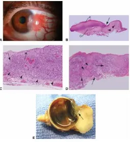
CONJUNCTIVAL- INTRAEPITHELIAL NEOPLASIA (CIN) AND SQUAMOUS CELL CARCINOMA (NON PIGMRNTED)
Epithelial neoplasia of the ocular surface is a disease complex, having a spectrum of changes
within the epithelial layers. Previously known as Bowen’s disease and ocular surface
squamous neoplasia. Conjunctival- corneal intraepithelial dysplasia (CCIN) is used if the
cornea is also involved. The earliest change is dysplasia, limited by the underlying epithelial
basement membrane. Increasing degrees of dysplasia occurs if process is not stopped
resulting in transgression of the basement membrane and invasion of underlying space &
structures. This invasion of subjacent tissue is hallmark of squamous cell carcinoma.
Histologic examination is needed for definitive diagnosis.
FIGURE 22: DIAGRAMMATIC REPRESENTATION OF HISTOPATHOLOGY OF VARIOUS CONJUNCTIVAL NEOPLASIA
Incidence and Etiology
CIN occurs in elderly, with light- complexioned men having extensive actinic exposure...
The incidence of SCC of the eye has been calculated to increase 49% for every 10-degree
39
from ultraviolet light, other reported risk factors are previous H/O skin cancer, smoking,
ocular trauma, petroleum derivative exposure. Human papilloma virus (HPV), types 16 and
18, has been found in conjunctival epithelial neoplasia by immunohistochemical and other
molecular analysis. The role of HPV in the etiology of CIN is unclear. Atypical, rapidly
progressive CIN in younger patients is associated with HIV. CIN has been found in younger
organ transplant patients using long- term cyclosporine. Xeroderma pigmentosum is an AR
disorder characterized by inadequate repair of DNA damage due to ultraviolet radiation.
Affected patients are prone to epithelial cancers, those of the conjunctiva & cornea. The
limbus is the transitional zone, from columnar conjunctival to stratified squamous corneal
epithelium. Within crypts of Vogt are the limbal stem cells. This area is similar to the uterine
cervix. Similar to cervical tissue, the corneal limbus is the site of origin for the majority of
dysplastic & neoplastic changes of the ocular surface.
Clinical Presentation
The patient with CIN presents with ocular irritation /complaints of redness /“growth on the
eye.” Vision is not affected. Differentiation of benign from malignant surface tumors is
difficult in slit lamp examination even for the experienced. Examination reveals a vascular
limbal mass, within interpalpebral area. The affected area is thick, and may appear gelatinous
/velvety. Gelatinous thickening, with superficial blood vessels, is common. CIN may mimic
diffuse chronic conjunctivitis with mild thickening. Other less common presentations are
sclerokeratitis, which is a focal corneal or scleral thinning with inflammation without any
tumor mass. Sclerokeratitis is similar to interstitial keratitis or Mooren’s ulcer. Biopsy is
needed in atypical /chronic scleritis / conjunctivitis unresponsive to standard treatment.
Hyperkeratosis is a characteristic feature of CIN and manifest as a white surface plaque, /
“leukoplakia”, which has no diagnostic significance. Neoplastic cells invades corneal
40
migrate centrally. Affected epithelium looks translucently gray, with sharply demarcated
border. Finger- like protrusions, and isolated islands are found on the leading edge. The
epithelium inside the lesion is thickened, and blood vessels are present, especially close to
limbus. Spontaneous regression may occur.
Marked thickening of a limbal lesion and fixation of the mass to underlying tissue, suggests
squamous cell carcinoma. Biopsy may yield helpful diagnostic information. In exfoliative
cytology and impression cytology, superficial cells are removed, fixed and stained with a
Papanicolaou technique, and studied by a cytopathologist. In exfoliative cytology cells are
taken away with a sterile platinum spatula, but in impression cytology cells are retained in
cellulose acetate paper strips, that are pressed against the area in doubt. Cells are graded on
degree of atypia, (including size, shape, nuclear and nucleolar characteristics, and mitotic
figures). Cytology is positive in 77% of biopsy- proven cases.
Differential Diagnosis
i) Benign epithelial lesion– associated thickening and surface keratin.
ii) Irritation caused by stromal inflammation or by pinguecula or pterygia may cause
pseudoepitheliomatous hyperplasia, and thickening of the epithelium and leukoplakia. Biopsy
reveals the benign acanthosis and hyper keratosis without dysplasia. Simple excision is done.
iii) Actinic keratosis of the ocular surface is due to UV radiation. These lesions occur in
older, light complexioned people with previous exposure to sunlight, the same population at
higher risk for CIN. Thickening with hyperkeratosis is present. Histopathologic examination
may show, mild acanthosis with hyperkeratosis &inflammation, to marked acanthosis with
cellular pleomorphism.
41
v) Chronic conjunctivitis
vi) Inflammation of a pinguecula or pterygium
FIGURE 23: OSSN – GROSS IMAGE
Pathology
Histologic examination is needed for definitive diagnosis.In small lesion, excisional biopsy
and removal of the entire mass is done. Incisional biopsy of the atypical area is performed for
larger lesions. Staining with hematoxylin- eosin and PAS is sufficient for the diagnosis of
CIN lesions. Biopsy helps to determine the depth of the lesion, & whether the margins are
free of tumor. The transition from normal to abnormal epithelium is sudden.
Specimens are graded based on location and degree of atypia.
Full- thickness dysplasia indicates carcinoma- in- situ.
Extension of tumour to sub epithelial space (substantia propria, sclera, or cornea) indicates
squamous cell carcinoma. Advanced tumors may enter into ciliary body, iris, and trabecular
42
Mucoepidermoid carcinoma is uncommon in the conjunctiva. Clinically it is difficult to
differentiate from CIN. Histopathology shows epidermoid and mucus- secreting cells.
Special stains for mucin, like Alcian blue / mucicarmine are used in diagnosis.As
mucoepidermoid carcinoma of the conjunctiva is locally aggressive than CIN, this diagnosis
needs proper follow up for invasion and recurrence.
Spindle cell carcinoma is aggressive variant of conjunctival squamous cell carcinoma arising
on the ocular surface. Clinical differentiation of this tumor is difficult. Pathologically,
pleomorphic spindle cells are seen, arranged in fascicles. Immuno histochemical analysis
using cytokeratin stains demonstrates the epithelial origin of spindle cell carcinoma.
[image:49.595.74.329.410.688.2]Transmission electron microscopy may be used in difficult cases.
43
Treatment
Surgical excision of suspicious area is the approach to therapy, if the entire lesion can be
removed in toto. Staining with rose Bengal will highlight abnormal epithelium. A wide
margin of 2 to 3 mm around the visible tumor is seen.
Frozen section control can shoe lateral surgical margins, not helpful with the deep margins.
Cryotherapy with a nitrous oxide probe done after surgical excision decreases the recurrence
rate of CIN. After removal of the lesion with 2- to 3-mm free margins, freezing the remaining
conjunctival margins and the sublesional base is done with formation of ice ball for 6
seconds, followed by slow thaw. A double freeze- thaw technique is usually done; but three
cycles are better if inadequate removal of tumor is suspected. Cryotherapy destroys tumor
cells by ‘thermal disruption’ & resultant local ischemia. Side effects of cryotherapy are
elevation or decrease in IOP, corneal scarring, iris atrophy, and destruction of retina. Local
application of beta- irradiation is used as primary treatment of squamous epithelial tumors, &
treatment of incompletely excised squamous tumors. Other side effects of irradiation are dose
related and include cataract, secondary glaucoma, local scarring, dry eye, & loss of cilia. The
threshold dose to prevent cataract of surface strontium 90 is estimated to be 5,000 rads.
Therapy with antimetabolite agents is beneficial in the adjunctive treatment of partially
excised corneal epithelial neoplasia & initial therapy in recurrent disease, extensive disease
having ill- defined borders/ situations in which excessive conjunctiva may be taken off,
leading to severe dry eye /limbal stem cell deficiency. “The rationale is use the highest
possible dose against the smallest amount of tumor”. Both MM-C and 5-FU have a selective
effect on rapid growing tumor cells. Due to dose- related local toxicity, intervening rest
periods between topical chemotherapy are employed. Rest intervals spares the limbal stem
cells. Punctal plugs needed to protect the NL system during therapy to reduce systemic
44
topical treatment to eradicate tumor cells located in the subepithelial space is an area of
concern in possible squamous cell carcinoma with scleral invasion. Long- term observation
is needed, as squamous cell carcinoma may recur. Mitomycin C (MMC) is a
chemotherapeutic antibiotic from Streptomyces caespitosus, which is used as an alkylating
agent and inhibits DNA synthesis. MMC also has an effect on fibroblasts and stem cells.
Therapeutic applications include 0.02% or 0.04% MMC QID for 14-day courses; 0.02%
MMC TDS for 14 days; and 0.04% MMC QID for 1 week cycles. A combination of excision,
cryotherapy, and topical MMC is effective in recurrent CIN. Adverse reactions to topical
drug mild hyperemia and tearing, photophobia, hyperemia, PEK, and blepharospasm. Pain
occurs if used more than 14 days; stoppage of therapy leads to decrease of pain. More severe
side effects, including scleral ulceration and perforation. 5--FU drug has antimetabolite
properties, rapidly growing cells accumulate lethal amounts of 5-FU. 5-FU is useful as an
adjunct in recurrent or incompletely excised squamous cell carcinoma. 1% 5-FU in artificial
tear base TDS- QID daily for 2- to 3-week cycles, until epithelium sloughs off. Clinical
improvement or resolution of intraepithelial neoplasia seen in all cases.
Recombinant interferon- alfa-2b
It has been used successfully in the treatment of CCIN, with an initial injection of 3 million
international units (IU), followed by “topical” interferon- alfa-2b drops (1 million IU/mL)
QID daily. If clinical response was noted by 1 week, topical therapy was continued until
resolution of the CIN. If response seen at 1week, subconjunctival and perilesional injections
are given three times weekly until clinical resolution. No complications seen with topical
therapy, though fever and myalgia may occur with subconjunctival dose.
Intralesional interferon alfa-2b is also successful in the treatment of squamous cell and basal
45
topical therapy with cidofovir (2.5 mg/mL), one drop 2 hourly initially, with a weekly taper
in frequency over the next 6 weeks. A residual focus needed excision, followed by
cryothe